THESE
THESE
En vue de l'obtention du
DOCTORAT DE L’UNIVERSITÉ DE TOULOUSE
DOCTORAT DE L’UNIVERSITÉ DE TOULOUSE
Délivré par l'Université Toulouse III - Paul Sabatier Discipline ou spécialité : Immunologie
JURY
Pr Joost van Meerwijk (Président du jury) Dr Daniel Speiser (Rapporteur)
Dr Hans Yssel (Rapporteur) Dr Loïc Dupré (Directeur de thèse)
Ecole doctorale : Ecole Doctorale Biologie - Santé - Biotechnologies Unité de recherche : INSERM, Unité de recherche 563
Directeur(s) de Thèse : Dr Loïc Dupré Rapporteurs : Dr Daniel Speiser & Dr Hans Yssel
Présentée et soutenue par Julie De Meester Le 28 Janvier 2011
Titre : Role of the Wiskott-Aldrich syndrome protein in human CD8+ T cell activation and effector function
ACKNOWLEDGEMENTS 7
ABSTRACT 8
RESUME 9
ABBREVIATIONS 11
I. INTRODUCTION 13
1. GENERAL INTRODUCTION ON THE IMMUNE SYSTEM 13 2. T CELL STIMULATION BY APC AT THE IMMUNOLOGICAL SYNAPSE 16
2.1 MODEL FOR IMMUNOLOGICAL SYNAPSE FORMATION 17
2.2 FUNCTIONS OF THE IS 18
3. T CELL ACTIVATION AND EFFECTOR FUNCTIONS: OLD FRIENDS AND NEW
ACQUAINTANCES 19
3.1 CD8+CYTOTOXIC T LYMPHOCYTES 22
3.2 CD4+T LYMPHOCYTES 22
a)Conventional T helper cells 22
b)CD4+ regulatory T cells 24
3.3 γ:δT CELLS 25
3.4 INVARIANT NKT CELLS 25
4. A CLOSER LOOK AT CD8+T CELL ACTIVATION AND EFFECTOR FUNCTIONS 26
4.1 CONTACT-DEPENDENT DESTRUCTION OF TARGET CELLS 28
a)Granzyme and perforin-mediated cytotoxicity 28
b)Receptor-mediated cytotoxicity 32
4.2 CONTACT-INDEPENDENT DESTRUCTION OF TARGET CELLS 33
a)IFN-γ 33
b)TNF-α 33
5. FAILURE OF THE IMMUNE SYSTEM 33
6. THE WISKOTT-ALDRICH SYNDROME 35
7. GENETIC DEFECTS 36
8. CLINICAL MANIFESTATIONS 37
8.1 BLEEDING DISORDERS DUE TO MICROTHROMBOCYTOPENIA 37
8.5 HEMATOLOGIC MALIGNANCIES 40
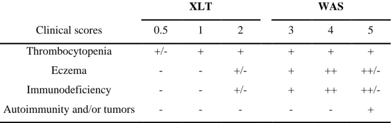
8.6 SCORING SYSTEM 40
9. THERAPY 41
a)Symptomatic therapy 41
b)Curative therapy 42
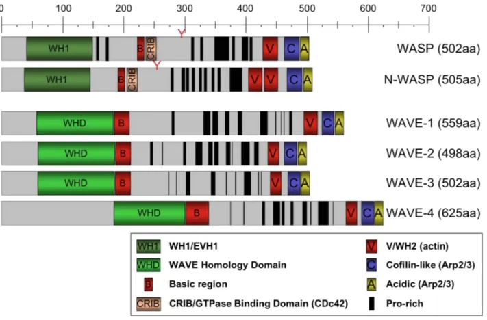
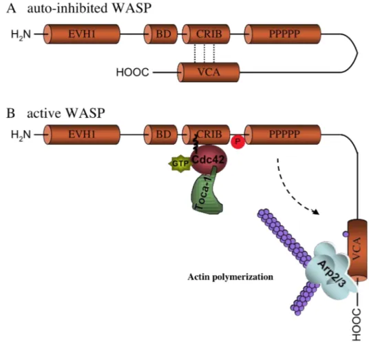
10. THE WISKOTT-ALDRICH SYNDROME PROTEIN 45
10.1 CHARACTERISTICS 45
a)Verprolin homology, central, acidic domain (VCA domain) 48
b)Poly-proline rich region 48
c)Cdc42 and Rac-interactive binding domain 48
d)Basic domain 48
e)Ena/ VASP homology 1 domain (EVH1 domain) 50
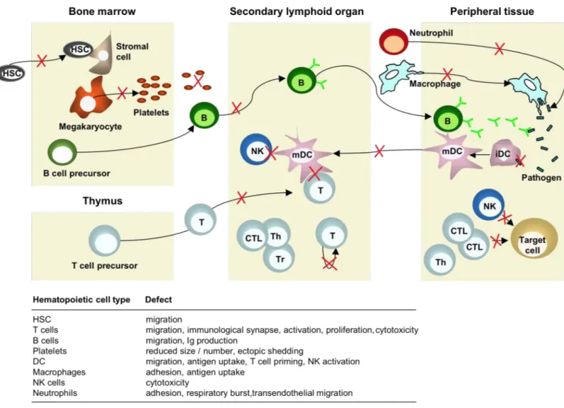
10.2 CELLULAR DEFECTS MEDIATED THROUGH WASP 52
a)Hematopoietic stem cells 52
b)Natural killer cells 54
c)Macrophages 54
d)Dendritic cells 55
e)T lymphocytes 55
f)B lymphocytes 58
II. THE STUDY OF CTL IN WAS: AIM, RATIONALE AND WORKING
HYPOTHESIS 60
1. AIM AND RATIONALE 60
2. WORKING HYPOTHESIS 61
III. DESIGN AND ORGANIZATION OF THE RESULTS PRESENTED IN THE
THESIS 62
1. ROLE OF WASP AT THE T CELL SIDE 62
1.1 EXPERIMENTAL DESIGN PART I 62
1.2 EXPERIMENTAL DESIGN PART II 63
DRIVEN T CELL PROLIFERATION BUT UNAFFECTED TARGET CELL LYSIS
UPON WISKOSTATIN TREATMENT 65
1. ABSTRACT 65
2. INTRODUCTION 67
3. CELLS, MATERIAL AND METHODS 69
3.1 CELLS AND MEDIA 69
3.2 BUFFY COATS 69
3.3 GENERATION OF ALLOGENEIC PBMC 69
3.4 GENERATION OF DENDRITIC CELLS 69
3.5 LOADING OF DC OR JY 70
3.6 GENERATION OF EFFECTOR CD8+T CELLS 70
3.7 FREEZING AND THAWING OF DC, CD8+T CELLS AND PBMCS 70
3.8 FLOW CYTOMETRY 71
a)Phenotype DC, JY and CD8+ T cells 71
b)Annexin V and propidium iodide staining 71
c)Measurement of TCR down-regulation 71
d)Measurement of T cell proliferation 72
e)Cytolytic assay 72
3.9 CONFOCAL MICROSCOPY 72
4. RESULTS 74
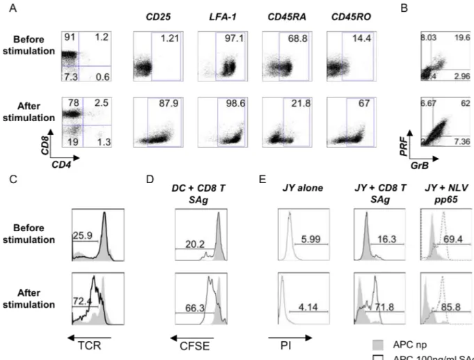
4.1 OPTIMIZATION OF A HUMAN AUTOLOGOUS SUPERANTIGEN-SPECIFIC SYSTEM 74
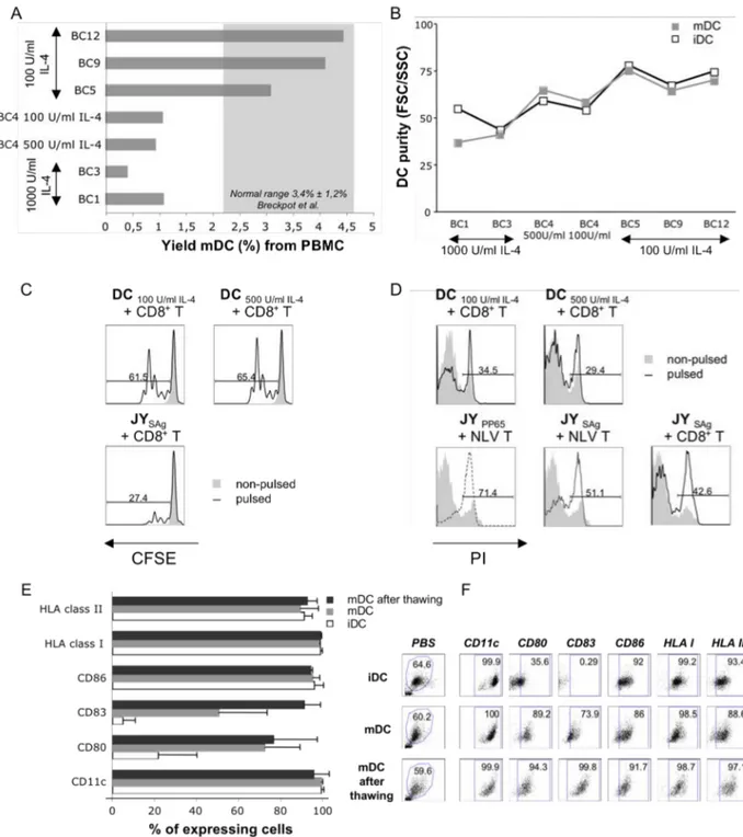
a)Generation of monocyte-derived dendritic cells 74
b)Naïve CD8+ T lymphocytes are efficiently activated by autologous SAg-loaded mDC
and become potent cytotoxic T cells 77
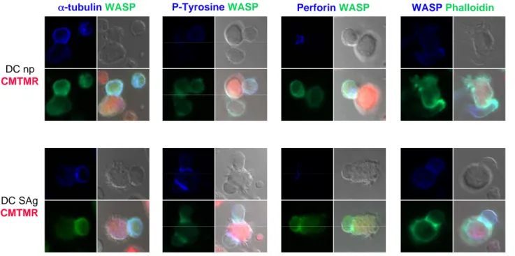
c)SAg-specific stimulation of CD8 T cells induces polarization of MTOC and perforin
towards the APC contact site 79
4.2 WISKOSTATIN TREATMENT INHIBITS PROLIFERATION OF ALLO-PBMC AND CD8+T CELLS IN RESPONSE TO ANTI-CD3/ANTI-CD28 STIMULATION 81 4.3 WISKOSTATIN HAS NO EFFECT ON CTL-MEDIATED CYTOTOXICITY 83 4.4 WISKOSTATIN IS TOXIC WHEN USED IN LONG-TERM ASSAYS 85
LYMPHOMA TARGETS 91
1. ABSTRACT 91
2. INTRODUCTION 94
3. PATIENTS, MATERIAL AND METHODS 96
3.1 PATIENTS 96
3.2 TRANSFORMED CELL LINES AND UNTRANSFORMED CD8+T CELL LINES 96
3.3 CHARACTERIZATION OF CD8+ T CELL LINES 97
3.4 MEASUREMENT OF TCR DOWN-REGULATION AND PROLIFERATION 97
3.5 DETECTION OF INTRACELLULAR CYTOKINES 98
3.6 CYTOLYTIC ASSAY 98
3.7 WESTERN BLOT 99
3.8 CONFOCAL MICROSCOPY 99
3.9 LENTIVIRAL VECTOR TRANSDUCTION OF CD8+T CELLS 100
4. RESULTS 101
4.1 CD8+T CELLS FROM WAS PATIENTS DIFFERENTIATE INTO ARMED EFFECTOR CTL
UPON STIMULATION WITH SAG-LOADED APC 101
4.2 REDUCED CYTOKINE PRODUCTION BY WAS CD8+T CELLS UPON APC STIMULATION 103 4.3 EXOCYTIC ACTIVITY AND LYTIC POTENTIAL OF WAS CTLS 105 4.4 WASP EXPRESSION IS REQUIRED FOR EFFICIENT KILLING OF B CELL LYMPHOMA CELLS
BY CTL 107
4.5 INCOMPLETE POLARIZATION OF WAS CTL LYTIC GRANULES TOWARD TUMORAL
TARGETS 109
4.6 CORRECTION OF ANTI-TUMORAL CTL CYTOTOXITY WITH A GENE THERAPY LENTIVIRAL
VECTOR 112
5. DISCUSSION 114
VI. INCREASED RESISTANCE OF WASP-DEFICIENT EBV-TRANSFORMED B
CELLS TO CTL-MEDIATED KILLING 120
1. INTRODUCTION 120
2. MATERIALS AND METHODS 122
2.1 PATIENTS 122
2.5 CYTOLYTIC ASSAY 123
3. RESULTS 124
3.1 DOWN-MODULATION OF WASP IN TARGET EBV-B CELLS DOES NOT SEEM TO ALTER
THEIR SUSCEPTIBILITY TO CTL-MEDIATED KILLING 124
3.2 WAS PATIENT TARGET B CELLS ACTIVATE NORMALLY CD8+T CELLS BUT ARE
RESISTANT TO CTL LYSIS 126
4. DISCUSSION 128
VII. GENERAL DISCUSSION 130
1. TECHNICAL ASPECTS AND RELEVANCE OF THIS WORK 130
1.1 WORKING ENVIRONMENT 130
1.2 SET-UP OF CELLULAR MODELS 130
1.3 ADVANTAGES AND DRAWBACKS OF THE CELLULAR MODELS 131 1.4 RELEVANCE OF INVESTING EFFORTS ON THE STUDY OF WAS 132
2. DIFFERENT APPROACHES TO LOOK AT WASP FUNCTION 133
3. MEANING OF THE SCIENTIFIC FINDINGS 134
3.1 MOLECULAR LEVEL 134
a)TCR-mediated T cell activation 134
b)Proliferative- and cytokine response to T cell activation 135 c)Cytolytic effector function upon target cell recognition 136
3.2 CELLULAR LEVEL 137
a)Cytokine production and proliferation 137
b)Cytotoxicity 139
3.3 EXTRAPOLATION TO THE SITUATION IN THE WAS PATIENTS 145
I would like to thank all the people with whom I’ve been working together the last few years!
Thank you Loïc and Salvatore for having given me the opportunity to perform my PhD here in Toulouse.
When I arrived in April 2006, it was Sabina who took me under her wings and taught me a lot of new techniques that were indispensable for the elaboration of this work, so thanks Sabina for all your help!
A few months after me, Ronan arrived to join our team. Besides being a great colleague, helping me with technical problems and with whom discussions brought along solutions, I would like to thank him for his friendship and motivation during these couple of years far away from our family and friends.
Of course, I also thank all the other team-leaders, students, PhD students, post-docs, etc. from the immunology department!
A special thank you for Fatima, Valérie, Sophie and Daniel for their help and patience!
I also acknowledge our collaborators that helped us for this work, in particular Ségolène Clayessens, Anna Villa, Alessandro Aiuti and Maria-Grazia Roncarolo for providing patient samples. I also thank Anne Galy for preparing and providing the lentiviral vectors used in this work. And finally I would like to thank Jean-Jacques Fournié for some good ideas that made this work far more interesting!
Thank you also to Daniel Speiser and Hans Yssel for the reviewing of this manuscript.
But most importantly, I want to thank my parents for their support in my choices and for their continuous help and encouragements. I also thank my husband, Christophe, for his love, kindness but mostly for his patience and constant motivation. Without him, I would never have had the courage to finish this work. And last but not least, I want to thank my family and friends who were always there for me when I needed a break or just some good time!!
The Wiskott-Aldrich syndrome protein (WASP) is confined to hematopoietic cells and regulates Arp2/3-mediated actin polymerization. Absence or impaired expression of WASP leads to the Wiskott-Aldrich syndrome (WAS), characterized by the triad of eczema, microthrombocytopenia and severe immune deficiency. Cellular defects have been described in a variety of cells, but how these defects contribute to the clinical phenotype, remains poorly understood. Patients with residual WASP expression, as in X-linked thrombocytopenia (XLT), have generally less severe clinical symptoms and do not develop hematologic malignancies. To date, bone marrow transplantation is the only curative therapy for WAS. However, new hope is nurtured by the implementation of gene therapy strategies, based on the administration of gene-corrected hematopoietic stem cells, when no compatible donor is available.
Since WAS patients are highly susceptible to infections, autoimmune diseases and tumors, we reasoned that defects in their CD8+ cytolytic T cells (CTL) might contribute to a state of reduced immune-surveillance. Therefore we investigated the role of WASP in this T cell subset. We identified defects in cytokine production (IL-2, IFN-γ and TNF-α) and proliferation of CD8+ T cells in response to stimulation with superantigens presented on allogeneic EBV-transformed B cells. This defect however was milder than the defects described in response to anti-CD3/anti-CD28 stimulation. In addition, the cytolytic capacity of CD8+ CTL against SAg-loaded EBV-transformed B cells was reduced, especially when those B cells were of tumoral origin. Although WAS CTL expressed normal levels of the lytic molecules, granzyme B and perforin, they appeared not to properly polarize these molecules toward the target cells. Reconstitution of WASP expression in CD8+ T cells by lentiviral transduction led to restoration of the lytic defect against tumoral B cells, confirming a direct role for WASP in CTL-mediated cytotoxicity. WASP-deficient EBV-transformed B cells appeared to exacerbate defective CTL-mediated killing, despite their normal stimulatory capacity.
Our data reveal a role for WASP in regulating the threshold for CD8+ T cell activation and lytic function in response to APC stimulation, likely contributing to the severe immune deficiency observed in WAS patients.
La protéine du syndrome de Wiskott-Aldrich (WASP) est exprimée uniquement dans les cellules hémapoïetiques dans lesquelles elle régule la polymérisation de l’actine médiée par Arp2/3. L’absence d’expression de WASP ou une expression réduite est la cause du syndrome de Wiskott-Aldrich (WAS), caractérisé par de l’eczéma, une microthrombocytopénie et une déficience immunitaire sévère. Des défauts cellulaires ont été décrits dans une multitude de cellules. Cependant, la contribution relative de ces défauts au phénotype clinique reste en grande partie inconnue. Les patients avec une expression résiduelle de WASP, comme dans la thrombocytopénie liée à l’X (XLT), ont généralement des symptômes cliniques moins graves et ne développent pas de tumeurs malignes hématologiques. Jusqu’à présent, la transplantation de moelle osseuse est la seule thérapie curative pour WAS. Cependant, un nouvel espoir justifie la mise en œuvre de stratégies de traitement par thérapie génique, basée sur l’administration de cellules souches hématopoïétiques avec le gène corrigé, quand un donneur compatible n’est pas disponible.
En raison de la susceptibilité accrue aux infections, maladies auto-immunes et tumeurs, nous avons émis l’hypothèse que les lymphocytes T CD8+ cytolytiques pourraient jouer un rôle dans le déficit immunitaire observé chez les patients WAS. Par conséquent, nous avons étudié le rôle de WASP dans cette population lymphocytaire T. Nous avons identifié des défauts dans la production cytokinique (IL-2, IFN-γ and TNF-α) et la prolifération des cellules CD8+ T en réponse à une stimulation par des superantigènes présentés par des cellules B transformées par l’EBV. Ce défaut a toutefois été plus modéré que ceux décrits dans la littérature en réponse à la stimulation avec des anticorps dirigé contre le CD3 et le CD28. En outre, la capacité cytolytique des cellules T CD8+ contre les cellules B transformées par EBV était réduite, en particulier lorsque ces cellules B sont d’origine tumorale. Bien que les cellules T CD8+ de patients WAS expriment des niveaux normaux de molécules lytiques, la polarisation des granules lytiques vers les cellules cibles s’est avérée incomplète. La reconstitution de l’expression de WASP dans les cellules CD8+ T conduit à la correction du défaut lytique contre les cellules tumorales B, confirmant un rôle direct pour WASP dans la cytotoxicité médiée par les CTL. Une déficience de WASP du côté des lymphocytes B transformés par l’EBV semble agraver la
Nos données révèlent donc un rôle de WASP dans la régulation du seuil d’activation des lymphocytes T CD8+ et de la fonction lytique en réponse à une stimulation avec des APC. Le défaut d’activation et de fonction des lymphocytes T CD8+ pourrait contribuer à la déficience immunitaire sévère observée chez les patients WAS.
ADA Adeosine-deaminase
Ag Antigen
APC Antigen presenting cell Arp2/3 Actin-related protein 2/3 BCR B cell receptor
CD Clusters of differentiation CIP4 Cdc42-interacting protein-4
CFSE Carboxyfluorescein diacetate succinimidyl ester CMFDA Chloromethylfluorescein diacetate (cellTracker Green)
CMTMR Chloromethyl benzoyl amino tetramethylrhodamine (cellTracker Red) CMV Cytomegalo virus
CRIB Cdc42-and-Rac interactive binding site CSF-1 Colony-stimulating factor 1
cSMAC Central supra-molecular activatory cluster CTL Cytotoxic T lymphocyte
DC Dendritic cell
dSMAC Distal supra-molecular activatory cluster EAE Experimental autoimmune encephalitis EBV Epstein-Barr virus
EVH1 Ena/ VASP homology domain 1 FACS Fluorescence activated cell sorter FasL Fas-Ligand
FHL Familial hemophagocytic lymphohistiocytosis Foxp3 Fork-head winged helix transcription factor 3
GrB Granzyme B
GTP Guanosine triphosphate GVHD Graft-versus-host disease
HD Healthy donor
HLA Human leucocyte antigen HLA:p HLA:peptide complex HSC Hematopoietic stem cell
ICAM-1 Intra-cellular adhesion molecule-1 (CD54)
Ig Immunoglobulin
IL Interleukin
IFN-γ Interferon gamma
iNKT Invariant natural killer T cell IS Immunological synapse IU International units
KD Knock-down
KO Knock-out
Lamp Lysosomal-associated membrane protein LFA Leukocyte functional antigen
MHC Major histocompatibility complex
MHC:p Major histocompatibility peptide complex MMLV Murine Moloney leukaemia virus
MMRD Mismatched related donor MOI Multiplicity of infection MTOC Microtubule-organizing center MUD Matched unrelated donor
MZ Marginal zone
NFAT Nuclear factor of activated T cells NK Natural killer
OMIM Online Mendelian Inheritance in Man PBMC Peripheral blood mononuclear cells PHA Phytohemagglutinin
PI Propidium iodide
PID Primary immune deficiency
PIP2 Phosphatidylinositol(4,5)biphosphate PKC Protein kinase C
PRF Perforin
pSMAC Peripheral supra-molecular activatory cluster
SCID Severe combined immune deficiency SEB Staphylococcus enterotoxin B
SEE Staphylococcus enterotoxin E TCR T cell receptor
Tfh Follicular T helper
TGFβ Transforming growth factor beta
Th T helper
TNF-α Tumor necrosis factor alfa Treg Regulatory T cell
TSST-1 Toxic shock syndrome toxin-1
VCA Verprolin homology central acidic region WAS Wiskott-Aldrich syndrome
WASP Wiskott-Aldrich syndrome protein WIP WASP interactive protein
X-CGD X-linked chronic granulomatous disease XLN X-linked neutropenia
I.
Introduction
The primary aim of the PhD research project presented in this manuscript has been to characterize the T cell subpopulation of CD8+ cytotoxic T lymphocytes (CTL) from patients with Wiskott-Aldrich syndrome (WAS). This rare disease, caused by inherited gene mutations in the WAS gene, leading to WAS protein (WASP) deficiency, can turn out life-threatening when left untreated due to severe hemorraghes and immune deficiency. Since CTL are crucial in immune responses to fight infection and cancer and, in addition, can contribute to the onset of autoimmunity, three important features in WAS, we believe that these cells from WAS patients need to be studied at the level of their physiological behavior in terms of activation, differentiation and effector function. In the next pages, I will give an overview on the immune system, and in particular the mounting of an adaptive T cell-mediated immune response with a specific emphasis on the activation and role of CTL. I will then present WAS and explain the rationale for studying CTL in WAS.
1. General introduction on the immune system
Leukocytes, or white blood cells, form a heterogeneous group of immune cells that have one common goal: protecting the host from disease. They all stem from a common pluripotent hematopoietic precursor cell that differentiates into more specialized progenitor cells in the bone marrow. Neutrophils, monocytes, macrophages and immature dendritic cells (DC) originate from common myeloid precursor cells, whereas B- and T lymphocytes and natural killer (NK) cells arise from common lymphoid progenitors. These differentiated leukocytes circulate in the bloodstream or reside in peripheral tissues to scan the environment for danger or respond to it through different effector mechanisms1.
Maturation of most immune cells, including B cells, occurs within the bone marrow. In contrast, T cell precursors migrate to the thymus, a central lymphoid organ, where they develop and undergo maturation. The thymus plays an important role in the development of tolerance. Indeed, T cells capable of recognizing self-MHC are positively selected while T cells with self-reactive T cell receptors (TCR), reacting strongly to self-antigens are purged from the repertoire by clonal deletion1,2 or by the induction of anergy1,3 to prevent autoimmune reactions.
The immune system comprises two components that work closely together and protect the host from disease, despite the constant presence of potentially pathogenic organisms: the innate and the adaptive immune systems. The innate immune system largely involves phagocytic cells, such as macrophages and neutrophils. It provides immediate defense mechanisms against infection in a non-specific manner and does not confer long-lasting or protective immunity to the host. In contrast, the adaptive immune system comprises the action of B- and T lymphocytes, providing the immune system with the ability to recognize and remember specific pathogens and to mount stronger attacks each time the pathogen is encountered. It is adaptive in the sense that the body's immune system prepares itself for future challenges1.
When a microorganism succeeds in crossing the epithelial barriers of the host and starts to replicate, the innate immune system is activated. Very often intruded pathogens are immediately recognized by macrophages that guard peripheral tissues. Binding of molecular patterns associated with pathogens to receptors on the surface of macrophages triggers them to engulf and destroy them. Pathogen destruction occurs either through the fusion of the phagosome with lysosomes that contain antimicrobial compounds or through the generation of a “respiratory burst”. In addition, activated macrophages secrete cytokines and chemokines, causing inflammation and the recruitment of other immune cells. Briefly, inflammatory responses increase the local blood flow, reduce blood flow velocity and induce the up-regulation of adhesion molecules on activated endothelial cells. Together these actions promote the adhesion and consequent extravasation of circulating leukocytes into the infected tissues. Neutrophils are the first to arrive at the site of infection, followed by monocytes that differentiate into more tissue-macrophages. In case of viral infection or infection with some intracellular pathogens, NK cells will as well be activated. These cells attack harmful cells by the directed release of lytic molecules leading to target cell destruction. Inflammatory responses also increase the flow of antigens from infected tissue to draining lymph nodes, a mechanism that will help the mounting of an adaptive immune response later on1.
In addition to the actions of innate immune cells, microbial products will activate the complement system. This humoral compound of innate immunity is composed of a series of plasma proteins that react with one another to opsonize pathogens. Due to increased vascular permeability during inflammation, these plasma proteins will facilitate pathogen removal by phagocytes in infected tissues. Macrophage-induced
cytokines will also induce activation of the clotting system, inhibiting the spread of infection to the bloodstream. Either the innate immune response clears infection, either it controls infection by preventing dissemination, while the adaptive immune response develops1.
The adaptive immune system is activated by the innate immune system. Since immature DC are recruited already during innate responses, they serve as a “link” between innate and adaptive immunity. Upon capture of antigens (Ag), substances that stimulate the production of antibodies, in infected tissues, immature DC migrate to secondary lymphoid organs. During this migration, and promoted by proinflammatory cytokines, DC undergo maturation characterized by the up-regulation of costimulatory molecules. In addition, on their route to lymphoid tissues DC degrade Ag-derived peptides to load them on MHC class I or II molecules expressed on their membrane. Intracellular pathogens, such as viruses and some intracellular bacteria, are degraded into peptides in the proteasome and loaded on MHC class I molecules (MHC-peptide complex = MHC:p). Intravesicular and extracellular pathogens are degraded in lysosomal vesicles and loaded on MHC class II molecules1,4. Once arrived in secondary lymphoid organs mature DC, now highly professional antigen presenting cells (APC), present Ag to recirculating T cells. Pathogen-derived Ag also arrives in the secondary lymphoid tissues through the lymph as result of inflammation enabling other APC, such as macrophages and B cells, to capture and present Ag to naïve T cells. Lymphocytes that completed their maturation leave the thymus via the bloodstream until reaching a peripheral lymphoid organ. In the absence of cognate Ag, they migrate through it, regaining the bloodstream. However, upon recognition of MHC:p via their specific TCR and simultaneous costimulation, naïve T lymphocytes get activated. Activation signals induce their clonal expansion and differentiation into effector cells. CD8+ T lymphocytes and CD4+ T helper (Th) 1 cells, being the soldiers mediating cellular immunity and CD4+ Th2 cells mediating humoral immunity. The activation of naïve T cells upon Ag-recognition, or “T cell priming” is known as the primary immune response. After having exerted their effector function, most T lymphocytes go into apoptosis and a small part survives as memory T cells. This pool of cells will rapidly re-differentiate into effector T cells when the host encounters the same pathogen on a
later time point, providing the adaptive immune system with immunological memory1,5.
B cells also recirculate through lymphoid organs, and unlike T cells, B cells can recognize soluble pathogens with their B cell receptor (BCR) without prior degradation1,4. Upon internalization and degradation peptides can be loaded on MHC class II molecules in order to activate naïve CD4+ T cells. However, B cells are very poor in initiating adaptive immune responses. In contrast, upon activation B cells can differentiate into very powerful antibody-producing plasma cells and contribute to the humoral immune response. The most direct way in which antibodies protect the host against pathogens is by the neutralization of viruses and bacterial toxins. A second mechanism comprises the action of antibodies to opsonize bacteria that are invisible to the immune system because of an outer coat, lacking molecular patterns recognized by phagocytes. Opsonized bacteria however will be ingested and destroyed by phagocytes. A third action of antibodies is complement activation that will strengthen the action of antibodies in promoting the coating of pathogens to subsequently be removed and destroyed by phagocytes1.
Thanks to the high variety of lymphocytes with unique TCR recognizing specific pathogen-derived peptides, and the powerful action of antibody-producing B cells, the adaptive immune response conquers almost every infection and we become ill only very rarely. However, tumor cells and certain pathogens have developed defense mechanisms protecting themselves from being recognized by the host’s immune system making their clearance more difficult, and facilitating their survival and replication in the host. Additionally, in some cases the immune system, whether it is the innate or adaptive system or a combination of both, does not work properly. Together, these events can eventually contribute to latent infections with periodical outbreaks and the development of chronic diseases, autoimmunity and malignancies.
2. T cell stimulation by APC at the immunological synapse
APC, and in particular DC, that migrate from inflamed tissues to peripheral lymphoid tissues will secrete chemokines to attract naïve T cells. Chemokine signaling activates a cascade of signal transduction leading to cytoskeletal rearrangements and polarization of T cells, displaying a leading edge, the lamellipodium highly sensitive to MHC:p complexes, and a trailing edge or uropod. Independently of the presence of antigen, APC will scan a large number of migrating naïve T cells in order to find one
or more T cells recognizing the Ag presented by the APC. Only when the TCR is engaged, Ag-specific T cells can stop migrating and can be fully activated6. At saturating Ag doses, the stop signal results in a tight interaction with APC and low T cell motility7, whereas at lower, more physiological Ag doses the stop signal is more transient and permits the T cell to crawl over the APC to sustain T cell signaling8,9. APC, and in particular DC, can engage several T cells simultaneously10,11, and these APC:T cell interactions can last for several hours6,10, or can be dynamic, short-lived and sequential as has been demonstrated between stromal cells and thymocytes12 and between DC and T cells13.
Upon Ag recognition, a multi-step process leading to the formation of the immunological synapse (IS), a specialized junction between a T lymphocyte and an APC14, occurs. An intact actin cytoskeleton is required to initiate TCR clustering and signaling8,15. Indeed, the actin cytoskeleton plays a very important role in Ag-recognition. First, it allows the movement of molecules on the surface of T cells, permitting T cells to scan APC and vice versa. Second, actin filaments serve as scaffolds for signaling complexes and might also be involved in the recruitment and stabilization of membrane molecules involved in T cell activation16.
2.1 Model for immunological synapse formation
The first step of synapse formation begins with the adhesion between two cells. Initial interactions are mediated through large adhesion molecules, such as the integrin leukocyte functional antigen-1 (LFA-1), expressed at high levels on T lymphocytes, and its ligand intracellular adhesion molecule-1 (ICAM-1) on APC17. LFA-1 requires activatory signals to modify its conformation so that ligation with ICAM-1 can be initiated. This activation is provided, at least in part, by the chemokine-signaling cascade. After establishment of a close cell-cell contact, smaller molecules, such as CD2 and its ligand CD58 on APC, can interact. Only when the T cell recognizes a specific antigen presented on the MHC molecules of the APC, a stable IS is formed. TCR bind with high affinity to the MHC:p complexes, resulting in the formation of a central supra-molecular activatory cluster (cSMAC), while integrins are forced to move to the periphery, resulting in the formation of a peripheral SMAC (pSMAC) 14. Finally, the outer ring of the IS contains very large molecules, such as CD45, and is called the distal SMAC (dSMAC) 18. This structure with a segregation of the cSMAC,
pSMAC and dSMAC, is called the “mature” IS and is maintained for several hours6. However, the formation of this prototypical IS is only observed in specific experimental conditions, such as the use of very high, non-physiological Ag concentrations, the use of B cell lines18, or the use of surrogate lipid-bilayers instead of APC14. Indeed, other types of synapses with a multi-focal character (multiple distinct TCR clusters) have been described in more physiological systems for example between naïve T cells and DC19, between CD4+ T cells and B cell lymphomas at low Ag dose20, and between thymocytes and thymic epithelial cells21. It is now well established that the type of IS depends on the activation state of T cells, the nature of the APC and the strength and quality of antigenic stimulation9. A lot of controversy exists to explain how the IS between a T cell and an APC can trigger and sustain the T cell activation process. Indeed, to some the IS is a stable adhesive junction between a polarized T cell and an Ag-bearing APC6,16,22, whereas to others the IS is a dynamic event, characterized by serial TCR triggering8,23-25.
2.2 Functions of the IS
Since the cSMAC contains TCR, CD28 and a variety of signaling molecules, it was suggested that the mature IS functions as a microenvironment facilitating TCR:MHC:p and costimulatory interactions, indispensable for T cell activation. The IS is believed to allow a continuous scanning of the T cells to find a maximum of MHC:p complexes to ensure optimal T cell activation16,22,26. However, the mature IS does not seem to be required for T cell activation, since TCR signaling occurs before this molecular structure is formed between macrophages and T cells27 or between B cells and T cells28. Moreover, TCR signaling appears also maximal before the mature IS is formed and is weak at its center. Therefore, it has been proposed that the cSMAC arises from the convergence of TCR microclusters that are formed upon initial contact formation, and are actively transported to the center29. Additionally, it appeared that these microclusters sustained TCR signaling during T cell activation15. Recently, it has been suggested that signaling components would be degraded in the cSMAC and thus the cSMAC would be the place of extinction of TCR signaling, whereas intensive TCR triggering would take place in the periphery15,30. The current view on the role of the cSMAC in T cell signaling postulates that at high antigenic stimulation, signal transduction occurs in the cSMAC, although, it is accompanied by
a high rate of degradation of TCR and signaling compounds. In contrast, for weak antigenic stimuli, the rate of TCR degradation in the cSMAC is slower and thus promotes sustained signaling as result of TCR accumulation and its interaction with cognate MHC:p complexes9.
Early observations that the microtubule-organizing center (MTOC) polarizes at the interface between cytolytic cells and susceptible targets31,32, and that lytic granules are exocytosed within this synaptic cleft (region of contact between a T cell and another immune cell)32 suggested a role for the mature IS in polarized secretion. This structure could retain the secreted granules to protect bystander cells from killing33. In contrast to naïve CD8+ T cells, it appears that a cSMAC is formed at the interface of effector CD8+ T cells and opposing target cells34. Later it has been demonstrated that a stable pSMAC is required for effective CD8+ CTL-mediated lysis of target cells35. In the mature synapse, accumulated F-actin and adhesion receptors are thought to form a ring in the pSMAC through which perforin and other lytic granule contents are secreted36. However, it has been described that at low antigen dose, the “lytic synapse” does not display segregated SMACs as observed for the “activatory synapse” at high antigen dose, required for full T cell activation37. In addition, it has been demonstrated that CTL-mediated cytotoxicity does not require a stable mature IS38.
In the same line, exocytosis of cytokines, such as IL-2, IFN-γ and IL-10 is thought to occur in the synaptic cleft39.
3. T cell activation and effector functions: old friends and new
acquaintances
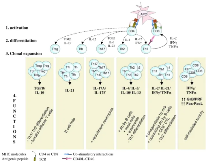
T lymphocytes are executive soldiers in the adaptive immune response. Indeed, as result of gene rearrangements during their development, each individual T lymphocyte expresses a unique TCR, responding to a specific Ag-derived peptide. This mechanism ensures that most pathogens can be recognized and removed from the body. T lymphocytes are characterized by the simultaneous expression of the lineage marker CD3 in combination with CD4 or CD8, molecules that are intimately tied to target recognition. In this group we find the CD8+ cytotoxic T lymphocytes, CD4+ Th1 and CD4+ Th2 cells, Th17 cells, follicular Th cells and a heterogeneous group of regulatory T cells (Figure 1). There exist however, additional T lymphocyte
subpopulations that have a more limited diversity of receptors and unlike the adaptive T lymphocytes, do not require clonal expansion before responding to Ag-recognition. They are somehow “intermediates” between innate and adaptive immunity. In that group we find amongst other the γ:δ T cells, and invariant NKT cells. The current picture of T cell biology is that of a very diversified family of cell subsets that display some level of differentiation plasticity and that carry either effector or regulatory functions at a specific time and location during an immune response.
Figure 1: Schematic representation of T cell activation and differentiation in response to
APC stimulation. TCR stimulation by recognition of HLA:p complexes in combination with
costimulatory signals and cytokines provided by APC, triggers the activation and differentiation of naïve CD4+ and CD8+ T cells. CD4+ T cells can differentiate into conventional (Th1, Th2, Th17 and Tfh) or regulatory T cells, depending on the cytokine milieu in which activation occurs. After activation and differentiation, effector T cells undergo clonal expansion to assure host defense.
3.1 CD8+ Cytotoxic T lymphocytes
Naïve CD8+ T cells have no cytotoxic potential but must undergo prior activation, a process requiring 1-3 days for maximal activity. They recognize peptides derived from dangerous cells presented in the context of HLA class I molecules on APC. Upon activation, Ag-specific CD8+ T cells will expand several orders of magnitude to form an army of effector cells42. They then up-regulate the lytic molecules, granzymes and perforin, and Fas-Ligand (FasL) expression levels transforming into armed and highly specific cytotoxic T lymphocytes14,32,43,44. CTL are the snipers of the cellular arm of the adaptive immune response and eliminate their target cells in several ways42,45. It is thought that the granzyme/perforin pathway dominates the class I elimination pathway46,47, whereas the Fas-FasL pathway would dominate class II elimination by CD4+ Th1 cells48,49. In addition, upon activation CTL secrete high levels of TNF-α and IFN-γ, 2 cytokines with antimicrobial activity50. CTL also play an important role in the surveillance of viral-infected cells and tumor cells42,45. However, CTL have a dark side: when they direct their lytic machinery against cells from self, or foreign tissue after transplantation, they can cause severe autoimmune disorders and graft rejection. In addition, after transplantation, donor-derived CTL can cause graft-versus-host disease (GVHD), by directing their lytic machinery toward host cells.
3.2 CD4+ T lymphocytes
Upon recognition of Ag-derived peptides presented in HLA class II molecules of APC and simultaneous costimulation, CD4+ T cells undergo expansion and differentiation. In general, depending on the inflammatory milieu in which Ag-presentation takes place, they will differentiate into conventional (Th1, Th2, Th17, Tfh) or regulatory T cells1,51. Conventional Th cells mediate the adaptive immune response by activating CD8+ T cells, B cells and macrophages, whereas Treg suppress potential destructive actions of effector T cells (Figure 1).
a) Conventional T helper cells
Th1 and Th2 cells are specialized cytokine producers and skew the immune response in cellular or humoral direction. Th1 cells develop when activation signals are
delivered in a milieu rich in IL-12, provided by APC1,5,52. Thanks to their exquisite capacity to produce high amounts of IFNγ, Th1 cells are central players in the cellular immune response. They activate the bactericidal activities of macrophages and induce B cells to make opsonizing antibodies. In addition, they activate CD8+ CTL either by direct interactions or indirectly via cytokines and through CD40L-CD40 mediated interaction with DC1,5. Th1 cells can also differentiate into cytotoxic cells. Indeed, they appear to kill dangerous cells mainly by the Fas-FasL-mediated pathway48,49, but also through granzyme B/perforin-mediated mechanisms. They seem to play a role in lymphocyte homeostasis by killing anergized B cells53 and by the elimination of chronically activated cells recognizing self-antigens54.
Th2 cells develop under the influence of APC-derived IL-4 during CD4+ T cell activation1,5,52. They mediate the humoral arm of the immune response through the production of cytokines. Indeed, they activate naïve Ag-specific B cells to produce antibodies via the production of IL-4 and IL-13. Th2 cells also activate mast cells and eosinophils via 5, and inhibit the development of a Th1 response via 10 and IL-131,5.
Th17 cells represent a more recently discovered Th subset. Their differentiation from
naïve CD4+ T cells appears to involve signals from TGF-β, 6, 21, 1β and IL-2355-57. Their differentiation occurs early after infection, since DC are rapidly triggered to produce IL-6 and TGF-β upon activation by pathogens. They are characterized by the production of IL-17A and IL-17F. They can also produce IL-21, IL-22, TNF-α, IL-6 and GM-CSF, but usually not IFN-γ and IL-4. IL-21 signaling induces the expression of RORγt, a key transcription factor for Th17 cell differentiation. Th17 cells are thought to enhance inflammation by the recruitment of leukocytes to sites of infection for pathogen clearance58, and seem to be important in immune responses against extracellular bacteria and fungi56, which are not efficiently cleared by Th1 or Th2 cells56,57. Th17 cells are highly cytopathic and are involved in several autoimmune diseases55-57.
Finally, follicular Th cells (Tfh) represent a group of Th cells that specifically help B cells to produce high affinity, class-switched antibodies important for the clearance of pathogens after infection and for the establishment of humoral memory immunity. In contrast to Th1 and Th2 cells, expressing only transiently the chemokine receptor CXCR5, Tfh express CXCR5 constitutively, promoting their unique localization in
the CXCL13-rich germinal centres of B cell follicles in peripheral lymphoid tissues. B cell help is provided by direct interactions involving the costimulatory molecules CD40L and ICOS and by the secretion of IL-2159,60.
b) CD4+ regulatory T cells
Similar to Th17 cells, it is thought that regulatory T cells (Treg) also require TGF-β during activation, but provided in the absence of IL-6. The characterization of CD4+
Treg seems to be difficult, since the phenotypic markers for Treg, CD25, CTLA-4, GITR, CD127, LAG-3 and Foxp3, are not strictly Treg-specific, since their expression is also up-regulated by conventional T cells upon activation51. As their name suggests, Treg “regulate” the potentially deleterious activities of effector T cells (reviewed in 61). There is evidence that decreased numbers and/or function of Treg can lead to autoimmunity, allergy and graft rejection, whereas over-abundance of Treg would inhibit antitumor and antipathogen immunity62. The way these cells restrain effector cells to destroy Ag-bearing tissue is mediated through multiple mechanisms, including the direct interactions with effector and dendritic cells, the release of immunosuppressive cytokines, such as TGF-β and IL-10, and the generation of adenosine (commented in 55).
Nowadays, different subsets of Treg have been characterized. Besides a thymic-derived subpopulation with potent regulatory properties, the “natural” Treg, naïve CD4+ T cells can differentiate into Treg upon activation by Ag-bearing tolerogenic APC (reviewed in 62). To this group belong the IL-10-producing Tr1 cells, differentiation of which is driven by IL-1063.
It has to be noted that the classification of the different CD4+ T cell subsets into conventional versus regulatory T cells does not exclude regulatory functions for conventional T cells and immunostimulatory functions for Treg. Indeed, conventional T cells exert besides their effector functions suppressive activity. For example: the production of IFNγ by Th1 cells inhibits the development of Th2 cells, and conversely, Th2-derived IL-4 suppresses the development of Th1 cells51. In the same line, both IFNγ and IL-4 inhibit the development of Th17 cells whereas Th17 cell-derived IL-17 inhibits Th1 differentiation51,56.
Some regulatory T cells secrete high amounts of TGF-β, a cytokine with strong immunosuppressive properties. However, TGF-β has been shown to exert immunostimulatory actions as well. Indeed, it seems to specifically promote IgA and Ig2b isotype class switch in mouse B cells. In addition, some Foxp3 Treg have been shown to release IL17, a proinflammatory cytokine, important for immunity against extracellular bacteria, and normally produced by Th17 cells51.
3.3 γ:δ T cells
Unlike the majority of mature T cells expressing an αβ TCR, γδ T cells express a TCR composed of a γ and δ chain. This population represents 1-10% of circulating T cells and is abundantly present in the skin and gut mucosa within a population of lymphocytes, known as intraepithelial lymphocytes. γδ T cells display several characteristics that place them at the border between the innate and the adaptive immune system64. For example: γδ T cells can respond very rapidly to infection, and unlike adaptive αβ T cells do not require priming to do so (innate). On the other hand, they display TCR rearrangement as do conventional αβ T cells, and they confer immunological memory as in the adaptive immune response65. γδ T cells recognize non-conventional antigens, such as phosphorylated microbial metabolites and lipid antigens presented in MHC class I-like molecules and require costimulation provided by professional APC. Their effector mechanisms are not fundamentally different from αβ T cells, in the sense that they can kill infected cells by death receptor/death ligand (eg Fas-FasL) signaling or in a granzyme B/perforin-mediated manner and they are potent cytokine producers64. Besides their role in fighting infection, γδ T cells have also been shown to be involved in the onset and propagation of autoimmune disease.
3.4 Invariant NKT cells
Invariant NKT (iNKT) cells represent a peculiar thymic-derived T cell subset that express both NK cell markers in combination with a semi-invariant α:β TCR (strictly conserved α-chain paired with a limited diversity of β-chains). They act as immune sentinels, recognizing lipid antigens presented in the MHC class I-like molecule, CD1d. iNKT cells become very rapidly activated upon infection and are potent immunoregulatory cells by the production of cytokines profoundly affecting both the
innate and adaptive arms of the immune system. iNKT cells play an important role in the control of infection and cancer immune surveillance. In addition, they are implicated in the protection against some autoimmune diseases1,66,67.
4. A closer look at CD8
+T cell activation and effector functions
Although the focus of this work lays on the sniper function of effector CTL, it is worth to note that CD8+ T cells also exert other functions. Indeed, effector CD8+ T cells have a suppressor function, in the sense that they kill Ag-bearing DC in order to terminate an immune response45,68, whereas memory CD8+ T cells can provide “help” to effector cells of cellular immunity indirectly via stimulation of DC maturation and prolongation of Ag-presentation68.
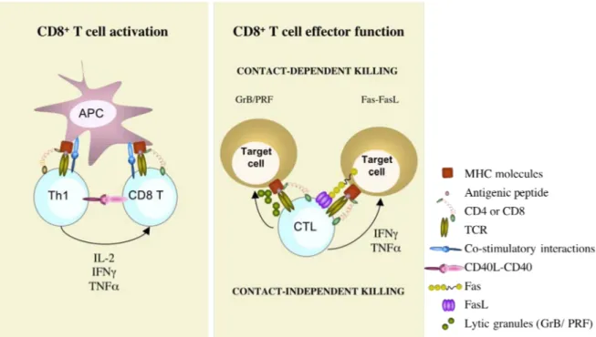
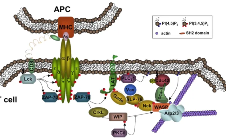
As mentioned above, CD8+ T cells recognize with their specific TCR pathogen-derived antigens presented in the groove of HLA class I molecules, expressed on the surface of APC, and leading to their activation (Figure 2). Those antigens are generally derived from pathogens that invade host cells and multiply in the cytoplasm. Proteins derived from these pathogens are broken down in the proteasome and peptides are loaded on the HLA class I molecules. However, CTL can also be implicated in fighting infections caused by extracellular bacteria or bacteria in intracellular vesicles, since DC have the ability to present Ag normally destined for HLA class II presentation in the groove of HLA class I, a process called “cross-presentation” 1,4. This makes CD8+ T cells extremely potent killers of cells infected with pathogens from different origin. Indispensable during T cell activation is simultaneous costimulation, provided by APC. Indeed, mature DC express high levels of costimulatory molecules such as the B7 molecules (CD80 and CD86) and 4-1BB that will bind their receptors (CD28 and 4-1BBL) expressed on T cells. CD8+ T cells also receive help from Th1 cells via cytokines, in particular IL-2 and IFN-γ69 that activate the cellular arm of the adaptive immune response or via direct or indirect CD40-CD40L interactions70-72.
Once properly activated, CD8+ T cells undergo clonal expansion. This expansion is driven by an enormous IL-2 production by the activated T cells themselves. The produced IL-2 works in an autocrine or paracrine fashion by binding the IL-2-receptor, that is up-regulated upon T cell activation. The army of Ag-specific CD8+ T cells then differentiates into effector soldiers armed with lytic molecules (granzymes, perforin, FasL) and leaves the lymphoid tissues to migrate to the site of infection.
CTL can kill compromised cells (I) by firing their lytic granules in a very polarized manner towards target cells14,31,32, (II) by ligation of death receptors expressed on target cells, and (III) by the secretion of cytotoxic cytokines with antiviral and antitumoral actions.
Figure 2: Naïve CD8+ T cells recognize peptides presented in HLA class I molecules with their specific TCR. Simultaneous costimulation will trigger their activation. CD8+ T cells also receive help from Th1 cells by means of direct interactions and cytokines (left panel). Upon activation, CD8+ T cells differentiate into highly cytotoxic T cells, up-regulating granzymes, perforin and FasL. CTL kill target cells in a contact-dependent manner by the polarized secretion of granzymes and perforin or by triggering receptors bearing death-domains expressed on target cells. In addition, CTL can exert cytolytic function without contacting target cells by the secretion of cytotoxic cytokines, such as IFN-γ and TNF-α ( contact-independent-killing) (right panel).
4.1 Contact-dependent destruction of target cells
The main weapons used by CTL to kill target cells is the granzyme/perforin-mediated pathway and in lesser extent the Fas-FasL-mediated pathway. These mechanisms are highly specific and require an intimate cell-cell contact.
a) Granzyme and perforin-mediated cytotoxicity
LYTIC GRANULE CONTENT
Lytic granules are specialized endo-lysozomes, containing multiple proteins:
Membrane-perturbing proteins: the pore-forming molecule “perforin” and granulysin. Granule serine proteases: in humans a family of 5 “granzymes” has been identified:
granzyme A, B, H, M and K.
Proteoglycan matrix: serglycin Perforin-inhibitor: calreticulin
Lysosomal enzymes or cathepsins with roles in granzyme processing (Cathepsin C)
and protecting CTLs against perforin (Cathepsin B)
Stored T effector molecules: Fas Ligand
Upon T cell activation, the transcription of the genes encoding perforin and granzymes starts. De novo synthesized proteins are non-functional and need to undergo post-translational modifications in order to take their biological active form. Once modified, they are assembled in lytic granules. Lytic granules have a low pH, keeping granzymes in an inactive state and contain lysosomal-associated mebrane glycoproteins (LAMPs) 1,2 and 345. These molecules are up-regulated upon CD8+ T cell activation and are associated with exocytosis of lytic granules73.
DELIVERY OF THE KISS OF DEATH
Recognition of dangerous cells has been shown to induce rapid reorientation of the MTOC, the golgi-apparatus and lytic molecules to the contact site with the target cell31,74,75. These observations led to the formulation of the “granule exocytosis pathway” in 198576. Upon target cell identification and conjugation, granules move towards the contact site, fuse with the plasma membrane and their content is released into the synaptic cleft at the target cell interface. In the presence of calcium, perforin polymerizes and enters the target cell. Originally, perforin was thought to directly
induce target cell apoptosis, by “perforating” the target cell membrane. However, perforin by itself does not induce DNA fragmentation in target cells77. Then, it was hypothesized that the pores induced by perforin polymerization, formed a portal for the entrance of other granule proteins to induce apoptosis. However, those pores are probably not large enough to allow entry of granzymes78. Additionally, granzyme A, B and H have been shown to enter target cells in the absence of perforin. Indeed, mannose-6-phosphate receptor-mediated endocytosis has been demonstrated for granzyme B. Moreover, it has been demonstrated that absence of this receptor prevented the rejection of mismatched tumor allografts, a finding that provides a mechanism for tumor immune evasion79. These findings have led to a more recent hypothesis for lytic granule delivery: after secretion of lytic granules by exocytosis, serglycin-bound-granzyme B binds to the mannose-6-phosphate receptor on target cells and is internalized80. Perforin would be involved in the release of serglycin-bound-granzyme B from the granules, since in the absence of perforin, granzyme B remains innocuously confined within the endo-lysosomal vesicles81,82. However, the exact role of perforin is still not fully understood. The group of Griffiths discovered that calcium-dependent directional exocytosis of lytic granules, present in the cytoplasm of killer cells occurs in a specialized secretory domain32 that lies next to the cSMAC18,83. The MTOC transiently contacts the plasma membrane to directly deliver lytic granules into the IS. Granule delivery seems to occur independently of actin, but requires minus-end directed movement along microtubules84. The group of de Saint Basile demonstrated that the effectors of the exocytic apparatus, Rab27a and hMunc13-4 and effectors of cytotoxic function, perforin and granzymes, are localized in distinct structures, endosomal and lysosomal vesicles, respectively. Both vesicles move to the contact site with the target cell where during a late maturation step both vesicles are likely to fuse, a mechanism indispensable for lytic granule exocytosis85. Mice lacking Rab27 expression display compromised granzyme/perforin-mediated killing due to defective granule exocytosis in effector T lymphocytes86.
HOW LYTIC MOLECULES INDUCE APOPTOSIS
Individual granzymes seem to have considerable functional redundancy, since mice that are deficient in one or several granzymes display less dramatic immune deficits, than do perforin-deficient mice87. Granzymes induce DNA fragmentation in the nucleus of target cells. Granzyme B is the best characterized and is believed to be a key player in the rapid induction of target cell death81,82. Indeed, granzyme B can cleave various caspases resulting in the activation of the cellular apoptotic cascade. However, granzyme B can also activate caspase-independent apoptosis pathways. One of them is granzyme B-induced mitochondrial collapse, resulting in the release of the pro-apoptotic protein, cytochrome c. Granzyme B has also been shown to directly activate nuclear DNAses by cleavage, contributing to target cell DNA apoptosis88. Granzyme A has a different substrate specificitiy and does not induce apoptosis by activation of caspases, as does granzyme B. Granzyme A induces slow cell death89 by destroying the nuclear envelope by targeting lamins and opening up DNA for degradation by targeting histones90, inducing single-stranded DNA breaks, rather than oligo-nucleosomal DNA degradation91.
The role of the other “orphan” granzymes (H, K and M) is largely unknown.
Once the “apoptotic switch” has been triggered the dying cell undergoes typical morphological transformation, characterized by:
• cell shrinkage and rounding because of the breakdown of the cytoskeleton by caspases
• dense cytoplasm with tightly packed organelles
• chromatin condensation (pyknosis) and fragmentation (karyorrhexis) • the nucleus breaks into several nucleosomal units due to DNA degradation • the cell membrane displays “ blebbing”
• and finally the cell breaks apart into apoptotic bodies, which are then cleared by phagocytes
RELEVANCE OF GRANZYME AND PERFORIN-MEDIATED KILLING
- Immune surveillance and defense against pathogens and tumors
The role of perforin-expressing cells, including CTL, NK cells, NKT cells, γδ T cells in immune surveillance92 has been strengthened by the observation that perforin-deficient mice develop spontaneous malignancies with age, in particular B cell
lymphomas93. Also in humans, it was suggested that perforin deficiency disturbed normal T cell homeostasis, favoring the uncontrolled proliferation of pre-malignant lymphoid cells, which in turn can acquire malignant phenotype94. Both CTL and NK cells share common cytolytic pathways that are implicated in the defense against virus-infected cells and transformed cells. As discussed above, the main pathway used by CTL is mediated through the directed release of cytolytic granules inducing apoptosis in the infected or malignant target cells. Studies in perforin-deficient mice have underlined the importance of this pathway in the response against viral infections and in tumorigenesis87.
- Termination of immune responses, T cell homeostasis and T cell memory: T lymphocytes with a certain Ag-specificity are present in low numbers in non-pathological conditions. However, upon antigen exposure these cells proliferate and differentiate to form reactive clones of effector T cells that efficiently kill infected target cells. Once the pathogen has been cleared, reactive T cells must undergo controlled deletion to reset cell numbers around initial levels (T cell homeostasis), leaving behind a pool of memory cells. Perforin seems to be implicated in the elimination of CD8+ T cells after acute exposure to foreign antigen. Moreover, perforin-deficient mice display increased clonal expansion and persistence of superantigen and virus-specific T cells95. The disappearance of effector T cells is essential as its persistence causes a fatal hemophagocytic syndrome resulting from uncontrolled activation and proliferation of T lymphocytes, leading to excessive macrophage activation and multiple deleterious effects96-98. Different mechanisms have been proposed to regulate T cell homeostasis. One explanation could be that CTL become infected themselves, making them targets for CTL-mediated fratricide. Indeed, some lymphotropic viruses (eg CMV, HTLV-1) naturally infect CD8+ T cells99. Another mechanism consists of a trans-activation of cytotoxic granules exerted against neighboring T cells if a high density of HLA:p is transferred from the stimulating APC to the surface of the target T cells, leading to fratricidal lysis100,101. Finally, trans-cytolytic action could also be exerted on APC. Perforin-dependent elimination of DC would act as a regulatory feedback mechanism to prevent the access of Ag-loaded DC to the lymph node and the further activation of Ag-specific T cells102.
- Autoimmunity
CD8+ T cells also seem to be implicated in the onset of some autoimmune disorders. This has been clearly illustrated in mouse models for induced or spontaneous diabetes and autoimmune encephalomyelitis (EAE). Indeed, perforin-deficiency eliminated or dramatically reduced the severity of autoimmune responses against β-cells without affecting lymphocyte infiltration of the pancreatic islets, and EAE symptoms103,104.
- Graft-versus-host disease
Finally, CD8+ T cells derived from transplanted tissues can direct their lytic machinery towards host cells, resulting in life threatening GVHD after bone marrow transplantation105. Besides Fas-FasL-mediated cytotoxicity, granzymes have been shown to be required for the lethal effects of GVHD induced by transferred allo-reactive CD8+ T cells87,89,106.
b) Receptor-mediated cytotoxicity
A second contact-dependent mechanism of CTL to kill dangerous cells involves ligation of death receptors expressed on the target cells. A well-characterized mechanism is the Fas-FasL-mediated pathway, inducing caspase-dependent apoptosis in target cells. This mechanism requires neither calcium nor lytic granules. Maximal expression of FasL requires prior activation of the effector cell107. Only little ligand is stored in CTL and for maximal killing the induction of new ligand synthesis is needed and requires TCR stimulation108. Lymphocyte homeostasis is thought to be regulated in part through the up-regulation of Fas and FasL upon T cell activation and subsequent triggering of activation-induced cell death95,109. Defective Fas-mediated apoptosis could allow prolonged survival of lymphocytes, which could then become targets of transforming events, suggesting a role for Fas-FasL in tumor development110. Fas-FasL interactions are also implicated in GVHD and autoimmune diseases. Indeed, Fas-FasL-mediated killing would play a role in the elimination of cells chronically exposed to self-antigens, either by fratricide or by specialized suicide processes111,112. In addition, mutations in Fas or FasL are associated with onset of autoimmune phenomena113,114.
4.2 Contact-independent destruction of target cells
Upon activation, CTL secrete cytokines such as IFN-γ and TNF-α that exhibit cytotoxic action when secreted in the vicinity of target cells.
a) IFN-γ
IFN-γ is produced predominantly by NK cells, NKT cells, CD4+ Th1 cells and CD8+ CTL. Its critical role in immunity is illustrated by the occurrence of tumors, and inflammatory- and autoimmune phenomena when its production and/or secretion is defective. IFN-γ can directly inhibit viral replication and has antitumoral, immunostimulatory and immunomodulatory properties. IFN-γ promotes Ag-presentation and lysosomal activity by macrophages, APC activation, up-regulation of MHC class I molecules, adhesion and binding required for leukocyte migration, NK cell activity and Th1 differentiation (by up-regulating the transcription factor T-bet)(reviewed in 115,116).
b) TNF-α
TNF-α is an important regulator of the immune response. TNF-α is also able to induce apoptotic cell death and inflammation, and to inhibit tumorigenesis and viral replication. TNF-α is produced mainly by macrophages, but also by other immune competent cells, in particular, T lymphocytes. A local increase in concentration of TNF will cause the typical signs of inflammation: heat, swelling, redness and pain. Abnormal regulation of TNF-α production has been implicated in a variety of human diseases, as well as cancer117.Binding of TNF-α with the TNF-Receptor1 (expressed in most tissues) can lead to activation of death domains, inducing apoptosis in the target cell. Moreover, this pathway seems to be involved in activation-induced cell death of CD8+ T cells, regulating the termination of immune responses95,118. However, TNF-induced cell death plays only a minor role compared to its overwhelming functions in the inflammatory process.
5. Failure of the immune system
The pivotal role of a functional immune system is highlighted by the existence of immune deficiencies, pathologies in which the immune system does not work properly or is partially missing. Immune deficiencies are classified as primary or
secondary. Primary immune deficiencies (PID) are the consequence of mutations in genes that are involved in the control of immune responses, and are inherited. Secondary immune deficiencies are acquired as a consequence of other diseases (eg infection with HIV), or can be secondary to environmental factors (eg malnutrition), or adverse effects of medical intervention (eg chemotherapy). By examining which infections accompany specific immune disorders, it is possible to understand which components of the immune system contribute to the selective clearance of pathogens. In addition, PID reveal how interactions between different cell types contribute to the immune response and to the development of T and B lymphocytes. They lead to the identification of the defective gene, providing information for diagnosis, treatment and eventually gene therapy strategies1. More than hundred immune deficiencies have been described so far and are the result of defects in innate and/or adaptive responses1.
A large proportion of PID are caused by recessive mutations in the X-chromosome. Since male subjects have only 1 chromosome, all males who inherit an X-chromosome carrying a defective gene will manifest disease. In contrast, female carriers with one defective X-chromosome are usually healthy because the mutated X chromosome is inactivated.
This work is focusing on a specific X-linked primary immune deficiency, the Wiskott-Aldrich syndrome.
6. The Wiskott-Aldrich syndrome
In 1937, an early paper written by the German paediatrician, Alfred Wiskott, described for the first time a ‘familial and innate thrombopathia’, affecting 3 brothers. The boys suffered from thrombocytopenia, bloody diarrhea, eczema and recurrent ear infections. All three of them died as result of intestinal hemorrhages and sepsis119. Seventeen years later, in 1954, the group of Aldrich investigated this syndrome in a family where 16 out of 40 males were affected, and they concluded that the disease was X-linked120.
Nowadays, the Wiskott-Aldrich syndrome (WAS; OMIM: 301000) is described as an X-linked primary immunodeficiency characterized by thrombocytopenia with small platelet size, eczema, recurrent infections and increased incidence of autoimmunity and hematologic malignancies, especially B cell lymphomas121-125.
Linkage studies were employed to map the WAS gene to the small arm of the X-chromosome (Xp11.22-p11.23)126. In 1994, the gene was isolated by positional cloning and found to encode an intracytoplasmatic protein that counts 1823 basepairs and is organized in 12 exons127. The Wiskott-Aldrich syndrome protein (WASP) participates in the dynamic regulation of actin polymerization, primarily through activation of the actin-related protein (Arp) 2/3 complex and is involved in cellular processes, such as a) cell signaling and subsequent cytoskeleton reorganization128 b) formation of podosomes, actin-rich structures identified in motile primary cells like macrophages129,130 and DC131,132, c) migration and cell trafficking of lymphocytes, DC, and granulocytes133.
The incidence of classical WAS has been estimated between 1 and 10 individuals in 1 million newborns, but might be much higher due to difficult early diagnosis in mild and evolutive cases. Clinical symptoms, such as petechiae, bruises, bloody diarrhea and infections manifest already early after birth. Due to microthrombocytopenia and severe disturbance in both the humoral and cellular immune system, affected patients develop life-threatening hemorraghes, infections, autoimmune diseases and/or tumors. The life expectancy of boys affected by WAS is estimated around 15 years in the absence of treatment123-125,134.