HAL Id: tel-01124101
https://tel.archives-ouvertes.fr/tel-01124101
Submitted on 30 Oct 2015HAL is a multi-disciplinary open access archive for the deposit and dissemination of sci-entific research documents, whether they are pub-lished or not. The documents may come from teaching and research institutions in France or abroad, or from public or private research centers.
L’archive ouverte pluridisciplinaire HAL, est destinée au dépôt et à la diffusion de documents scientifiques de niveau recherche, publiés ou non, émanant des établissements d’enseignement et de recherche français ou étrangers, des laboratoires publics ou privés.
ROS/RNS modulation in Systemic sclerosis treatment
Wioleta Marut
To cite this version:
Wioleta Marut. ROS/RNS modulation in Systemic sclerosis treatment. Human health and pathology. Université René Descartes - Paris V, 2012. English. �NNT : 2012PA05T079�. �tel-01124101�
UNIVERSITÉ PARIS DESCARTES
FACULTÉ DE MÉDECINE PARIS DESCARTES – SITE COCHIN
THÈSE
pour obtenir le grade de
DOCTEUR
Sciences de la Vie et de la Santé
Ecole Doctorale : Gc2iD
Discipline: Immunologie
présentée et soutenue publiquement
par madame Wioleta Marut
le 15 novembre 2012
ROS/RNS modulation in Systemic sclerosis treatment
Jury:
Dr Bernard WEILL, Président
Pr Frédéric BATTEUX, Directeur de thèse
Dr Phillipe Guilpain, Rapporteur
Pr Gilbert Kirch, Rapporteur
Pr Claus Jacob, Examinateur
Dr Amelie Servettaz, Examinateur
2
I dedicate this thesis to my husband and my beloved animals
3 ACKNOWLEDGEMENTS
I would like to thank all those people who made this thesis possible and an unforgettable experience for me.
I would like to especially thank to my PhD supervisor, Frederic Batteux for supporting me during these past three years. Frederic is the funniest advisor and one of the smartest people I know.
Also I would like to express my very sincere gratitude to Mr. Bernard Weill for the essential contribution to this work.
I am very grateful to Frederic and Mr. Weill for their scientific advice, knowledge, and a lot of useful suggestions.
I also have to thank the members of my PhD committee, Professors Gilbert Kirsch and Philippe Guilpain for reviewing this work.
I will forever be thankful to Carole Nicco for her help, suport and friendship. I could always count on her when I had a problems. She is my best role model for a scientist and mentor.
Remecrciemnts également à Christiane Chereau, sans qui ce travail n’aurait pas abouit. Je la remercie pour sa gentillesse permanente et son soutien.
I also thank my best friend Mathilde for providing support and friendship that I needed.
The best outcome from these past three years is finding my best friend, soul-mate, and husband. Grzes has been a true and great supporter and has unconditionally loved me during my good and bad times.
4
TABLE OF CONTENTS
ABBREVIATIONS ... 7
PATHOGENESIS OF SYSTEMIC SCLEROSIS (SSC)... 10
I.1 Fibroblast dysfunction ... 10
I.1.1 Intrinsic factors implicated in the fibroblast dysfunction ... 11
I.1.1.1 Activation of pro-collagen gene via TGF-β signalling ... 11
I.1.1.2 CTGF - Connective tissue growth factor ... 14
I.1.1.3 Early growth response genes ... 14
I.1.1.4 Molecules involved in extracellular matrix degradation ... 15
I.1.1.5 Implication of ERK1/2 in fibrosis ... 16
I.1.1.6 Synthesis of free radicals ... 17
I.1.1.7 Synthesis of cytokines and chemokines ... 17
I.1.2 Extrinsic factors implicated in the fibroblast dysfunction ... 18
I.1.2.1 The role of cytokines ... 18
Interleukin-4 ... 18 Interleukin-13 ... 19 Interleukin-17 ... 19 Interferon γ ... 20 I.1.2.2 Endothelin 1 ... 20 I.1.2.3 TGF-β ... 21 I.1.2.4 PDGF ... 21 I.1.2.5 Serotonin ... 23 I.1.2.6 Angiotensin II ... 24
I.1.2.7 Notch proteins ... 25
I.2 Endothelium dysfunction ... 26
I.1.3 Vasculogenesis and angiogenesis ... 26
I.1.4 Dysregulation of vasculogenesis ... 27
I.1.4.1 Endothelin-1 ... 27
I.1.4.2 Nitric oxide (NO) ... 27
I.1.5 Dysregulation of angiogenesis ... 28
I.1.6 Local recruitment of leukocytes ... 30
I.1.7 The coagulation/fibrinolysis balance in systemic sclerosis ... 30
Intervention of the immune system ... 30
5
I.1.9 Cellular adaptive immunity ... 32
I.1.10 Humoral adaptive immunity ... 33
I.1.10.1 Elements contributing to the activation of B cells ... 34
I.1.10.2 Role of autoantibodies in SSc ... 35
I.1.10.3 Autoantibodies relatively specific for SSc ... 36
I.1.11 Targeted therapy directed against B lymphocytes ... 39
Genetic and environmental factors ... 40
I.1.12 Genetic factors ... 40
I.1.13 Environmental Factors ... 42
Animal models of SSc ... 42
I.1.14 Inducible models of SSc ... 43
I.1.15 Spontaneous models ... 47
ROLE OF REACTIVE OXYGENE SPECIES (ROS) ... 48
II.1 Sources of ROS ... 48
The superoxide anion O•-2 ... 48
Hydrogen peroxide (H2O2) ... 51
Hydroxyl radical (OH•) ... 51
Nitrogen oxide (NO•) ... 52
II.1 Role and consequences of ROS ... 52
I.1.16 Action on proteins ... 52
I.1.17 Effect on lipids ... 53
I.1.18 Action on DNA ... 53
I.1.19 Roles and effects of ROS in cellular metabolism ... 53
I.1.20 Antioxidants systems ... 55
I.1.20.1 Examples of enzymatic antioxidants systems ... 55
I.1.20.2 Examples of non-enzymatic antioxidant systems ... 56
I.1.21 The role of ROS/RNS in SSc ... 56
PERSONAL WORK ... 60
III.1 Article 1 ... 62
III.2 Article 2 ... 93
6
DISCUSSION ... 154
CONCLUSIONS AND PERSPECTIVE ... 163
APPENDICES ... 165
7 ABBREVIATIONS
Ab Antibody
AOPP Advanced Oxidation Protein Products AT II Angiotensin II
AT1R Angiotensin II type 1 receptor
CTGF Connective Tissue Growth Factor DPTTS Dipropyltetrasulfide
EC Endothelial cells EP Epithelial cells
Egr Early Growth Response Factor ET1 Endothelin-1
GSH Glutathion
GVHD Graft-versus-host Disease HIF1 Hypoxia-Induced Factor 1 HOCl Hypochlorus acid
IFN Interferon IL Interleukine KO Knock-Out LB Lymphocyte B LPS Lipopolysaccharide LT Lymphocyte T
MCP-1 Monocyte Chemoattractant Protein-1 MMP Matrix Metalloproteinase
NO Nitric oxide
NKT Natural Killer T Cells
OSCs Organosulfur compounds
pDC plasmacytoid Dendritic Cell PDGF Platelet Derived Growth Factor PDGFR Platelet Derived Growth Factor
Receptor
(PHTE)2
RNS Reactive nitrogen species
NQ 3-bis(phenyltellanyl)naphthoquinone ROS Reactive Oxygen Species
8
SCID Severe Combined ImmunoDeficiency SSc Systemic Sclerosis
SMA Smooth Muscle Actin
Smad
TGF Transforming Growth Factor Th1 lymphocyte T helper 1
Th2 lymphocyte T helper 2
TIMP Tissue Inhibitor of Metalloproteinase TKI Tyrosine Kinase Inhibitor
TLR Toll like Receptors TNF Tumor Necrosis Factor TSK Tight Skin
VEGF Vascular Endothelial Growth Factor VEGFR Vascular Endothelial Growth Factor
9 FIGURE LIST
Figure 1. Link between dysfunction of fibroblasts, endothelial cells, and immune cells in
systemic sclerosis from J Varga, Nature Reviews Rheumatology, 2012. ... 11
Figure 2. TGFβ Smad and non-Smad signaling pathways from initiation to nucleus. (a)
Signal initiation (b) Smad-dependent pathway (c) Smad-independent pathway from Souchelnytskyi, Experimental Oncology, 2012. ... 13
Figure 3. Link between Ras and ROS by Gabrielli, The Journal of biological Chemistry,
2005. ... 16
Figure 4. IL-13 binding to the IL-13 receptor induces the activation of fibroblasts,
differentiation into myofibroblasts, increased ECM deposition, and fibros is from
Fuschiotti, Cytokine, 2011. ... 19
Figure 5. Possible profibrotic pathways by angiotensin II by Dobrev, Circulation: Arrythmia
and Electrophysiology, 1995. ... 25
Figure 6. Endothelial receptor tyrosine kinases involved in angiogenesis adapted from
Alitalo, The Journal of Cell Biology, 1995. ... 29
Figure 7. Angiogenesis in systemic sclerosis from Giacomelli, Autoimmunity Review, 2011.
... 30
Figure 8. Animal models of inducible and spontaneous SSc by Batteux, Current Opinion in
Rheumatology, 2011. ... 47
Figure 9.
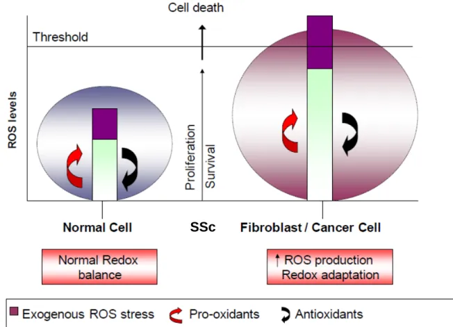
Figure 10. Regulation of cancer cells and SSc fibroblasts proliferation adapted from P.
Huang, Nature Reviews, Drug Discovery, 2009...58
Figure 11.
Sources of ROS by Dawes, Trends in Cell Biology, 2005...52
Induction of murine SSc through daily intradermal injections of HOCl generating
10
Pathogenesis of systemic sclerosis (SSc)
Systemic sclerosis (SSc) is a rare connective tissue disease characterized by fibrosis of skin
and internal organs, autoimmunity, and microcirculatory abnormalities 1 [ , 2]. There are
two widely recognized subsets of SSc, limited cutaneous (lcSSc) and diffuse SSc (dSSc). The main difference between these two subsets is the speed of disease progression, and the extent and severity of skin and internal organs. In lcSSc, skin thickening is confined to the face and extremities. In dcSSc, disease progression is very rapid, with the skin changes extending on the trunk, thighs, and upper arms. dcSSc is a more serious subset of the disease, with fatal visceral organ involvement.
SSc affects approximately 75.000-100.000 individuals in the United States. It affect four times more woman than man, and is more frequent in African-Americans than Caucasians [3]. Pathogenesis of SSc is complex and poorly understood. Factors causing dysfunction of fibroblasts, endothelial cells, and cells of the immune system have to be clarified (Figure 1). Among environmental factors, certain viruses, like cytomegalovirus [4] and certain chemicals such as solvents, silica, or pesticides may play a role in triggering the disease. In addition, several studies have shown that reactive oxygen species (ROS) [5-7] or TGF-β [8, 9] may play a role in the initiation and progression of the disease. These new pathophysiological tracks open new therapeutic perspectives for this disease.
I.1 Fibroblast dysfunction
In SSc the excessive connective tissue fibrosis is the most characteristic pathological manifestation of the disease. In fibrotic tissue, normal architecture is replaced with a largely acellular, collagen-rich, stiff connective tissue, resulting in loss of functional
11 integrity
Figure 1. Link between dysfunction of fibroblasts, endothelial cells, and immune cells in
systemic sclerosis from J Varga, Nature Reviews Rheumatology, 2012.
. The key cellular moderator of fibrosis are myofibroblasts which are a differentiated and activated form of fibroblasts [10]. Myofibroblasts are responsible for increased collagen synthesis and deposition. Biochemical analysis of SSc skin shows increased accumulation of the main fibrillar collagens (Type I and III). Moreover, lesional skin has abundant fibrillin and elastin fibrils, and elevated levels of enzymes such as lysyl hydroxylase and lysyl oxidase. These enzymes catalyze post-translation collagen modifications [11].
SSc fibroblasts are characterized by abnormal growth, secretion of cytokines and chemokines, expression of cell-surface integrin adhesion molecules, and receptors for PDGF, CCL2, and TGF-β. They differentiate into myofibroblasts which express alpha smooth muscle actin (α-SMA) which is strongly associated with tissue fibrosis [12].
I.1.1 Intrinsic factors implicated in the fibroblast dysfunction
I.1.1.1 Activation of pro-collagen gene via TGF-β signalling
The family of transforming growth factor-β (TGF-β) contain a large number of signaling proteins including TGF-isoforms, the factors that includes bone morphogenic proteins and activins. There are three isoforms of TGF-β encoded by three different genes (TGF-β 1, 2, and 3). TGF-β 1 is synthesized by endothelial cells (EC), hematopoietic cells and connective tissue cells. TGF-β 2 is synthesized by epithelial and neuronal cells, and TGF-β 3 by mesenchymal cells. After secretetion, all isoforms are stored in the extracellular
12
matrix as a complex of TGF-β, the propeptide, and a protein called latent TGF-β–binding
protein. TGF-β is released in vivo from the complex by the multifunctional matrix
glycoprotein thrombospondin-1, and may also be activated by plasmin-mediated cleavage
of the complex [13]. TGF-β can signal through intracellular Smad proteins or through
receptor serine/threonine kinases.
• TGF-β signalling through Smad pathway:
The interaction of TGF-β with its specific transmebrane receptors (like TGFβRI)with
serine-threonine kinase activity, induces a signal transduction involving intra-cytoplasmic Smad proteins. Ligand-induced phosphorylation of Smad2 and Smad3 allows them to form an heterocomplex with Smad4. This complex migrate into the nucleus where its regulates transcription of certain genes including type I collagen, plasminogen activator inhibitor-1, α-SMA, and CTGF (Figure 2). Ligand-induced signal transduction through the Smad proteins is tightly controlled by endogenous inhibitors such as Smad7. Compared to healthy fibroblasts, fibroblasts from SSc patients are at the site of an abnormal nuclear accumulation of phosphorylated smad3 and a lack of smad7 expression. The lack of
Smad7 helps to keep an abnormal production of collagen in SSc fibroblasts 14 [ ]. The
imbalance of Smad 3 and Smad 7 in SSc fibroblasts might contribute to the initiation of the abnormal fibrogenic response.
Smad proteins are the most potent mediators of COL1A2 gene activation in SSc fibroblasts. Overexpression of Smad3 and Smad4 leads to the activation of COL1A2 promoter in normal fibroblasts via interaction with SBE (Smad Binding Element), and thus transcription of the COL1A2 gene.
13 Figure 2. TGFβ Smad and non-Smad signaling pathways from initiation to nucleus. (a)
Signal initiation (b) Smad-dependent pathway (c) Smad-independent pathway from Souchelnytskyi, Experimental Oncology, 2012.
• TGF-β signaling through non Smad pathway
Non-smad molecules activated by TGF-β include protein kinase (the MAPKs p38 and
JNK, focal adhesion kinase [FAK], and TGF-β activated kinase 1), lipid kinases (such as
PI3K and its downstream target, AKT), and the calcium-dependent phosphatase calcineurin (Figure 2) [15, 16]. Non-Smad pathway is involved in regulating cell proliferation, cytoskeletal rearrangement, ECM synthesis, and apoptosis.
• Role of TGF-β in SSc
The expression of TGF-β is elevated in scleroderma dermal fibroblasts, as well as in monocyte/macrophages and other infiltrating inflammatory cells [17]. TGF-β is one of the most potent inducers of synthesis of extracellular matrix, and plays a central role in the process of fibrosis observed in SSc and other fibrosis diseases [18].
The importance of TGF-β in SSc has been confirmed by DNA microarray studies of SSc
skin and SSc fibroblasts, in which TGF-β signatures correlated with certain
gene-expression subsets (34, 35). Moreover Milano et al. (36) have shown that the TGF-
β-responsive signature occurred only in a subset of skin biopsies from patients with diffuse Several teams have demonstrated increased expression of type I and type II TGF-β receptors in the fibroblasts from SSc patients [19, 20]. Pannu et all have shown that aberrantly expressed TGF-β-RI may drive an autocrine loop involved in the up-regulation of collagen and other matrix-related genes in SSc fibroblasts. In addition they demonstrated that imatinib mesylate blocks TGF-β-induced activation of Smad1 and ERK-1/2 pathways in control fibroblasts and induces phosphorylation of Smad1 and ERK-1/2 in SSc fibroblasts [16].
14 SSc. It was not increased in the patients with limited SSc, which suggests that fibrosis in these patients is driven by a different mechanism [21].
I.1.1.2 CTGF - Connective tissue growth factor
Connective tissue growth factor (CTGF) or CCN2 is a member of the CCN early-response gene family. CTGF is involved in essential cellular functions such as the maintenance of homeostasis, wound healing, control of cell growth, as well as the regulation of angiogenesis. Serum levels of CTGF are elevated in SSc patients, and SSc fibroblasts demonstrate overexpression of CTGF [22, 23]. Expression of CTGF can be regulated by TGF-β and by many other factors (cAMP, angiotensin II) [24]. In vitro, CTGF can stimulate proliferation, chemotaxis and adhesion of fibroblasts, and increase synthesis of collagen. In animal models of SSc, CTGF level is associated with the development of fibrosis. Some authors have suggested that CTGF is essential to the maintenance of fibrosis, after it has been induced by a signal mediated by TGF-β [23].
I.1.1.3 Early growth response genes
Egr-1 is the prototypical member of a family of zinc finger transcription factors. Expression of Egr-1 is induced at sites of acute injury and implicated in cell proliferation, differentiation, and survival. Egr-1 regulates gene expression of several pro-fibrotic factors
like: TGF-β, platelet-derived growth factor (PDGF), CTGF, vascular endothelial
growth factor (VEGF), fibronectin, tissue inhibitor of metalloproteinase (TIMP-1), and osteopontin. In Hep-G2 cells, Egr-1 induces epithelial-mesenchymal transition (EMT) by upregulating the expression of Snail. Abnormal expression of Egr-1 was found in several animal models of fibrosis. In normal skin and lung fibroblasts, forced expression of Egr-1 is sufficient to induce the increase of COL1A2 promoter activity and increase stimulation induce by TGF-β. Subcutaneus injection of bleomycin induces fibrosis into the mice. This
15 leads to the accumulation of Egr-1 in skin fibroblasts and enhances TGF-β signaling.
Transgenic fibroblasts expressing Egr-1 have an eight-fold increase expression of
NADPH oxidase-4 (NOX-4), which is involved in the generation of ROS. Egr-1 increase levels of NOX-4onic hypoxia condition. Some authors suggest that in SSc patients, hypoxia and TGF-β might be responsible for overexpression of Egr-1 resulting in a increase activity of NOX-4 and production of ROS, which contribute to the progression of fibrosis. In addition, high levels of Egr-1 in peripheral mononuclear cells were found in SSc patients and is associated with pulmonary hypertension [25].
I.1.1.4 Molecules involved in extracellular matrix degradation
Fibroblasts are able of both synthesis and degradation of extracellular matrix (ECM). The amount of ECM in the tissue is controlled through the balance of ECM degradation enzymes - metaloproteinases (MMPs) and tissue inhibitors of metalloroteinases (TIMPs). MMPs and TIMPs are produced by mesenchymal cells (fibroblasts, myofibroblasts, endothelial cells), innate immunity cells (macrophages, monocytes, neutrophils), and metastatic cancer cells. MMPs
26-28
are a family of at least 23 calcium-activated and zinc-dependent endopeptidases. They are essential for various biological processes such as
embryonic development, morphogenesis, and tissue reproduction [ ]. MMP-1 to 8 are
the collagenases and play a major role in the digestion of type 1 collagen 29[ ]. Too low or
too high accumulation of ECM may results in different diseases such as connective tissue disorders or organ fibrosis. Numerous studies have shown overexpression of MMPs and TIMPs in SSc patients. In one study, authors have shown that SSc patients have higher serum concentrations of MMP-9 and TIMP-1. Moreover, they demonstrated that serum concentrations of MMP-9 is significantly higher in the diffuse type than the limited type of SSc [30]. Futhermore, expression of TIMP-1 in SSc fibroblasts is elevated and correlated
16 with disease severity in SSc patients. Auto-antibodies blocking anti-MMP1 and -3 were found in SSc patients, and their concentrations are correlated with the extent of cutaneous fibrosis, pulmonary and renal arterioles [28, 29, 31]. All these data suggest that an imbalance of MMP / TIMP could cause the development of fibrosis in SSc patients.
I.1.1.5 Implication of ERK1/2 in fibrosis
ERK 1/2 (extracellular signal-regulated kinases 1/2) is constitutively activated in SSc lungs and skin fibroblasts. This activation is accompanied by significant production of ROS via
NADPH oxidase, and an abnormal accumulation of Ha-Ras protein in the cytoplasm.
Accumulation of Ha-Ras proteins could be the result of a inhibition of their degradation by a proteasome as a result of ERK 1/2 activation. Svegliati et al highlight the importance of ERK 1/2, ROS and Ha-Ras protein in the induction and maintenance of "Scleroderma" phenotype fibroblasts (Figure 3). They have shown that in vitro, "scleroderma" phenotype fibroblasts obtained from SSc patients are rapidly reversible (after 1-2 days). In more recent work, the authors linked ERK1/2 activation to PDGF receptor signaling. They showed that extrinsic activation of PDGF receptor is required to maintain long-term activation of ERK and "scleroderma" phenotype, associated with
activation of antibody (Ab) anti-receptor PDGF
Figure 3.
[32, 33]. Another studies have shown activation of ERK1/2 by TGF-β in many cells type, and its important role for the CTGF and collagen stimulation [34].
Link between Ras and ROS by Gabrielli, The Journal of biological Chemistry, 2005.
17 I.1.1.6 Synthesis of free radicals
SSc fibroblasts spontaneously synthesize a large quantities of free radicals (superoxide
anions O•-2 and H2O2
7
), molecules that stimulate fibroblast proliferation and collagen synthesis [ ]. This synthesis appears to be independent from IL4, CTGF and TGF-β, and could be partly dependent on the platelet derived growth factor (PDGF) [33]. Moreover, this increase oxidative stress could results from an inbalance between prooxidants and antioxidants system observed in SSc fibroblasts.
I.1.1.7 Synthesis of cytokines and chemokines
Fibroblasts are involved in the recruitment of leukocytes in the tissues by secreting chemokines and cytokines (IL-1, IL-6, tumor necrosis factor-α (TNF-α), IL-8, monocyte chemo-attractant protein-1 (MCP-1), and PDGF). They promote the migration of circulating lymphocytes through the endothelial barrier and their accumulation in the dermis [35-37].
In vitro data suggest that some cytokines secreted by fibroblasts are directly involved in the dysfunction of fibroblasts by the autocrine activation.
- Interleukin-1α: Fibroblasts from scleroderma patients aberrantly synthesize the IL1α active precursor form (pro-IL-1α). The blocking of the synthesis of IL-1 results in a reduction of IL-6 and collagen synthesis, and reduces the transcription of PDGF in these fibroblasts [38].
- MCP 1: This chemokine also appears to have a direct effect in stimulating the synthesis of collagen, according to data obtained in a murine model of bleomycin-induced fibrosis [35].
18
I.1.2 Extrinsic factors implicated in the fibroblast dysfunction
I.1.2.1 The role of cytokines
Interleukin-4
Interleukin-4 (IL-4) is a cytokine associated with B cell activation and T cell polarization to the Th2 phenotype. IL-4 have distinct roles in the regulation of tissue remodeling and fibrosis. Recent study suggest that IL-4 isa major factor of fibrosis induction [39]. The level of IL-4 is found increased in the bronchoalveolar lavage fluids of patients with idiopathic pulmonary fibrosis (IPF) [40], in the pulmonary interstitium of individulas with cryptogenic fibrosis alveolitis [41], and in peripheral blood mononuclear cells of those suffering from periportal fibrosis [42]. Increased serum levels of IL-4 [43] and IL-4– producing T cells were observed in patients with SSc [44] as well as in mice with bleomycin-induced skin sclerosis and lung fibrosis [45]. TSK-1 mice harboring the tight skin (TSK) mutation. TSK-1 spontaneously develop skin fibrosis and express a large quantities of IL-4 receptors. As a consequences, TSK-1 mice have a constitutive activation of IL- 4 signaling pathways [46]. This excess of IL-4 seems to have a pathogenic role, since treatment of TSK mice with anti IL-4 prevents collagen deposition in the dermis [47]. In SSc, IL-4 is mostly produced by CD4 + T cells and monocytes. Mice deficient for T-bet, a Th1 initiating factor, show increased and faster development of fibrosis with an exaggerated immune response and a constitutive elevation of IL-13 (45, 46 revue from bleo hocl). In humans, IL-4 stimulates proliferation of fibroblasts, increases deposition of extracellular matrix in the tissues, and is involved in the pathogenesis of SSc [48]. In addition, IL-4 stimulates the synthesis of pro-fibrotic cytokines such as MCP-1 by fibroblasts [39].
19
Interleukin-13
IL-13 is an immunoregulatory cytokine secreted by activated Th2 cells. IL-13 is involved
in the pathogenesis of many fibrotic diseases, including SSc, and appears to be necessary
in the effector phase of fibrosis and inflammation. IL -13 and IL-4 share many functional
activities, because they exploit the same signaling pathways (IL-4Ra/Stat6) [49]. IL-13, like IL-4, can induce the expression of collagen type 1 in the skin fibroblasts. Thus, the pro-fibrotic effects of IL-13 are due to direct activation of fibroblast or mediated by TGF-β
Figure 4. IL-13 binding to the IL-13 receptor induces the activation of fibroblasts,
differentiation into myofibroblasts, increased ECM deposition, and fibrosis from
Fuschiotti, Cytokine, 2011.
, whereas the maintenance of the type-2 immune response and the activation of
proinflammatory mediators are important contributors to the chronic nature of fibrotic
disorders (Figure 3).
IL-13 stimulate macrophages to produce TGF-β by several different mechanism including the production of latent and up-regulation of MMPs and also through IL-13Rα2 signaling. The transgenic IL-13 mice have exacerbated pulmonary fibrosis with high levels of TGF-β [50]. Moreover, in SSc-bleomycin mice, mRNA levels of IL-13 were increased in the damaged skin, and expression of IL-13 receptor - IL-13R-2 was increased in mononuclear cells and macrophages infiltratingthe skin. Finally, IL-13 deficient mice did not develop skin fibrosis after daily injection of bleomycin, showing involvement of IL-13 in the development of
Interleukin-17
cutaneous fibrosis [50].
Recent study suggests that patients with SSc have strikingly increased frequencies of Th17 cells. Th17 cells are cells that secrete Interleukin-17 (IL-17). IL-17 is cytokine family of six members (IL-17 A–F). IL-17A and IL-17F are implicated in a variety of autoimmune
20 diseases, such as rheumatoid arthritis, multiple sclerosis, inflammatory bowel disease, asthma, and psoriasis. IL- 17A and IL-17F have similarities in their amino acid sequence, and both bind to the same receptor: IL-17R type A. The effect of IL-17 in SSc is still poorly understood. It was recently shown that expression of IL-17A, but not IL-17F was significantly increased in the skin and serum of SSc patients [65]. On the contrary, the IL-17R type A is under-expressed in skin fibroblasts from SSc patients compared to normal fibroblasts, which is caused by activation of the intrinsic TGF-β pathway in these cells. According to Nakashima study, IL-17A has an anti-fibrotic effect via down-regulation of type I collagen and CTGF through the microRNAs. IL-17A receptor signaling is
suppressed by the intrinsic activation of TGF-β1 in SSc fibroblasts, which may amplify the
increased collagen accumulation and fibrosis in SSc [51]. By contrast, Yoshizaki et al have shown that IL-17 has a pro-fibrotic effect
Interferon γ
. They demonstrated in bleomycine-induced fibrosis mice that L-selectin and ICAM-1 regulate Th17 cell accumulation into the skin and lung, leading to the development of fibrosis [52].
Interferon-gamma is produces by Th1 lymphocytes and is negative regulator of fibroblast activation. IFN-γ inhibit the proliferation of fibroblast and differentiation into myofibroblast. There is a lot of studies showing capability of IFN-γ to selectively inhibit collagen synthesis [53, 54]. Another studies in SSc have shown that some fibroblasts from SSc patients are resistant to the inhibitory effect of IFN-γ.
I.1.2.2 Endothelin 1
Endothelin-1 (ET-1) is a potent vasomodulatory peptide produced by endothelial cells, macrophages, fibroblasts, and other type of cells. In addition to its vasomodulatory effects, ET-1 can induce the transformation of fibroblasts into myofibroblasts via the endothelin
21 receptors A (ETRA) and B (ETRB) [55]. ET-1 can induce collagen synthesis by activation of CTGF. The expression of ET-1 and its receptors is elevated in the skin from SSc
patients [56]. Serum concentrations of endothelin-1 is increased in SSc patients 57 [ ].
Moreover, blockade of the endothelin receptors (ET-A and ETB) with bosentan
significantly reduces overexpression of α-SMA and ECM concentration by SSc fibroblasts
[58].
I.1.2.3 TGF-β
TGF-β is a pleiotropic cytokine, recognized as a master regulator of pathological fibrosis.
This cytokine is clearly involved in the activation and survival of fibroblasts, and synthesis of collagen [59]. TGF-β can lead to transformation of normal fibroblasts into activated
myofibroblasts and increase the synthesis of extracellular matrix [8]. Moreover TGF-β
increases the synthesis of CTGF by fibroblasts and as a consequence the synthesis of
collagen [60]. Nevertheless, SSc fibroblasts do not produce themselves TGF-β [61] and the
level expression of TGF-β is variable [62]. TGF-β is originally secreted by endothelial
cells, or by inflammatory cells present in the dermis in the first stage of the disease. It has also been shown that the skin of patients with SSc contained varying amounts of three
isoforms of TGF-β according the stage of disease [63]. Isoforms 1 and 2 are found in a
large amount in the dermis at an early stage of the disease, a coexistinginfiltration of the dermis by inflammatory cells. In fibrosis stage, these isoforms are absent, as in the skin of
control subjects. It seems likely that TGF-β plays a role in the induction of fibrosis at an
early stage of the disease, but is less involved in the fibrosis [63, 64].
I.1.2.4 PDGF
Platelet-derived growth factor (PDGF) is a major mitogen for many cell types of mesenchymal origin, e.g. fibroblasts and smooth muscle cells. PDGF is a dimeric peptide
22 secreted by various cell types such as platelets, fibroblasts, and smooth muscle cells. PDGF induces migration, differentiation and transformation of various cell types. PDGF participates in the regulation of apoptosis in part through the generation of ROS. Five isoforms of PDGF are described: PDGF AA, AB, BB, CC, DD. Many cells express PDGF, but the degree of expression is variable from one cell type to the other (Figure 3) [65]. For example, PDGF B is mainly synthesized by endothelial cells, megakaryocytes and neurons [66]. PDGF A and PDGF C are mainly secreted by epithelial cells, muscle cells and neural progenitors. PDGF D appears to be mainly synthesized by fibroblasts and smooth muscle cells. The PDGF receptors are formed by the association of two chains with tyrosine kinases: two α chains, two β chains or a heterodimer formed by α and β chain [65]. Activation of PDGFR induces phosphorylation of MAP kinases (Erk1/2, p38, JNK), Ras activation and the expression of genes such as Egr-1, c-fos, c-jun. Abnormalities of PDGF or their receptors have been identified in tumor process, various vascular diseases (atherosclerosis, pulmonary hypertension) and fibrosis [66, 67]. Svegliati et al have shown that stimulatory antibodies to the PDGFR selectively induced the Ha-Ras-ERK1/2 pathway and the stimulation of ROS cascades. This cascade of events increase the production of collagen type I and conversion of normal fibroblasts into activated myofibroblast [32, 33]. In mice, activation of PDGFR leads to progressive fibrosis of the skin suggesting the role of this signaling pathway in the development of fibrosis [68]. Fibroblasts from SSc patients constitutively synthesize PDGF AA under the autocrine stimulation by IL1α [38], whereas normal fibroblasts do not synthesize PDGF AA, stimulated with mitogens in vitro. PDGF AA concentrations were found increased in the alveolar lavage of SSc patients [69]. In addition, expression of PDGF receptor α was increased on the surface of fibroblasts from SSc patients. Activation of PDGFR could be inhibited by synthetic molecules inhibiting tyrosine kinases (TKI), including imatinib, axitinib, sunitinib, dasatinib, sorafenib and the
23 nilotinib. These molecules were initially developed for cancer treatment to inhibit signals proliferation via bcr/abl and c-kit and for some of them by inhibiting VEGFR and noangiogenesis. Imatinib has been successfully tested in a murine model of lung hypertension [70]. Others have shown that imatinib could reduce the synthesis of collagen and fibronectin in human skin fibroblasts [71, 72]. Recently, our team provided a new data on the activation of PDGFR in SSc and the effectiveness of certain TKI molecules to conteracts fibrosis, vascular abnormalities and activation of immune system in mouse model of SSc [73].
I.1.2.5 Serotonin
Serotonin (5-Hydroxytryptamine, 5-HT) is a hormone released upon the activation of platelets. Normally, plasma level of 5-HT is very low because circulating 5-HT is stored in platelets, but in SSc patients upon platelets activation, plasma level of 5-HT is significantly increased [74]. Serotonin is known to increase proliferation of fibroblasts and synthesis of collagen I. For example, 5-HT stimulates the proliferation of fibroblasts isolated from pulmonary hypertensive animals [75]. Seven distinct families of 5-HT receptors have been identified (5-HT1–5-HT7). Among them, two receptor subtypes, 5-HT2A and 5-HT2B, have been shown to play a role in the lung fibrosis. Large amounts of serotonin 5-HT2B receptor is expressed in the lung of patients with pulmonary fibrosis, and 5-HT2A and B receptors are induced in the lungs after intratracheal instillation of bleomycin, a molecule inducing pulmonary fibrosis. Inhibition of these receptors decreases fibrosis induced by bleomycin. Moreover, inhibition of the serotonin receptors on human fibroblasts reduce synthesis of collagen by inhibiting one of TGF-β dependent pathways involving Smad proteins. In 2011, studies conducted in different murine models of SSc demonstrated that serotonin
24 induced platelet synthesis of extracellular matrix via activation of 5-HT2B by a TGF-β dependent mechanism [76]. Thus, serotonin can be a mediator linking vascular injury, platelet activation, and fibrosis in SSc.
I.1.2.6 Angiotensin II
Angiotensin II (ANG II) is a profibrotic component of the renin angiotensin system (RAS) and plays a critical role in controling of cardiovascular homeostasis. The RAS is a critical regulator of sodium balance, extracellular fluid volume, vascular resistance, and, ultimately, arterial blood pressure.
ANG II is profibrotic moleculr produced by activated fibroblasts and macrophages and exerts its effect by inducing NADPH oxidase activity and by stimulation of TGFβ1 production. This leads to the proliferation of fibroblast and then differentiation into active myofibroblasts. Moreover, ANG II increases TGFβ1 signaling by increasing the level of smad 2/3 [77, 78]. In addition to its implification in various cardiovascular diseases, ANG II is also involved in cardiac fibrosis and in hypertensive heart disease [79]. ANG II plays an important role in the development of hepatic, lung, and renal fibrosis [80, 81].
Angiotensin II acts by binding to two receptor subtypes, angiotensin type I (AT1R) and type II (AT2R) receptors. AT1R mediateds the profibrotic effects of ANG II by stimulating fibroblasts proliferation, cardiomycates hypertrophy and by increasing apoptosis of endothelial cells [82]. AT1R can induce fibrosis through the Shc/Grb2/SOS protein complex which activates Ras protein. This phenomenon initiates mitogen-activated protein kinase (ERK 1/2, p38, JNK) phosphorylation cascades which are centrally involved in fibrosis remodeling [83]. The JAK/STAT pathway, activating transcription factors such as activator protein-1 (AP-1) and nuclear factor-κB (NF-κB) are also activated by ANG
25 II(Figure 4). Both pathways lead to the proliferation and differentiation of fibroblasts, and synthesis of collagen.
Figure 5. Possible profibrotic pathways by angiotensin II by Dobrev, Circulation:
Arrythmia and Electrophysiology, 1995.
I.1.2.7 Notch proteins
The Notch pathway is an evolutionary conserved signalling mechanism that regulates cellular fate and development in various cell types. The main function of the Notch proteins is the regulation of many processes such as proliferation, differentiation, and apoptosis. Notch mutation has been demonstrated in Drosophila in the early twentieth century. "Partial loss of function" of Notch had resulted in the formation of notches ("Notch") at the end of the wings of fruit flies. At the end of the 30s, Poulson showed that the total loss of Notch function resulted in embryonic lethal phenotype characterized by an overproduction of neurons, called phenotype "neurogenic" [73]. Subsequently, four Notch proteins (Notch1 to 4) have been described in mammals. They have non-redundant functions during embryogenesis. An increasing amount of evidence suggests that the Notch pathway is implicated in the development of fibrosis that characterizes SSc in both rodent and human. Indeed, Notch1 is activated in the lesional skin of SSc patients and in their fibroblasts. Mice with ROS-induced SSc, bleomycin-induced SSc and Tsk1-mice also display elevated levels of intracellular portion of Notch receptors NICD (Notch Intra-Cellular Domain) in skin and lungs. This accumulation of NICD is associated with the overactivation of ADAM17 (TACE), a proteinase involved in Notch activation through the
first cleavage of the Notch receptor. Moreover, treating mice with DAPT, a γ-secretase
26 and the production of autoantibodies, thus preventing the development of SSc in the different mouse models. Similarly, treating SSc-mice with Notch siRNA prevented dermal thickening and fibrosis [225].
I.2 Endothelium dysfunction
Vascular injury appears to be one of the earliest clinical manifestations in SSc and can be a crucial initiating event in the pathology of SSc. More than 90% of SSc patients present Raynaud's phenomenon and reversible and transient digital ischemia. This cause changes in the cellular metabolism and the generation of reactive oxygen species (ROS) by ischemia-reperfusion. Early apoptosis of endothelial cells is highlighted in the skin biopsies of patients, resulting in a loss of integrity of the endothelial barrier. Endothelial dysfunction has also consequences for the recruitment of inflammatory cells, changes in the coagulation / fibrinolysis balance, and abnormal vascular tone. In addition, endothelial abnormalities may contribute to the phenomenon of fibrosis in SSc [84].
I.1.3
Vasculogenesis and angiogenesis
In adults, blood vessels formation and repair are mediated via two different processes:
vasculogenesis and angiogenesis. Vasculogenesis is describes by the formation of new vessels by circulating EP (bone marrow-derived endothelial progenitor cells) independent from pre-existing vessels [85]. Angiogenesis is a process of sprouting of differentiated EC from pre-existing vessels. It involves the proliferation and migration of mature endothelial cells. Stabilization of vessel wall by pericytes is the final process of angiogenesis and leads to a functional network of new capillary [86].
27
I.1.4
Dysregulation of vasculogenesis
I.1.4.1 Endothelin-1
Endothelin-1 (ET-1) is a typical endothelial cell-derived product. ET-1 receptors are expressed by various cells, including fibroblasts, monocytes and endothelial cells. ET-1 promotes fibroblasts synthesis of collagen, and thus provide the link between fibrosis and vasculopathy [87]. ET-1 exerts vasoconstrictor action through stimulation of ETA receptors in vascular smooth muscle and vasodilator action through stimulation of ETB receptors in endothelial cells. In SSc patients, increased production of ET-1 and decreased production nitric oxide and PGI2
88
suggest contribution of ET-1 to the vasoconstrictive
tendency in SSc [ ]. However, a number of other studies have shown no difference in
endothelin levels in patients with Raynaud's phenomenon [89]. This indicates that increased endothelin levels may be rather a marker of endothelial damage than a reflect of enhanced susceptibility to vasoconstriction .
I.1.4.2 Nitric oxide (NO)
Nitric oxide is an important vasodilator of vascular smooth muscle, constitutively secreted by endothelial cells. NO inhibits vascular smooth muscle cell proliferation via inhibition of
mitogenic proteins TGF-β and PDGF, and elevation of cyclic guanosine monophosphate
(cGMP) [90]. The role of NO in the pathogenesis of SSc is complex. Plasma values of NO measured in patients are discordant from one study to another [91]. Moreover, the role of NO is ambivalent because it may interact with other reactive oxygen forms like superoxide anion to form the higly toxic species peroxynitrite. By contrast, NO can also limits cytokine-induced endothelial cell activation, adhesion of monocyte, and inhibits the release of inflamatory cytokine like IL-6 and IL-8 from endothelial cells. Therefore, NO
28 production in SSc may contribute to the pathogenesis of arteriolar intimal proliferation and may have a prominent role in pathogenesis of the disease.
I.1.5
Dysregulation of angiogenesis
Angiogenesis is a multistep process under the control of angiogenesis inducers and inhibitors. Under normal conditions, the levels of angiogenesis inducers and inhibitors are balanced and angiogenesis does not occur in healthy tissues. Under inflammatory states or in hypoxic environment like Systemic sclerosis, angiogenic growth factors are induced which leads to the initiation of angiogenesis. Tissue ischemia leads to the expression of vascular endothelial growth factor (VEGF), PDGF, and basic fibroblast growth factor (bFGF), which have been characterized as key molecules in angiogenesis (Figure 5).
VEGF is a pro-angiogenic factor, which controls several molecular and cellular steps in the angiogenesis process. It causes proliferation and migration of endothelial cells, and stabilization of the lumina to form new vessels. Patients with early SSc have elevated level of VEGF in the serum [123]. VEGF and its receptors VEGFR-1 and VEGFR-2 are overexpressed in the skin of SSc patients [124]. However, the role of VEGF in the pathogenesis of SSc is unknown, although some authors have suggested that local hypoxia in the skin could be a contributing factor to increase of VEGF in some patients [125]. Other pro-angiogenic mediators like PDGF "Placental Growth Factor" (PIGF) and "Fibroblast Growth Factor-2" (FGF-2) also have elevated plasma levels compared to healthy subjects:
29 Figure 6. Endothelial receptor tyrosine kinases involved in angiogenesis adapted from
Alitalo, The Journal of Cell Biology, 1995.
Resent study have shown that the chemokine stromal-cell derived factor-1 (SDF-1) has a major role in the recruitment and retention of endothelial progenitor cells expressing the The concentration of anti-angiogenic factors is also increased in the serum of some SSc patients, like: angiostatin, CXCL4, thrombospondin, and IL-4 [126]. It remains to determine whether deregulation of pro and anti-angiogenic factors is cause or consequence of vascular disease in SSc.
Angiogenic growth factors such as GM-CSF or VEGF can stimulate the release of endothelial progenitor cells (EPC) from the bone marrow into the blood in response to stress or damage related signals. EPC migrate through the bloodstream to the sites of vascular injury, where they contribute to the formation of neovessels or the repair of damaged vessels.
30 SDF-1 receptor, and probably has an important role in triggering cell arrest and emigration into the angiogenic niches (Figure 6).
I.1.6 Local recruitment of leukocytes
Several recent studies have evaluated involvement of endothelial progenitor cells in SSc but the results on quantification and properties of these cells are contradictory, probably because the surface markers used in experiments are not the same in each study. Del Papa at al have shown that the rate of endothelial progenitor cells CD45-CD133 + was significantly lower in SSc patients that in healthy subjects [127]. Other team has shown increased angiogenic potency of monocytic endothelial progenitor cells in SSc patients [92].
After in vitro activation endothelial cells from scleroderma patients, are capable to secrete MCP-1 and RANTES, two chemokines that attract monocytes and macrophages [93].
I.1.7 The coagulation/fibrinolysis balance in
An increase of coagulability is observed in SSc patients. Many clotting factors are present in excess in plasma of SSc patients like: dermatan sulfate, thrombin, antithrombin, and von Willebrand factor
systemic sclerosis
(
94
vWF). Meanwhile, there is a deficiency in D-dimer, suggesting a defect
in fibrinolysis in these patients [ ].
Figure 7. Angiogenesis in systemic sclerosis from Giacomelli, Autoimmunity Review, 2011.
Innate immunity and adaptive immunity play an important role in the pathogenesis of SSc. At an early phase of the
Intervention of the immune system
sclerotic process, leukocyte activation is visible in the peripheral blood and damaged tissue, where it is directly involved in the induction of tissue damage.
31
I.1.8 Innate immunity
Recently accumulating and mounting evidence points toward the importance of immune cells in SSc pathogenesis.
Monocytes and macrophages have been demonstrated in the skin of patients in the first
stage of SSc 95 [ ]. These cells were also observed in a mouse model of SSc secondary to
Graft versus Host Disease 96[ ]. Monocytes and macrophages express on their surface a
large amounts of antigen presenting molecules and they can induce the activation of LT. Mast cells are increased in the skin of patients at an early phase of the sclerotic process
compared to healthy skin 97[ ]. These cells release IL-4 and histamine, two compound
involved in the fibrotic process. In murine models of fibrosis induced by bleomycin, a mast cell deficiency delayed the appearance of skin fibrosis [98]. Overexpression of CD163, a marker for M2 macrophages found on TLR4 activated monocytes in SSc patients with interstital lung disease along with the increase production of CCL18 and IL-10 [99]. Interestingly, the expression of CD163 is also increase in diseased SSc skin [100]. Another study identifided a circulating TLR4 ligand in SSc sera [101]. They have shown that ligation of TLR4 induces an incresed production of IL-10 in SSc monocyte-derived dendritic cells [102]. Moreover, overproduction of IL-10 is partly related to the increased level of profibrotic CCL-18. In SSc patients, the expression of TLR3 in the skin is higher that in healthy controls. In vivo administration of several TLR ligands, especially TLR3, throught a transcutaneous pump, induces fibrosis progression in mouse model of SSc [103]. Activation of TLR stimulates several inflammatory mediators, such as IL-1, IL-6 and TNFα in macrophages and dendritic cells. Activation of these and other inflammatory mediators, through TLR sensors, may colaborate to upregulate fibrotic mediators such as TGFβ and IL-13.
32
I.1.9 Cellular adaptive immunity
Several arguments favor the role of T lymphocyte in the pathogenesis of SSc, especially in the initial phase of the disease. Infiltrating T cells, predominately CD4+, are the major lymphocytes seen in the involved skin of Systsemic sclerosis patient [104]. The importance of this cells is correlated with a short diseased period and high inflamatory skin score. In fact, skin biopsies performed at the late stage of the disease with advanced fibrosis showed only moderate inflammatory infiltrates [63]. The perivascular infiltrates are predominantly
CD3+ and CD4+
105
T cells that express the activation markers - HLA-DR and CD25, and
secrete fibrogenic cytokines and chemokines [ ]. T cells found in the affected tissues
have restricted TCR specificities, suggesting an oligoclonal T cells expansion [106]. However, it is unclear whether these clones are activated nonspecificaly by cytokines or chemokines or specifically by unknown antigens. In the skin, the recruitment of lymphocytes occurs through adhesion molecules. In 2010, Sato's team studied the role of various adhesion molecules in mice trated with bleomycin induced-SSc [52]. They shown that the L-selectin and ICAM-1 regulates accumulation of Th2 and Th17 cells in the skin and lungs. Both, Th2 and Th17 had a pro-fibrotic properties. In contrast, Th1 infiltrate appears to be regulated by P-selectin and PSGL-1, and have the property of inhibiting the development of fibrosis in this model. Th1/Th2 cytokine balance in SSc patients is still unclear. Several studies have shown conflicting results. On the one side, high levels of IL-4 were found in the serum of SSc patients [107]. In addition, a microarray analysis of the transcriptome of circulating lymphocytes of patients with scleroderma in 2006 showed high levels of GATA-3, a factor involved in the Th2 polarization. On the other side a profile of Th1 gene expression was observed in peripheral blood mononuclear cells of SSc patients [108]. Finally, proteomic analysis of bronchoalveolar lavage of patients confirms the predominance of Th2-type cytokines. Moreover, mice deficient in T-bet, a transcription
33 factor, specific for the activation of Th1 lymphocytes, develop exacerbated fibrosis in response to bleomycin injections [109].
Inducible costimulator ICOS, expressed on activated T Cells, and its ligand ICOSL on antigen-presenting cells, has recently been implicated in the pathogenesis of SSc. Indeed, ICOS deficiency reduces lung and skin fibrosis induced by bleomycin injection. In contrast, the ICOS-ligand deficiency increases the fibrotic process. The authors have shown that the severity of fibrosis was correlated with the expression levels of ICOS-ligand in B lymphocytes and macrophages [110]. Thus, ICOS-ICOS-ligand plays a regulatory role in the activation of antigen-presenting cells in bleomycin-induced fibrosis.
Other studies have shown the role of activated T lymphocyte upon contact with fibroblast, in the production of collagen type I by cultured dermal fibroblasts from patients with diffuse systemic sclerosis (SSc) and healthy controls. They found that production of type I
collagen by skin fibroblasts is inhibited by membranes from activated T cells 111 [ ]. This
inhibitory effect is mediated by a direct contact between LT and fibroblasts, and dependent on a prior in vitro activation of T Cells. The same team has recently shown that Th-1 cells inhibit collagen production by skin fibroblasts, through the membrane associated tumor necrosis factor alpha [112]. Only anti-CD3 activated T Cells can reduce collagen synthesis by fibroblasts in SSc patients. These two last works shown the complexity of cellular interactions in SSc.
I.1.10 Humoral adaptive immunity
Abnormalities of B lymphocytes in SSc have been known for several years. They are characterized by the hypergammaglobulinemia, the production of autoantibodies, and the hyperactivity of B lymphocytes. More recently, anomalies on various B cells populations were identified.
34 I.1.10.1 Elements contributing to the activation of B cells
The role of B lymphocyte in the development of fibrosis has been explored by studies in several animal model of SSc. Those mice produce autoantibodies against SSc-specific target autoantigens including topoisomerase I (scl70), fibrillin 1, type I collagen, and Fc-receptors [40]. Saito et al have shown that in bleomycin-induced fibrosis mice, deficiency of CD19 suppresses fibrosis and autoantibodies production by inhibiting TLR4 signals. Constitutive CD19 tyrosine phosphorylation is increased in TSK B cells compared with wild-type B cells . Chronically activated TSK B cells in vivo
113
, and the cytokines produced by those cells may participate in the induction of skin fibrosis in TSK mice. In this animal
model, the down-regulation of B-cell function decreased skin thickness [ , 114]. Saito et
al. shown that CD19 expressed by B lymphocytes is abnormally activated and overexpress in SSc patients [115].
Furthermore, memory B cells from SSc patients show overexpression of CD80 and CD86, that are critical co-stimulatory molecules of B cells activation. To up-regulate the expression of CD80 and CD86, activation of B cells is required. This results indicate, that SSc memory B cells are chronically activated in vivo, possibly due to overexpression of CD19.
Another molecule, BAFF, is also involved in B cell homeostasis. BAFF is overexpressed in the blood and skin of SSc patients. Indeed, as patients with systemic lupus erythematosus or Sjögren's syndrome, SSc patients have elevated serum level of soluble BAFF compared to healthy control [116-118]. The level of BAFF correlate with the severity of skin disease. Further, expression of BAFF receptor on B cells is increased in SSc patients. SSc B cells overexpressing BAFF receptor, have a higher ability to produce IgG and IL-6, showing that BAFF signaling might contribute to the development of SSc. The evidence of T cell polarization toward a Th2 response play an important role in the
35 development of tissue fibrosis. The activation of B lymphocyte and humoral response are facilitated by the Th2 envirronnement characterized by overproduction of IL-4, IL-5, IL-6, IL-10 and IL-13. These cytokines promote the production of antibodies by the B lymphocyte, and as we have already seen, they stimulate synthesis of collagen by
fibroblasts. Moreover, B cells might influence the Th1⁄Th2 balance by regulating dendritic
cells function. Activated B cells produces IL-10 which inhibits the production of IL-12 by dendritic cells, and promotes Th2 differentiation. That suggest a critical role of B cells for the development of Th2 response. IL-6 produced by B lymphocyte can also induce the production of collagen and glycosaminoglycans by dermal fibroblasts. Several studies have shown a high level of IL-6 in the skin and in the serum of SSc patients. This results indicate that this cytokine may plays a role in the development of fibrosis via its pro-inflammatory activity [119]. Fibroblasts from scleroderma patients produce four times more IL-6 than fibroblasts from healthy controls. In Tsk1 mice, B lymphocytes stimulated with anti-IgM Ig and CD40-Ig produced significantly higher levels of IL-6 than B lymphocytes from control mice [113]. The B lymphocytes also secretes activated TGF-β, which amplify the excessive production of extracellular matrix proteins by fibroblasts [120].
I.1.10.2 Role of autoantibodies in SSc
Autoimmune disorders in SSc patients are characterized by activation of the immune system. This activation leads to lymphocyte activation and cytokines secretion, and to the production of specific autoantibodies. SSc patients have specific autoantibodies against a variety of self antigens which are not associated with other autoimmune diseases. Antinuclear autoantibodies are observed at the first diagnosis in more than 95% of SSc patients. Some of the autoantibodies are associated with distinct disease severity and
36 activity, incuding skin involvement, internal organ manifestations and prognosis. Anti-topoisomerase I antibodies (anti-topo I) and anti-centromere antibodies (ACA) are the classic autoantibodies associated with SSc [121]. Futhermore, antibodies against several other structures (including U3RNP, PM/Scl, snRNP, mitochondrial components, antigens present on endothelial cells and fibroblasts, and extracellular antigens) can also be detected in SSc serum [122]. However, the contribution of SSc-specific autoantibodies in the pathogenesis of SSc is not clearly demonstrated.
I.1.10.3 Autoantibodies relatively specific for SSc
• Anti-centromere antibodies
Anti-centromere antibodies (ACA) were described in 1980 by Moroi et al. ACA are detected in the serum of 20 to 30% of scleroderma patients. The presence of these antibodies is correlated with certain clinical signs: limited cutaneous involvment, calcinosis, and Raynaud's syndrome. The presence of ACA can develop pulmonary hypertension but not pulmonary fibrosis. ACA-positive SSc patients have a more promising prognosis than patients with autoantibodies related to diffuse form of the disease. So far, no pathogenic role of ACA has been demonstrated [123, 124]
• Anti-topoisomerase I antibodies
.
Autoantibodies against a chromatin-associated protein were described for the first time in SSc patients in 1979. Anti-topoisomerase I antibodies (ATA) are highly associated with the dcSSc disease subset. ATA have been found in approximately 40% of patients with dSSc, and in less than 10% of lcSSc patients [125]. ATA are associated with pulmonary fibrosis, cardiac and musculoskeletal involvement, increased mortality, and with higher risk of developing malignancies. SSc patients with undetectable ATA have milder disease and better survival. Recent studies have shown that ATA positive patients earlier
37 developed Raynaud's phenomenon syndrome and have approximately double rate of lung fibrosis compared with anti-CENP patients [126]. Another recent studies have shown that anti-topoisomerase I is able to bind to the fibroblasts membrane, and induce adhesion of
monocyte to fibroblasts and their activation 121 [ ].
• Anti-RNA polymerase III antibodies
These antibodies are detected approximately in 15% of patients with SSc. They are associated with the diffuse form of the disease, especially with renal involvement, and a poor prognosis. Their presence may be associated with the anti-DNA topoisomerase-1. Their determination in the serum of patients may have a prognostic value in the context of scleroderma renal crisis. Their pathogenicity has not
• Anti-Th ⁄To antibodies
been demonstrated yet [127].
Antibodies to Th/To are directed against subunits of the ribonuclease mitochondrial RNA processing and ribonuclease P complexes. Those antibodies were recently detected in
patients with localized scleroderma [128]. Anti-Th ⁄To are present in 2 to 5% of SSc
patients and are relatively specific for SSc. They are associated with limited skin involvement but high risk o organ involvement and therefore a worse prognosis.
• Anti-endothelial cell antibodies (AECA)
Autoantibodies against endothelial cell antigens were found in 25–85% of patients with SSc, but were also detected in other connective tissue diseases. They are more frequently found in patients with a diffuse form of SSc, digital ulcers, severe Raynaud's phenomenon, pulmonary fibrosis and / or cardiac disease. Their antigenic targets are being identified. Several studies have demonstrated a pathogenic role of AECA. In SSc patients, AECA can induce EC activation in vitro, resulting in the expression of adhesion molecules and release
38 • Fibroblasts antibodies (AFA)
AFA were identified in the serum of SSc patients in the 1980s [130]. Since then, AFA have been well characterized. They can be detected by ELISA in the serum from 46 to 58% of SSc patients [131]. They are more common in patients with diffuse form. They can be also detected by indirect immunofluorescence and flow cytometry. After binding to their ligands, some of AFA are internalized. In vitro, they induce the expression of adhesion molecules such as ICAM-1, and cytokines (IL1, IL6) by fibroblasts [132]
• Fibrillin-1 antibodies
. A recent study revealed alpha-enolase as a primary target antigen of AFA from SSc patients [133].
Fibrillin is a major component of the extracellular matrix microfibrils. A polymorphism in the fibrillin gene (FBN1) is associated with development of SSc in some populations. Anti-fibrillin-1 antibody can be detected in patients with SSc most commonly in dcSSc but also in lcSSc or MCTD. The prevalence of these Acts effect varies according to the ethnic background of patients, since they are present in 94% of Choctaw Indians, 87% of Japanese patients and only 4% of subjects of African-American origin. Sera from patients with diffuse SSc or calcinosis, Raynaud's, or mixed connective tissue disease also had significantly higher frequencies of anti-fibrillin-1 Abs than sera from healthy controls or patients with other
• Anti-platelet-derived growth factor receptor antibodies (anti-PDGFR) connective tissue diseases [134].
PDGFR autoantibodies might have a pathogenic role in SSc patients. PDGFR expression is
increased by pathological TGF-β signaling, and binding of anti-PDGFR to the PDGFR
ligand results in amplification of the Ras-extra-cellular signal-regulated kinase (ERK) and 1/2-reactive oxygen species (ROS) cascade. Thus, it leads to increased collagen production
39 [135]. Recent study reported autoantibodies against PDGFR in patients with SSc, but not in healthy controls or patients with rheumatoid arthritis, idiopathic pulmonary fibrosis or primary Raynaud’s phenomenon [32]. Moreover, h
• Vascular receptor antibodies
igher levels of those autoantibodies were detected among those with skin or lung fibrosis.
Angiotensin II and endothelin-1 induce synthesis of collagen through target receptor stimulation in fibroblasts. Anti-angiotensin II type 1 receptor (anti-AT1R) and endothelin-1 type A receptor (anti-ETAR) autoantibodies were detected in the sera of SSc patients. Higher levels of both autoantibodies
136
were associated with more severe disease
manifestations and predicted mortality [ ].
I.1.11 Targeted therapy directed against B lymphocytes
Recently, four clinical trials and four case studies suggest the possible use of the chimeric monoclonal anti-CD20 (Rituximab) in SSc patients [137-140]. This study included patients with the diffuse form of SSc and most of them were positive for anti-Scl 70 autoantibodies (anti-DNA-topoisomerase-1). In 3 of 4 trials, a significant improvement of Rodnan score in histological (reduction of B cell infiltrate in the skin, myofibroblasts and collagen deposition) and biological changes (serum concentration of IL-6 and BAFF ) has been reported. Those patients had relatively heterogeneous clinical forms (disease duration, severity, lung / heart involvment). They have received different immunosuppressive treatments in the past, and Rituximab doses used in those trials were highly variable. Therefore further studies, with more homogeneous groups of patients should be performed to precise the conclusions about the effectiveness of Rituximab in SSc.
40
Genetic and environmental factors
I.1.12 Genetic factors
SSc is not a disease determined only by genetic factors. Nevertheless, it has been suggested that genetic risk factors predispose individuals to the onset of SSc [141, 142]. Many genes might interact to promote or influence on the clinical and serological manifestations of the disease (diffuse or limited cutaneous, vascular or fibrotic, anti-centromere Ab or anti-DNA topoisomerase-1). Fibrillin 1 is one of candidate genes that may promote fibrosis. Some polymorphisms in this gene were identified in the North American Indians Choctaw population, where the prevalence of SSc is very high [143]. Moreover, this gene is duplicated in TSK-1/+ mice which spontaneously develop cutaneous fibrosis [144]. However, this polymorphism was not found in other populations. Gene mutations encoding type II receptor of "bone morphogenetic proteins" (BMP) have been identified in familial forms of idiopathic pulmonary arterial hypertension. Gene mutations encoding "activin receptor-like- kinase-1 "(ALK-1) have been identified in pulmonary hypertension associated with Rendu-Osler Syndrome [145]. So far, these mutations have not been demonstrated in the SSc. There is no gene of the major histocompatibility complex (MHC) which confers increased susceptibility to develop SSc. However, many studies have shown associations between certain alleles of MHC genes and certain subgroups of SSc patients. In 2009, GENISOS (Genetic versus Environment In Scleroderma Outcome Study) demonstrated that the HLA DRB1*0802 and DQA1*0501 allels were death predictive factors in SSc patients. A polymorphism at position 945 of the CTGF gene promoter has recently been demonstrated in two different groups in patients with SSc [146]. Study from few years, shows several single nucleotide polymorphisms
41 (SNPs) enriched in SSc patients compared with healthy controls. The SNPs were usually selected based on their locus in a gene involved in SSc pathogenesis. Using these
strategies, several novel genetic risk factors were identified: STAT4, CD247, IRF5,
Hepatocyte Growth Factor (
CD247 play an important role in the immune system by encoding the T-cell coreceptor
CD3 zeta, a component of the T-cell receptor complex. Low expression of CD247 may lead to defect in the immune response [
HGF), and OX40L [147]. .
STAT4 is a transcription factor playing a role in the differentiation of T lymphocyte. It
also induces the transcription of type I IFN in activated monocytes. Its role as a mediator of inflammation in SSc was confirmed by development a moderate fibrosis after induction of SSc through the injections of bleomycin into STAT4 -/- mouse.
148].
IRF5 isa member of the IFN regulatory factors, which are critical components of virus-induced immune activation and type I IFN regulation. IRF5 was identified as a susceptibility gene for SSc. IRF5 rs4728142 [149]. IRF5 rs2004640 T allele was found to be associated with SS in the French Caucasian population [150, 151].
HGF is an antifibrotic factor involved in regenerating damaged tissue by promoting
angiogenesis and by inhibiting fibrosis. A recent Japanese study have shown that the HGF −
OX40L is tumor necrosis factor superfamily-4, natural ligand of OX40 present on T cells
and influences T-cell survival and expansion. OX40L binding partner OX40 is over-expressed in the serum of patients with SSc than in healthy controls [
1652 TT genotype occurs significantly more frequently in SSc patients with end-stage lung disease compared with healthy controls (41% vs 8%).
147]. In addition, analysis of clinical phenotypes demonstrated associations of OX40L variants with lcSSc and dcSSc, antitopoisomerase antibody and anticentromere antibody profiles [152].
42
I.1.13 Environmental Factors
There is a lot of evidences of the association between systemic sclerosis and certain environmental risk factors, including exposures to vinyl chloride, silicone breast implants, organic solvents, and other agents such as epoxy resins and pesticides [153]. However, a meta-analysis did not find association between particular silicone breast implants and
connective tissue 154 [ ].
Two viruses have attracted particular attention in SSc and are the subjects of several works: the Parvovirus B19 and the cytomegalovirus (CMV). DNA from parvovirus B19 was detected in the skin and bone marrow of SSc patients. However this finding was made
in a small number of patients and no viral protein was found in those patients 154 [ , 155].
Nevertheless, this virus remains a major candidate due to its capacity to fibroblasts
activation 156 [ ]. More convincing arguments suggest a possible involvement of CMV in
SSc. There is a structural homology between the viral protein UL94 and NAG 2, a membrane proteins expressed by human EC and normal fibroblasts [4]. It has been shown that anti-UL94 Ab were able to bind to EC and fibroblasts and induce their activation with
transcription of adhesion molecules, cytokines, TGF-β, and CTGF [157]. In addition, these
Abs induced apoptosis of EC. Structural homology was also observed between another
protein of CMV (UL 70) and the DNA topoisomerase I 158 [ ].
Animal models of SSc
Animal models of systemic sclerosis (SSc) have provided valuable insights into the causative mechanisms and the pathogenesis of SSc [159]. Several mouse models are available to study different aspects of the SSc disease related to fibrosis, vasculopathy, inflammation and autoimmunity (Figure 8). Since none of these models can reproduce all