HAL Id: dumas-03018956
https://dumas.ccsd.cnrs.fr/dumas-03018956
Submitted on 23 Nov 2020HAL is a multi-disciplinary open access archive for the deposit and dissemination of sci-entific research documents, whether they are pub-lished or not. The documents may come from teaching and research institutions in France or abroad, or from public or private research centers.
L’archive ouverte pluridisciplinaire HAL, est destinée au dépôt et à la diffusion de documents scientifiques de niveau recherche, publiés ou non, émanant des établissements d’enseignement et de recherche français ou étrangers, des laboratoires publics ou privés.
Monitorage du débit cardiaque par une méthode
d’analyse du contour de l’onde de pouls
(ProAQT/Pulsioflex®) en post opératoire de chirurgie
cardiaque
Cécile Estève
To cite this version:
Cécile Estève. Monitorage du débit cardiaque par une méthode d’analyse du contour de l’onde de pouls (ProAQT/Pulsioflex®) en post opératoire de chirurgie cardiaque. Médecine humaine et pathologie. 2018. �dumas-03018956�
AVERTISSEMENT
Ce document est le fruit d'un long travail approuvé par le
jury de soutenance et mis à disposition de l'ensemble de la
communauté universitaire élargie.
Il n’a pas été réévalué depuis la date de soutenance.
Il est soumis à la propriété intellectuelle de l'auteur. Ceci
implique une obligation de citation et de référencement
lors de l’utilisation de ce document.
D’autre part, toute contrefaçon, plagiat, reproduction illicite
encourt une poursuite pénale.
Contact au SID de Grenoble :
bump-theses@univ-grenoble-alpes.fr
LIENS
LIENS
Code de la Propriété Intellectuelle. articles L 122. 4
Code de la Propriété Intellectuelle. articles L 335.2- L 335.10
http://www.cfcopies.com/juridique/droit-auteur1 UNIVERSITÉ GRENOBLE ALPES
UFR DE MÉDECINE DE GRENOBLE
Année : 2018
Monitorage du débit cardiaque par une méthode d’analyse du contour
de l’onde de pouls (
ProAQT/Pulsioflex
®) en post opératoire de chirurgie
cardiaque.
THÈSE
PRÉSENTÉE POUR L’OBTENTION DU TITRE DE DOCTEUR EN MÉDECINE DIPLÔME D’ÉTAT
Cécile Marie ESTEVE
THÈSE SOUTENUE PUBLIQUEMENT À LA FACULTÉ DE MÉDECINE DE GRENOBLE
Le : 22/11/2018
DEVANT LE JURY COMPOSÉ DE :
Président du jury : M. le Professeur Pierre ALBALADEJO Membres :
M. le Docteur Michel DURAND, Directeur de thèse M. le Professeur Jean-François PAYEN
M. le Professeur Pierre BOUZAT
L’UFR de Médecine de Grenoble n’entend donner aucune approbation ni improbation aux opinions émises dans les thèses ; ces opinions sont considérées comme propres à leurs auteurs.
2
E
NSEIGNANTS DE LA FACULTE DE MEDECINECORPS NOM-PRENOM Discipline universitaire
PU-PH ALBALADEJO Pierre Anesthésiologie réanimation PU-PH APTEL Florent Ophtalmologie
PU-PH ARVIEUX-BARTHELEMY Catherine Chirurgie générale PU-PH BAILLET Athan Rhumatologie PU-PH BARONE-ROCHETTE Gilles Cardiologie PU-PH BAYAT Sam Physiologie
PU-PH BENHAMOU Pierre Yves Endocrinologie, diabète et maladies métaboliques PU-PH BERGER François Biologie cellulaire
MCU-PH BIDART-COUTTON Marie Biologie cellulaire MCU-PH BOISSET Sandrine Agents infectieux
PU-PH BONAZ Bruno Gastro-entérologie, hépatologie, addictologie PU-PH BONNETERRE Vincent Médecine et santé au travail
PU-PH BOREL Anne-Laure Endocrinologie, diabète et maladies métaboliques
PU-PH BOSSON Jean-Luc Biostatistiques, informatique médicale et technologies de communication MCU-PH BOTTARI Serge Biologie cellulaire
PU-PH BOUGEROL Thierry Psychiatrie d'adultes PU-PH BOUILLET Laurence Médecine interne PU-PH BOUZAT Pierre Réanimation
MCU-PH BRENIER-PINCHART Marie Pierre Parasitologie et mycologie PU-PH BRICAULT Ivan Radiologie et imagerie médicale
PU-PH BRICHON Pierre-Yves Chirurgie thoracique et cardio- vasculaire MCU-PH BRIOT Raphaël Thérapeutique, médecine d'urgence
MCU-PH BROUILLET Sophie Biologie et médecine du développement et de la reproduction PU-PH CAHN Jean-Yves Hématologie
PU-PH CARPENTIER Françoise Thérapeutique, médecine d'urgence PU-PH CARPENTIER Patrick Chirurgie vasculaire, médecine vasculaire PU-PH CESBRON Jean-Yves Immunologie
PU-PH CHABARDES Stephan Neurochirurgie
PU-PH CHABRE Olivier Endocrinologie, diabète et maladies métaboliques PU-PH CHAFFANJON Philippe Anatomie
PU-PH CHARLES Julie Dermatologie
PU-PH CHAVANON Olivier Chirurgie thoracique et cardio- vasculaire PU-PH CHIQUET Christophe Ophtalmologie
Doyen de la faculté : Pr Patrice MORAND
3
PU-PH CHIRICA Mircea Chirurgie générale
PU-PH CINQUIN Philippe Biostatistiques, informatique médicale et technologies de communication MCU-PH CLAVARINO Giovanna Immunologie
PU-PH COHEN Olivier Biostatistiques, informatique médicale et technologies de communication PU-PH COURVOISIER Aurélien Chirurgie infantile
PU-PH COUTURIER Pascal Gériatrie et biologie du vieillissement
PU-PH CRACOWSKI Jean-Luc Pharmacologie fondamentale, pharmacologie clinique PU-PH CURE Hervé Oncologie
PU-PH DEBILLON Thierry Pédiatrie
PU-PH DECAENS Thomas Gastro-entérologie, Hépatologie PU-PH DEMATTEIS Maurice Addictologie
MCU-PH DERANSART Colin Physiologie PU-PH DESCOTES Jean-Luc Urologie MCU-PH DETANTE Olivier Neurologie
MCU-PH DIETERICH Klaus Génétique et procréation MCU-PH DOUTRELEAU Stéphane Physiologie
MCU-PH DUMESTRE-PERARD Chantal Immunologie
PU-PH EPAULARD Olivier Maladies Infectieuses et Tropicales PU-PH ESTEVE François Biophysique et médecine nucléaire MCU-PH EYSSERIC Hélène Médecine légale et droit de la santé PU-PH FAGRET Daniel Biophysique et médecine nucléaire PU-PH FAUCHERON Jean-Luc Chirurgie générale
MCU-PH FAURE Julien Biochimie et biologie moléculaire PU-PH FERRETTI Gilbert Radiologie et imagerie médicale PU-PH FEUERSTEIN Claude Physiologie
PU-PH FONTAINE Éric Nutrition
PU-PH FRANCOIS Patrice Epidémiologie, économie de la santé et prévention
MCU-MG
GABOREAU Yoann Médecine Générale
PU-PH GARBAN Frédéric Hématologie, transfusion PU-PH GAUDIN Philippe Rhumatologie
PU-PH GAVAZZI Gaétan Gériatrie et biologie du vieillissement PU-PH GAY Emmanuel Neurochirurgie
MCU-PH GILLOIS Pierre Biostatistiques, informatique médicale et technologies de communication MCU-PH GRAND Sylvie Radiologie et imagerie médicale
PU-PH GRIFFET Jacques Chirurgie infantile PU-PH GUEBRE-EGZIABHER Fitsum Néphrologie
MCU-PH GUZUN Rita Endocrinologie, diabétologie, nutrition, éducation thérapeutique PU-PH HAINAUT Pierre Biochimie, biologie moléculaire
PU-PH HENNEBICQ Sylviane Génétique et procréation PU-PH HOFFMANN Pascale Gynécologie obstétrique PU-PH HOMMEL Marc Neurologie
PU-MG IMBERT Patrick Médecine Générale PU-PH JOUK Pierre-Simon Génétique
4
PU-PH KAHANE Philippe Physiologie
MCU-PH KASTLER Adrian Radiologie et imagerie médicale PU-PH KRACK Paul Neurologie
PU-PH KRAINIK Alexandre Radiologie et imagerie médicale PU-PH LABARERE José Epidémiologie ; Eco. de la Santé
MCU-PH LABLANCHE Sandrine Endocrinologie, diabète et maladies métaboliques MCU-PH LANDELLE Caroline Bactériologie - virologie
MCU-PH LAPORTE François Biochimie et biologie moléculaire MCU-PH LARDY Bernard Biochimie et biologie moléculaire MCU-PH LARRAT Sylvie Bactériologie, virologie
MCU - PH
LE PISSART Audrey Biochimie et biologie moléculaire PU-PH LECCIA Marie-Thérèse Dermato-vénéréologie
PU-PH LEROUX Dominique Génétique
PU-PH LEROY Vincent Gastro-entérologie, hépatologie, addictologie PU-PH LEVY Patrick Physiologie
PU-PH LONG Jean-Alexandre Urologie
PU-PH MAGNE Jean-Luc Chirurgie vasculaire
MCU-PH MAIGNAN Maxime Thérapeutique, médecine d'urgence PU-PH MAITRE Anne Médecine et santé au travail
MCU-PH MALLARET Marie-Reine Epidémiologie, économie de la santé et prévention MCU-PH MARLU Raphaël Hématologie, transfusion
MCU-PH MAUBON Danièle Parasitologie et mycologie PU-PH MAURIN Max Bactériologie - virologie MCU-PH MC LEER Anne Cytologie et histologie
PU-PH MERLOZ Philippe Chirurgie orthopédique et traumatologie PU-PH MORAND Patrice Bactériologie - virologie
PU-PH MOREAU-GAUDRY Alexandre Biostatistiques, informatique médicale et technologies de communication PU-PH MORO Elena Neurologie
PU-PH MORO-SIBILOT Denis Pneumologie PU-PH MOUSSEAU Mireille Cancérologie
PU-PH MOUTET François Chirurgie plastique, reconstructrice et esthétique ; brûlologie MCU-PH PACLET Marie-Hélène Biochimie et biologie moléculaire
PU-PH PALOMBI Olivier Anatomie
PU-PH PARK Sophie Hémato - transfusion PU-PH PASSAGGIA Jean-Guy Anatomie
PU-PH PAYEN DE LA GARANDERIE Jean-François Anesthésiologie réanimation MCU-PH PAYSANT François Médecine légale et droit de la santé MCU-PH PELLETIER Laurent Biologie cellulaire
PU-PH PELLOUX Hervé Parasitologie et mycologie PU-PH PEPIN Jean-Louis Physiologie
PU-PH PERENNOU Dominique Médecine physique et de réadaptation PU-PH PERNOD Gilles Médecine vasculaire
PU-PH PIOLAT Christian Chirurgie infantile PU-PH PISON Christophe Pneumologie
5
PU-PH PLANTAZ Dominique Pédiatrie PU-PH POIGNARD Pascal Virologie PU-PH POLACK Benoît Hématologie PU-PH POLOSAN Mircea Psychiatrie d'adultes PU-PH PONS Jean-Claude Gynécologie obstétrique PU-PH RAMBEAUD Jacques Urologie
PU-PH RAY Pierre Biologie et médecine du développement et de la reproduction PU-PH REYT Émile Oto-rhino-laryngologie
PU-PH RIGHINI Christian Oto-rhino-laryngologie PU-PH ROMANET Jean Paul Ophtalmologie PU-PH ROSTAING Lionel Néphrologie
MCU-PH ROUSTIT Matthieu Pharmacologie fondamentale, pharmaco clinique, addictologie MCU-PH ROUX-BUISSON Nathalie Biochimie, toxicologie et pharmacologie
MCU-PH RUBIO Amandine Pédiatrie
PU-PH SARAGAGLIA Dominique Chirurgie orthopédique et traumatologie MCU-PH SATRE Véronique Génétique
PU-PH SAUDOU Frédéric Biologie Cellulaire PU-PH SCHMERBER Sébastien Oto-rhino-laryngologie PU-PH SCHWEBEL-CANALI Carole Réanimation médicale
PU-PH SCOLAN Virginie Médecine légale et droit de la santé
MCU-PH SEIGNEURIN Arnaud Epidémiologie, économie de la santé et prévention PU-PH STAHL Jean-Paul Maladies infectieuses, maladies tropicales PU-PH STANKE Françoise Pharmacologie fondamentale
MCU-PH STASIA Marie-José Biochimie et biologie moléculaire PU-PH STURM Nathalie Anatomie et cytologie pathologiques PU-PH TAMISIER Renaud Physiologie
PU-PH TERZI Nicolas Réanimation MCU-PH TOFFART Anne-Claire Pneumologie
PU-PH TONETTI Jérôme Chirurgie orthopédique et traumatologie PU-PH TOUSSAINT Bertrand Biochimie et biologie moléculaire PU-PH VANZETTO Gérald Cardiologie
PU-PH VUILLEZ Jean-Philippe Biophysique et médecine nucléaire
PU-PH WEIL Georges Epidémiologie, économie de la santé et prévention PU-PH ZAOUI Philippe Néphrologie
PU-PH ZARSKI Jean-Pierre Gastro-entérologie, hépatologie, addictologie PU-PH : Professeur des Universités et Praticiens Hospitaliers
6
Table des matières
REMERCIMENTS ... 7
INTRODUCTION... 10
2.1 Abréviations ... 10
2.2 Contexte ... 10
2.3 Techniques d’analyse du contour de l’onde de pouls... 11
2.4 Le ProAQT/ Pulsioflex® ... 13 OBJECTIF DE L’ETUDE ... 16 4.1 Objectif principal ... 16 4.2 Objectifs secondaires ... 16 ARTICLE ... 17 5.1 Tittle ... 17 5.2 Abbreviations ... 17 5.3 Abstract ... 18 5.4 Introduction ... 19
5.5 Materials and methods ... 20
5.5.1 Patients ... 20 5.5.2 Study design ... 20 5.5.3 Data collection ... 22 5.5.4 Statistical analyses ... 22 5.6 Results ... 24 5.6.1 Patient characteristics ... 24
5.6.2 Comparison of the CI measured continuously by Continuous thermodilution and ProAQT/ Pulsioflex® 24 5.6.3 Volume expansion ... 25
5.6.4 Evolution of the marker of contractility ... 25
5.7 Discussion ... 26
5.7.1 CI absolute values... 26
5.7.2 Fluid responsiveness ... 28
5.7.3 dP/dt max ... 28
5.7.4 The strong points ... 29
5.7.5 Limitations ... 29
5.8 Conclusions ... 30
5.9 Tables and figures ... 31
REFERENCES ... 40
6.1 Références de l’article ... 40
6.2 Références de l’introduction ... 43
CONCLUSIONS GENERALES ... 45
7
REMERCIMENTS
Aux membres de mon jury :
Au président du jury, Monsieur le professeur Pierre Albaladejo,
Je vous remercie pour la qualité de votre enseignement lors de ma formation.
Votre écoute et votre présence auprès des internes est très appréciable et rassurante. Merci pour votre investissement au cours de ce travail et pour vos nombreuses relectures. Merci de me faire l’honneur de présider cette thèse.
A mon directeur de thèse, Monsieur le docteur Michel Durand,
Je te remercie pour tout le temps que tu m’as consacré pour ce travail ainsi que pour tes précieux conseils.
Merci pour ta présence, ton aide et ton soutien lorsque je suis passé en réanimation cardiovasculaire et thoracique.
J’admire cette façon que tu as d’exercer la médecine et de gérer ton service.
Ta gentillesse et ta bienveillance auprès des malades et des équipes soignantes est épatante.
Monsieur le professeur Jean-François Payen,
Je vous remercie pour le travail formidable que vous faites pour assurer une formation de qualité aux internes grenoblois.
Merci pour votre présence au cours de ces cinq années d’internat. Merci d’avoir accepté de juger ce travail.
Monsieur le professeur Pierre Bouzat,
Je te remercie pour tes conseils précieux, ton humour, ta patience et ta disponibilité lors de mon passage au déchocage.
8
A ceux qui m’ont aidé dans l’élaboration de cette thèse :
Toute l’équipe médicale et paramédicale de réanimation cardio-vasculaire et thoracique :
Merci pour votre accueil chaleureux lors de mon passage chez vous. Ce fut un réel plaisir de travailler avec vous.
Vous formez une équipe soudée, qui allie bonne humeur et efficacité. Merci pour les inclusions et les recueils de données.
Aux ARC, Samia, Pauline :
Merci pour votre aide, toujours avec le sourire, pour la recherche des données manquantes sur la base de données.
À Yann Leguen :
Merci pour ton aide précieuse, grâce à tes formules magiques.
Remerciements personnels :
A toutes les équipes d’anesthésie et de réanimation de Grenoble, de Chambéry et d’Annecy :
Merci pour tout ce que vous m’avez appris durant ces 5 années d’internat.
Merci pour votre patience, votre envie de transmettre, votre bonne humeur, qui m’ont permis d’avoir une formation de qualité et une vie d’interne heureuse.
Et tout particulièrement, à l’équipe médicale et paramédicale de l’HCE :
Merci pour votre accueil chaleureux. Je suis très heureuse de venir travailler avec vous.
Aux CCA et aux internes du PAR :
Merci pour tous ces moments passés à l’hôpital comme à l’extérieur.
Et plus particulièrement, À Thomas et Sam:
Merci pour toutes ces soirées depuis la P2. Merci thomas pour toute la patience que tu as eu pour m’apprendre l’écho cœur.
À Eloise :
Merci pour ta joie et ta bonne humeur contagieuse. Ta motivation permanente pour aller skier ou grimper. Je suis si heureuse que tu sois revenue. Tu m’as manqué cette année !
À Fefe et JoJo, Max, Caro, Josquin, Thomas et Audrey, Quentin, Manu, Romain, Julie, Ludo, Mathilde, Sarah, Océane…
Merci pour tous ces bons moments passés à l’hôpital ou en dehors ; sur un bateau, au ski ou sur une paroi d’escalade…
À mes parents :
Merci d’avoir toujours été là pour moi, d’avoir toujours cru en moi malgré mes doutes. Papa, merci de m’avoir donné goût à la montagne, en prenant le temps de m’emmener skier et grimper dès mon plus jeune âge.
Maman, de toujours te plier en quatre pour notre bonheur ! Je vous aime.
9
À mes frères Antoine, Benoît :
A mes deux petits frères préférés, pour votre gentillesse, notre complicité malgré la distance qui nous sépare. Je suis si fière d’être votre sœur.
Merci à mes grands-parents :
Spéciale dédicace à « mamie de la côte » : Tu es une grand-mère formidable. Tu es un exemple pour moi. J’admire ta simplicité et ta générosité.
Merci à Mamie Marthe, Papi de Pau, Papi de la côte, Pascal, Patrick et Jean Pierre, partis trop
vite, vous garderez toujours une place dans mon cœur.
À toute la famille de « sur la côte » :
Je vous remercie de m’apporter le bonheur des choses simples. Nous avons la chance d’être une famille unie. Le fameux lieu-dit « sur la côte » aux Villards est mon havre de paix et de tranquillité grâce à vous.
À tata France, Roger et toute la famille des Pyrénées :
Merci pour tous ces moments.
À ma belle-famille :
Merci de m’avoir accueilli à bras ouverts, je suis heureuse de faire partie de votre famille. Merci de faire tous ces kilomètres pour venir randonner avec nous. « Promis, on est bientôt arrivé, il ne reste plus qu’un petit quart d’heure pour le sommet… »
À Florianne et Emile :
Mes amies d’enfance. Malgré la vie qui nous éloigne parfois, nous sommes toujours aussi proches et c’est toujours une joie de vous retrouver.
Aux frassiens :
Sans vous je ne serais pas ce que je suis devenue. Merci pour toutes ces soirées mémorables aux Frasses, merci pour tous ces moments de bonheur, merci de m’avoir supporté et encouragé durant ces longues études. Je ne peux pas écrire en quelques lignes tout ce que je ressens pour vous, mais je vous aime.
À Marine :
Mon binôme de choc pour l’internat ! Sans toi cette sixième année n’aurait pas été la même ! Merci d’être une super amie et merci d’être toujours là pour moi !
À Marie :
Ma cousine et mon amie d’enfance.
À tous les grenoblois :
Juju le Don Juan, la croziflette family, Estelle, Gautier, la coloc’ du Jean Achard, Clairousse, Papino et tous les autres…
À Moumousse :
Merci pour ton soutien, ta tendresse et ton affection qui me comble chaque jour. Merci d’être toujours là pour moi, dans les bons moments, comme dans les moments plus durs. Tu es extraordinaire et j’ai vraiment de la chance de t’avoir à mes côtés.
10
INTRODUCTION
2.1 Abréviations
CPI : Puissance cardiaque indexée PA : pression artérielle
ΔPP : Variation de la pression pulsée en fonction des cycles respiratoires PAM : Pression artérielle moyenne
PF : ProAQT/Pulsioflex®
RVS : Résistances vasculaire systémique SGc : cathéter de Swan Ganz
VES : volume d’éjection systolique
ΔVVE : Variation du volume d’éjection systolique en fonction des cycles respiratoire
2.2 Contexte
La chirurgie cardiaque est une chirurgie à haut risque, associée à une morbi-mortalité importante.
En post opératoire de chirurgie cardiaque, la situation hémodynamique est particulièrement complexe. En effet, l’état pro-inflammatoire lié à la circulation extra-corporelle, le temps d’ischémie reperfusion, la durée importante de la chirurgie et la fragilité des patients sont autant de facteurs associés à une instabilité hémodynamique importante.
Le débit cardiaque est l’un des déterminants essentiels du transport en oxygène aux tissus. Ainsi, un débit cardiaque insuffisant peut conduire à une dysfonction d’organe voire à un syndrome de défaillance multiviscérale.
Le remplissage vasculaire fait partie de l’arsenal thérapeutique à notre disposition pour corriger une diminution du débit cardiaque. Cependant, un excès de remplissage peut conduire à un œdème interstitiel et à une hémodilution délétère pour le patient. (1)
11
Un monitorage hémodynamique adéquat pour détecter précocement une chute du débit cardiaque, afin de proposer une stratégie adaptée et surveiller l’efficacité et la tolérance des thérapeutiques engagées apparait donc primordial.
La méthode actuelle de référence de monitorage du débit cardiaque est la thermodilution bolus par un cathéter de Swan Ganz (2).
Cependant, cette technique de monitorage n’est pas sans risque. (3) (4). De plus son utilisation n’a jamais été associée à une diminution de la mortalité. (5) (6) (7).
La Swan Ganz est donc réservée à certaines indications bien précises. Citons par exemple l’insuffisance ventriculaire gauche ou droite, l’hypertension artérielle pulmonaire sévère, l’infarctus du myocarde récent, la chirurgie redux, la chirurgie combinée, les valvulopathies sévères.
De nouvelles techniques de monitorage du débit cardiaque plus simples et moins invasives ont
fait leur apparition depuis quelques années.
Parmi elles, plusieurs méthodes utilisent l’analyse du contour de l’onde de pouls de la courbe de pression artérielle (PA).
2.3 Techniques d’analyse du contour de l’onde de pouls
Ces techniques estiment le volume d’éjection systolique (VES) à partir de l’analyse du contour de l’onde de pouls.
Deux principes ont été développés pour estimer le VES (8,9):
• La première technique se base sur l’étude statistique de la forme de l’onde de pouls. Le VES est calculé à partir de l’écart type des valeurs instantanées de la pression artérielle :
12
• La deuxième consiste à modéliser l’arbre artériel en lui attribuant des propriétés mécaniques :
o Moniteur PICCO® (Pulsion Medical Systems, Munich, Germany), PulseCO® (LiDCO
Ltd, London, UK), ProAQT/Pulsioflex®
Le VES n’est pas le seul déterminant de la courbe de l’onde de pouls. Les caractéristiques physiques de l’arbre artériel vont également influencer de manière significative la courbe de l’onde de pouls.
Historiquement, la modélisation de l’arbre artériel dérive du modèle de "Windkessel”, décrit la première fois en 1899 par Otto Franck. Dans ce modèle, la transformation d’un débit discontinu pulsé en un débit continu est attribuée à la compliance artérielle. La distension des gros vaisseaux en systole permet un stockage de sang qui est restitué pendant la diastole.
La complexification du modèle a permis l’intégration d’autres paramètres comme les résistances artérielles périphériques et l’impédance aortique pour aboutir au « modèle de
13
2.4 Le ProAQT/ Pulsioflex®
Le ProAQT/ Pulsioflex® (capteur de pression Pulsioflex®, 4.0 et ProAQT PV8810 ; Pulsion Medical Systems) est un outil de monitorage semi invasif du débit cardiaque.
Il permet le calcul de l’index cardiaque en intégrant les données dans l’algorithme modifié de Wesseling (10,11) :
IC : index cardiaque
« cal » : facteur de calibration individuel, basé sur les caractéristiques des patients et un algorithme connu par le constructeur uniquement, basé sur l’âge, le poids, la taille, la pression artérielle et fréquence cardiaque.
FC : fréquence cardiaque
P(t)/RVS : surface sous la courbe de pression systolique P(t) : Pression artérielle en temps réel
RVS : Résistance vasculaire systémique
dP/dt : forme de la partie systolique de la courbe
C(p) : compliance aortique (application du modèle Windkessel), algorithme connu par le constructeur uniquement
SC : surface corporelle
Le capteur (ou transducteur) ProAQT™ s’installe entre un cathéter artériel radial ou fémoral standard et une tête de pression.
C’est un système non calibré, cependant une calibration externe est aussi possible, avec l’échographie par exemple.
Le ProAQT/Pulsioflex® permet également les calculs des indices de précharges dépendances dynamiques :
• Variation du volume d’éjection systolique en fonction des cycles respiratoires (ΔVVE) : o ΔVVE (%) : (VES maximum + VES minimum) / VES moyen
14
o Un ΔVVE supérieur à 9,5% est un indice prédictif de réponse au remplissage, avec une sensibilité de 79% et une spécificité de 93% (13).
• Variation de la pression pulsée en fonction des cycles respiratoires (ΔPP) :
o La pression pulsée est la différence entre pression artérielle systolique et la pression artérielle diastolique. Elle est proportionnelle au volume d’éjection systolique, et inversement proportionnelle à la compliance artérielle. (14) o Sa variation lors des cycles respiratoires peut être prédictive d’une réponse au
remplissage, par la formule suivante (15):
ΔPP (%) = (PP max – PP min) / ((PP max + PP min) ⁄ 2) × 100
PP max et PP min sont les valeurs de pression pulsée maximum et minimum durant un cycle respiratoire.
o Une variation de pression pulsée supérieure à 13% est prédictive d’une réponse au remplissage avec une sensibilité de 94% et une spécificité de 96% (16) Il permet par ailleurs le calcul d’indices de post charge :
• Résistances vasculaire systémique (RVS) : o Elles sont calculées selon la formule :
RVS (dynes.sec.cm-5) = (PAM-PVC) / DC x80
• Pression artérielle moyenne (PAM)
Et enfin, il permet une approche de la contractilité myocardique par :
• Le dP/dtmax :
o Il correspond à la vitesse maximale de développement de la pression
ventriculaire gauche. Sa valeur normale est de 900 à 1200 mmHg/s.
o C’est un paramètre global d’inotropisme, il est par contre imparfait car il
15
o Il n’est pas influencé par la post charge car il survient lors de la contraction
isovolumétrique.
Figure 1 dP/dtmax
B.D. Guth et al. / Journal of Pharmacological and Toxicological Methods (2015)
• Puissance cardiaque indexée (PCI) :
Exprimée en Watt/m², elle est le produit de l’index cardiaque par la pression artérielle moyenne
multipliée par 0,0022. Sa valeur normale est de 0.5 à 0.7 W/m2..
L’étude de Fincke et al (18) a démontré que la puissance cardiaque indexée est le paramètre hémodynamique le mieux corrélé à la mortalité chez les patients en choc cardiogénique.
16
OBJECTIF DE L’ETUDE
4.1 Objectif principal
• Comparaison de la mesure de l’index cardiaque par le ProAQT/ Pulsioflex® avec la méthode de thermodilution continue par cathéter de Swan Ganz en post opératoire de chirurgie cardiaque.
4.2 Objectifs secondaires
• Etude de la fiabilité du Pulsioflex/ProAQT® pour la réponse au remplissage. • Evolution des indices de contractilité myocardique au cours du temps.
17
ARTICLE
5.1 Tittle
Cardiac output monitoring with uncalibrated pulse contour
method (ProAQT/Pulsioflex®) after major cardiac surgery.
Department of Anesthesiology and Critical care, Grenoble-Alpes University Hospital, 38000 Grenoble, France.
5.2 Abbreviations
CI: cardiac index CO: cardiac output CPI: cardiac power index CVP: central venous pressure
dP/dtmax: the maximum value of the first derivative of left ventricular ICU: Intensive cardiac unit
LV: left ventricular
PaOP: pulmonary artery occlusion pressure PA: arterial pressure
PF: ProAQT/Pulsioflex®
PRAM: pressure record analytic method SGc: Swan Ganz catheter
SvO2: mixed venous oxygen saturation TD: Intermittent bolus thermodilution
18
5.3 Abstract
Background:
The ProAQT/ Pulsioflex® (PF) is an arterial pressure-derived continuous cardiac output monitoring technique. This study aimed to evaluate the new device after major cardiac surgery. Methods:
We realized an ancillary study of SUCCES study, a prospective randomized study whose purpose
was to compare the hemodynamic effect of a fluid challenge with hypertonic saline or lactated
ringer®. Inclusion criteria were patients requiring a Swan Ganz catheter (SGc) for cardiac surgery and requiring fluid challenge during the post-operative period.
Cardiac Index (CI) was monitored simultaneously in patients by PF or by a SGc continuous
thermodilution. CI from ProAQT/ Pulsioflex® monitor system was obtained directly from the monitor every 12 seconds, CI from SGc was obtained every 2 minutes from our ICU software (Clinisoft; CentricityTM Critical Care, GE Healthcare®): the CI values from ProAQT/ Pulsioflex® were averaged every 2 minutes and synchronized with SGc values; data were compared during 24 hours. Statistical analysis was performed using linear regression and Bland and Altman analysis. Inter-method agreement during volume expansion was assessed using Kappa statistic.
Results:
The data of 22 patients were reviewed. We analyzed 12734 pairs of measurements. Values of CI were slightly but significantly different between SGc system (2.52 ± 0.52, 95% CI of the mean: 2.51-2.53 L/min/m²) and Pulsioflex® (2.58 ± 0.72, 95% CI of the mean: 2.57-2.59 L/min/m²), p< 0.001. Correlation coefficient between PF and SGc was R = 0.86 (95 % CI = 0.84 to 0.88, p <0.001); the bias was -0.06L / min, and limits of agreement were -1.18-1.05L/min, with a percentage error of 44%. Response to volume expansion was considered as positive for 12 (75%) cases in SGc and for 2 (12%) cases in PF (Weighted Kappa -0.09; 95% CI= -0.35 to 0.17)
19
Discussion:
The concordance between ProAQT/ Pulsioflex® and SGc is moderate after cardiac surgery. ProAQT/Pulsioflex® didn’t track correctly cardiac index changes compared to SGc during a fluid expansion.
5.4 Introduction
Cardiac output (CO) is a main determinant of oxygen transport (1). CO measurement following cardiac surgery allows assessment of cardiac function and optimization of tissue perfusion. Early treatment of low CO is a crucial factor for preventing multiple organ failure (2). Hemodynamic optimization in cardiac surgery reduces mortality and length of hospital stay (3).
Intermittent bolus thermodilution (TD) using a Swan-Ganz catheter (SGc) is still the reference
method to measure CO (4) (5). Continuous measurement of CO by TD is easier to use in clinical
practice. In addition, reliability of this method was validated experimentally, (6) clinically (7) and following cardiac surgery (8).
This device is invasive and can cause severe damage (9) (10) (11), and the benefit-risk balance is still debated (12) (13).
Some less invasive methods have been developed using arterial pulse contour waveform to
estimate CO. Among these methods, a new device, the ProAQT/ Pulsioflex® (Pulsioflex monitor,
4.0 and ProAQT PV8810 pressure sensor; Pulsion Medical Systems®) is an uncalibrated system,
measuring CO beat per beat, with arterial pulse contour waveform from radial or femoral arterial catheter. Few studies have been realized on this device, especially in cardiac surgery (14). Moreover, in most studies, PF measurement was obtained with femoral artery catheters. In only one study, radial artery catheters were used (15).
20
This study aimed to compare cardiac index (CI) estimated by ProAQT/ Pulsioflex® with a radial
artery catheter or estimated by the SGc continuous thermodilution after major cardiac surgery. A secondary objective was to evaluate if the ProAQT/ Pulsioflex® accurately tracked CI changes induced by volume expansion.
5.5 Materials and methods
5.5.1 Patients
We realized an ancillary study of SUCCES study (clinical trial number: 02782520), which is a prospective randomized study conducted from August 2016 to October 2017 in an intensive cardiac unit (ICU). The study was approved by the Ethics Committee of our hospital (MEC 2014-210), and informed consent was obtained from each patient the day before surgery.
Patients were included in SUCCES study to compare the hemodynamic effect of a fluid challenge after major cardiac surgery.
Inclusion criteria were patients requiring a SGc for cardiac surgery and requiring a fluid challenge in the post-operative period.
Exclusion criteria were pregnancy, under the age of 18, pulmonary arterial hypertension with a systolic pulmonary pressure >60 mmHg, preoperative arrhythmia, urgent surgery, chronic renal replacement therapy, severe hypernatremia.
5.5.2 Study design
Figure 1 represents the study design.
The use of a SGc (177F75N, Edwards Lifesciences, Irvine, CA ®) was decided preoperatively
21
The need of volume expansion was defined by the presence of at least 2 of the following criteria: Cardiac index < 2L/min/m²; mixed venous oxygen saturation (SvO2) < 60 %; pulse pressure variation > 13 %; central venous pressure (CVP) < 12 mmHg and/ or pulmonary artery occlusion pressure (PaOP) < 12 mmHg. A fluid challenge response was considered positive if the CI increased by more than 15 %. Fluid challenge was performed over 20 min, using hypersaline or
crystalloids fluids (with hypertonic saline (3 ml/kg) or lactated ringer® (10 ml/kg)).
Arterials catheters were inserted before surgery. Arterial catheter site implantation depended on the results of Allen maneuver and the need to leave the radial artery free for a coronary
graft. A SGc (Edwards Lifesciences, Irvine, CA, USA ®) was inserted in internal jugular vein with
an 8.5 F introducer (Fast-Cath™, St Jude Medical, USA®); that permits the measurement of CO,
mixed venous blood saturation and right ventricle ejection fraction (CCOmbo/CEDV/VIP,
777HF8). Catheter position was intraoperatively confirmed by pulmonary artery pressure
tracing and in postoperative time by chest film.
After arrival in ICU, arterial pressure sensor was connected to a PulsioFlex® monitor, 4.0
(ProAQT PV8810 pressure sensor; Pulsion Medical Systems®) in all patients. All were in sinus rhythm and none had a significant tricuspid regurgitation highlighted by a high V wave on central venous pressure curve. SGc was connected to a Vigilance II monitor system.
After arrival in ICU, patients were maintained sedated thanks to a continuous infusion of propofol and remifentanil. Patients were ventilated with an assisted control mode with a Tidal volume of 7 ml per kg of predicted body weight and a positive end-expiratory pressure of 7
cmH2O. Extubation was performed after rewarming and hemodynamic stabilization.
22
5.5.3 Data collection
CI from the ProAQT/ Pulsioflex® monitor system was obtained every 12 seconds directly from the monitor, CI from the SGc was obtained every 2 minutes from our ICU software (Clinisoft; CentricityTM Critical Care, GE Healthcare Europe, Helsinki, Finland): CI values from the PF were averaged every 2 minutes and synchronized with SGc values; data were compared during 18 to 24 hours until SGc withdrawal.
Secondary endpoints were to assess concordance of the cardiac output variation during a fluid challenge and to assess evolution of left ventricular function using 2 parameters obtained by PF: the dP/dt max which measures the slope of the pressure change during systolic period (Normal value = 900-1200 mmHg) and the cardiac power index (CPI = CI x mean arterial pressure
x 0.0022, Normal value = 0.5-0.7 W/m2).
5.5.4 Statistical analyses
Data were expressed median (25 to 75th interquartile range) or mean ± standard deviation.
For statistical analyses, only the CI values from the ProAQT/ Pulsioflex® with a percentage of error included between the 5th and the 95th percentile were analyzed. Comparison of the two measurement methods of CI was performed by linear regression and by calculating Pearson correlation coefficient.
Bland-Altman difference plots (16), (17), (18) were used to determine the degree of difference defined as the average difference between the two methods, and the limits of concordance defined as the difference ± the standard deviation of the difference.
23 For each CI value, we calculated the percentage of error defined by 1,96 times the standard deviation of the bias divided by mean cardiac output of the two methods (19). A percentage of error below 30% was retained as a criterion for equivalence of the two methods, as suggested by Critchley and Critchley (19).
The concordance of cardiac output variations was evaluated using Cohen Kappa test, 15 and 30 minutes after the beginning of the fluid challenge: a kappa value of 0.75 or higher was considered to represent an excellent agreement and a value of less than 0.40 indicated a poor agreement; a value between 0.40 and 0.75 represented a moderate agreement and values ≤ 0
indicated no agreement (20). The Kolmogorov-Smirnov test was used to assess the normality of
the continuous data.Evolution of dP/dt max and CPI were assessed by ANOVA for repeated measures. The Kolmogorov-Smirnov test was used to assess the normal distribution of continuous variables.
The statistical analyses were performed using MedCalc® software (MedCalc Software, Ostend, Belgium).
24
5.6 Results
5.6.1 Patient characteristics
Twenty-two patients were included, aged 65 (58-73) years. The characteristics are summarized in Table 1.
All arterial catheters were implanted in radial arteries. Surgical procedures were coronary bypass surgery (n = 10), Bentall surgery (n = 2), mitral valve repair surgery (n = 5), aortic valve repair surgery (n = 2), aortic valve replacement (n=3). Extubation was performed, on average, 301 ± 197min after patient arrival in ICU.
Flow chart represented by the figure 2.
5.6.2 Comparison of the CI measured continuously by Continuous thermodilution and ProAQT/ Pulsioflex®
Continuous TD was compared to ProAQT/ Pulsioflex®output measurements during 24 h, every
2 minutes, 14151 pairs of CI measurements were obtained, after rejecting 1417 most extreme
values (10 %), 12734 pairs of measurements were analyzed. 579 ± 241 pairs of measurements were analyzed for each patient.
SGc CI values (2.52 ± 0.52, 95% CI of the mean: 2.51-2.53 L/min/m²) were slightly but statistically different from CI ProAQT/ Pulsioflex® (2.58 ± 0.72, 95% CI of the mean: 2.57-2.59 L/min/m²), p< 0.001. The correlation coefficient between PF and SGc was significant (R = 0.86; 95 % CI = [0.84 to 0.88], p <0.001) (Figure 3). The bias measured by Bland-Altman was -0.06L / min, and the limits of agreement were -1.18-1.05 L/min (Figure 4).
25
The percentage of error between ProAQT/Pulsioflex® and continuous TD was 44%. The distribution of differences between continuous TD and ProAQT/Pulsioflex® was not considered as normal (p < 0.0001).
5.6.3 Volume expansion
A fluid challenge was performed in all patients at the beginning of the study (T0 min).
We analyzed data of 16 patients (Figure 5), data from the last six patients were not collected because ProAQT/Pulsioflex® was connected later.
At 15 minutes, (table 3), fluid challenge response was positive in 9 (56%) cases for SGc and in 7 (44%) cases for PF (Weighted Kappa 0.51; IC: -0.10 to 0.91) with a percentage of agreement of 75 %. At 30 minutes, (table 4) fluid challenge was positive in 12 (75%) cases for SGc and in 2
(13%) cases for ProAQT/Pulsioflex® (Weighted Kappa -0.09; IC: -0.35 to 0.17). The percentage
of agreement was 25 %.
5.6.4 Evolution of the marker of contractility
Figures 6 and 7 represent evolution of dp/dt max and cardiac power index (CPI) during volume expansion. The dP/dt max was low at arrival in ICU and improved significantly with time (p=0.007). A significant rise in dP/dt max compared to baseline value was observed 3, 6 and 18 hours after volume expansion.
The CPI was low at arrival in ICU and improves significantly with time (p=0.002). CPI increased significantly 6 and 18 hours after volume expansion (p = 0.03) but the rise was not significant at 30 minutes (p = 0.06).
26
5.7 Discussion
5.7.1 CI absolute values
The aim of this study was to compare cardiac index obtained by ProAQT/Pulsioflex® and by continuous thermodilution with Swan Ganz catheter after major cardiac surgery. A moderate agreement was found between both methods. The percentage of error with ProAQT/
Pulsioflex® system compared to the reference method TD (44%) was over the limit usually
considered as acceptable according to Critchley and Critchley’s standards (<30%). (19). However, Peyton and Chong (21) suggest that a percentage of error below 45% could be acceptable in clinical practice. Indeed, in their meta-analysis, they reviewed four different minimally invasive
methods compared with thermodilution. None of them reach the 30% limits.Specifically, for
the pulse contour’s method, they analyzed 714 patients and found no mean bias, but a large agreement limit at 1.22 l / min and percentage of error of 41.3%. Once again, one must remember that this approach depends on the intrinsic precision of the gold standard, whose error can extend up to 15 % for continue thermodilution. (22)
Only few studies evaluated reliability of ProAQT/Pulsioflex®; their results are conflictual. Smetkin et al.(23) compared reliability of ProAQT/Pulsioflex® with transpulmonary
thermodilution in 20 patients during pomp coronary bypass graftings. They found a good
correlation between the two methods (error of 31%), however the ProAQT/Pulsioflex® doesn’t have the ability to follow trends in cardiac output (concordance rate of 74%).
On the contrary, Van Drumpt et al. (24), studied the Pulsioflex® in 25 patients, after major
cardiac surgery using the same reference method; they concluded that CI absolute value
measured by ProAQT/Pulsioflex® was inaccurate compared to transpulmonary thermodilution.
27 and moderate when it was connected to femoral artery (the four quadrant concordance rates were 74 and 75% respectively).
Monnet et al. (25), compared ProAQT/Pulsioflex®, Flotrack®/ Vigileo® and transpulmonary
thermodilution, in critically ill patients; they found a percentage error of 40% for ProAQT/Pulsioflex®.
Our results obtained by ProAQT/Pulsioflex® system can be compared with other non-calibrated minimally invasive methods to assess cardiac output with use of arterial pressure waveform analysis. The most used methods are probably the Vigileo (Flotrack®/Vigileo [FV]; Edwards Lifesciences, Irvine, CA) and the Pressure Record Analytic Method (PRAM).
Vasdev et al. (26) compared the 3rd generation of Vigileo® and Swan Ganz TD method. The
authors found a mean bias of -0.28 L/min with a percentage error of 20%. However, a poor agreement was found with Swan Ganz catheter in patients with septic shock or during liver
transplantation (27)(28).A meta-analysis (29) including five studies with the third generation of
Vigileo® found a percentage of error of 47 % and a bias of -0.77 l/min.
The other available method is the Pressure Record Analytic Method (PRAM). The studies comparing PRAM versus other methods reported very contrasting results with either very good
or very poor results. PRAM seems to perform well in 2 studies during cardiac surgery (30), (31)
and in a study after cardiac surgery (32). On the contrary, two post cardiac surgery studies (33)
(34) found only mediocre results. Reasons for these differences are unclear.
In our study, most of patients receiving an infusion of norepinephrine and/or dobutamine and it’s known that the methods based on pulse contour analysis are less efficient when there is a vasomotor tone modification, which may account our results (35–37).
Bland and Altman’s chart show that the bias increase with the high cardiac index values. This effect can be explained by the large number of cardiac output values, it has already been
28
described in the study by Yang et al.(38) which compared cardiac output measured by
transpulmonary thermodilution with aortic flow probe in pig.
5.7.2 Fluid responsiveness
Based on the results of our study; agreement of ProAQT/Pulsioflex® and SGc to track cardiac
index changes during fluid expansion was modest. These results are in disagreement with
Monnet et al. study (25) and Van Drumpt et al. study (24) witch both conclude that the ability
of ProAQT to track fluid induced was reliable.
The ability of Vigileo® to track cardiac output changes is also reported. A study of Suehiro et al (39) found that Vigileo® performed poorly in monitoring changes in CO induced by changes in
vasomotor tone. In everyday practice, ability of these mini-invasive methods to detect cardiac output changes is as important as to give accurate absolute values. This is a real limit of our current methods; a definitive conclusion cannot be given because of variability of the results. On the other side, the choice of the SGc continuous technique as the reference method to track changes in cardiac output is questionable: the SGc using an electric filament wire heater is cycled every 52 seconds; furthermore, the Vigilance II averages each measure in order to reduce the variability; therefore, SGc continuous technique can take up to 12 minutes to detect a change in CO (40).
5.7.3 dP/dt max
Dp/dt max is the maximum value of the first derivative of left ventricular (LV) pressure over time during isovolumic contraction (41–44) and is a non-invasive marker of (LV) systolic function. We observe that the marker of contractility dp/dt max is load dependent; in fact, dp/dt max increases with volume expansion. These results are in agreement with the study of Thiele et al.
29
(41). The 2 markers of contractility dp/dt max and CPI increased with time as expected after cardiac surgery.
5.7.4 The strong points
This is the only study to our knowledge published on the ProAQT/Pulsioflex® that analyzed a large number of unselected measurements (12734); other studies reported around 200 pairs of selected measurements. A recent meta-analysis about five mini-invasive methods included 43
studies, with a total of 5780 measurements (29). Moreover, we included unstable patients after
cardiac surgery needing volume expansion. We studied the validity of ProAQT/Pulsioflex® in radial position, which is the most common site used for arterial cannulation. Another point is that we had included hemodynamically unstable patients.
5.7.5 Limitations
This study has several limitations: the number of subjects was not calculated a priori.
The choice of SGc as the reference method is questionable; however it is still generally considered as the standard method (45) and permitted us to have a large number of pair measurements.
Our choice to have excluded 10% of the most divergent paired values, is also questionable. When bolus TD is the reference method, it is common to discard values varying by more than
20% of the preceding values. Another limit of our study is the choice of included patients only
after cardiac surgery; the results of this study cannot be applied to septic patients for example. Moreover, eight patients were lost to follow-up in the study, because of a failure to record the data of ProAQT/Pulsioflex®’s monitor.
30
5.8 Conclusions
In our study, mean ProAQT/Pulsioflex® CI measurements were only barely different of the results of the SGc, but the percentage of error was at the upper limit. Moreover, the ability of the ProAQT/Pulsioflex® to track fluid responsiveness was modest and needs further studies.
31
5.9 Tables and figures
Figure 1 Study design 32
Figure 2 Flow Chart 33
Figure 3: Linear regression between continuous thermodilution method versus Pulsioflex® 34 Figure 4 Bland-Altman plots for continuous thermodilution method versus Pulsioflex® 34 Figure 5 Cardiac index measured by continuous thermodilution and Pulsioflex® during volume
expansion 35
Figure 6 Evolution of the maximum value of first derivative of left ventricular pressure over time during isovolumic contraction (dP/dt max) 36
Figure 7 Evolution of cardiac power index during volume expansion 36
Table 1: Patients Characteristics 37
Table 2 Hemodynamic data 38
Table 3 Fluid challenge at 15 minutes 39
32
Figure 1: Study design
Values are number
33
Figure 2: Flow Chart
34
Figure 3: Linear regression between continuous thermodilution method versus ProAQT/Pulsioflex®
Figure 4: Bland-Altman plots for continuous thermodilution method versus ProAQT/Pulsioflex®
35
Figure 5: Cardiac index measured by continuous thermodilution and ProAQT/Pulsioflex® during volume expansion:
36
Figure 6: Evolution of the maximum value of first derivative of left ventricular pressure over time during isovolumic contraction (dP/dt max)
37
Table 1: Patients Characteristics
Gender Male 17 Female 5 Age (years) 65 (58-73) Weight (kg) 79 (68-83) Height (cm) 170 (165-175) BMI (kg/m²) 27 (24-30) Euroscore 1,8 (1-2,7)
Duration of ECC (min) 131 (93-141)
Type of surgery Coronary artery bypass 10
Aortic valve replacement
biologic 3
Mitral valve repair 5
Aortic valve repair 2
Bentall Biologic 2
Performed extubation (min) 240 (210-340)
Volume expansion hypertonic saline 12
lactated ringer® 10
Filling criteria IC < 2l/min/m² 17
SvO2 < 60 % 7
Pulse pressure variation >
13 17
PaOP and / or CVP < 12 21
Values are number(n), mean ± standard deviation or median (25 to 75th percentile)
Abbreviations: CI: Cardiac Index, SvO2: mixed venous oxygen saturation; PaOP: pulmonary artery occlusion pressure, CVP central venous pressure, BMI: Body Mass Index, ECC: Extra Corporeal Circulation
38
Table 2: Hemodynamic data
Values are number (n), mean ± standard deviation or median (25 to 75th percentile)
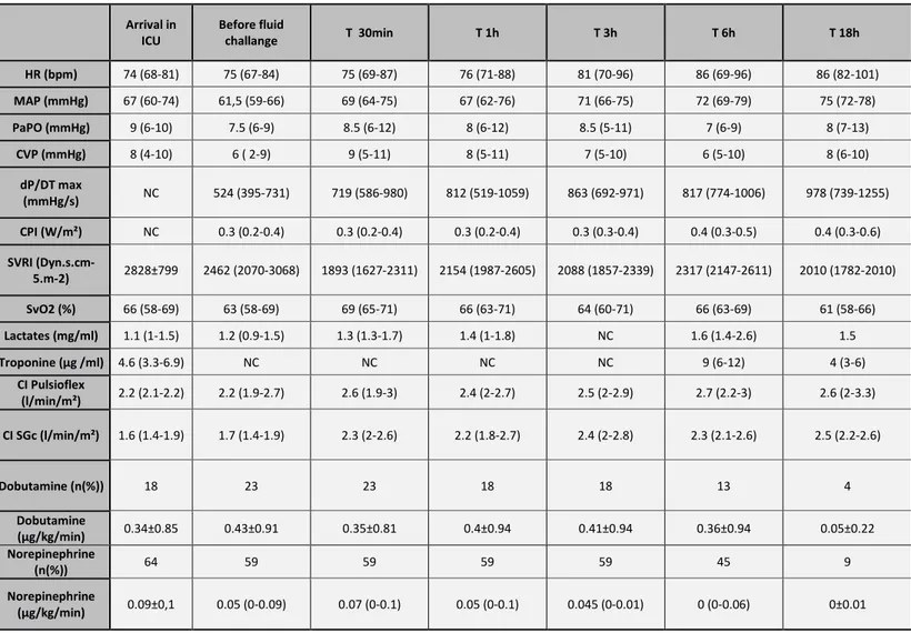
Abbreviations: HR: Heart Rate, MAP: Mean Arterial Pressure; PaOP: pulmonary artery occlusion pressure, CVP central venous pressure, dP/DT max: maximum value of first derivative of left ventricular pressure over time during isovolumic contraction max, CPI: Cardiac Power Index, SVRI: systemic vascular resistance index, , SvO2: mixed venous oxygen saturation CI: Cardiac Index
. Arrival in ICU Before fluid challange T 30min T 1h T 3h T 6h T 18h HR (bpm) 74 (68-81) 75 (67-84) 75 (69-87) 76 (71-88) 81 (70-96) 86 (69-96) 86 (82-101) MAP (mmHg) 67 (60-74) 61,5 (59-66) 69 (64-75) 67 (62-76) 71 (66-75) 72 (69-79) 75 (72-78) PaPO (mmHg) 9 (6-10) 7.5 (6-9) 8.5 (6-12) 8 (6-12) 8.5 (5-11) 7 (6-9) 8 (7-13) CVP (mmHg) 8 (4-10) 6 ( 2-9) 9 (5-11) 8 (5-11) 7 (5-10) 6 (5-10) 8 (6-10) dP/DT max (mmHg/s) NC 524 (395-731) 719 (586-980) 812 (519-1059) 863 (692-971) 817 (774-1006) 978 (739-1255) CPI (W/m²) NC 0.3 (0.2-0.4) 0.3 (0.2-0.4) 0.3 (0.2-0.4) 0.3 (0.3-0.4) 0.4 (0.3-0.5) 0.4 (0.3-0.6) SVRI (Dyn.s.cm-5.m-2) 2828±799 2462 (2070-3068) 1893 (1627-2311) 2154 (1987-2605) 2088 (1857-2339) 2317 (2147-2611) 2010 (1782-2010) SvO2 (%) 66 (58-69) 63 (58-69) 69 (65-71) 66 (63-71) 64 (60-71) 66 (63-69) 61 (58-66) Lactates (mg/ml) 1.1 (1-1.5) 1.2 (0.9-1.5) 1.3 (1.3-1.7) 1.4 (1-1.8) NC 1.6 (1.4-2.6) 1.5 Troponine (µg /ml) 4.6 (3.3-6.9) NC NC NC NC 9 (6-12) 4 (3-6) CI Pulsioflex (l/min/m²) 2.2 (2.1-2.2) 2.2 (1.9-2.7) 2.6 (1.9-3) 2.4 (2-2.7) 2.5 (2-2.9) 2.7 (2.2-3) 2.6 (2-3.3) CI SGc (l/min/m²) 1.6 (1.4-1.9) 1.7 (1.4-1.9) 2.3 (2-2.6) 2.2 (1.8-2.7) 2.4 (2-2.8) 2.3 (2.1-2.6) 2.5 (2.2-2.6) Dobutamine (n(%)) 18 23 23 18 18 13 4 Dobutamine (µg/kg/min) 0.34±0.85 0.43±0.91 0.35±0.81 0.4±0.94 0.41±0.94 0.36±0.94 0.05±0.22 Norepinephrine (n(%)) 64 59 59 59 59 45 9 Norepinephrine (µg/kg/min) 0.09±0,1 0.05 (0-0.09) 0.07 (0-0.1) 0.05 (0-0.1) 0.045 (0-0.01) 0 (0-0.06) 0±0.01
39
Table 3: Fluid challenge at 15 minutes
Values are number
Table 4: Fluid challenge at 30 minutes
40
REFERENCES
6.1 Références de l’article
1. Tavernier B, Fischer MO, Lorne E, Fellahi JL. Monitorage du débit cardiaque en anesthésie : quelles techniques? Quelles limites? :17.
2. Shoemaker WC, Patil R, Appel PL, Kram HB. Hemodynamic and oxygen transport patterns for outcome prediction, therapeutic goals, and clinical algorithms to improve outcome. Feasibility of artificial intelligence to customize algorithms. Chest. nov 1992;102(5 Suppl 2):617S-625S.
3. Aya HD, Cecconi M, Hamilton M, Rhodes A. Goal-directed therapy in cardiac surgery: a systematic review and meta-analysis. Br J Anaesth. avr 2013;110(4):510‑7.
4. Hamilton WF, Moore JW, Kinsman JM, Spurling RG. Simultaneous determination of the pulmonary and systemic circulation times in man and of a figure related to the cardiac output. Am J Physiol-Leg Content. 1 mars 1928;84(2):338‑44.
5. Sakka SG, Reinhard K, Wegscheider K, Meier-Hellmann A. Is the placement of a pulmonary artery catheter still justified solely for the measurement of cardiac output? J Cardiothorac Vasc Anesth. 1 avr 2000;14(2):119‑24.
6. Thrush D, Downs JB, Smith RA. Continuous thermodilution cardiac output: agreement with Fick and bolus thermodilution methods. J Cardiothorac Vasc Anesth. août 1995;9(4):399‑404.
7. Mihaljevic T, von Segesser LK, Tönz M, Leskosek B, Seifert B, Jenni R, et al. Continuous versus bolus thermodilution cardiac output measurements--a comparative study. Crit Care Med. mai
1995;23(5):944‑9.
8. Kaukinen S, Kööbi T, Bi Y, Turjanmaa VM h. Cardiac output measurement after coronary artery bypass grafting using bolus thermodilution, continuous thermodilution, and whole-body impedance cardiography. J Cardiothorac Vasc Anesth. avr 2003;17(2):199‑203.
9. Swan HJ, Ganz W, Forrester J, Marcus H, Diamond G, Chonette D. Catheterization of the heart in man with use of a flow-directed balloon-tipped catheter. N Engl J Med. 27 août
1970;283(9):447‑51.
10. Evans DC, Doraiswamy VA, Prosciak MP, Silviera M, Seamon MJ, Rodriguez Funes V, et al.
Complications associated with pulmonary artery catheters: a comprehensive clinical review. Scand J Surg SJS Off Organ Finn Surg Soc Scand Surg Soc. 2009;98(4):199‑208.
11. Ferretti GR, Thony F, Link KM, Durand M, Wollschläger K, Blin D, et al. False aneurysm of the pulmonary artery induced by a Swan-Ganz catheter: clinical presentation and radiologic management. AJR Am J Roentgenol. oct 1996;167(4):941‑5.
12. Connors AF, Speroff T, Dawson NV, Thomas C, Harrell FE, Wagner D, et al. The effectiveness of right heart catheterization in the initial care of critically ill patients. SUPPORT Investigators. JAMA. 18 sept 1996;276(11):889‑97.
13. Schwann NM, Hillel Z, Hoeft A, Barash P, Möhnle P, Miao Y, et al. Lack of effectiveness of the pulmonary artery catheter in cardiac surgery. Anesth Analg. nov 2011;113(5):994‑1002.
41 14. Broch O, Carbonell J, Ferrando C, Metzner M, Carstens A, Albrecht M, et al. Accuracy of an
autocalibrated pulse contour analysis in cardiac surgery patients: a bi-center clinical trial. BMC Anesthesiol. 26 nov 2015;15:171.
15. Monnet X, Vaquer S, Anguel N, Jozwiak M, Cipriani F, Richard C, et al. Comparison of pulse contour analysis by Pulsioflex and Vigileo to measure and track changes of cardiac output in critically ill patients. Br J Anaesth. févr 2015;114(2):235‑43.
16. Romano SM, Pistolesi M. Assessment of cardiac output from systemic arterial pressure in humans. Crit Care Med. août 2002;30(8):1834‑41.
17. Bland JM, Altman DG. Statistical methods for assessing agreement between two methods of clinical measurement. Lancet Lond Engl. 8 févr 1986;1(8476):307‑10.
18. Bland JM, Altman DG. Agreement between methods of measurement with multiple observations per individual. J Biopharm Stat. 2007;17(4):571‑82.
19. Critchley LA, Critchley JA. A meta-analysis of studies using bias and precision statistics to compare cardiac output measurement techniques. J Clin Monit Comput. févr 1999;15(2):85‑91.
20. Gisev N, Bell JS, Chen TF. Interrater agreement and interrater reliability: key concepts, approaches, and applications. Res Soc Adm Pharm RSAP. juin 2013;9(3):330‑8.
21. Peyton PJ, Chong SW. Minimally invasive measurement of cardiac output during surgery and critical care: a meta-analysis of accuracy and precision. Anesthesiology. nov 2010;113(5):1220‑35. 22. Rödig G, Keyl C, Liebold A, Hobbhahn J. Intra-operative evaluation of a continuous versus
intermittent bolus thermodilution technique of cardiac output measurement in cardiac surgical patients. Eur J Anaesthesiol EJA. mars 1998;15(2):196.
23. Smetkin AA, Hussain A, Kuzkov VV, Bjertnæs LJ, Kirov MY. Validation of cardiac output monitoring based on uncalibrated pulse contour analysis vs transpulmonary thermodilution during off-pump coronary artery bypass grafting. Br J Anaesth. juin 2014;112(6):1024‑31.
24. van Drumpt A, van Bommel J, Hoeks S, Grüne F, Wolvetang T, Bekkers J, et al. The value of arterial pressure waveform cardiac output measurements in the radial and femoral artery in major cardiac surgery patients. BMC Anesthesiol. 14 2017;17(1):42.
25. Monnet X, Vaquer S, Anguel N, Jozwiak M, Cipriani F, Richard C, et al. Comparison of pulse contour analysis by Pulsioflex and Vigileo to measure and track changes of cardiac output in critically ill patients. Br J Anaesth. févr 2015;114(2):235‑43.
26. Vasdev S, Chauhan S, Choudhury M, Hote MP, Malik M, Kiran U. Arterial pressure waveform derived cardiac output FloTrac/Vigileo system (third generation software): comparison of two monitoring sites with the thermodilution cardiac output. J Clin Monit Comput. avr
2012;26(2):115‑20.
27. Tsai Y-F, Su B-C, Lin C-C, Liu F-C, Lee W-C, Yu H-P. Cardiac output derived from arterial pressure waveform analysis: validation of the third-generation software in patients undergoing orthotopic liver transplantation. Transplant Proc. mars 2012;44(2):433‑7.
28. Biancofiore G, Critchley LAH, Lee A, Yang X, Bindi LM, Esposito M, et al. Evaluation of a new software version of the FloTrac/Vigileo (version 3.02) and a comparison with previous data in cirrhotic patients undergoing liver transplant surgery. Anesth Analg. sept 2011;113(3):515‑22.
42 29. Schlöglhofer T, Gilly H, Schima H. Semi-invasive measurement of cardiac output based on pulse
contour: a review and analysis. Can J Anaesth J Can Anesth. mai 2014;61(5):452‑79.
30. Giomarelli P, Biagioli B, Scolletta S. Cardiac output monitoring by pressure recording analytical method in cardiac surgery. Eur J Cardio-Thorac Surg Off J Eur Assoc Cardio-Thorac Surg. sept 2004;26(3):515‑20.
31. Romano SM, Scolletta S, Olivotto I, Biagioli B, Gensini GF, Chiostri M, et al. Systemic arterial
waveform analysis and assessment of blood flow during extracorporeal circulation. Perfusion. mars 2006;21(2):109‑16.
32. Zangrillo A, Maj G, Monaco F, Scandroglio AM, Nuzzi M, Plumari V, et al. Cardiac index validation using the pressure recording analytic method in unstable patients. J Cardiothorac Vasc Anesth. avr 2010;24(2):265‑9.
33. Paarmann H, Groesdonk HV, Sedemund-Adib B, Hanke T, Heinze H, Heringlake M, et al. Lack of agreement between pulmonary arterial thermodilution cardiac output and the pressure recording analytical method in postoperative cardiac surgery patients‡. Br J Anaesth. 1 avr
2011;106(4):475‑81.
34. Maj G, Monaco F, Landoni G, Barile L, Nicolotti D, Pieri M, et al. Cardiac index assessment by the pressure recording analytic method in unstable patients with atrial fibrillation. J Cardiothorac Vasc Anesth. juin 2011;25(3):476‑80.
35. Monnet X, Anguel N, Naudin B, Jabot J, Richard C, Teboul J-L. Arterial pressure-based cardiac output in septic patients: different accuracy of pulse contour and uncalibrated pressure waveform devices. Crit Care Lond Engl. 2010;14(3):R109.
36. Meng L, Tran NP, Alexander BS, Laning K, Chen G, Kain ZN, et al. The impact of phenylephrine, ephedrine, and increased preload on third-generation Vigileo-FloTrac and esophageal doppler cardiac output measurements. Anesth Analg. oct 2011;113(4):751‑7.
37. Monnet X, Anguel N, Jozwiak M, Richard C, Teboul J-L. Third-generation FloTrac/Vigileo does not reliably track changes in cardiac output induced by norepinephrine in critically ill patients. Br J Anaesth. avr 2012;108(4):615‑22.
38. Yang XX, Critchley LA, Rowlands DK, Fang Z, Huang L. Systematic error of cardiac output measured by bolus thermodilution with a pulmonary artery catheter compared with that measured by an aortic flow probe in a pig model. J Cardiothorac Vasc Anesth. déc 2013;27(6):1133‑9.
39. Suehiro K, Tanaka K, Funao T, Matsuura T, Mori T, Nishikawa K. Systemic vascular resistance has an impact on the reliability of the Vigileo-FloTrac system in measuring cardiac output and tracking cardiac output changes. Br J Anaesth. août 2013;111(2):170‑7.
40. Haller M, Zöllner C, Briegel J, Forst H. Evaluation of a new continuous thermodilution cardiac output monitor in critically ill patients: a prospective criterion standard study. Crit Care Med. mai 1995;23(5):860‑6.
41. Thiele RH, Durieux ME. Arterial waveform analysis for the anesthesiologist: past, present, and future concepts. Anesth Analg. oct 2011;113(4):766‑76.
42. Little WC. The left ventricular dP/dtmax-end-diastolic volume relation in closed-chest dogs. Circ Res. juin 1985;56(6):808‑15.
43 43. Ishikawa K, Chemaly ER, Tilemann L, Fish K, Ladage D, Aguero J, et al. Assessing left ventricular
systolic dysfunction after myocardial infarction: are ejection fraction and dP/dt(max)
complementary or redundant? Am J Physiol Heart Circ Physiol. 1 avr 2012;302(7):H1423-1428. 44. Furnival CM, Linden RJ, Snow HM. Inotropic changes in the left ventricle: the effect of changes in
heart rate, aortic pressure and end-diastolic pressure. J Physiol. déc 1970;211(2):359‑87. 45. Mihm FG, Gettinger A, Hanson CW, Gilbert HC, Stover EP, Vender JS, et al. A multicenter
evaluation of a new continuous cardiac output pulmonary artery catheter system. Crit Care Med. août 1998;26(8):1346‑50.
6.2 Références de l’introduction
1. Al-Ghamdi AA. Intraoperative fluid management: Past and future, where is the evidence? Saudi J Anaesth. 2018;12(2):311‑7.
2. Ganz W, Donoso R, Marcus HS, Forrester JS, Swan HJ. A new technique for measurement of cardiac output by thermodilution in man. Am J Cardiol. avr 1971;27(4):392‑6.
3. Swan HJ, Ganz W, Forrester J, Marcus H, Diamond G, Chonette D. Catheterization of the heart in man with use of a flow-directed balloon-tipped catheter. N Engl J Med. 27 août
1970;283(9):447‑51.
4. Evans DC, Doraiswamy VA, Prosciak MP, Silviera M, Seamon MJ, Rodriguez Funes V, et al.
Complications associated with pulmonary artery catheters: a comprehensive clinical review. Scand J Surg SJS Off Organ Finn Surg Soc Scand Surg Soc. 2009;98(4):199‑208.
5. Cecconi M, Grounds M, Rhodes A. Methodologies for assessing agreement between two methods of clinical measurement: are we as good as we think we are? Curr Opin Crit Care. juin
2007;13(3):294‑6.
6. Sakka SG, Reinhard K, Wegscheider K, Meier-Hellmann A. Is the placement of a pulmonary artery catheter still justified solely for the measurement of cardiac output? J Cardiothorac Vasc Anesth. 1 avr 2000;14(2):119‑24.
7. Hamilton WF, Moore JW, Kinsman JM, Spurling RG. Simultaneous determination of the pulmonary and systemic circulation times in man and of a figure related to the cardiac output. Am J Physiol-Leg Content. 1 mars 1928;84(2):338‑44.
8. Chatti R, Cholley B. Les nouvelles techniques de monitorage du débit cardiaque: gadgets ou avancées réelles? Réanimation. avr 2007;16(2):156‑62.
9. Ouattara A, Biais M. Quel monitorage hémodynamique au bloc opératoire? :28.
10. Thiele RH, Durieux ME. Arterial waveform analysis for the anesthesiologist: past, present, and future concepts. Anesth Analg. oct 2011;113(4):766‑76.
11. Wesseling KH, Jansen JR, Settels JJ, Schreuder JJ. Computation of aortic flow from pressure in humans using a nonlinear, three-element model. J Appl Physiol Bethesda Md 1985. mai 1993;74(5):2566‑73.
44 12. Pinsky MR. Advances in Hemodynamic Monitoring, An Issue of Critical Care Clinics. Elsevier Health
Sciences; 2014. 201 p.
13. Berkenstadt H, Margalit N, Hadani M, Friedman Z, Segal E, Villa Y, et al. Stroke volume variation as a predictor of fluid responsiveness in patients undergoing brain surgery. Anesth Analg. avr
2001;92(4):984‑9.
14. Lamia B, Chemla D. Interprétation de la courbe de pression artérielle au cours des états de choc. Réanimation. avr 2006;15(2):96‑102.
15. Goedje O, Hoeke K, Lichtwarck-Aschoff M, Faltchauser A, Lamm P, Reichart B. Continuous cardiac output by femoral arterial thermodilution calibrated pulse contour analysis: comparison with pulmonary arterial thermodilution. Crit Care Med. nov 1999;27(11):2407‑12.
16. Michard F, Boussat S, Chemla D, Anguel N, Mercat A, Lecarpentier Y, et al. Relation between respiratory changes in arterial pulse pressure and fluid responsiveness in septic patients with acute circulatory failure. Am J Respir Crit Care Med. juill 2000;162(1):134‑8.
17. Little WC. The left ventricular dP/dtmax-end-diastolic volume relation in closed-chest dogs. Circ Res. juin 1985;56(6):808‑15.
18. Fincke R, Hochman JS, Lowe AM, Menon V, Slater JN, Webb JG, et al. Cardiac power is the
strongest hemodynamic correlate of mortality in cardiogenic shock: a report from the SHOCK trial registry. J Am Coll Cardiol. 21 juill 2004;44(2):340‑8.