Université de Montréal
Cardiac cell fate control
by the imidazoline I
1receptor/nischarin:
Application in cardiac pathology
par
Henry Adolfo Aceros Muñoz
Département de Pharmacologie Faculté de Médicine
Thèse présentée à la Faculté de Médicine en vue de l’obtention du grade de doctorat
en Pharmacologie
option Pharmacologie Intégrative Cardiovasculaire
Août, 2014
Université de Montréal
Faculté des études supérieures et postdoctorales
Cette thèse intitulée :
Cardiac cell fate control
by the imidazoline I
1receptor/nischarin:
Application in cardiac pathology
Présenté par :
Henry Adolfo Aceros Muñoz
A été évaluée par un jury composé des personnes suivantes :
Dr Éric Thorin, Président-rapporteur
Dre Suhayla Mukaddam-Daher, Directeur de Recherche Dr Nicolas Noiseux, co-directeur de recherche
Dr Ashok Srivastava, membre du jury Dr Ghassan Bkaily, examinateur externe
Résumé
La moxonidine, un médicament antihypertenseur sympatholytique de type imidazolinique, agit au niveau de la médulla du tronc cérébral pour diminuer la pression artérielle, suite à l’activation sélective du récepteur aux imidazolines I1 (récepteur I1, aussi nommé nischarine). Traitement avec de la moxonidine prévient le développement de l’hypertrophie du ventricule gauche chez des rats hypertendus (SHR), associé à une diminution de la synthèse et une élévation transitoire de la fragmentation d’ADN, des effets antiprolifératifs et apoptotiques. Ces effets se présentent probablement chez les fibroblastes, car l’apoptose des cardiomyocytes pourrait détériorer la fonction cardiaque. Ces effets apparaissent aussi avec des doses non hypotensives de moxonidine, suggérant l’existence d’effets cardiaques directes. Le récepteur I1 se trouvé aussi dans les tissus cardiaques; son activation ex vivo par la moxonidine stimule la libération de l’ANP, ce qui montre que les récepteurs I1 cardiaques sont fonctionnels malgré l’absence de stimulation centrale.
Sur la base de ces informations, en plus du i) rôle des peptides natriurétiques comme inhibiteurs de l’apoptose cardiaque et ii) des études qui lient le récepteur I1 avec la maintenance de la matrix extracellulaire, on propose que, à part les effets sympatholytiques centrales, les récepteurs I1 cardiaques peuvent contrôler la croissance-mort cellulaire.
L’activation du récepteur I1 peut retarder la progression des cardiopathies vers la
défaillance cardiaque, en inhibant des signaux mal adaptatifs de prolifération et apoptose.
Des études ont été effectuées pour :
1. Explorer les effets in vivo sur la structure et la fonction cardiaque suite au traitement avec moxonidine chez le SHR et le hamster cardiomyopathique.
2. Définir les voies de signalisation impliquées dans les changements secondaires au traitement avec moxonidine, spécifiquement sur les marqueurs inflammatoires et les voies de signalisation régulant la croissance et la survie cellulaire (MAPK et Akt). 3. Explorer les effets in vitro de la surexpression et l’activation du récepteur I1 sur la
4. Rechercher la localisation, régulation et implication dans la croissance-mort cellulaire du récepteur I1 in vitro (cardiomyocytes et fibroblastes), en réponse aux stimuli associés au remodelage cardiaque : norépinephrine, cytokines (IL-1β, TNF-α) et oxydants (H2O2).
Nos études démontrent que la moxonidine, en doses hypotensives et non-hypotensives, améliore la structure et la performance cardiaque chez le SHR par des mécanismes impliquant l’inhibition des cytokines et des voies de signalisation p38 MAPK et Akt. Chez le hamster cardiomyopathique, la moxonidine améliore la fonction cardiaque, module la réponse inflammatoire/anti-inflammatoire et atténue la mort cellulaire et la fibrose cardiaque. Les cellules HEK293 surexprimant la nischarine survivent et prolifèrent plus en réponse à la moxonidine; cet effet est associé à l’inhibition des voies ERK, JNK et p38 MAPK. La surexpression de la nischarine protège aussi de la mort cellulaire induite par le TNF-α, l’IL-1β et le H2O2.
En outre, le récepteur I1 s’exprime dans les cardiomyocytes et fibroblastes, son activation inhibe la mort des cardiomyocytes et la prolifération des fibroblastes induite par la norépinephrine, par des effets différentiels sur les MAPK et l’Akt.
Dans des conditions inflammatoires, la moxonidine/récepteur aux imidazolines I1 protège les cardiomyocytes et facilite l’élimination des myofibroblastes par des effets contraires sur JNK, p38 MAPK et iNOS.
Ces études démontrent le potentiel du récepteur I1/nischarine comme cible anti-hypertrophique et anti-fibrose à niveau cardiaque. L’identification des mécanismes cardioprotecteurs de la nischarine peut amener au développement des traitements basés sur la surexpression de la nischarine chez des patients avec hypertrophie ventriculaire. Finalement, même si l’effet antihypertenseur des agonistes du récepteur I1 centraux est salutaire, le développement de nouveaux agonistes cardiosélectifs du récepteur I1 pourrait donner des bénéfices additionnels chez des patients non hypertendus.
Mots-clés : Récepteur aux imidazolines I1, moxonidine, hypertension, hypertrophie du ventricule gauche, défaillance cardiaque, cardiomyocyte, fibroblaste, HEK293, norépinephrine, cytokines.
Abstract
Moxonidine, an antihypertensive sympatholytic imidazoline compound, reduces blood pressure by selective activation of non-adrenergic imidazoline I1-receptors (also known as nischarin) in brainstem medulla. Moxonidine prevents left ventricular hypertrophy development in hypertensive rats, associated with reduced cardiac DNA synthesis and early transient increase in DNA fragmentation. It is likely that the anti-proliferative and apoptotic effects occur in fibroblasts, as cardiomyocyte apoptosis may deteriorate cardiac function. The effects also occurred to sub-hypotensive doses, suggesting a blood-pressure-independent mechanism and pointing to a local cardiac action. Imidazoline I1-receptors have been identified in cardiac tissues, and their ex vivo activation by moxonidine stimulates ANP release, demonstrating that cardiac imidazoline I1-receptors are functional without the contribution of the central nervous system.
Based on the above studies and on i) the role of natriuretic peptides in inhibition of myocardial cell apoptosis and ii) studies linking imidazoline I1-receptors to the maintenance of the extracellular matrix and PC12 cell survival, we propose that apart from centrally-mediated sympatholytic function, imidazoline I1-receptors in the heart may control cell growth and
death. Activation of imidazoline receptors may delay the progression of cardiac pathologies into heart failure by inhibition of maladaptive proliferative signalling and downstream apoptotic pathways.
In order to test this hypothesis studies were performed to:
1. Explore the in vivo effects of moxonidine on cardiac structure and function in SHR and cardiomyopathic hamsters.
2. Define the pathways involved in the observed changes following moxonidine treatment, specifically, on inflammatory markers and pathways involved in LVH and cardiac cell survival/death (MAPK and Akt).
3. Explore in vitro the effect of imidazoline I1-receptor activation by moxonidine, on cell survival by over-expressing nischarin in HEK293 cells, to circumvent the lack of specific imidazoline I1-receptor agonists and antagonists.
4. Investigate in vitro, imidazoline I1-receptor localization (cardiomyocytes and fibroblasts), regulation and implication in cell growth/death in response to cardiac remodelling-associated stimuli: norepinephrine, cytokines (IL-1 ,
TNF-), and oxidants (H2O2).
The studies reveal that hypotensive and sub-hypotensive concentrations of moxonidine improve cardiac structure and performance in SHR by mechanisms that involve inhibition of cytokines, p38MAPK, and Akt signalling pathways. In cardiomyopathic hamsters moxonidine improves cardiac performance, in association with differential inflammatory/anti-inflammatory responses that culminate in attenuated cardiomyocyte death and fibrosis and altered collagen type expression. HEK293 cells, transfected with nischarin cDNA, show increased viability/proliferation in response to moxonidine. The overall survival response is associated with moxonidine’s inhibition of ERK, JNK, and p38MAPK. Nischarin also opposes the reduced cell viability in response to oxidative stimuli (TNF-α, IL-1β and H2O2), with differential responses to moxonidine. Furthermore, the imidazoline I1-receptor is expressed in cardiac fibroblasts and myocytes and its activation inhibits norepinephrine-induced cardiomyocyte death and fibroblast proliferation, through differential effects on MAPKs and Akt. Moxonidine/imidazoline I1-receptor protects cardiomyocytes and facilitates elimination of myofibroblasts in inflammatory conditions, through opposite effects on JNK, p38MAPK and iNOS activity.
These studies emphasize the potential importance of imidazoline I1-receptor/nischarin as an anti-hypertrophic and anti-fibrotic target. Identification of the cardio-protective mechanisms of cardiac nischarin could result in specifically-tailored cell/gene-driven nischarin treatments, which could be important for patients with heart disease. Also, while the antihypertensive action of centrally acting compounds is appreciated, new cardiac-selective I1-receptor agonists may confer additional benefit.
Keywords: Imidazoline I1-receptor, moxonidine, hypertension, left ventricular hypertrophy, heart failure, cardiomyocyte, fibroblast, HEK293, norepinephrine, cytokines.
Table of contents
Chapter 1. Introduction ... 16
1.1 The burden of cardiovascular disease. Focus on hypertension and heart failure ... 16
1.2 The heart ... 18
1.2.1 The heart as a pump ... 19
1.2.2 The endocrine heart... 22
1.3 The continuum hypertension-LVH-heart failure ... 25
1.4 Factors involved in the development of LVH and heart failure ... 27
1.4.1 Mechanical Factors ... 27
1.4.2 Biochemical Factors... 33
1.5 Current pharmacological treatment options for hypertension and heart failure ... 49
1.6 Moxonidine ... 52
1.6.1 Moxonidine in hypertension ... 53
1.6.2 Moxonidine in heart failure ... 54
1.7 The imidazoline I1-receptor ... 55
1.7.1 Characterization of the imidazoline I1-receptor ... 55
1.7.2 Structure of imidazoline I1-receptor/nischarin ... 58
1.7.3 Signalling through the imidazoline I1-receptor/nischarin ... 59
Chapter 2. Hypothesis and aims... 64
Chapter 3. Results ... 66
3.1 Contributions of co-authors ... 66
3.2 Article 1. Moxonidine improves cardiac structure and performance in SHR through inhibition of cytokines, p38 MAPK, and Akt ... 68
3.3 Article 2. Functional and molecular effects of imidazoline receptor activation in heart failure ... 109
3.4 Article 3. Nischarin over-expression opposes cell-death induced by oxidative stress . 145 3.5 Article 4. Moxonidine modulates cytokine signalling and effects on cardiac cell viability ... 162
3.6 Supplementary data:... 208
4.1 Moxonidine improves cardiac function in hypertensive and heart failure models, in association with anti-inflammatory and anti-oxidative actions ... 211 4.2 In vitro imidazoline I1-receptor activation by moxonidine protects cardiomyocytes against inflammatory and oxidative stressors. Opposite effects occur in fibroblasts ... 220 4.3 Conclusion ... 225 4.4 Perspectives... 227
List of figures
Figure 1: Schema showing the central role of the sympathetic nervous system in the control of cardiac output (Silverthorn, 2013). ... 22 Figure 2: Mechanisms involved in integrin-mediated mechanotransduction in cardiomyocytes.
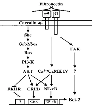
Of note the link with cytoskeletal proteins and signalling pathways inducing hypertrophy. For details see text (Ross, 2002). ... 29 Figure 3: Signalling pathways stimulated by α5β1 integrin leading to an increase in Bcl-2
expression. Grb2: Growth factor receptor-bound protein 2; Ras: Rat sarcoma protein; PI3-K: Phosphatidylinositol-3 kinase; CREB: cAMP response element-binding protein; FKHR: Forkhead in rhabdomyosarcoma protein; FAK: Focal adhesion kinase; CaMK IV: Calcium calmoduline dependant kinase IV (Lee & Ruoslahti, 2005). ... 32 Figure 4: Steps involved in catecholamine synthesis. Of note, different catecholamines are
produced in different sites depending on the local availability of enzymes (Becker, 2012). ... 35 Figure 5: Schema illustrating the interaction between the β-adrenergic receptor and the β1
integrin signalling in cardiomyocytes. β-AR: Beta adrenergic receptor; ECM: extracellular matrix; MMP-2: Matrix metalloproteinase 2; EUB: Extracellular ubiquitin; PI3-K: phosphatidylinositol-3 kinase; FAK: Focal adhesion kinase; GSK: glycogen synthase kinase (Amin et al., 2011). ... 37 Figure 6: Signalling pathways involved in adrenergic receptor-mediated regulation of
cardiomyocyte apoptosis. AR: adrenergic receptors; JNKs: c-Jun-N-terminal kinase; GSK-3β: glycogen synthase kinase-3β; ROS: reactive oxygen species; PKA: protein kinase A; AC: adenylyl cyclase; Gs: stimulatory G protein; Gi: inhibitory G protein; Gq: phospholipase C activating G protein; ATP: adenosine triphosphate; cAMP: cyclic adenosine monophosphate; CaMKIIδ: calcium calmodulin kinase IIδ; PI-3K: phosphatidylinositol 3-kinase. Modified from Amin et al. (2011) and Sing et al. (2001) (Amin et al., 2011; Singh et al., 2001). ... 38 Figure 7: Current view of the renin-angiotensin-aldosterone system. ACE, angiotensin
and 2; Mas, Mas receptor; MR, mineralocorticoid receptor. Reviewed by Fournier et al., 2012 (Fournier, Luft, Bader, Ganten, & Andrade-Navarro, 2012). ... 40 Figure 8: Opposing effects of TNFR1 and TNFR2 over cardiac contractility. Of note, the
predominant effect in most of the cases is the TNFR1-mediated negative inotropic effect. TNFR: TNF-α receptor; MSK1: Mitogen‐ and stress‐activated protein kinase‐1; cPLA2: Cytoplasmic phospholipase A2; PKCζ: Protein kinase C ζ; CaMKII: Calcium calmodulin dependant protein kinase II; T17-PLB: Phospholipase B phosphorylated at threonine 17; ROS: Reactive oxygen species (Defer, Azroyan, Pecker, & Pavoine, 2007). ... 44 Figure 9 : Protective effects of natriuretic peptides in cardiac pathology (reviewed by
Mukaddam-Daher, 2006) (Mukaddam-Daher, 2006). ... 48 Figure 10 : Structure of imidazoline agonists clonidine and moxonidine. ... 52 Figure 11 : Domain map of nischarin. For details see text (Sun et al., 2007). ... 58 Figure 12 : Imidazoline I1-receptor mediated production of PGE2 in PC12 cells (Ernsberger,
1998). ... 60 Figure 13 : Signalling through the imidazoline I1-receptor. For details see text (Edwards et al.,
2012). PC-PLC: Phosphatidylcholine-selective phospholipase C; AA: Arachidonic acid; DAG: diacylglycerol ... 62 Figure 14: Time-dependent ANP release to the culture medium (DMEM) from cultured
neonatal rat cardiomyocytes following incubation with the imidazoline I1-receptor agonist moxonidine (10-5 M), alone or in combination with the imidazoline I1-receptor antagonist efaroxan (10-5 M) or the α2-adrenergic receptor antagonist yohimbine (10-5 M). * P<0.01 vs. medium; $ P<0.01 vs. moxonidine. ... 208 Figure 15 : ANP release following perfusion of isolated rat hearts with the imidazoline I1-receptor agonist moxonidine (10-6 M) or control Krebs buffer. ... 209 Figure 16: Cardioprotective effects of in vivo moxonidine treatment in hypertensive SHR and
Bio14.6 hamsters: ... 218 Figure 17: Flow cytometry images showing the percentage of GFP positive cardiomyocytes or
fibroblasts after lentiviral infection (2.5x10⁴ infectious units (IFU) and 2.5x10⁵ IFU). 228 Figure 18: Three different clones of nischarin knockout embryonic stem cells, originated in
overnight to the blastocyst stage of embryonic development, and then transferred into pseudo-pregnant females that had been mated to vasectomised males. Pups were born 16-19 days after surgery and chimeras were identified by coat color. In the images the change in coat color (arrows) characteristic of chimeric animals is observed and contrasted with white-coated littermates. ... 230 Figure 19: PCR gel image of normal and heterozygous nischarin genomic DNA samples
List of abbreviations
ACE: Angiotensin converting enzyme ANP: Atrial natriuretic peptide
ARB: Angiotensin receptor blocker ATP: Adenosine triphosphate BNP: Brain natriuretic peptide
CaMKII: Calcium/calmodulin-dependent protein kinase II cAMP: Cyclic adenosine monophosphate
cGMP: Cyclic guanosine monophosphate CNP: C-type natriuretic peptide
CVD: Cardiovascular disease DAG: Diacylglycerol
ECM: Extracellular matrix EDV: End diastolic volume
eNOS: Endothelial nitric oxide synthase ERK: Extracellular signal-regulated kinases FAK: Focal adhesion kinase
GFP: Green fluorescent protein
GIRK: G-protein inwardly rectifying potassium channel GTP: Guanosine triphosphate
HEK293: Human embryonic kidney cells IFU: Infectious unit
ILK: Integrin linked kinase INF-γ: Interferon γ
iNOS: Inducible nitric oxide synthase IRAS: Imidazoline receptor antisera selected IRS: Insulin receptor substrate
JNK: c-Jun N-terminal kinase LKB1: Liver kinase B1
LVH: Left ventricular hypertrophy
MAPK: Mitogen activated protein kinases miRNA: Microribonucleic acid
mTOR: Mammalian target of rapamycin
NADPH: Nicotinamide adenine dinucleotide phosphate NE: Norepinephrine
NF-κB: Nuclear factor κB NO: Nitric oxide
NOS: Nitric oxide synthase NPR: Natriuretic peptide receptor PAK: p21-activated kinase
PC12: Rat pheochromocytoma cell line
PC-PLC: Phosphatidylcholine-selective phospholipase C PDGF: Platelet derived growth factor
PGE2: Prostaglandin E2
PKB: Protein kinase B PKC: Protein kinase C
RAAS: Renin-angiotensin-aldosterone system ROS: Reactive oxygen species
RVLM: Rostro-ventro lateral medulla S1P: Sphingosine 1 phosphate
SERCA: Sarco(endo)plasmic Ca2+-ATPase SHR: Spontaneously hypertensive rat SNS: Sympathetic nervous system TGF-β: Transforming growth factor-β TNF-α: Tumor necrosis factor α
VEGF: Vascular endothelial growth factor
A mis padres, Nayibe y Henry, quienes sin reserva me han apoyado en este camino. Gilles, Suzon, Maxime, ma famille au Canada, votre support inconditionnel a fait un peu plus facile d’être loin de mon pays. Dr. Gabriel Pascual (Q.E.P.D.) Gracias por contaminarme con su deseo de siempre saber más. Ese deseo ha orientado mi carrera en una dirección completamente inesperada.
Remerciements
Thank you Dr Suhayla Mukaddam-Daher for opening your laboratory to a complete stranger. Without your constant supervision, help, and support this work would not have been possible.
Thank you Dr Nicolas Noiseux, your words of advice, teaching, and example set a very high bar for the continuation of my career.
To the other members of the laboratory, Ahmed, Pierre-Alexandre, Georges and Mélanie, I learned a lot from each one of you, both from a technical and a human point of view.
Al departamento de ciencias fisiológicas de la Pontificia Universidad Javeriana, gracias por darme mi primera oportunidad en la academia, la cual se ha convertido en una opción de vida.
Chapter 1. Introduction
Cardiovascular pathologies, including hypertension, coronary artery disease, and heart failure, are the most common cause of death in the western world today (World Health Organization, 2014). According to the World Health Organization (WHO), more people die of cardiovascular diseases (CVD) yearly than any other cause or pathological condition, with approximately 17.3 million deaths due to CVDs in 2008, a number which is predicted to rise to 23.6 million by the year 2030 (Mathers & Loncar, 2006). In Canada, over 28.6% of total deaths per year are due to major cardiovascular diseases (Statistics Canada, 2012); in fact at every 7 minutes someone is suffering from acute myocardial infarct in Canada. The cost of cardiovascular diseases in Canada is of around $20.9 billion per year (Heart and Stroke Fundation of Canada, 2013), and this burden and cost will most likely increase with the aging of the population.
1.1 The burden of cardiovascular disease. Focus on hypertension
and heart failure
High blood pressure is the leading risk factor for cardiovascular disease mortality, causing more than 7 million deaths every year worldwide (World Health Organization, 2014). Hypertension is also a growing problem. In Canada alone, the prevalence of hypertension has grown from 12.5% in 1988/1989 to 19.6% for the year 2007/2008; in the same year, in the age group of 75 to 79 years, 69.5% had hypertension (Robitaille et al., 2012). This increasing trend most likely will accelerate in time, due to the ageing of the population (Employment and Social Development Canada, 2014). In spite of all the efficacious antihypertensive treatments, one third of Canadian adults with hypertension have uncontrolled blood pressure (Campbell, McAlister, & Quan, 2013; Danaei et al., 2011).
Uncontrolled hypertensive patients develop left ventricular hypertrophy (LVH) (Ferrara, Vaccaro, Cardoni, Mancini, & Zanchetti, 2004). Using echocardiography, a sensitive method for LVH detection, a survey in Italy revealed that around 30 to 50% of hypertensive patients had LVH (Cuspidi et al., 2011). In hypertension, the degree of hypertrophy is directly
correlated to the severity of blood pressure elevation, being present in all patients with severe hypertension (Belenkov, Vikhert, Belichenko, & Arabidze, 1992). LVH is an independent cardiovascular risk factor (Chobanian et al., 2003) and lowering it using antihypertensive treatments lowers both cardiovascular and total mortality (Devereux et al., 2004).
A second major complication of hypertension is heart failure (Chobanian et al., 2003). The Framingham study showed that 75% of heart failure patients detected in the cohort had hypertension (defined as a blood pressure above 160 mmHg systolic or 95 mmHg diastolic). Additionally, heart failure risk is 10 times higher in hypertensive patients with LVH, linking these two complications (Kannel, Castelli, McNamara, McKee, & Feinleib, 1972). Heart failure has a very high mortality rate, with roughly 50% of patients dying during the first 5 years of diagnosis, even with modern treatments (Levy et al., 2002; Roger et al., 2004). Noteworthy, effective treatments of hypertension, which reduce LVH, also reduce the incidence of heart failure (Larstorp et al., 2012), pointing towards the importance of treatment of LVH as a preventive measure for heart failure.
Hypertension treatment usually involves the use of 2 or more medications from different pharmacological groups. Antihypertensive medications include thiazide diuretics, calcium channel antagonists, blockers of the renin-angiotensin-aldosterone system and peripheral or centrally acting antagonists of the sympathetic nervous system (Chobanian et al., 2003; Hackam et al., 2013) (see section 1.5 Current pharmacological treatment options for hypertension and heart failure for details). Despite this plethora of options, around 15% of treated patients will not have blood pressure within normal limits following the use of 3 or more different medication classes, thus being classified as having resistant hypertension (Daugherty et al., 2012; Khan et al., 2014).
Better understanding of possible interactions of systems that control blood pressure and/or cardiac function may lead to innovative ways of treatment to prevent cardiac pathologies progression, avoiding the pathophysiological consequences of heart failure.
A common feature in cardiovascular diseases, including hypertension and heart failure, is over-activity of the sympathetic nervous system (SNS) (Meredith et al., 1993; Schlaich et al., 2003). The SNS is implicated in the regulation of blood pressure and cardiac function.
However SNS over-activity contributes to the pathogenesis and consequences of cardiovascular diseases, by activating signaling events intimately linked to vascular and cardiac cell growth and death (Dorn, 2002; Osadchii, 2007). Imidazoline I1-receptors in the brainstem are involved in the control of SNS activity, and consequently its deleterious effects on the heart, through mechanisms that inhibit the release of the neurotransmitter, norepinephrine (Bousquet, Feldman, & Schwartz, 1984; Raasch, Jungbluth, Schafer, Hauser, & Dominiak, 2003). Imidazoline I1-receptors are also found in the heart (El-Ayoubi, Gutkowska, Regunathan, & Mukaddam-Daher, 2002; El-Ayoubi, Menaouar, Gutkowska, & Mukaddam-Daher, 2003, 2004) and are involved in atrial natriuretic peptide (ANP) release (Mukaddam-Daher, Lambert, & Gutkowska, 1997; Mukaddam-Daher, Menaouar, & Gutkowska, 2006), yet their cellular localization and function are not fully elucidated. Investigation of heart imidazoline I1-receptor interaction with hypertension- and heart failure-associated control mechanisms may lead to new treatments of cardiovascular disease. We have enough evidence to propose that cardiac imidazoline I1-receptors are involved in cardiac cell growth and death, thus may be a new therapeutic target.
1.2 The heart
For the ancient Egyptians, the heart was one of the main organs of the body, being the center of emotions and intelligence as well as responsible for transport of blood (Ziskind & Halioua, 2004). This spiritual point of view has evolved towards the present view of the heart as a structure with multiple and tightly controlled functions on the cardiovascular system. The heart is involved in the maintenance of blood pressure and blood flow towards the different organs (Silverthorn, 2013; West, 1990). It acts as a pump that propels blood towards the vasculature, and as an endocrine organ that produces natriuretic hormones involved in blood pressure and volume control (de Bold, Borenstein, Veress, & Sonnenberg, 1981). Furthermore, both pump and endocrine activities are under homeostatic control of the sympathetic nervous system, as well as circulating and locally produced neurohormones and cytokines (Silverthorn, 2013; West, 1990). Abnormalities in the control mechanisms lead to
altered heart structure and function and consequently impair cardiac function (Cacciapuoti, 2011; Chaggar, Malkin, Shaw, Williams, & Channer, 2009).
1.2.1 The heart as a pump
The heart contracts and relaxes in a cyclical way (Iaizzo, 2009; West, 1990). During contraction, the phase known as systole, the heart contracts, pumping into the aorta a quantity of blood. This pumping function works against a resistance, generating pressure, an effect that maintains blood flow. Blood pressure is the product of flow (or cardiac output) and the resistance to flow, also known as arterial resistance (Fukuta & Little, 2008; Iaizzo, 2009). Cardiac output is an indicator of total blood flow, because all the blood that leaves the heart flows through the tissues. Since the needs of the body are variable, blood flow (cardiac output) adapts to accommodate different situations. Cardiac output, a common measure of cardiac function, directly varies with the ejected volume per contraction (stroke volume, SV) and by the number of cardiac cycles (or beats per minute, heart rate, HR) in a relationship shown in the formula: CO = SV x HR (reviewed by West, 1990) (West, 1990). This relationship that appears simple is extremely complex, as many factors are involved in the regulation of each parameter.
Heart rate is under the control of the autonomic nervous system, decreasing by active parasympathetic nervous system and increasing by active SNS and epinephrine (Bonow, Mann, Zipes, Libby, & Branwald, 2012). Increased heart rate may be maladaptive under pathological circumstances (Bonow et al., 2012; Brunton, Chabner, & Knollmann, 2011), and thus could be a target for treatment with sympatholytics.
On the other hand, stroke volume varies with the amount of blood that returns from the tissues to the heart. This amount of blood (venous return) is controlled by muscle pump, respiratory pump as well as by sympathetic innervation of veins, which constricts the veins, squeezing more blood out of them and into the heart (Levy, Koeppen, & Stanton, 2006; Silverthorn, 2013). With a larger ventricular volume at the beginning of the next contraction (end diastolic volume, EDV), the muscle fibers stretch, and then contract more forcefully, ejecting more
blood. The degree of myocardial stretch before contraction begins is called the preload on the heart, because this stretch represents the load placed on cardiac muscles before they contract. Changes in preload change contractility in the same sense, so, an increase of the preload will increase contractility and stroke volume, a phenomenon known as Frank-Starling mechanism (Bombardini, 2005; Shiels & White, 2008). The Frank-Starling mechanism fails in heart failure, when the heart is unable to respond to preload changes, thus elevating the pressure in the venous system, and increasing the symptoms of congestion (Kemp & Conte, 2012). This is the rationale for the use of medications that reduce preload, like diuretics and some venous dilators, in the current in treatment of heart failure (Hunt et al., 2009).
Stroke volume is also influenced by afterload, which is the combined load of EDV and arterial resistance created by blood filling the arterial system during ventricular contraction. For all practical purposes, unless there is an obstruction to blood flow, afterload is directly related to the systolic pressure (Fukuta & Little, 2008; Silverthorn, 2013) and clinically, arterial blood pressure is often used as an indirect indicator of afterload. As blood pressure is inversely related to the vessel radius, a small decrease in vessel radius will induce marked increase in blood pressure that increases afterload and might affect cardiac output (Levy et al., 2006; West, 1990).
Vessel radius (and thus afterload) is mainly controlled by the sympathetic nervous system (SNS) and the renin-angiotensin-aldosterone system (RAAS). Through corresponding release of neurohormones and hormones, these vasoconstrictive, anti-natriuretic, anti-diuretic and growth promoting systems influence vessel radius and tone and consequently cardiac function (Kemp & Conte, 2012; Silverthorn, 2013; West, 1990). In cardiovascular pathologies, such as hypertension or heart failure, afterload is usually elevated, increasing cardiac wall stress and work (Kemp & Conte, 2012; Lam et al., 2013). This is the rationale for treatment with medications that act through blocking the activities of the RAAS and the SNS (Hackam et al., 2013; Hunt et al., 2009).
Ventricular contraction occurs due to the entry from the extracellular space of small amounts of calcium, which in turn induce the release of large amounts of calcium from the endoplasmic reticulum of cardiomyocytes, permitting the cross-link between actin and myosin
modified by mechanical (preload) or endocrine/paracrine factors, including catecholamines, which increase the available calcium, thus increase contractility. Medications that modify sympathetic tone also modify contractility (Bombardini, 2005; Brunton et al., 2011; Shiels & White, 2008; Silverthorn, 2013).
The second component of cardiac function, relaxation (called lusitropism), depends on the active extrusion of calcium (including its rate). Once the cytoplasmic calcium concentration decreases, the cross-link between actin and myosin disappears and the tension decreases, starting the diastole (Bombardini, 2005; Levy et al., 2006; West, 1990). Relaxation also depends on the static properties of the myocardium, specifically the amount of collagen in the tissue. In pathological conditions, the lack of relaxation can disturb ventricular function by impeding the filling of the heart (Bonow et al., 2012; Diez et al., 2002).
From the above description, it is clear that the in vivo determinants of cardiac function act in concert to maintain cardiac output following a wide range of changes, and that the SNS plays an important role in the neurohormonal control of each of these determinants (Figure 1).
Figure 1: Schema showing the central role of the sympathetic nervous system in the control of cardiac output (Silverthorn, 2013).
1.2.2 The endocrine heart
The heart itself is in a privileged position to sense the hemodynamic conditions present in the circulatory system. This sensory system responds to volume changes by secreting diverse substances, including cardiac natriuretic hormones and other paracrine/autocrine factors like adrenomedullin and endothelin-1 (Ogawa & de Bold, 2014).
Adrenomedullin is mostly produced in the adrenal medulla, but it’s also detectable in the heart, both in atria and ventricles (Ichiki et al., 1994). Adrenomedullin can act locally to decrease angiotensin II-induced cardiomyocyte protein synthesis, a marker of hypertrophy, thus cardiac production of adrenomedullin may counter-regulate cardiac hypertrophy (Ogawa & de Bold, 2014; Tsuruda et al., 1998).
fibroblasts and endothelial cells (Sakurai et al., 1991; van Wamel, Ruwhof, van der Valk-Kokshoom, Schrier, & van der Laarse, 2001; Yanagisawa et al., 1988). Endothelin-1 induces cardiomyocyte hypertrophy and fibrosis (Drawnel, Archer, & Roderick, 2013), thus it is involved in the development of LVH. On the other hand, endothelin-1 induces cardiac secretion of natriuretic peptides (Horio, Kohno, & Takeda, 1993), a counter-regulatory protective effect.
The natriuretic peptide family, atrial natriuretic peptide (ANP), brain natriuretic peptide (BNP), and C-type natriuretic peptide (CNP), are structurally and functionally related peptide hormones that are involved in blood pressure regulation and body fluid homeostasis under normal and pathological conditions, such as heart failure and hypertension (Ogawa & de Bold, 2014). They are primarily of cardiac origin, but also produced by several extracardiac tissues, including the gastrointestinal tract and the brain (Gower et al., 1994; Teran, Rodriguez-Iturbe, Parra, & Gutkowska, 1991). In the heart, ANP is synthesized and stored in granules in cardiac atrial cardiomyocytes (de Bold et al., 1981). ANP release into the circulation is continuous, increasing in response to stimuli that result in atrial distension (Dietz, 2005; McGrath & de Bold, 2005; Thibault, Amiri, & Garcia, 1999). ANP secretion is stimulated by increasing venous return or increasing aortic pressure, as well as by vasoactive factors, neurotransmitters, and pro-inflammatory cytokines (Interleukin (IL)-1β, IL-6, tumor necrosis factor-α) (Ambler & Leite, 1994; Dietz, 2005; Mukaddam-Daher, 2006; Schiebinger & Greening, 1992). Nitric oxide, calcitonin gene-related peptide, and histamine inhibit ANP release (Dietz, 2005; Li et al., 2003; Piao, Cao, Han, Kim, & Kim, 2004).
BNP is co-stored with ANP in some atrial and ventricular granules (Nakamura et al., 1991); it is believed that BNP is secreted predominantly from cardiac ventricles (myocytes and fibroblasts), yet most of regulated BNP secretion, in response to increased ventricular stretch and endothelin, happens in the atria (Ogawa & de Bold, 2014; Ogawa, Vatta, Bruneau, & de Bold, 1999). BNP levels are increased in cardiovascular diseases, including ischemia, arrhythmias, fibrosis, cardiac hypertrophy, coronary endothelial dysfunction and heart failure (Mukaddam-Daher, 2006; Ogawa & de Bold, 2014). CNP is synthesized and secreted by the coronary endothelium and by cardiac myocytes and fibroblasts (Del Ry et al., 2011).
cytokines and lipopolysaccharide, and inhibited by insulin and VEGF. Myocardial production of CNP is increased in chronic heart failure. Plasma CNP, which is normally very low or undetectable, is significantly increased in patients with chronic heart failure, cirrhosis, and sepsis (reviewed by Mukaddam-Daher, 2006)(Mukaddam-Daher, 2006).
The natriuretic peptides elicit a number of vascular, renal and endocrine effects that help maintain blood pressure and extracellular fluid volume, and are involved in neuronal cardiac regulation, influencing contractility and beating rate (Antunes-Rodrigues, de Castro, Elias, Valenca, & McCann, 2004; Mukaddam-Daher, 2006). Circulating ANP and BNP can produce hypotensive effects, particularly in elderly patients (Hausdorff, Clark, Shannon, Elahi, & Wei, 1995), due to their diuretic, natriuretic, and vasodilatory properties (Koda, Sakamoto, & Ogawa, 2005; Melo, Steinhelper, Pang, Tse, & Ackermann, 2000). These effects are attained by direct inhibition of renal tubular sodium reabsorption, particularly in the distal tubule (Zhao, Pandey, & Navar, 2010), by inhibition and counteracting the effects of the sympathetic nervous system and the renin-angiotensin-aldosterone system, as shown in KO mouse models and in patients (Kasama et al., 2007; Melo et al., 2000), and by action on brain regulatory sites to suppress thirst, and inhibit vasopressin and ACTH release and sympathetic outflow (Antunes-Rodrigues et al., 2004). Natriuretic peptides inhibit the production of endothelin and secretion of renin and aldosterone. CNP is considered as a neuropeptide and an endothelium-derived autocrine/paracrine regulator exerting a hypotensive effect, positive inotropic action, but no significant diuretic or natriuretic actions; however, it inhibits aldosterone synthesis in a similar manner to ANP & BNP (reviewed by Mukaddam-Daher, 2006)(Mukaddam-Daher, 2006).
The important role of ANP in chronic blood pressure regulation has been shown in transgenic mouse model overexpressing ANP, where elevated plasma ANP was associated with a 25-30 mmHg blood pressure reduction, an opposite effect is observed in ANP KO, where blood pressure is around 30 mmHg higher when compared to wild type animals (Melo et al., 2000). Knockout mice with a homozygous disruption of the pro-ANP gene (-/-) are incapable of producing ANP and are hypertensive relative to their wild-type siblings, developing salt-sensitive hypertension after prolonged feeding of a high salt diet, in part due to a synergistic
plasma renin activity (Melo et al., 1999). The mechanism mediating the chronic relaxant effect of ANP is indirect, since the resistance vasculature is relatively insensitive to direct cGMP-mediated relaxation by ANP. Melo et al., have shown in ANP knockout mouse that the chronic hypotensive effect of ANP is mediated by attenuation of tonic cardiovascular sympathetic tone (Melo et al., 1999).
The actions of natriuretic peptides ANP and BNP are mediated by a common transmembrane cell surface guanylyl cyclase receptor (NPR-A), and CNP through NPR-B, which act through generation of cGMP and subsequent activation of cGMP-dependent protein kinase G (PKG) (Ogawa & de Bold, 2014). All 3 peptides bind to a more abundantly expressed receptor (NPR-C), which lacks the cytoplasmic guanylyl cyclase domain. NPR-C promotes the peptide clearance from the circulation and mediates inhibition of endothelin release and antagonism of the renin-angiotensin-aldosterone via Gi and inhibition of adenylyl cyclase/cyclic adenosine monophosphate (cAMP) (El Andalousi, Li, & Anand-Srivastava, 2013; Li, Sarkar, Brochu, & Anand-Srivastava, 2014). Natriuretic peptide receptors are ubiquitous, present in kidneys, vasculature, lungs, brain, thymus, macrophages etc., and recently demonstrated in the heart, where local actions of natriuretic peptides may occur (Lin, Hanze, Heese, Sodmann, & Lang, 1995). In the heart, the mRNAs for all three NPRs are found in fibroblasts with 80% of the NPR-C subtype. NPR-A is abundant in ventricular myocytes, whereas NPR-B is abundant in fibroblasts and endothelial cells (Lin et al., 1995).
Based on the previously described findings, it is clear that the heart protects itself through its endocrine functions, both systemically, by regulating blood pressure, and locally, by counter-regulating hypertrophic and fibrotic stimuli.
1.3 The continuum hypertension-LVH-heart failure
During exercise, the heart is required to pump facing increases in preload (for example in swimming), afterload (weightlifting), or both (rowing) (Ekblom & Hermansen, 1968; Pelliccia, Maron, Spataro, Proschan, & Spirito, 1991; Weeks & McMullen, 2011). The ventricle increases its force of contraction to maintain adequate stroke volume, increasing the
mechanism are activated, causing cardiovascular stress (reviewed by Weeks and McMullen, 2011) (Weeks & McMullen, 2011). However, normal, short term cardiovascular stresses, such as bursts of exercise, have beneficial effects on the cardiovascular system and are an integral part of preventive and treatment strategies in hypertension and heart failure (Chobanian et al., 2003; Hackam et al., 2013; Hunt et al., 2009). In contrast, chronic cardiovascular stress (such as chronically increased afterload as occurs in hypertension) cause myocardial cells to hypertrophy, resulting in increased thickness of the ventricular wall, leading to maladaptive changes such LVH (Weeks & McMullen, 2011). In hypertension, the degree of hypertrophy is directly correlated to the severity of blood pressure elevation, being present in all patients with severe hypertension (Belenkov et al., 1992).
The hypertrophic response of the heart is a dynamic process that involves progressive changes in gene expression that creates structural, hemodynamic, and cellular alterations (Carreno, Apablaza, Ocaranza, & Jalil, 2006; Weeks & McMullen, 2011). LVH involves cardiomyocyte hypertrophy, cardiac fibroblast proliferation, increased synthesis and deposition of collagen, and progression of interstitial and perivascular fibrosis (Cacciapuoti, 2011). Initially, these alterations contribute to lowering of the ventricular wall stress and preservation of contractile function; a stage known as adaptive or compensatory. Eventually, the remodeling process leads to ventricular stiffness, secondary to a decrease in the active extrusion of calcium after the contraction of the myocardium (i.e. reduced relaxation), and alterations of the static properties of the myocardium, secondary to the increased amount of collagen in the tissue. Cardiac stiffness can impede the filling of the heart, hence, results in reduced force of contraction (Frank-Starling mechanism) and subsequent reduction in ejection fraction, stroke volume, and cardiac output (Bonow et al., 2012; Kemp & Conte, 2012).
The kidneys respond to reduced cardiac output and subsequently reduced renal perfusion by altering their hemodynamic milieu through a number of physical and neurohumoral mechanisms, including activated renin-angiotensin-aldosterone system (RAAS) and vasopressin, as well as stimulated catecholamines and renal nerve activity. These vasoconstrictive and sodium and water retaining mechanisms lead to volume expansion and to further deterioration in cardiac performance (Bonow et al., 2012; Kemp & Conte, 2012). The result is a failing heart that cannot
which results in systemic and pulmonary edema, as well as progressive apoptosis and fibrosis (Kemp & Conte, 2012; Ward, Crossman, & Cannell, 2011). Exaggerated apoptosis, a mode of cell death in which the cell participates in its own demise, may account for the loss of contractile cardiomyocytes in the hypertensive left ventricle, which may further progress towards heart failure (Diez, Fortuno, & Ravassa, 1998; Ikeda, Hamada, & Hiwada, 1999; Li, Bing, Long, Robinson, & Lakatta, 1997).
In fact, LVH is linked to unfavorable prognoses and various deleterious sequelae of cardiovascular diseases, with conditions, such as coronary heart disease, stroke, congestive heart failure and sudden death, being aggravated by LVH (Devereux et al., 2004; Larstorp et al., 2012). Importantly, LVH can often be reversible. It is not surprising, therefore, that numerous therapeutic approaches have been pursued with the aim of regressing/preventing LVH and, hence, reducing both cardiovascular and total mortality (Devereux et al., 2004).
1.4 Factors involved in the development of LVH and heart failure
Similar to all cells in the body, cardiomyocytes and fibroblasts sense and respond to their mechanical and neurohormonal environment (Bernardo, Weeks, Pretorius, & McMullen, 2010; Haggart, Ames, Lee, & Holmes, 2014). The mechanical environment includes mechanical stretch as well as the composition and stiffness of the extracellular matrix (ECM), a network of cellular and extracellular constituents that comprises the left ventricular myocardium (Jane-Lise, Corda, Chassagne, & Rappaport, 2000). In vivo cardiac remodelling, including cytoskeleton reorganization, results from a combination of the effects of mechanical and biochemical factors in close interaction with extracellular matrix (ECM) proteins, integrins, and proteins secreted locally, such as metalloproteinases (Bernardo et al., 2010).
1.4.1 Mechanical Factors
Pressure overload in hypertension or volume overload, for example, in mitral valve insufficiency, result in mechanical stretch, which influences many basic cellular processes such as growth, remodeling, apoptosis, and gene expression (Liu et al., 1992; Sadoshima,
Jahn, Takahashi, Kulik, & Izumo, 1992; Wilson, Mai, Sudhir, Weiss, & Ives, 1993), as well as differentiation (Reusch, Wagdy, Reusch, Wilson, & Ives, 1996), rearranged cytoskeleton or focal contacts (Carver, Nagpal, Nachtigal, Borg, & Terracio, 1991; Davies, Robotewskyj, & Griem, 1994; Girard & Nerem, 1995; Moore et al., 1994), or altered composition of the extracellular matrix (Jane-Lise et al., 2000).
Mechanical stretch induces protein synthesis and hypertrophy, both in isolated cardiomyocytes and in papillary muscle preparations (Haggart et al., 2014; Lammerding, Kamm, & Lee, 2004; Wang, Wu, Cheng, & Shyu, 2013), induces secretion or synthesis of bioactive molecules from cardiomyocytes (Shyu, 2009), and increases production of extracellular matrix proteins by fibroblasts, the principal cell type responsible for extracellular matrix synthesis during growth and pathophysiological conditions (Jugdutt & Amy, 1986; Kuwahara et al., 2003; Weber, Brilla, & Campbell, 1992).
Mechanical stretch is detected by cardiac cells through multiple mechanisms, including mechanosensitive ion channels, which are stretch activated channels that permit the handling of sodium and calcium (Bonow et al., 2012; Lammerding et al., 2004) as well as by cell surface adhesion receptors, termed integrins (Lammerding et al., 2004).
Integrins form the primary link between extracellular matrix ligands and cytoskeletal structures, which is important for maintaining the architecture of tissues (Hynes, 1992; Lammerding et al., 2004; Ruoslahti, 1991). In myocardial tissue, integrins play a role in transmitting and distributing the mechanical force generated by the contraction of each myocyte to the extracellular matrix, and preventing myocytes from overstretching by elevated tension (Borg, Johnson, & Lill, 1983; Winegrad & Robinson, 1978).
Integrins are a family of transmembrane glycoproteins that function as mechanotransducers, converting mechanical forces to biochemical signals. The integrin large extracellular domain binds directly to ECM proteins, like collagen or fibronectin, among others (Evans & Calderwood, 2007; Lammerding et al., 2004), playing a crucial role in ECM organization. The integrin cytoplasmic tail does not have an intrinsic enzymatic activity (Lammerding et al., 2004), but binds to cytoskeletal proteins, such as F-actin, through the interaction of integrin tails with talin (Ross, 2002; Ross et al., 2013), as well as to signalling proteins, including
integrin linked kinase (ILK), focal adhesion kinase (FAK), the small guanosine triphosphate (GTP) binding proteins Rho and Rac1, with downstream activation of mitogen activated protein kinases (MAPK), p21-activated kinase (PAK), nuclear factor-κB (NF-κB) and Akt/protein kinase B (PKB), pathways that have been linked to hypertrophy, proliferation, and apoptosis in different cell types, including cardiomyocytes and fibroblasts (Figure 2) (Aikawa et al., 2002; Bettink et al., 2010; Krishnamurthy, Subramanian, Singh, & Singh, 2007; Lammerding et al., 2004; Ross, 2002).
Figure 2: Mechanisms involved in integrin-mediated mechanotransduction in cardiomyocytes. Of note the link with cytoskeletal proteins and signalling pathways inducing hypertrophy. For details see text (Ross, 2002).
Rac1 is a small guanosine triphosphatase catalytic subunit of nicotinamide adenine dinucleotide phosphate (NADPH) oxidase (Hordijk, 2006). Rac1 activation increases intracellular reactive oxygen species (ROS), such as superoxide (∙O2−) and H2O2, into which ∙O2 is rapidly converted. ROS are important mediators of cellular signal transduction cascades such as proliferation, migration, and apoptosis (Maack et al., 2003). Excess ROS production damages DNA, protein and lipids, causing cardiac cell death. Activation of Rac1 and its effector protein NADPH oxidase can also regulate proliferative signals through mechanisms that include NF-κB stimulation (Hordijk, 2006), mediated by increasing intracellular ROS or through phosphatidylinositol-3 kinase (PI3-K)/Akt. The Rac1–ROS signaling pathway is a
decreases NADPH oxidase-related production of ROS in cardiomyocytes and reduces cardiac hypertrophy (Maack et al., 2003), demonstrating the importance of Rac1 activity in cardiac pathologies. The direct involvement of Rac1 in cardiovascular disorders has been confirmed by both cardiac-specific transgenic and knockout mice. Mice overexpressing the constitutively active form of Rac1 clearly evidence a dramatic cardiomyopathy phenotype (Sussman et al., 2000). In contrast, in cardiac-specific Rac1 knockout mice, infusion of angiotensin II increases systolic blood pressure but does not induce cardiac hypertrophy in association with the inhibition of NADPH oxidase-dependent generation of O2- and NF-κB transcriptional activity (Satoh et al., 2006).
Mammalian integrins comprise 18α and 8β subunits (Ross & Borg, 2001). Cardiac myocytes predominantly express β1 integrin, the main integrin subtype that mediates adhesion to the extracellular matrix (Ross & Borg, 2001). β1 integrin heterodimers significantly contribute to cardiomyocyte adhesion to the ECM, and inhibition of β1 integrin-mediated adhesion leads to ventricular dilatation of the normal heart, and plays a significant role in the progression of adverse myocardial remodeling that leads to heart failure (Stewart, Gardner, Brower, & Janicki, 2014). In addition, β1 integrin signaling protects cardiomyocytes against β-adrenergic receptor-stimulated apoptosis in vitro and left ventricular remodeling and myocyte apoptosis in vivo (Krishnamurthy et al., 2007). In vivo infusion with β-adrenergic receptor agonist isoproterenol increases cardiomyocyte apoptosis in mice with partial knock-out of β1 integrin subunit, in association with reduced ventricular function and overactivation of c-Jun N-terminal kinase (JNK), when compared to wild type animals subjected to the same treatment (Krishnamurthy et al., 2007). Over-expression of β1 integrins, in neonatal rat ventricular myocytes, enhances the hypertrophic effects of α1-adrenergic stimulation, and inhibition of β1 integrin function and signaling reduces the hypertrophic response (Ross et al., 1998).
The α5 integrins, on the other hand, modulate cardiac neural crest proliferation and survival, and are required for cardiac morphogenesis and, in particular, for the formation of the cardiac outflow tract (Mittal, Pulina, Hou, & Astrof, 2010, 2013). The expression of α1, α3 and α5 integrins is low in normal cardiomyocytes, but is upregulated by myocardial infarction in rats (Nawata et al., 1999). Integrin α5 subunit is also over-expressed in LVH (Ross, 2002).
factors and cytokines (Nawata et al., 1999). Platelet derived growth factor (PDGF) upregulates the expression of the α5 integrin subunit in cardiomyocytes. Transforming growth factor-β (TGF-β) and inflammatory cytokines, such as interleukin 1β (IL-1β), tumor necrosis factor α (TNF-α) or interferon γ (IFN-γ) enhance the expression of both α1 and α5 integrin subunits (Gailit, Xu, Bueller, & Clark, 1996; Nawata et al., 1999). In fibroblasts, the expression of α5 integrin is relatively higher in cells obtained from exercised rat hearts and lower in those from hypertensive hearts when compared to control, and these cells migrate on fibronectin at higher and lower rates respectively, demonstrating a direct correlation between the expression of α5 integrin and fibroblast migration (Burgess, Terracio, Hirozane, & Borg, 2002).
The β1 integrin forms dimers with multiple α subunits. Each specific dimer serves as a receptor for a specific extracellular matrix protein. For example α1β1 and α2β1 integrin complexes can bind to collagen and laminin, α4β1 and α5β1 to fibronectin, while α3β1 binds collagen, laminin, and fibronectin (Ross & Borg, 2001).
The α5β1 integrin (fibronectin receptor) is present in cardiomyocytes, but predominantly in fibroblasts (Clark et al., 2005). It supports migration in several cell types (Alahari, Lee, & Juliano, 2000) and its mutation or knock-out results in reduction or abolition of migration (Huttenlocher, Sandborg, & Horwitz, 1995). Increased expression of α1β1 and α5β1 heterodimers are found in association with increased adhesion during the compensatory hypertrophy secondary to renovascular hypertension (Terracio et al., 1991).
Over-expression of α5β1 integrin protects cell lines against apoptotic stimuli, in part by modulating the expression of the anti-apoptotic protein Bcl2, by activating the PI3-K/Akt pathway (Figure 3) (Lee & Ruoslahti, 2005; Lee & Juliano, 2000).
Figure 3: Signalling pathways stimulated by α5β1 integrin leading to an increase in Bcl-2 expression. Grb2: Growth factor receptor-bound protein 2; Ras: Rat sarcoma protein; PI3-K: Phosphatidylinositol-3 kinase; CREB: cAMP response element-binding protein; FKHR: Forkhead in rhabdomyosarcoma protein; FAK: Focal adhesion kinase; CaMK IV: Calcium calmoduline dependant kinase IV (Lee & Ruoslahti, 2005).
Furthermore, modulation of the α5β1 receptor ligand, fibronectin, is implicated in cardiac remodeling. Loss of fibronectin leads to further deterioration of cardiac function in a mouse model of myocardial infarction, due to impaired wound healing and reduced progenitor cell recruitment (Konstandin, Toko, et al., 2013). In contrast, genetic conditional ablation of the fibronectin gene in adult mice blunts cardiomyocyte hypertrophy upon pressure overload induced by transverse aortic constriction, and delays development of heart failure and improves survival (Konstandin, Volkers, et al., 2013). Noteworthy, induction of fibronectin expression upon myocardial infarction is very quick, while in the pressure overload model, fibronectin continuously increases over time at a lower level of expression (Konstandin, Toko, et al., 2013; Konstandin, Volkers, et al., 2013). Accordingly, it appears that the duration and intensity might impact the ultimate outcome of fibronectin expression.
Taken together, these studies show that mechanical overload leads to hypertrophic remodeling, mediated by mechanotransducing integrins, including α5β1 fibronectin receptor
and activation of Rac1 and subsequent ROS production. Of interest to our studies, imidazoline I1-receptor/nischarin associates with the α5 subunit of the α5β1 integrin and reduces cell proliferation and migration through inhibition of Rac1 (Alahari et al., 2000). The association with integrins puts imidazoline I1-receptors/nischarin at the heart of ECM control and cardiac remodelling, a function that may go beyond its established central and peripheral sympatholytic effect.
1.4.2 Biochemical Factors
The hypertensive heart undergoes significant structural changes that occur in response to biochemical stress, induced by activated sympathetic nervous system (SNS), the renin-angiotensin-aldosterone system (RAAS), growth factors, and pro-inflammatory cytokines. In addition to their hemodynamic activity, these factors initiate and maintain left ventricular hypertrophy through a complex array of signaling events, with cross-talk and positive and/or negative interaction, and contribute to regression to a fetal program of gene expression (reviewed by Cacciapuoti, 2001; Sugden and Clerk, 1998)(Cacciapuoti, 2011; Sugden & Clerk, 1998). On the other hand, the heart produces natriuretic peptides (ANP and BNP), vasodilatory, diuretic and natriuretic hormones, and more importantly, with anti-hypertrophic, anti-proliferative, anti-inflammatory, and sympatholytic actions (reviewed by Ogawa and de Bold, 2014) (Ogawa & de Bold, 2014).
1.4.2.1 The sympathetic nervous system
The sympathetic nervous system (SNS) plays an important role in the regulation of blood pressure and cardiac function (Grassi, Seravalle, & Quarti-Trevano, 2010). It is involved in minute-to-minute control of blood pressure because it responds to pressure alterations within seconds (Silverthorn, 2013; West, 1990). SNS also influences long term pressure control through activation of the renin-angiotensin-aldosterone system (RAAS). Renal nerves stimulate renin release and positively enhance angiotensin II, which directly stimulates muscle sympathetic nerve activity and facilitates adrenergic sympathetic transmission (Brunton et al., 2011;
Sympathetic nervous activity is regulated in centers in the brainstem and transmitted to organs and blood vessels that are innervated by sympathetic nerve endings (Brunton et al., 2011). Overactivation of the sympathetic outflow to the heart, kidneys and skeletal muscle vasculature is commonly present in young patients with essential hypertension, where it leads to the development of cardiac risk factors, such as development of left ventricular hypertrophy, predisposing to ventricular arrhythmias, increasing insulin resistance and accelerating atherogenesis (Grassi et al., 2010). The level of sympathetic drive to the heart is a major determinant of prognosis in patients with hypertension and heart failure, and a decrease in heart rate and nerve activity by treatments that directly block or oppose the sympathetic nervous system has been shown to be beneficial for long-term prognosis in these patients (Frohlich, Gonzalez, & Diez, 2011).
The effects of the SNS are mediated by the neurotransmitter norepinephrine, a catecholamine synthesized in the neurons from tyrosine. The rate-limiting step for this synthesis is in the transformation of tyrosine to L-dopa, by the enzyme tyrosine hydroxylase (Figure 4) (Becker, 2012; Brunton et al., 2011). Norepinephrine is then stored at nerve endings until released by exocytosis upon stimulation. At the adrenal medulla, which is considered as a sympathetic ganglion, norepinephrine undergoes methylation producing epinephrine, which has similar actions to norepinephrine, but binds with similar affinity to all adrenergic receptor subtypes, while norepinephrine binds with higher affinity to β1- than β2- adrenergic receptors (Brunton et al., 2011).
Figure 4: Steps involved in catecholamine synthesis. Of note, different catecholamines are produced in different sites depending on the local availability of enzymes (Becker, 2012).
Norepinephrine signals via its interaction with α- and β-adrenergic receptors, a family of G protein-coupled receptors (Becker, 2012; Brunton et al., 2011). Norepinephrine controls its own release by acting on α2-adrenergic receptors present on the pre-synaptic membrane, inhibiting further release (Brunton et al., 2011; Kalant, Grant, & Mitchell, 2007). Norepinephrine release is also inhibited by activation of imidazoline I1-receptors, non-adrenergic receptors expressed in brainstem medulla and pre-synaptic membranes (Chan, Burke, Zhu, Piletz, & Head, 2005; Molderings & Gothert, 1998). The effects of norepinephrine are terminated through two mechanisms: reuptake by the norepinephrine transporter, and diffusion followed by degradation by extracellular enzymes (Brunton et al., 2011; Kalant et al., 2007).
The released norepinephrine causes vasoconstriction through binding to vascular α1-adrenergic receptors (Hosoda et al., 2005; Zacharia, Hillier, & MacDonald, 2004). In the kidney, stimulated SNS activity/norepinephrine constricts afferent arterioles and reduces glomerular filtration rate by decreasing blood flow into glomerular capillaries (DiBona, 2002).
On the heart, norepinephrine acutely increases heart rate and contractility by activating β1-adrenergic receptors, the main β1-adrenergic receptor subtype (80%) present in the heart, specifically in cardiomyocytes (Brunton et al., 2011; Singh, Xiao, Remondino, Sawyer, & Colucci, 2001). Short term (10 minutes) activation of the β1-adrenergic receptor increases cardiomyocyte contractility by inducing an increase in calcium transients, which are dependent on the activation of protein kinase A (PKA) (Wang et al., 2004). Longer exposure (24 h) to β1-adrenergic stimulation of isolated adult rat cardiomyocytes switches the increase in contractility and calcium transients from PKA dependency towards dependency on the activity of the calcium/calmodulin-dependent protein kinase II (CaMKII), an enzyme that has also been linked to adrenergic-induced apoptosis (Wang et al., 2004; Zhu et al., 2003).
In heart failure, the expression of β1-adrenergic receptor in cardiomyocytes is down-regulated and uncoupled, leading to a decrease in contractility (Bristow et al., 1986; Lamba & Abraham, 2000; Osadchii, 2007; Parati & Esler, 2012). The down regulation of β1-adrenergic receptors gives importance to the activity of β2- and α1-adrenergic receptors (Lamba & Abraham, 2000; Osadchii, 2007). During β1-adrenergic receptor down regulation, α1-adrenergic receptors sustain adrenergic function (Lamba & Abraham, 2000).
In vivo and in vitro studies demonstrate that prolonged exposure to norepinephrine causes ventricular hypertrophy, with cardiomyocyte hypertrophy and death, fibroblast proliferation, and an increase in collagen accumulation (Bhambi & Eghbali, 1991; Lamba & Abraham, 2000; Singh et al., 2001), increasing the risk of cardiac failure. In genetically engineered mice, deletion of α2A- and α2C-adrenergic receptors leads to cardiac hypertrophy and failure due to chronically enhanced catecholamine release (Hein, Altman, & Kobilka, 1999) and sympathectomy prevents cardiac fibrosis in hypertensive rats independent of blood pressure (Perlini et al., 2006). Also, mice that are unable to synthesize norepinephrine exhibit less cardiac hypertrophy and preserved ventricular function after aortic banding (Esposito et al., 2002). Overexpression of β1-adrenergic receptors in the heart produces a cardiomyopathic phenotype (Seeland et al., 2007). Prolonged in vitro exposure to norepinephrine or isoproterenol increase the number of apoptotic myocytes via stimulation of the β1-adrenergic receptor pathway (Communal, Singh, Pimentel, & Colucci, 1998; Iwai-Kanai et al., 1999), and
β1-adrenergic receptor selective antagonists completely prevent norepinephrine-stimulated apoptosis (Morisco, Zebrowski, Vatner, Vatner, & Sadoshima, 2001; Osadchii, 2007).
The β1-adrenergic receptor, coupled to Gs, exerts a pro-apoptotic action via a cAMP/PKA-dependent mechanism, which appears to involve mitochondria and ROS, and is associated with the activation of glycogen synthase kinase-3β /JNK and p38 MAPK. Pro-apoptotic action of β1-adrenergic receptor may also involve activation of calcium/calmodulin-dependent protein kinase II (CaMKII). (Lamba & Abraham, 2000; Singh et al., 2001; Zaugg & Schaub, 2004). Amin et al. (2011) have shown that chronic β-adrenergic stimulation causes cardiomyocyte apoptosis and myocardial remodeling, and that the involvement of β1 integrin has protective effects via the FAK and PI3-kinase/Akt pathways (Figure 5) (Amin, Singh, & Singh, 2011).
Figure 5: Schema illustrating the interaction between the β-adrenergic receptor and the β1 integrin signalling in cardiomyocytes. β-AR: Beta adrenergic receptor; ECM: extracellular matrix; MMP-2: Matrix metalloproteinase 2; EUB: Extracellular ubiquitin; PI3-K: phosphatidylinositol-3 kinase; FAK: Focal adhesion kinase; GSK: glycogen synthase kinase (Amin et al., 2011).
The β2-adrenergic receptor couples to Gs, Gi, or Gq depending on its functional status (Zaugg & Schaub, 2004). Similarly to β1-adrenergic receptor, β2-adrenergic receptor coupling to Gs exerts a pro-apoptotic action via cAMP pathways. Alternatively, β2-adrenergic coupling to Gi exerts an anti-apoptotic action which is mediated by PI3-kinase/Akt. Thus, in some cases, β2-adrenergic receptor activity in cardiomyocytes may oppose some of the deleterious effects of long-term β1-adrenergic receptor stimulation (Singh et al., 2001; Zaugg & Schaub, 2004). The α1-adrenergic receptor signals through Gq/G11-Phospholypase C pathway, leading to an increase in intracellular calcium and an increase in contractility (Lamba & Abraham, 2000; Shannon & Chaudhry, 2006; Woodcock, 2007; Zaugg & Schaub, 2004). Overexpression or activation of α1-adrenergic receptors also results in cardiomyocyte hypertrophy and reduced apoptosis, through extracellular signal-regulated kinases (ERK) activation (Singh et al., 2001; Woodcock, 2007). So, depending on the balance between the activities of α1-, β2- and β1-adrenergic receptors, β1-adrenergic activation can be pro- or anti-apoptotic for the cardiomyocyte (Figure 6).
Figure 6: Signalling pathways involved in adrenergic receptor-mediated regulation of cardiomyocyte apoptosis. AR: adrenergic receptors; JNKs: c-Jun-N-terminal kinase; GSK-3β: glycogen synthase kinase-3β; ROS: reactive oxygen species; PKA: protein kinase A; AC: adenylyl cyclase; Gs: stimulatory G protein; Gi: inhibitory G protein; Gq: phospholipase C activating G protein; ATP: adenosine triphosphate; cAMP: cyclic adenosine monophosphate; CaMKIIδ: calcium calmodulin kinase IIδ; PI-3K: phosphatidylinositol 3-kinase. Modified from
Chronic adrenergic stimulation of 1-week-old rat heart fibroblasts induces proliferation and stimulates collagen production (Bhambi & Eghbali, 1991; Lai, Sanderson, & Yu, 2009), through β1- and α1-adrenergic receptors, respectively (Lai et al., 2009). On the other hand, exposure of adult rat fibroblasts to norepinephrine causes cell proliferation, by a β2- but not β1- or α1-adrenergic receptor action (Leicht, Greipel, & Zimmer, 2000). This proliferative effect is mediated through ERK activation and production of interleukin (IL)-6, being decreased by blocking any one of these two mechanisms (Leicht, Briest, & Zimmer, 2003; Leicht et al., 2000). Intriguingly, β2-adrenergic receptor activation in adult rat fibroblasts results in reduced collagen I deposition, secondary to an increase in degradation. The importance of this effect in vivo remains to be demonstrated, but it is tempting to view this effect as a possible counterbalancing mechanism mediated by β2-adrenergic receptors (Aranguiz-Urroz et al., 2011).
In general, fibroblasts respond to noxious stimuli by morphologically and functionally changing to myofibroblasts, which are characterized by the expression of α-smooth muscle actin (Baum & Duffy, 2011; Rohr, 2011). These cells are metabolically active and contractile, have the ability to migrate, and locally produce inflammatory factors, including IL-1β, TNF-α and nitric oxide (NO) (Baum & Duffy, 2011; Turner et al., 2009). Noteworthy, fibroblasts in culture show markers of myofibroblast differentiation as early as the first passage (Rohr, 2011; Santiago et al., 2010), indicating that, in the literature, most of the in vitro studies on cardiac fibroblasts essentially refer to myofibroblasts (Baum & Duffy, 2011; Rohr, 2011).
Taken together, these studies demonstrate that excess catecholamines accelerate left ventricular remodelling and worsen myocardial function. The effects are mediated through direct activation of adrenergic receptors present in the heart. Furthermore, left ventricular remodelling and regulation of cardiac function may also be mediated indirectly, through catecholamine activation of the renin-angiotensin-aldosterone system, inflammatory cytokines, and oxidative stress.