HAL Id: dumas-03007831
https://dumas.ccsd.cnrs.fr/dumas-03007831
Submitted on 16 Nov 2020HAL is a multi-disciplinary open access archive for the deposit and dissemination of sci-entific research documents, whether they are pub-lished or not. The documents may come from teaching and research institutions in France or abroad, or from public or private research centers.
L’archive ouverte pluridisciplinaire HAL, est destinée au dépôt et à la diffusion de documents scientifiques de niveau recherche, publiés ou non, émanant des établissements d’enseignement et de recherche français ou étrangers, des laboratoires publics ou privés.
Distributed under a Creative Commons Attribution - ShareAlike| 4.0 International License
Cerebrospinal fluid lactate does not differentiate
ventriculostomy related infections from aseptic
meningitis in patients with aneurysmal subarachnoid
hemorrhage and ventriculostomy
Alexandre Mas
To cite this version:
Alexandre Mas. Cerebrospinal fluid lactate does not differentiate ventriculostomy related infections from aseptic meningitis in patients with aneurysmal subarachnoid hemorrhage and ventriculostomy. Human health and pathology. 2018. �dumas-03007831�
UNIVERSITE DE MONTPELLIER
FACULTE DE MEDECINE MONTPELLIER-NIMES
THESE
Pour obtenir le titre de
DOCTEUR EN MEDECINE
Présentée et soutenue publiquement Par
Alexandre MAS
Le 12 octobre 2018
Directeur de thèse : Dr Kevin CHALARD, Chef de Clinique Assistant
JURY
Président :
Pr Gérald CHANQUES
PROFESSEUR DES UNIVERSITES - Anesthésiologie-réanimation
Assesseurs :
Pr Vincent LE MOING
PROFESSEUR DES UNIVERSITES - Maladies infectieuses ; maladies tropicales
Pr Pierre-François PERRIGAULT
PROFESSEUR ASSOCIE – Anesthésiologie-réanimation ; médecine d’urgence
Dr Kevin CHALARD
CHEF DE CLINIQUE ASSISTANT – Anesthésiologie-réanimation
“Cerebrospinal fluid lactate does not differentiate
ventriculostomy related infections from aseptic meningitis
in patients with aneurysmal subarachnoid
hemorrhage and ventriculostomy”
REMERCIEMENTS
Au Professeur Gérald Chanques, merci de m’avoir accordé l’honneur de présider le jury de
cette soutenance, de ton implication dans la recherche et la pédagogie pour la faculté de médecine de Montpellier, ton dévouement est une chance et un exemple pour nous tous.
Au Professeur Pierre-François Perrigault, merci pour l’accueil chaleureux et la place que tu
m’as accordée dans le département. C’est un réel plaisir de travailler à tes côtés et j’ai hâte de profiter autant que possible de tout l’enseignement scientifique et humain que tu sais si bien prodiguer.
Au Professeur Vincent Le Moing, merci d’avoir si gentiment et spontanément accepté de
siéger dans ce jury, votre reconnaissance et votre expertise sont chers à mes yeux.
Au Docteur Kevin Chalard, merci pour ta patience, ta rigueur, ton dévouement et ton écoute
tout au long de ce travail. Tu as su porter la blouse de Chef de Clinique du département avec aisance et professionnalisme, j’espère arriver à la hauteur quand viendra mon tour.
A Virginie,
Ma Princesse, merci pour tous ces moments. Ceux passés, présents et à venir où nous tissons cette relation qui nous correspond et nous épanouit tous les deux. Merci pour ton amour de la vie que tu partages sans compter, ton optimisme et ta passion récente pour le wakeboard. Nos
prochaines aventures s’annoncent de plus en plus incroyables et je suis impatient d’y goûter.
A ma famille,
Maman, merci pour tout l’amour que tu nous as donné, merci de nous montrer tous les jours que
la sensibilité est une grande qualité humaine, merci pour ton écoute toujours attentive. Ta générosité n’a pas de limites, j’espère te rendre aussi fière que je le suis de t’avoir comme maman.
Papa, merci de m’avoir en grande partie construit tel que je le suis maintenant. Merci d’avoir
toujours été derrière moi et de m’avoir fait grandir au travers de nos débats, matchs de sport et de nous avoir donné un goût si prononcé pour l’aventure. Merci même pour les exercices de
mathématiques, j’espère bientôt pouvoir allumer la Martinique !
Eric, mon petit frère, merci d’être quelqu’un d’aussi authentique, franc et intègre. Je suis
vraiment heureux de la complicité qui nous lie et vivement les prochaines discussions à n’en plus finir.
Juliette, ma petite sœur, véritable rayon de soleil. Merci pour tout le bonheur que tu fais
Joséphine et Jean-François Ramay, merci beaucoup d’être présents ce soir, et de m’accorder
votre confiance pour prendre soin de votre fille. Je prends cette tâche très à cœur.
Anne Sophie, Laurent, Aloïse et Maëlys, merci pour votre gentillesse et vos idées, j’attends
avec impatience notre croisière aux îles Grenadines.
A mes futurs confrères,
Du DAR C : Frédérique, Stéphane, Myriam, Frederic, Flora, merci pour la confiance que vous m’accordez, c’est avec un immense plaisir que je viens travailler et apprendre à vos côtés.
A tous ceux qui m’ont construit jusqu’à présent, vos enseignements scientifiques et humains font aujourd’hui partie de ma pratique, à mon tour de transmettre l’amour de notre métier.
A mes amis,
Ardavan, mon huissier, toi qui te souviens de tout et sur qui nous pouvons toujours compter,
merci de me compter parmi tes amis. Merci Ileana pour faire de notre chat un vrai Shah (ou bien l’inverse).
Remy, j’ai beau contredire tout le monde, personne mieux que toi n’arrive à me recontredire…
Je t’estime énormément. Stéphanie, prends soin de notre immense copain.
Valentine, c’est un bonheur de profiter de ta sagesse, ta vivacité et de ta poésie, un grand merci
d’être aussi unique et de nous ramener Freek le chic type.
Manon, pour m’avoir appris à maîtriser la Sévillane, pour être toujours à l’écoute, pour tous les
moments de rire à venir. Merci à Pierre pour toute ta gentillesse et ta culture pop.
Célia, pour tous ces bons moments passés et les nombreux à venir, merci.
Laurie, parce qu’un instant passé avec toi vaut mieux que dix tu l’auras, et merci à Chouchou
de prendre si bien soin de toi.
Thomas Quinaux, la légende vivante du XXIème siècle, merci pour absolument tous les
moments en ta compagnie. Tous mes vœux de bonheur avec Camille.
Guillaume, tant de souvenirs partagés, en espérant en créer de nouveaux avec Wendy aussi. Vincent, les sous colles n’auraient jamais été les mêmes sans toi, au plaisir de finir celle de
dermato.
Camille, mon ami, mon frère, pour le rire qui m’anime ne serait-ce que quand je pense à tous ces
moments passés ensemble… merci.
François-Xavier, un immense merci pour ces conversations enrichissantes, toujours dans la
bonne humeur.
Gautier et Audrey, Eric, Grégoire, Julien, Chloé, Charles, Célia, Nicolas et Céline, Anaïs…
les lignes manquent pour tous vous remercier et évoquer tous les bons moments passés ensemble.
Adrien, pour faire perdurer notre vieille et belle amitié.
Jeremy, pour notre belle amitié également, et parce que chez moi, c’est toujours chez toi. Kevin, car c’est toujours un immense plaisir de croiser ta route.
Christopher, un grand merci pour tout l’enthousiasme que tu partages allègrement avec tes
amis, même quand ils ne sont pas d’accord avec toi, j’espère que ce paragraphe te convient. Merci Marine, pour faire de Christopher un homme tolérable.
Mathieu, pour les grandes discussions, les échanges musicaux, les canons, et les prochaines
aventures à venir, avec Delphine dans un bouchon Lyonnais.
Kassim et Suzy, d’être sans pareils, toujours aussi vivants pétillants.
Kevin, toujours aussi attentif, charmant, gentil et prêt pour la fête, à tous les bons diners que je te
dois bientôt.
A mes très chers amis Montpellierains sans qui ces années n’auraient pas été aussi belles :
Ouahida, Camille Brot, Gaëlle, Camille et Leo, Manu et Anaïs, Alice, Robin, Mika.
Et aussi tous ceux que j’ai pris plaisir à connaître plus récemment : Marion, Eric et Margaux,
Yann, Aline, Leo et Amandine, Thomas et Juliette, Aymeric, Leticia, Yohan et Fiona, François et Karolina, Xavier et Mélanie.
A mes amis de promotion, et même les autres
Travailler à vos côtés, c’est surtout un plaisir : Maxime, Béranger, Charlotte, Timothée, Julie,
Anaïs, Julien, Aurore, Jessie, Sophia, Benjamin Garnaud, Yann, Mathieu, Yassir, Benjamin Mounet, Habib, Pierre, Simon, Clément, Jacques, Philippe, Nicolas…
A cette équipe hors du commun qui sévit au DAR A : Orianne, Camille, Marina, Elie, Pauline,
Jonathan, Arthur, Pablito, Maud, Hugo, Hugues, Xavier, Mehdi Fox et bien sur toute l’équipe paramédicale.
A tous ceux que j’ai pu rencontrer dans mon cursus et qui ont contribué à faire de moi le médecin que je deviens jour après jour.
1
SUMMARY
Introduction………. 2
Material and Methods………. 3
Patients and CSF samples inclusion criteria 3
Measures and variables 4
Statistical analysis 5
Results……….…. 5
Descriptive analysis 5
Comparison of VRI and AM groups 6
Microorganisms found 7
Association between lactate values and other parameters 7
Discussion………. 7
Conclusion………...……. 9
References………...………. 10
Conflicts of Interest……….………... 12
Tables and Figures………..……… 13
Table 1………13 Figure 1………16
Table 2………14 Figure 2………17
Table 3………15 Figure 3………18
2 INTRODUCTION
External ventricular drains (EVD) are commonly used in neurologic intensive care units (ICU). They allow drainage of cerebrospinal fluid (CSF) and are useful to monitor intracranial pressure (ICP). Despite the frequency of this procedure, complications such as hemorrhage, malposition and infection can occur.
The incidence of ventriculostomy related infections (VRI) ranges from 3 to 23.2% [1-7] and factors associated with increased risk of VRI are well known: extended EVD duration, frequent handling of the device and CSF leakage [1-3]. In a recent study, subarachnoid hemorrhage (SAH) seemed to be an independent risk factor of VRI as well [4].
The diagnosis of VRI is usually based on the association of clinical symptoms, abnormal CSF biological parameters and positive CSF culture [5]. But the time to obtain CSF culture results can be source of delay for diagnosis, overuse of antibiotics and in the end, longer hospital length of stay.
Patients with critical SAH often need EVD during their stay to treat acute hydrocephalia and control ICP elevations. However, usual CSF markers (low CSF glucose level, high CSF proteins level and CSF pleocytosis) do not seem appropriate to diagnose VRI in post-neurosurgical context and for patients with SAH [5-12]. Moreover, clinical signs and blood markers may lack of specificity in the ICU [13-15].
Usually, when a CSF sample has abnormal findings, antibiotics are prescribed without waiting for culture results. The most triggering diagnostic question is therefore to discriminate aseptic meningitis and bacterial meningitis.
Some studies and meta-analysis suggested that CSF lactate measure could diagnose VRI with good predictive values [16-22]. Nevertheless, these works were focused on heterogeneous populations and few of them included patients with SAH or patients
3
admitted in ICU. In this specific population, CSF lactate might be useful to diagnose VRI [23].
We tested in this study the diagnosis value of CSF lactate to distinguish aseptic meningitis from bacterial meningitis in patients with SAH and EVD.
MATERIAL AND METHODS
Patients and CSF samples inclusion criteria
We conducted a retrospective transversal study in an academic center in Montpellier. Patients with spontaneous aneurysmal SAH and EVD hospitalized in the neurological ICU from January 2010 to December 2016 were recruited. All patients underwent a computed tomography angiography at admission to confirm SAH caused by aneurysm rupture. EVD were inserted by a neurosurgeon to patients who presented intraventricular hemorrhage with acute hydrocephalia or who were at risk of developing intracranial hypertension.
A CSF sample was systematically collected at EVD placement. Then, CSF samples were only collected in case of VRI suspicion (fever, delayed neurologic impairment, CSF leakage). CSF samples were collected at proximal site of the EVD circuit in high suspicion of VRI but a significant proportion of samples were collected at reservoir bag. All positive CSF cultures associated with abnormal CSF biological parameters were treated with antibiotic therapy for more than 3 days.
We screened all CSF samples made during the ICU stay and excluded samples from patients undergoing an antibiotic therapy within 48 hours before sampling, samples without CSF lactate analysis, or samples from patients with central nervous system infection already diagnosed in the month before inclusion (CSF culture with redundant microorganism identification in ICU or patients with infection diagnosed before ICU admission).
4
Measures and variables
All bacterial cultures were placed on specific growth mediums for a period of 15 days, microorganism’s identification was made using phenotypic biochemical procedures before January 2016 and using MALDI-TOF spectrometer after January 2016.
The following data were recorded for each patient: age, sex, Simplified Acute Physiology Score II (SAPS II), World Federation of Neurosurgeon Scale (WFNS), Fisher score, EVD duration, length of stay and survival in ICU. CSF values of lactate, CSF glucose, CSF proteins, CSF leucocyte count (quantitative measure and qualitative measure) and CSF red cell count were collected. Concomitant plasmatic values of leucocyte count, C-Reactive Protein (CRP) and procalcitonin (PCT) were described when available.
Each CSF sample was classified according to the definition proposed by Lozier et al [5]:
- Normal CSF: Negative CSF culture and normal CSF biological parameters (CSF glucose >0,6g/L and CSF proteins <0,35g/L and CSF leucocytes <10/mm3)
- Contamination: Positive CSF culture and normal CSF biological parameters.
- Aseptic meningitis (AM): Negative CSF culture and abnormal CSF biological parameters
(CSF glucose ≤0,6g/L and/or CSF proteins ≥0,35g/L and/or CSF leucocytes ≥10/mm3)
- Ventriculostomy related infection (VRI): Positive CSF culture associated with abnormal CSF biological parameters resulting in an antibiotic therapy for more than 3 days.
Patients could have multiple diagnosis during their stay but only one CSF sample showing VRI was included per patient (see exclusion criteria above).
5
Statistical analysis
All patients and samples were described using usual statistics with mean (± standard deviation) or median [interquartile range] according to the data distribution. Differences between VRI and AM groups were tested using a chi square test or a Fisher exact test for categorical variables, and Student’s t Test or Wilcoxon Mann-Whitney test for continuous variables. Tests were considered statistically significant at p-value <0.05.
Statistical analyses were used to compare the results between VRI and AM samples. The discriminative ability of different measures for the diagnostic of VRI or AM was assessed using ROC curves.
The association between CSF lactate values and other variables along the ICU stay were assessed with Spearman’s correlation coefficients for quantitative values or Kruskal-Wallis tests for semi-quantitative values. The association between initial CSF lactate measure (first CSF sample of the ICU stay) and the severity of SAH, was assessed with Wilcoxon Mann-Whitney test.
RESULTS
Descriptive analysis
The flow chart of the study design is shown in Figure 1. Five hundred and sixty-seven CSF samples from 207 patients with SAH and EVD were screened for this study. Three hundred and fifteen samples were excluded. Hence, 252 CSF samples were analyzed, in 120 patients.
Overall population characteristics are summarized in Table 1. Eighty-three (40%) patients were men. One hundred seventy-one patients (83%) were in poor neurologic condition at admission (WFNS ≥ III).
6
The mean duration of catheter insertion was 18 ± 10 days, ranging from 1 to 57 days. Twenty-nine patients (14%) had one or more EVD changes during the stay and 29 (14%) needed ventriculoperitoneal shunt.
Thirty-nine CSF samples were classified as normal, 40 CSF samples as contamination, 147 samples as AM and 26 as VRI (Figure 1). The incidence rate of VRI was 2.09 per 1000 catheter days.
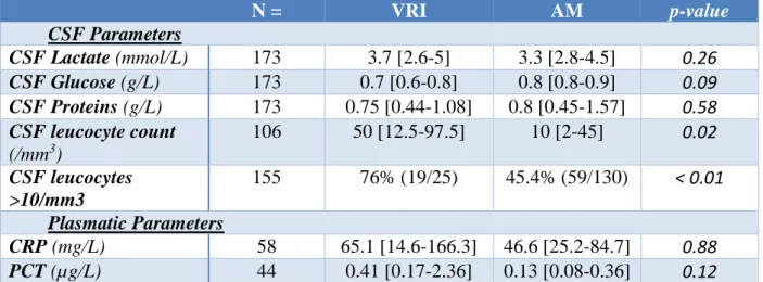
Comparison of VRI and AM groups
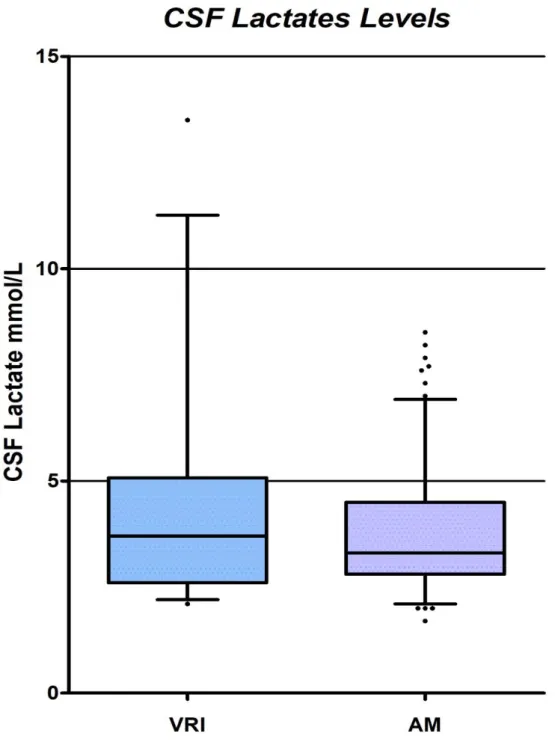
Comparison of CSF and plasmatic parameters between the VRI and AM groups is shown in Table 2. Median CSF lactate levels were 3.7 [2.6-5] mmol/L in the VRI group and 3.3 [2.8-4.5] mmol/L in the AM group, p=0.26. Figure 2 shows these CSF lactate values represented in box plots. There was no significant difference in CSF lactate levels between both groups.
The diagnostic ability of CSF lactate to diagnose VRI is shown on the ROC curve in Figure
3. Values of area under curve were low (AUC = 0.57), making the estimation of cut-off
values impossible for clinical use.
Results for CSF leucocyte count are given in Table 2. Data are shown either as qualitative or quantitative result according to the type of report by the bacteriological laboratory. Proportion of CSF leucocyte count > 10/mm3 was significantly different between the VRI and AM groups, respectively 76% and 45.4%, p<0.01. Looking at quantitative values, the respective median for CSF leucocyte count were 50 [12.5-97.5] per mm3 and 10 [2-45] per mm3, p=0.02.
There was no significant difference regarding CSF glucose (p=0.09), CSF proteins (p=0.58), plasmatic PCT (p=0.12) and plasmatic CRP (p=0.88) between both groups (Table 2).
7
ROC curves for CSF glucose, CSF proteins and CSF quantitative leucocyte count are shown in Figure 3. AUC was very low for CSF protein and glucose. AUC for CSF leucocytes count was 0.68; IC 95% [0.52-0.82]. For a leucocyte count of >30/mm3, sensitivity and specificity for VRI were respectively 69% and 63%.
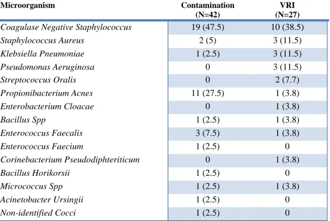
Microorganism found
We noticed that the proportion of positive CSF cultures raises up from 23.8% (41/172) to 32.5% (26/80) with setting up of MALDI-TOF spectrometer (before/after January 2016). Microorganisms found in the CSF cultures are described in Table 3. Far ahead other species, Coagulase Negative Staphylococcus was the most common microorganism responsible for VRI (38.5%) whereas the most common germs in contaminated CSF cultures were Coagulase Negative Staphylococcus (47.5%) and Propionibacterium Acnes (27.5%).
Association between lactate values and other parameters
Higher lactate values were significantly associated with higher CSF proteins (Spearman’s correlation coefficient = 0.52, p<0.01), lower CSF glucose (Spearman’s correlation coefficient = -0.19, p=0.01) and higher red cell count (p=0.01) (see Appendix
1 and Appendix 2). CSF leucocyte count and Fisher score at admission were not associated
with CSF lactate values (see Appendix 1 and Appendix 3).
DISCUSSION
This study shows that CSF lactate analysis cannot discriminate VRI from AM in patients admitted in ICU with SAH and EVD. We found same conclusions concerning the analysis of CSF proteins, plasmatic CRP and PCT. The only biological parameter that was associated with the presence of VRI was CSF leucocyte count.
8
The ROC curves show low AUC for each CSF biological parameter analyzed. Considering the AUC for CSF lactate, we thought it would not be statistically pertinent to determine cut off values that wouldn’t be useful for clinical practice.
These results are not consistent with literature. In a study published in 2008 by Wong et al [21] including patients with intraventricular hemorrhage and EVD, CSF lactate permitted to diagnose VRI with a good negative predictive value. A cut-off of 4 mmol/L was used, but the population was small (16 patients). A meta-analysis published in 2016 by Xiong
Xiao et al [23] also shown the possible utility of CSF lactate to distinguish bacterial or aseptic meningitis in post-neurosurgical patients, but it reviewed only 5 studies (404 patients) with a significant heterogeneity index and cut-offs values which were determined a priori in 2 of the 5 studies. As compared to our study, the populations were also heterogenous.
To date there is no standard definition of VRI. The literature review from Lewis et al found 16 unique definitions [24], which can explain the large range of VRI frequency across studies. For this study we decided to use diagnosis criteria described by Lozier et al [25] which are used in regular practice in our unit. The lack of standard definition and indeed the use of different criteria to diagnose VRI explains the heterogeneity for CSF lactate’s results and interpretations in different studies.
According to a recent study [26], meningitis associated with SAH is a common and normal inflammatory response, unrelated to infection in a majority of cases, and proportional with the volume of spilt blood. This explains why the proportion of patients with AM is high in this population and it makes the diagnosis of VRI challenging before CSF culture shows definitive results. Since normal CSF parameters and contamination do not require any treatment and are easily diagnosed, we decided to focus on the distinction between VRI and AM.
According to B. Venkatesh et al, red cells in CSF cause significant increases in lactate concentrations, and even more when CSF is exposed to air [27]. We suggest that CSF collection from a reservoir bag should be avoided. Moreover, the association between CSF
9
lactate levels and CSF red cells count found in our study should be considered when interpreting lactate in bloodstained CSF. Since our population was exclusively made of people suffering from subarachnoid and intraventricular bleeding, this might explain why we did not find results comparable to previous studies.
In another study by K. Mori et al looking at CSF lactate evolution following SAH in groups with and without vasospasm and excluding patients who had meningitis, CSF lactate was always high at day one and decreased following the pathology evolution, but showed new increase in patients who had vasospasms [28]. This should be taken in consideration when interpreting CSF lactate.
Our study has several limits. First, due to its retrospective design there was a significant number of missing data, such as for plasmatic PCT and CRP. But, even if we were not able to show statistical difference between groups for these parameters, they don’t seem specific enough of VRI to be used in ICUs. Secondly, a large number of samples were excluded due to antibiotic therapy administered in the 48 hours before sampling. There are many reasons that can lead to antibiotic prescription in ICU, such as aspiration pneumonia related to coma or orotracheal intubation procedure. Third, the lack of a standard definition for VRI can be a limit since different definitions can change the frequency of VRI and the distribution between groups. We believe that there is a real need to find a universal definition of VRI in order homogenize research and to evaluate infection rates in future studies.
CONCLUSION
CSF lactate could not differentiate VRI from AM in patients with SAH and EVD. The only parameter that showed statistical difference between groups was leucocyte count. Until now, no usual biological parameter was able to show good enough specificity and sensitivity to diagnose VRI. Cerebrospinal fluid culture remains the gold standard but it still takes notable time to accomplish. Further research needs to focus on new markers or multiple markers correlation to enhance the diagnosis possibilities for this challenging situation.
10
REFERENCES
1. Hoogmoed, J., van de Beek, D., Coert, B. A., Horn, J., Vandertop, W. P., & Verbaan, D. (2016). Clinical and Laboratory Characteristics for the Diagnosis of Bacterial Ventriculitis After Aneurysmal Subarachnoid Hemorrhage. Neurocritical Care, 1-9.
2. Williamson, R. A., Phillips-Bute, B. G., McDonagh, D. L., Gray, M. C., Zomorodi, A. R., Olson, D. M., ... & James, M. L. (2014). Predictors of extraventricular drain–associated bacterial ventriculitis. Journal of critical care, 29(1), 77-82.
3. Mounier, R., Lobo, D., Cook, F., Martin, M., Attias, A., Aït-Mamar, B., ... & Plaud, B. (2015). From the skin to the brain: pathophysiology of colonization and infection of external ventricular drain, a prospective observational study. PloS one, 10(11), e0142320.
4. Bota, D. P., Lefranc, F., Vilallobos, H. R., Brimioulle, S., & Vincent, J. L. (2005). Ventriculostomy-related infections in critically ill patients: a 6-year experience. Journal of
neurosurgery, 103(3), 468-472.
5. Lozier, A. P., Sciacca, R. R., Romagnoli, M. F., & Connolly Jr, E. S. (2002). Ventriculostomy-related infections: a critical review of the literature. Neurosurgery, 51(1), 170-182
6. Muttaiyah, S., Ritchie, S., Upton, A., & Roberts, S. (2008). Clinical parameters do not predict infection in patients with external ventricular drains: a retrospective observational study of daily cerebrospinal fluid analysis. Journal of medical microbiology, 57(2), 207-209.
7. Schade, R. P., Schinkel, J., Roelandse, F. W., Geskus, R. B., Visser, L. G., van Dijk, M. C., ... & Kuijper, E. J. (2006). Lack of value of routine analysis of cerebrospinal fluid for prediction and diagnosis of external drainage–related bacterial meningitis. Journal of
neurosurgery, 104(1), 101-108.
8. Pfisterer, W., Mühlbauer, M., Czech, T., & Reinprecht, A. (2003). Early diagnosis of external ventricular drainage infection: results of a prospective study. Journal of
Neurology, Neurosurgery & Psychiatry, 74(7), 929-932.
9. Walti, L. N., Conen, A., Coward, J., Jost, G. F., & Trampuz, A. (2013). Characteristics of infections associated with external ventricular drains of cerebrospinal fluid. Journal of
Infection, 66(5), 424-431.
10. Citerio, G., Signorini, L., Bronco, A., Vargiolu, A., Rota, M., Latronico, N., & Infezioni LIquorali Catetere Correlate Study Investigators. (2015). External ventricular and lumbar drain device infections in ICU patients: a prospective multicenter Italian study. Critical
11
11. Beer, R., Lackner, P., Pfausler, B., & Schmutzhard, E. (2008). Nosocomial ventriculitis and meningitis in neurocritical care patients. Journal of neurology, 255(11), 1617-1624.
12. Pfausler, B., Beer, R., Engelhardt, K., Kemmler, G., Mohsenipour, I., & Schmutzhard, E. (2004). Cell index–a new parameter for the early diagnosis of ventriculostomy (external ventricular drainage)-related ventriculitis in patients with intraventricular hemorrhage?. Acta neurochirurgica, 146(5), 477-481.
13. Mounier, R., Lobo, D., Cook, F., Fratani, A., Attias, A., Martin, M., ... & Bloc, S. (2015). Clinical, biological, and microbiological pattern associated with ventriculostomy-related infection: a retrospective longitudinal study. Acta neurochirurgica, 157(12), 2209-2217.
14. Berger, C., Schwarz, S., Schaebitz, W. R., Aschoff, A., & Schwab, S. (2002). Serum procalcitonin in cerebral ventriculitis. Critical care medicine, 30(8), 1778-1781.
15. Martinez, R., Gaul, C., Buchfelder, M., Erbguth, F., & Tschaikowsky, K. (2002). Serum procalcitonin monitoring for differential diagnosis of ventriculitis in adult intensive care patients. Intensive care medicine, 28(2), 208-210.
16. Laifer, G., Wasner, M., Sendi, P., Graber, P., Gratzl, O., Huber, P., ... & Zimmerli, W. (2005). Dynamics of serum procalcitonin in patients after major neurosurgery. Clinical
Microbiology and Infection, 11(8), 679-681.
17. Lewis, A., Wahlster, S., Karinja, S., Czeisler, B. M., Kimberly, W. T., & Lord, A. S. (2016). Ventriculostomy-related infections: The performance of different definitions for diagnosing infection. British journal of neurosurgery, 30(1), 49-56.
18. Maskin, L. P., Capparelli, F., Mora, A., Hlavnicka, A., Orellana, N., Díaz, M. F., ... & Del Castillo, M. (2013). Cerebrospinal fluid lactate in post-neurosurgical bacterial meningitis diagnosis. Clinical neurology and neurosurgery, 115(9), 1820-1825.
19. Leib, S. L., Boscacci, R., Gratzl, O., & Zimmerli, W. (1999). Predictive value of cerebrospinal fluid (CSF) lactate level versus CSF/blood glucose ratio for the diagnosis of bacterial meningitis following neurosurgery. Clinical infectious diseases, 29(1), 69-74.
20. Huy, N. T., Thao, N. T., Diep, D. T., Kikuchi, M., Zamora, J., & Hirayama, K. (2010). Cerebrospinal fluid lactate concentration to distinguish bacterial from aseptic meningitis: a systemic review and meta-analysis. Critical care, 14(6), R240.
21. Wong, G. K., Poon, W. S., & Ip, M. (2008). Use of ventricular cerebrospinal fluid lactate measurement to diagnose cerebrospinal fluid infection in patients with intraventricular haemorrhage. Journal of Clinical Neuroscience, 15(6), 654-655.
22. Li, Y., Zhang, G., Ma, R., Du, Y., Zhang, L., Li, F., ... & Kang, X. (2015). The diagnostic value of cerebrospinal fluids procalcitonin and lactate for the differential diagnosis of post-neurosurgical bacterial meningitis and aseptic meningitis. Clinical
12
23. Xiao, X., Zhang, Y., Zhang, L., Kang, P., & Ji, N. (2016). The diagnostic value of cerebrospinal fluid lactate for post-neurosurgical bacterial meningitis: a meta-analysis. BMC Infectious Diseases, 16(1), 483.
24. Allan R. Tunkel, Rodrigo Hasbun,Adarsh Bhimraj,Karin Byers, Sheldon L. Kaplan, W. Michael Scheld,Diederik van de Beek,Thomas P. Bleck,Hugh J. L. Garton,and Joseph R. Zunt 2017 Infectious Diseases Society of America’s Clinical Practice Guidelines for Healthcare Associated Ventriculitis and Meningitis. Clin Infect Dis. 2017 Mar 15; 64(6): e34–e65
25. Sakushima, K., Hayashino, Y., Kawaguchi, T., Jackson, J. L., & Fukuhara, S. (2011). Diagnostic accuracy of cerebrospinal fluid lactate for differentiating bacterial meningitis from aseptic meningitis: a meta-analysis. Journal of Infection, 62(4), 255-262.
26. Maged D. Fam, Hussein A. Zeineddine, Javed Khader Eliyas, …& Issam A. Awad. CSF inflammatory response after intraventricular hemorrhage. Neurology Oct 2017, 89 (15) 1553-1560
27. Hall, J; Siebert, D; Morgan, TJ; Boots, RJ and Venkatesh, B. Interpreting CSF lactic acidosis: effect of erythrocytes and air exposure [online]. Critical Care and Resuscitation, Vol. 5, No. 3, 2003 Sep: 177-81
28. Mori, K., Nakajima, K. & Maeda, M. Long-term monitoring of CSF lactate levels and lactate/pyruvate ratios following subarachnoid haemorrhage Acta neurochir (1993) 125: 20.
Declaration of interests
Tables and Figures:
Table 1: Demographic and clinical factors of the 207 patients with subarachnoid hemorrhage and external ventricular drain
Table 1. Patients characteristics Total population (N= 207) VRI present (N= 26) VRI absent (N=94) Age, years 56 ± 12 59 ± 10 57 ± 11 Men, n (%) 83 (40) 8 (30) 41 (44) SAPS II 41 ± 11 40 ± 11 41 ± 11 WFNS scale, n (%) I II III IV V 17 (8) 19 (9) 12 (6) 76 (37) 83 (40) 2 (7.5) 4 (16) 2 (7.5) 12 (46) 6 (23) 10 (11) 6 (6) 8 (9) 35 (37) 35 (37) Fisher score, n (%) I II III IV 1 (0,5) 3 (1,5) 49 (24) 154 (74) 0 0 10 (38) 16 (62) 1 (1) 2 (2.1) 21 (22.4) 70 (74.5)
EVD duration, days 18 ± 10 25 ± 12 20 ± 10
EVD changes during stay, n (%) 29 (14) 11 (42) 14 (15) VPS insertions, n (%) 29 (14) 10 (38) 15 (16) CSF samples per patient 3 ± 2 6 ± 2 2 ± 1
ICU Length of stay,
days
29 ± 21 36 ± 14 33 ± 21
Deaths during stay,
n (%)
56 (27) 2 (8) 14 (15)
The first column describes the entire population screened with subarachnoid hemorrhage and external ventricular drain. The second column describes the 26 patients included who presented ventriculostomy related infection and the third column describes the 94 patients included who never presented ventriculostomy related infection during their stay in the intensive care unit.
Data are expressed as mean ± standard deviation. VRI indicated Ventriculostomy related infection, SAPSII, Simplified Acute Physiology Score II; WFNS, World Federation of Neurological Surgeons; EVD, External Ventricular Drain; VPS, Ventriculoperitoneal Shunt; CSF, Cerebrospinal fluid, ICU, Intensive Care Unit
14
Table 2: Comparison of cerebrospinal fluid and plasmatic parameters between the group with ventriculostomy related infection and the group with aseptic meningitis
N = VRI AM p-value CSF Parameters CSF Lactate (mmol/L) 173 3.7 [2.6-5] 3.3 [2.8-4.5] 0.26 CSF Glucose (g/L) 173 0.7 [0.6-0.8] 0.8 [0.8-0.9] 0.09 CSF Proteins (g/L) 173 0.75 [0.44-1.08] 0.8 [0.45-1.57] 0.58 CSF leucocyte count (/mm3) 106 50 [12.5-97.5] 10 [2-45] 0.02 CSF leucocytes >10/mm3 155 76% (19/25) 45.4% (59/130) < 0.01 Plasmatic Parameters CRP (mg/L) 58 65.1 [14.6-166.3] 46.6 [25.2-84.7] 0.88 PCT (µg/L) 44 0.41 [0.17-2.36] 0.13 [0.08-0.36] 0.12
Data are expressed as median and interquartile range. Because of the retrospective design some data were missing. VRI indicated Ventriculostomy Related Infections. AM, Aseptic Meningitis. CRP, C-Reactive Protein. PCT,
15
Table 3: Summary of microorganisms found in the 69 positive CSF cultures
Data are expressed as number of samples and percentage. VRI indicated Ventriculostomy related infections. CSF, Cerebrospinal Fluid. Two CSF samples presented contaminations with 2 different microorganisms. One CSF sample presented VRI with 2 different micro-organisms.
Microorganism Contamination
(N=42)
VRI (N=27)
Coagulase Negative Staphylococcus 19 (47.5) 10 (38.5)
Staphylococcus Aureus 2 (5) 3 (11.5) Klebsiella Pneumoniae 1 (2.5) 3 (11.5) Pseudomonas Aeruginosa 0 3 (11.5) Streptococcus Oralis 0 2 (7.7) Propionibacterium Acnes 11 (27.5) 1 (3.8) Enterobacterium Cloacae 0 1 (3.8) Bacillus Spp 1 (2.5) 1 (3.8) Enterococcus Faecalis 3 (7.5) 1 (3.8) Enterococcus Faecium 1 (2.5) 0 Corinebacterium Pseudodiphteriticum 0 1 (3.8) Bacillus Horikorsii 1 (2.5) 0 Micrococcus Spp 1 (2.5) 1 (3.8) Acinetobacter Ursingii 1 (2.5) 0 Non-identified Cocci 1 (2.5) 0
16
Figure 1: Flow Chart of the study design
CNS indicates central nervous system infection, AM, aseptic meningitis; VRI, ventriculostomy related infection. *Patients could have multiple diagnosis during their stay but only one cerebrospinal fluid sample showing ventriculostomy related infection was included per patient (total of patients superior to 120 patients included)
17
Figure 2: Box and Whiskers Plot (5-95th percentiles) comparing cerebrospinal fluid lactate
values between the group with ventriculostomy related infection and the group with aseptic meningitis
Data shown are the median (horizontal line within the box), 5th and 95th percentile (open boxes), upper and lower
fence (whiskers) and outlying values (circles). CSF indicates cerebrospinal fluid; VRI, ventriculostomy related infection; AM, aseptic meningitis
18
Figure 3: ROC curves for CSF Lactate, CSF Proteins and CSF Glucose
CSF indicates cerebrospinal fluid
CSF Lactate: AUC=0.57, IC95[0.45-0.70], Z=-1.12 CSF Proteins : AUC=0.53, IC95[0.41-0.66], Z=0.56 CSF Glucose : AUC=0.60, IC95[.48-0.72], Z=1.68 CSF Leucocytes : AUC=0=68, IC95[0.52-0.82], Z=-2.24
19
APPENDICES:
Appendix 1: Correlations between the variation of cerebrospinal fluid lactate values and other biological parameters
Spearman’s correlation coefficients
CSF Lactate
CSF Proteins CSF Glucose CSF Leucocyte count
0.52 - 0.19 0.10
p < 0.01 p = 0.01 p = 0.33
n = 173 n = 173 n = 100
CSF indicates cerebrospinal fluid. Table showing the significant correlation coefficients between CSF lactate and CSF proteins and CSF glucose. There was no significant correlation with CSF leucocyte count.
Appendix 2: Association between cerebrospinal lactate and cerebrospinal red cell count thresholds
Analysis variable : Lactate Red Cell Count
( / mm3) Number of samples CSF Lactate (mmol/L) < 100 21 3.3 ± 1.1 p = 0.01 100 – 1000 30 3.1 ± 0.9 1000 – 10 000 41 3.7 ± 1.3 > 10 000 73 4.1 ± 1.9
Data are expressed as mean ± standard deviation. Association were tested using a Kruskal – Wallis test CSF indicates cerebrospinal fluid.
Appendix 3: Association between Fisher’s score and cerebrospinal fluid lactates values
Analysis variable : Lactate
Fisher score Number of samples CSF Lactate (mmol/L)
4 47 4.07 ± 1.8 p = 0.42
<4 20 3.55 ± 1.3
Includes only samples with CSF lactate analysis available at the day of admission. Data are expressed as mean ± SD, Differences were tested using a Wilcoxon test
20
SERMENT
➢ En présence des Maîtres de cette école, de mes chers condisciples et
devant l’effigie d’Hippocrate, je promets et je jure, au nom de l’Etre
suprême, d’être fidèle aux lois de l’honneur et de la probité dans l’exercice
de la médecine.
➢ Je donnerai mes soins gratuits à l’indigent et n’exigerai jamais un salaire
au-dessus de mon travail.
➢ Admis (e) dans l’intérieur des maisons, mes yeux ne verront pas ce qui s’y
passe, ma langue taira les secrets qui me seront confiés, et mon état ne
servira pas à corrompre les mœurs, ni à favoriser le crime.
➢ Respectueux (se) et reconnaissant (e) envers mes Maîtres, je rendrai à
leurs enfants l’instruction que j’ai reçue de leurs pères.
➢ Que les hommes m’accordent leur estime si je suis fidèle à mes promesses.
Que je sois couvert (e) d’opprobre et méprisé (e) de mes confrères si j’y
manque.
RESUME
Introduction : En raison des modifications biologiques du liquide cérébrospinal (LCS) au cours
des hémorragies sous arachnoïdiennes (HSA), le diagnostic d’infection sur dérivation ventriculaire externe (DVE) est particulièrement complexe. D'après des études récentes, la mesure du lactate dans le LCS pourrait être un outil diagnostique intéressant.
L'objectif de cette étude est de démontrer la valeur diagnostique du lactate pour distinguer les infections bactériennes sur DVE des méningites aseptiques (MA) chez des patients atteints d'HSA et porteurs de DVE.
Méthodes : De 2010 à 2016, les patients hospitalisés en réanimation avec une HSA et chez qui
une DVE était implantée étaient inclus. L'infection sur DVE était définie comme des paramètres biologiques du LCS anormaux avec une culture positive et l'introduction d'une antibiothérapie d'au moins 3 jours, la MA par des paramètres biologiques anormaux et des cultures négatives.
Résultats : 207 patients ont été inclus, 567 prélèvements de LCS analysés et 315 prélèvements
exclus. Parmi les 252 prélèvements analysés ; 26 (12.6%) montraient une infection sur DVE, 147 (58.3%) une AM. 40 prélèvements étaient contaminés et 39 prélèvements étaient normaux. Les valeurs médianes du Lactate ne montraient pas de différences significatives entre les groupes infection sur DVE et MA (3.7 [2.6-5] mmol/L vs. 3.3 [2.8-4.5] mmol/L, p=0.26), tout comme les valeurs médianes de la glycorachie et de la proteinorachie. La valeur médiane de la leucorachie était significativement plus élevée dans le groupe infection sur DVE (50 /mm3 [12.5-97.5] vs. 10 /mm3 [2-45] p=0.02).
Conclusion : En conclusion, le lactate dans le LCS ne permet pas de distinguer l'infection sur
DVE des MA chez des patients atteints d'HSA et porteurs de DVE.
Mots clés : Méningite Nosocomiale, Hémorragie sous arachnoïdienne, Dérivation ventriculaire
externe, Lactate, Liquide cérébro-spinal, Méningite aseptique, Antibiothérapie, Etude rétrospective