HAL Id: tel-01127588
https://tel.archives-ouvertes.fr/tel-01127588
Submitted on 7 Mar 2015
HAL is a multi-disciplinary open access
archive for the deposit and dissemination of sci-entific research documents, whether they are pub-lished or not. The documents may come from teaching and research institutions in France or abroad, or from public or private research centers.
L’archive ouverte pluridisciplinaire HAL, est destinée au dépôt et à la diffusion de documents scientifiques de niveau recherche, publiés ou non, émanant des établissements d’enseignement et de recherche français ou étrangers, des laboratoires publics ou privés.
Role of the proinflammatory environment in the
immunogenicity of therapeutic factor viii in patients
with severe hemophilia A
Ivan Peyron
To cite this version:
Ivan Peyron. Role of the proinflammatory environment in the immunogenicity of therapeutic factor viii in patients with severe hemophilia A. Immunology. Université Pierre et Marie Curie - Paris VI, 2014. English. �NNT : 2014PA066455�. �tel-01127588�
THESE DE DOCTORAT DE L’UNIVERSITE PARIS VI PIERRE ET MARIE CURIE
Spécialité : Immunologie
Ecole doctorale : Physiologie et physiopathologie
Présentée par : Ivan Peyron Pour obtenir le grade de DOCTEUR DE L’UNIVERSITE PARIS VI
Sujet de la thèse
ROLE DE L’ENVIRONNEMENT INFLAMMATOIRE DANS
L’IMMUNOGENICITE DU FACTEUR VIII THERAPEUTIQUE CHEZ LES
PATIENTS HEMOPHILES A
Soutenue le 16 septembre 2014
devant le jury composé de :
M. Adrien SIX, Professeur, Université Paris VI (Président)
M. Jan VOORBERG, Directeur de recherche, Université d’Amsterdam (Rapporteur)
M. Antonino NICOLETTI, Directeur de Recherche, INSERM (Rapporteur)
Mme Véronique WITKO-SARSAT, Directrice de Recherche, INSERM (Examinateur)
Mlle Sophie SIBERIL, Maître de conférences universitaire, Université Paris VI (Examinateur)
2
RÉSUMÉ
L‟hémophilie A est une maladie hémorragique rare liée au chromosome X qui se traduit par un déficit en facteur VIII (FVIII) fonctionnel. Chez les patients atteints de la forme sévère de l‟hémophilie A, l‟apparition d‟hémorragies incontrôlées augmente la morbidité et affecte leur qualité de vie. Le traitement de choix permettant de prévenir ou de traiter les saignements consiste en l‟injection intraveineuse de FVIII thérapeutique. Cependant, chez 5 à 30% des patients, une réponse immunitaire dirigée contre le FVIII se développe. La réponse anti-FVIII est caractérisée par l‟apparition d‟anticorps qui inhibent l‟activité pro-coagulante du FVIII. Ainsi l‟apparition de ces anticorps, appelés « inhibiteurs » représente-t-elle la complication majeure du traitement des patients hémophiles A.
Plusieurs facteurs de risque ont été suggérés ou proposés comme affectant le développement de la réponse anti-FVIII. Parmi eux, supporté par la théorie du « danger », la présence de médiateurs pro-inflammatoires générés par les épisodes de saignement est considérée comme adjuvant dans la réponse immunitaire anti-FVIII. Cependant, les saignements induisent de nombreux mécanismes pro mais aussi anti-inflammatoires. Ainsi, l‟activation de l‟endothélium vasculaire induit-elle le recrutement et l‟activation des cellules effectrices appartenant aux compartiments innés et adaptatifs du système immunitaire.
Au cours de ma thèse, je me suis concentré sur le rôle des saignements dans le développement de la réponse immunitaire dirigée contre le FVIII. Ainsi, ai-je découvert une association entre la présence d‟un polymorphisme dans un gène induit par les saignements et la présence d‟inhibiteurs du FVIII dans une cohorte de patients hémophiles A sévères. J‟ai également étudié le développement de la réponse immunitaire dirigée contre le FVIII thérapeutique dans un modèle murin qui mime les saignements locaux observés chez les patients hémophiles A sévères. Finalement, j‟ai caractérisé l‟effet des formes réactives de l‟oxygène (FRO), qui sont générées au niveau du site de saignement, sur la structure, la fonction et l‟immunogénicité du FVIII.
Les résultats obtenus au cours de ma thèse démontrent que les saignements ne sont pas associés à un risque plus élevé de développer des inhibiteurs dans le modèle murin largement utilisé d‟hémophilie A sévère, contrairement à ce qui est communément admis. De plus, j‟ai démontré une association entre un polymorphisme présent dans le promoteur du gène HMOX1 et la survenue d‟inhibiteurs dans une cohorte de patients atteints d‟hémophilie A sévère. En parallèle, je décris pour la première fois une altération du statut oxydatif chez les souris déficientes en FVIII et je démontre que le contrôle du statut oxydatif in vivo permet de moduler la réponse immunitaire anti-FVIII. Au contraire, l‟exposition du FVIII aux FRO augmente son immunogénicité.
De fait, mes résultats suggèrent que, bien que les molécules pro-inflammatoires libérées suite aux saignements puissent affecter positivement la réponse immunitaire dirigée contre le FVIII, de puissants médiateurs anti-inflammatoires sont générés in vivo et amenuisent l‟effet potentiellement adjuvant des saignements. Également, ces résultats soulignent la balance complexe entre les médiateurs pro- et anti-inflammatoires qui sont générés consécutivement aux saignements ainsi que leurs effets sur la réponse immunitaire dirigée contre le FVIII thérapeutique.
3
ABSTRACT
Hemophilia A is a rare X-linked hemorrhagic disease consecutive to the lack of functional pro-coagulant factor VIII (FVIII). In patients with the severe form of hemophilia A, uncontrolled hemorrhages increase the morbidity and affect the quality of life. To prevent or treat bleeding episodes, therapeutic FVIII is administered intravenously. However, an anti-FVIII immune response develops in 5 to 30% of the patients. The anti-anti-FVIII immune response is characterized by the development of anti-FVIII IgG antibodies that inhibit FVIII activity. These antibodies, referred to as “FVIII inhibitors” represent the major complication in the treatment of hemophilia A patients.
Several risk factors have been suggested or proposed to predispose to the development of FVIII inhibitors. Amongst them, inspired by the “danger” theory, the presence of inflammatory mediators released upon bleeding episodes is thought to adjuvant the anti-FVIII immune response. Bleeding episodes trigger several pro and anti-inflammatory mechanisms. Thus, damage to the endothelium triggers the recruitment and activation of effector cells from the innate and adaptive compartments of the immune system.
During my PhD, I investigated the role of bleedings in the development of the anti-FVIII immune response. Thus, I discovered an association between a polymorphism in the promoter region of a gene that is induced in response to bleedings, and the presence of FVIII inhibitors in a cohort of patients with severe hemophilia A. I followed the development of the immune response to therapeutic FVIII in a mouse model that mimics the localized bleedings found in patients with severe hemophilia A. Ultimately, I characterized the effects of reactive oxygen species (ROS) that are potentially released at the bleeding site in view of the structure, function and immunogenicity of the FVIII molecule.
The results I obtained during my PhD demonstrate that bleedings are not associated with a higher risk for FVIII inhibitor development in the commonly used mouse model of severe FVIII deficiency. Additionally, I demonstrated an association between a polymorphism in the promoter of the HMOX1 gene and the presence of FVIII inhibitors. In parallel, I report for the first time an exacerbated oxidative status in FVIII-deficient mice as compared to control mice, and demonstrate that the control of the oxidative status in vivo reduces the anti-FVIII immune response. Conversely, the exposure of FVIII to ROS ex vivo increases the immunogenicity of the FVIII molecule.
Taken together, my results suggest that, although the pro-inflammatory molecules released upon bleeding may positively affect the FVIII immune response, several strong anti-inflammatory compounds are generated in vivo that dampen the potential adjuvant effect of bleedings. Ultimately, these results highlight the complex balance between the pro and anti-inflammatory mediators generated upon bleeding and their effect on the anti-FVIII immune response.
4
PUBLICATION LIST
This work is based on the following publications:
Article 1: Repessé Y*, Peyron I*, Dimitrov JD, Dasgupta S, Farrokhi Moshai E, Costa C, Borel-Derlon A, Guillet B, D‟Oiron R, Aouba A, Rothschild C, Oldenburg J, Pavlova A, Kaveri SV, Lacroix-Desmazes S. Development of inhibitory antibodies to therapeutic factor VIII in severe hemophilia A is associated with microsatellite polymorphism in the HMOX1 promoter, Haematologica 2013.
Article 2: Peyron I, Dimitrov JD, Delignat S, Gangadharan B, Planchais C, Kaveri SV, Lacroix-Desmazes S. Hemarthrosis and arthropathies do not favour the development of factor VIII inhibitors in Hemophilia A mice, Haemophilia (under revision)
Article 3: Peyron I, Dimitrov JD, Delignat S, Gangadharan B, Kaveri SV, Lacroix-Desmazes S. Restoration of oxidative balance by N-Acetyl-L-cysteine in FVIII-deficient mice reduces immune responses to therapeutic FVIII (in preparation)
Article 4: Peyron I, Dimitrov JD, Delignat S, Gangadharan B, Kaveri SV, Lacroix-Desmazes S. Oxidation of therapeutic factor VIII aggravates its immunogenicity in factor VIII-deficient mice (in preparation)
5
TABLE OF CONTENTS
I. INTRODUCTION ... 11
I.1. Hemophilia A ... 11
I.1.1. Generalities ... 11
I.1.1.1. Historical aspects ... 11
I.1.1.2. Phenotypic aspects ... 12
I.1.1.3. Genetic aspects ... 12
I.1.1.4 Bleeding complications in Hemophilia A ... 14
I.1.2 The coagulation cascade ... 15
I.1.2.1 Primary haemostasis ... 16
I.1.2.2 Secondary haemostasis ... 17
I.1.2.2.1 The extrinsic pathway ... 17
I.1.2.2.2 The intrinsic pathway ... 17
I.2.2.3 Pro-coagulant factor VIII ... 19
I.2.2.3.1 Synthesis, structure and post-traductional modifications ... 19
I.2.2.3.2 Activation of FVIII ... 20
I.2.2.3.3 Life cycle of FVIII, catabolic receptors, in vivo distribution, site of elimination ... 21
I.2.2.3.4 Catabolic receptors for FVIII ... 22
I.2.2.3.5 In vivo distribution of FVIII ... 24
I.1.3 Prevention or treatment of bleeding episodes in patients with hemophilia A ... 24
I.1.3.1 Factor VIII products ... 24
I.1.3.2 Infusion regiment ... 24
I.1.3.3 Complications of the treatment ... 25
I.1.3.4 Treatment of patients with FVIII inhibitors ... 26
I.2 The anti-factor VIII immune response ... 27
I.2.1 FVIII as seen by the immune system in patients with hemophilia A ... 27
I.2.2 Antigen-presenting cells ... 28
I.2.2.1 Dendritic cells ... 28
I.2.2.2 Macrophages ... 29
I.2.2.3 B cells ... 29
6
I.2.4 The anti-FVIII immune response in hemophilia A patients ... 30
I.2.4.1 T-cell activation ... 30
I.2.4.2 B-cell activation ... 31
I.3 Risk factors ... 32
I.3.1 The danger signal theory ... 32
I.3.1.1 Exogenous danger signals ... 33
I.3.1.2 Endogenous danger signals ... 33
I.3.2 Genetic risk factors ... 33
I.3.2.1 FVIII mutations ... 34
I.3.2.2 HLA haplotype ... 35
I.3.2.3 Polymorphisms in immune-related genes ... 35
I.3.3 Non-genetic risk factors ... 37
I.3.3.1 Age at the start of treatment ... 37
I.3.3.2 Mode and intensity of FVIII administration ... 37
I.3.3.3 State of the immune system at the time of FVIII infusion ... 38
I.4 Purpose of the PhD work ... 39
II. RESULTS ... 40
II.1. Development of inhibitory antibodies to therapeutic factor VIII in severe hemophilia A is associated with microsatellite polymorphism in the HMOX1 promoter ... 40
II.2. Hemarthrosis and arthropathies do not favor the development of factor VIII inhibitors in hemophilia A mice ... 49
II.3. Restoration of the oxidative balance in factor VIII-deficient mice reduces the immunogenicity of therapeutic factor VIII ... 71
II.4. Oxidation of therapeutic factor VIII aggravates its immunogenicity in factor-VIII deficient mice ... 80
III. DISCUSSION ... 104
IV. REFERENCES ... 116
7
FIGURE AND TABLES LIST
Figure 1. Physiopathology of hemophilic arthropathy 15
Figure 2. Primary hemostasis. 16
Figure 3. Secondary hemostasis. 18
Figure 4. FVIII structure. 20
Figure 5. Activation of FVIII. 21
Figure 6. FVIII endocytosis and presentation on major histocompatibility
class II (MHC II) molecules. 28
Figure 7. CD4+ T-cell activation by APC. 31
Figure 8. B-cell activation. 32
Figure 9. HMOX1 promoter organization. 41
Table 1. F8 gene mutations and risk for inhibitor development. 34
8
ABBREVIATION LIST
AAGE Advanced glycation end-product AOPP Advanced oxidation protein product APC Antigen-presenting cell
ATP Adenosine triphosphate
B
BCR B cell receptor
BDD-FVIII B domain-deleted factor VIII BHK Baby hamster kidney
C
CHO Chinese hamster ovary CORM-3 CO-releasing molecule-3 CRM Cross-reactive material
D
DAMP Danger-associated molecular pattern DC Dendritic cell
DNA Deoxyribonucleic acid
E
ELISA Enzyme-linked immunosorbent assay
F
F8 Factor VIII (gene)
FEIBA Factor eight inhibitor bypassing agent FRAP Ferric reducing ability of plasma FVIII Factor VIII (protein)
G
9 H
HAMSTeR Hemophilia A mutation, structures, test and resource HLA Human leukocyte antigen
HMGB1 High mobility group box 1 HMOX1 Heme oxygenase-1 (gene) HNE Hydroxynonenal
HO-1 Heme oxygenase-1 (protein) HSP Heat shock protein
HSPG Heparan sulphate proteoglycans
I
ICAM-1 Intercellular Adhesion Molecule 1 IgG Immunoglobulin G
Inv22 Inversion 22
ITI Immune tolerance induction IU International unit K kbp Kilobase pair kDa KiloDalton L LDL Low-density lipoprotein
LOX-1 Lectin-like oxidized low-density lipoprotein receptor-1 LPS Lipopolysaccharide
LRP Low-density lipoprotein receptor-related protein
M
MDA Malondialdehyde
MHC-II Major histocompatibility complex class II MPO Myeloperoxidase
10 N
NAC N-acetyl-L-cystein
NADPH Nicotinamide adenine dinucleotide phosphate NETs Neutrophil extracellular traps
O
ORAC Oxidant radical absorbance capacity
P
PAMP Pathogen-associated molecular pattern PBMC Peripheral blood monocyte cell
pdFVIII plasma-derived FVIII PKC Protein kinase C
R
RAGE Receptor for advanced glycation end product RAP Receptor associated protein
rFVIII Recombinant factor VIII ROI Reactive oxygen intermediates ROS Reactive oxygen species
S
Siglec-5 Sialic acid-binding Ig-like lectin 5 SNP Single nucleotide polymorphism SOD Superoxide dismutase
T
TCR T cell receptor TF Tissue factor TLR Toll-like receptor
V
VCAM-1 Vascular cell adhesion protein 1 VWF Von Willebrand factor
11
I.
INTRODUCTION
I.1. Hemophilia A
I.1.1. Generalities
Hemophilia A is the most frequent haemorrhagic disorder with an incidence of one newborn in ten thousands (Lenting et al., 1998). This X-linked genetic disease follows a recessive transmission pattern and is characterized by a qualitative or quantitative defect in functional pro-coagulant factor VIII (FVIII) (Hoyer, 1994). In the most severe forms of this pathology, abnormal levels of FVIII lead to spontaneous haemorrhages located mainly in joints and in soft tissues. Moreover, chronic bleedings in joints lead to the development of crippling arthropathies, decreasing the quality of life of the patients. In the most dramatic situation representing up to ten percent of the cases (Klinge et al., 1999; Daniele et al., 2011), haemorrhages develop in the brain, engaging the vital prognosis of the patient.
I.1.1.1. Historical aspects
The first written report describing a congenital affection of blood coagulation is found in the Talmud, two centuries BC (Rosner, 1969). This text reports that circumcision should be avoided if the two previous sons passed away following their circumcision. Thirteen centuries later, the famous Arabic medical doctor Abu Al-Qasim described a haemorrhagic disease which is transmittable amongst generations. Later on, in 1803, Dr. John Conrad Otto described a haemorrhagic condition existing in some families. Comforting his hypothesis, he was able to trace back the origin of the disease affecting his patients to a woman living in Plymouth in 1720. Ten years later, John Hay described that the affected males may pass the genetic trait to their unaffected daughter (HAY, 1813). The term “Haemorrhaphilia” appears only in 1828 in the PhD manuscript by Friedrich Hopff, and is rapidly contracted to “Hemophilia” as an association of two greek words “haïma” (blood) and “philia” (affection).
12
The first treatments of hemophilia were reported by Patek and Taylor, two medical doctors from Harvard. They succeed to correct the coagulation defect by adding a substance derived from the plasma. They named this substance „antihemophilic globulin”. However, the treatment of the affected patients with the plasma from healthy individuals was often inefficient, due to the very low concentration of the required coagulation factor. It is only in 1964 that Dr. Judith Pool discovered that the cryoprecipitation of normal plasma was generating a pellet enriched in coagulation factor (Pool et al., 1964).
I.1.1.2. Phenotypic aspects
The severity of hemophilia A is dependent on the residual activity of the endogenous circulating FVIII. Thus, hemophilia A is considered minor, when residual levels of FVIII are between 5 and 35 % of activity found in normal plasma, moderate with levels between 1 to 5% of normal and severe if FVIII levels are below 1% (Lenting et al., 1998). The repartition of the three degree of severity is not homogenous. Indeed, half of the hemophilia A population is affected by the minor form of the disease, 10% display the moderate form and 40% are affected by the severe form of hemophilia A (Antonarakis et al., 1995).
I.1.1.3. Genetic aspects
The FVIII encoding gene (F8 gene) was cloned between 1982 and 1984 by Gitschier and his team (Gitschier et al., 1984). The F8 gene is located at the distal part of the long arm of the X chromosome (Xq28). Its length is of 186 kbp, representing 0.1% of the total chromosome length. The coding part of the gene is spread amongst 26 exons. Once transcripted and spliced, the mature mRNA has a size of 9 kbp and represents 5% of the initial gene length. The 95% remaining sequences correspond to the intronic parts of the gene. Despite the fact that they do not encode the FVIII protein, the intronic segments can carry some mutations responsible for FVIII deficiency. The phenotypic heterogeneity of hemophilia A reflects the wide variety of the genetic abnormalities that can affect the patients‟ F8 gene.
13
These numerous mutations are listed in the HAMSTeR (the Hemophilia A Mutation, Structures, Test and Resource website; www.factorviii-db.org). Of note, some rare hemophilia A cases are due to mutations which are not directly affecting the F8 gene, but are affecting proteins involved in FVIII production, intracellular trafficking or secretion (Zhang et al., 2003; Oldenburg and El-Maarri, 2006). In most hemophilia A cases (about 90% of the patients), the genetic abnormality consists in a single nucleotide mutation which is affecting the FVIII encoding gene (Bhopale and Nanda, 2003). The term single nucleotide mutation engulf the missense mutations were a nucleotide substitution leads to a change in the incorporated amino acid, the non-sense mutations were a stop codon is integrated before the end of the normal gene, and the splicing mutations, when the addition or the removal of a splicing site leads to the generation of an abnormal mRNA, thus affecting the final amino-acid sequence of the FVIII protein. Small deletions or insertions and larges deletions (>50 bp) are found in 5 to 10% of hemophilia A patients. Most often, these mutations lead to a shift in the DNA reading frame, leading to a drastic modification of the protein and are generally associated with a severe form of hemophilia A (Oldenburg et al., 1998). Duplication and inversions are rare, excepted for the intron 22 (Inv22) which is found inverted in 45 to 50% of patients with severe hemophilia A (Lakich et al., 1993). Mutation Inv22 is classified as a “null” because it leads to the complete lack of the FVIII secretion. However, recent findings suggest that hemophilia A patients with Inv22 mutation produce the entire FVIII protein as two polypeptide chain (Pandey et al., 2013), although these findings remain controversial. Moreover, in some patients with missense mutations, the FVIII molecule is secreted at a normal concentration, but is not functional. This explains why 5% of hemophilia A patients do not have any residual FVIII activity but display normal levels of circulating FVIII. These patients are referred to as “cross-reactive-material (CRM)-positive” patients (Amano et al., 1998). Thus, the fact that hemophilia A patients have distinct genetic and phenotypic profiles
14
makes hemophilia A a very heterogeneous disease. Moreover, the type of mutation affecting the FVIII-encoding gene lead to the production of several types of FVIII protein in different patients. From an immunological point of view, it means that during the ontogeny of the immune system, hemophilia A patients in whom FVIII is partially secreted, even though not functional, will participate in the elimination of FVIII-specific T cells. Moreover, these patients will develop regulatory T cells capable of suppressing FVIII-specific T cells in the periphery. Altogether, these mechanisms may prevent the development of the anti-FVIII immune response after infusion of therapeutic FVIII. Conversely, in patients in whom no FVIII is secreted, and particularly in CRM-negative patients, the FVIII-reactive T cells are not eliminated during T-cell ontogeny, thus increasing the risk for FVIII inhibitor development after birth/later in life.
I.1.1.4 Bleeding complications in Hemophilia A
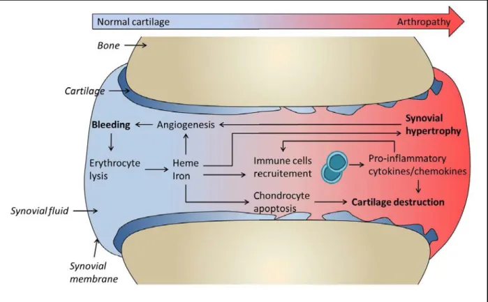
The lack of functional pro-coagulant FVIII leads to de development of uncontrolled haemorrhages. These haemorrhages may occur at any place in the body. Thus, deep internal bleedings in the muscles lead to swelling of the affected member. Swelling may press on nerves and lead to recurring pain. Ultimately, repetition of painful episodes may result in a reluctance to use the affected member. When located in a joint, the presence of non-clotted blood, referred to as hemarthrosis, leads to the progressive destruction of the joint. It has been demonstrated that extravasation of blood within the synovial space leads to an increased concentration of erythrocyte-derived haemoglobin and heme along with the recruitment of neutrophils, monocytes, macrophages and lymphocytes. On the bleeding site, the production of pro-inflammatory cytokines and chemokines (Lafeber et al., 2008), combined with the deleterious pro-oxidative effect of heme-derived iron, leads to the progressive destruction of the joint. Indeed, the pro-oxidative properties of iron may directly degrade the cartilage by inducing chondrocyte apoptosis (Roosendaal et al., 1999), resulting in an inhibition of the
15
turnover of the cartilage extracellular matrix protein (Jansen et al., 2007). Moreover, the pro-angiogenic property of heme leads to a sustained vascular neoplasia, provoking a synovial hypertrophy along with the loss of joint space (Arnold and Hilgartner, 1977). Of note, the vicious circle of joint destruction by hemophilic arthropathy is sustain by the neo-vascularization of the affected joint; indeed, the increase in the number of small vessels has been proposed to increasing the risk of future bleeding events at the same location (Lafeber et al., 2008). (Figure 1)
Figure 1. Physiopathology of hemophilic arthropathy. The presence of blood within the synovial space triggers several pro-inflammatory events. Thus, in the joint, the recruited immune cells release pro-inflammatory cytokines and chemokines. In combination with heme-derived iron, these pro-inflammatory mediators lead to synovial hypertrophy, chondrocyte apoptosis and progressive destruction of the joint.
I.1.2 The coagulation cascade
Any kind of alteration of the vascular endothelium integrity leads to the exposure of sub-endothelial structures/molecules such as collagen or tissue factor which, upon direct contact with blood, induce/activate primary and secondary haemostasis.
16
I.1.2.1 Primary haemostasis
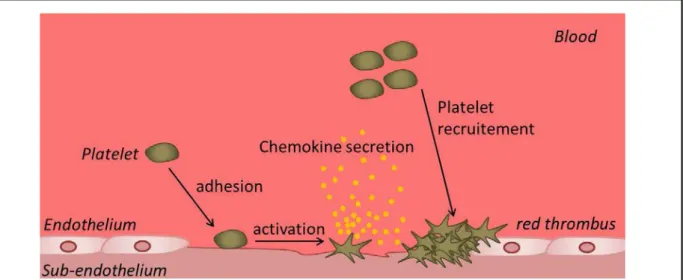
Upon vascular damage, a localized vasoconstriction occurs as a response to a reflex neurogenic mechanism and to the secretion of endothelium-derived vasoconstrictors such as endothelin. This phenomenon reduces the blood flow upstream of the vascular breach, thus limiting the blood loss and facilitating the interaction of the mediators required for the initiation of haemostasis. Immediately following the vasoconstriction, primary haemostasis occurs. Platelets are recruited and get activated, allowing their adhesion to the exposed sub-endothelial matrix. Platelet activation results in dramatic changes in their shape, along with the release of the content of secretory granules. The platelet-derived mediators recruit other platelets, causing more platelet to adhere to the sub-endothelial matrix and to aggregate to each other at the vascular breach. Platelet aggregation ultimately results in the formation of a primary haemostatic plug, referred to as the “red thrombus” (Figure 2).
Figure 2. Primary hemostasis. Upon binding to the exposed endothelium, platelets are activated, leading to morphological changes and release of platelet chemoattractant mediators. At the site of endothelium injury, the aggregation of platelets results in the formation of the red thrombus which is consolidate by the fibrin generated during the secondary hemostasis.
17
I.1.2.2 Secondary haemostasis
The goal of secondary haemostasis is to form a stable plug when the bleeding is severe. During secondary haemostasis, also known as the coagulation cascade, the activated endothelium in association with activated platelets will allow the initiation of a succession of reactions leading to the generation of an insoluble fibrin clot, referred to as the “white clot”. Two pathways have been identified as being responsible for the initiation of the coagulation cascade, the extrinsic and the intrinsic pathways.
I.1.2.2.1 The extrinsic pathway
Upon exposure of the sub-endothelial matrix proteins and ensuing platelet aggregation, a membrane-bound pro-coagulant factor called „tissue factor‟ (TF) is expressed, which complexes and activates coagulation factor VII. The activated form of factor VII (FVIIa) in turn activates factor X (FXa), which associates with the activated factor Leiden (also known as factor V, FVa) to form a complex called the pro-thrombinase complex. The pro-thrombinase complex activates pro-thrombin (FII) into thrombin (FIIa). Eventually, the thrombin generated allows the polymerisation of fibrin monomers and activates factor XIII. To achieve the white thrombus, the fibrin polymers are stabilised by the activated factor XIII giving rise to an insoluble fibrin clot, that act as a “net”, helping the platelets to stabilize at the site of bleeding.
I.1.2.2.2 The intrinsic pathway
The intrinsic pathway, also known as the “contact factor pathway” initiates upon binding of factor Hageman (factor XII) to the negatively charged surface provided by the activated sub-endothelium. This binding results in the auto-activation of FXII to FXIIa. FXIIa induces the conversion of pre-kallikrein to kallikrein and factor XI to FXIa. FXIa activates in turn FIX to FIXa (Scott et al., 1985). FIXa then associates with activated FVIII (FVIIIa) and with platelet-derived phospholipids (PL). In the presence of calcium, this complex, referred to
18
as the “tenase” complex, allows the conversion of FX to FXa that sustains the downstream generation of thrombin initiated by the extrinsic pathway.
Figure 3. Secondary hemostasis. Upon exposure of the sub-endothelial matrix proteins and ensuing platelet aggregation, extrinsic pathway starts with the expression of the pro-coagulant tissue factor (TF), which complexes and activates coagulation factor VII. The activated form of factor VII (FVIIa) in turn activates factor X (FXa), which associates with activated factor V, (FVa) to form a complex called the pro-thrombinase complex. The pro-thrombinase complex activates prothrombin (FII) into thrombin (FIIa). Eventually, the thrombin generated allows the polymerisation of fibrin monomers and activates factor XIII. Then, fibrin polymers are stabilised by the activated factor XIII giving rise to a stable fibrin clot, that act as a “net”, helping the platelets to stabilize at the site of bleeding. The intrinsic pathway, initiates upon binding of factor XII to the negatively charged surface provided by the activated sub-endothelium. This binding results in the auto-activation of FXII to FXIIa. FXIIa induces the conversion of pre-kallikrein to kallikrein and factor XI to FXIa. FXIa in turn activates FIX to FIXa. FIXa then associates with activated FVIII (FVIIIa) and with platelet-derived phospholipids (PL). In the presence of calcium, this complex, called “tenase”, allows the conversion of FX to FXa that sustains the downstream generation of thrombin initiated by the extrinsic pathway.
19
I.2.2.3 Pro-coagulant factor VIII
I.2.2.3.1 Synthesis, structure and post-traductional modifications
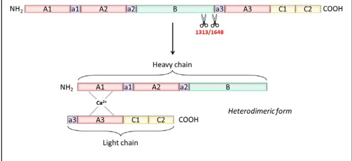
For years, the type of cells that are producing FVIII has been under debate. Initially, the secretion of FVIII was thought to be mainly mediated by hepatocytes (Ingerslev et al., 1988) and later on by liver sinusoidal endothelial cells (Hellman et al., 1989). However, recent advances strongly suggest that, while sinusoidal endothelial cells from the liver presumably account for an important part of FVIII synthesis, they might not be the exclusive source for its production (Jacquemin et al., 2006; Everett et al., 2014; Fahs et al., 2014). The primary structure of the molecule is a multi-domain protein containing 2351 amino acids translated in the endoplasmic reticulum. After a first maturation step, a 19 amino-acid signal peptide is cleaved, thus reducing the size of the protein to 2332 amino acids. In the endoplasmic reticulum, N-glycosylations are added to the FVIII molecule before been translocated to the Golgi apparatus. In the Golgi apparatus, complex carbohydrate modifications on N-linked sites, addition of carbohydrate to serine and threonine residues and sulfation of several tyrosine residues in the acidic regions of FVIII, occur (Kaufman et al., 1988). Eventually, a proteolysis step at residues Arg1313 and Arg1648 leads to the secretion of a protein with an apparent molecular weight of 280 kDa (Vehar et al., 1984; Lenting et al., 1998). Within the circulation, the mature form of FVIII corresponds to an heterodimer, composed of a heavy chain comprising the A1a1, A2a2 and B domains, and a light chain comprising the a3A3, C1 and C2 domains (Wood et al., 1984), linked by a divalent ion (Figure 4). The a3 acidic region, in association with the C2 domain, allows the binding of the FVIII protein to its chaperon molecule, the von Willebrand factor (VWF). The binding of VWF to the FVIII molecule prevents its premature degradation and allows the addressing of FVIII to the bleeding site (Kaufman et al., 1997).
20
Figure 4. FVIII structure. Following the translation of FVIII mRNA, the immature 2352 amino-acid form of FVIII is translocated to the endoplasmic reticulum. There, the proteolytic cleavage of the signal peptide reduces its size to 2332 amino-acids. After its translocation to the Golgi apparatus, a proteolytic cleavage at Arg1313 and Arg1648, lead to the formation of an heterodimer. The heavy chain comprises the domains A1-a1-A2-a2-B and the light chain the domains a3-A3-C1-C2. The two chains are linked through a divalent ion. Within the circulation, FVIII circulates in association with VWF.
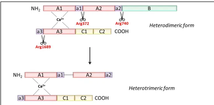
I.2.2.3.2 Activation of FVIII
FVIII is addressed to the bleeding site by its chaperon molecule VWF. Thrombin then activates the FVIII molecule through a proteolytic cleavage of the heavy chain (residues Arg372 and Arg740) and the light chain (Arg1689) (Figure 5). The cleavage of the light chain leads to the dissociation of the FVIII molecule from VWF. Once released from VWF, the FVIII molecules binds to phospholipids and a structural change in FVIII orientation occurs, allowing its interaction with FIXa and FX. Binding to FIXa involves a sequence from the A2 domain, spanning Ser558 to Gln565, and a part of the A3 domain, spanning Glu1811 to Lys1818 (Lenting et al., 1996; Mertens et al., 1999).
21
Figure 5. Activation of FVIII. Thrombin activates the FVIII molecule through a proteolytic cleavage of the heavy chain (residues Arg372 and Arg740) and the light chain (Arg1689). Activated FVIII adopts a heterotrimeric conformation and its affinity for VWF is reduced.
The activated form of FVIII (FVIIIa) is able to exert its co-factor activity towards FIXa within the tenase complex that is composed of FVIIIa, FIXa and FX. In association with the phospholipids exposed at the surface of the activated platelets or by the damaged endothelium, the tenase complex activates FX. The presence of FVIIIa in the tenase complex enhances FX activation by 105 folds (van Dieijen et al., 1981). Later on in the coagulation
cascade, FXa participates in the formation of the prothrombinase complex, involved in the generation of insoluble fibrin.
I.2.2.3.3 Life cycle of FVIII, catabolic receptors, in vivo distribution, site of elimination
Once released in the circulation, FVIII rapidly associates with VWF (Vlot et al., 1995; Dimitrov et al., 2012). The VWF-FVIII interaction increases FVIII half-life from about few minutes, as seen in patients with von Willebrand disease who lack FVIII-binding VWF, to 10-12 hours. VWF stabilizes the heterodimeric structure of FVIII, protects FVIII from proteolytic degradation, avoids binding to phospholipids and association to FIXa and prevents the cellular
22
uptake of FVIII (Lenting et al., 2007). After having played its role within the tenase complex, FVIII is inactivated through two distinct mechanisms: proteolytic degradation or spontaneous dissociation of the A2 domain. The proteolytic degradation of activated FVIII is mediated by activated FIX, activated FX and activated protein C (APC) and involves a cleavage in the acidic region a1 of the heavy chain at position 336 (Koedam et al., 1988; Rick et al., 1990). This proteolytic cleavage increases the dissociation rate of the A2 domain, thus impairing FVIII ability to bind and activate FX (Regan et al., 1996). APC also cleaves the A2 domain at position 562 thus disrupting the binding site of FVIII for FIXa (Fay et al., 1991). The spontaneous dissociation of FVIII is believe to be the consequence of the low affinity of the A2 domain for the metal ion-linked A1/A3-C1-C2 dimer (Persson et al., 1995).
Clearance studies have revealed that after injection to hemophilia A patients, the removal of FVIII from the circulation follows a biphasic model characterized by a fast phase and a slow phase (Saenko et al., 1999). Several catabolic receptors have been identified that mediate FVIII clearance.
I.2.2.3.4 Catabolic receptors for FVIII
The catabolism of FVIII implicates its uptake by specific receptors expressed on immune or non-immune cells. One of the first receptor identified as a catabolic receptor for FVIII uptake was the low density lipoprotein receptor-related protein (LRP, CD91) (Saenko et al., 1999). LRP is a major endocytic receptor which is expressed in the liver but is also present in the placenta, the brain and the lungs (Strickland et al., 1995). In vitro and in vivo studies have confirmed that LRP binds FVIII through the terminal part of the C2 domain in the light chain (Lenting et al., 1999) and the A2 domain of the heavy chain (Saenko et al., 1999) and leads to FVIII endocytosis and degradation. These observations have been confirmed by the use of a specific LRP inhibitor, the receptor-associated protein (RAP). Thus, the treatment of cells expressing LRP with RAP inhibited the uptake and the subsequent degradation of FVIII in
23
vitro. Subsequently, the intravenous administration of RAP into mice completely inhibited the fast phase of FVIII clearance (Saenko et al., 1999).
Few years later, the membrane-bound heparan sulphate proteoglycans (HSPG) receptor was found to participate in the LRP-mediated FVIII uptake. The uptake of FVIII by HSPG was proposed to be mediated through the heparin-binding domain of FVIII located in the A2 domain. The identification of HSPG as a catabolic receptor for FVIII led to the description of two distinct pathway for FVIII elimination: an dependent pathway and an LRP-independent pathway (Sarafanov et al., 2001). However, the fact that blockade of both LRP and HSPG receptors does not completely prevent FVIII endocytosis in vitro suggests the implication of other receptors (Dasgupta et al., 2008). In this respect, the mannose-ending sugars present on FVIII have been implicated in FVIII endocytosis through the „macrophage mannose receptor‟ or CD206 (Dasgupta et al., 2007). Other sugar residues, particularly within the B domain of FVIII have been found to mediate FVIII uptake through the asialoglycoprotein receptor, an endocytic receptor for lectins, following exogenous removal of capping sialic acids using neuraminidase (Bovenschen et al., 2005). Recently, the sialic acid capping moiety on N-linked carbohydrate has been found to mediate FVIII endocytosis through the sialic-acid binding immunoglobulin-like lectin 5 (Siglec-5) (Pegon et al., 2012). To date, the nature of the receptors involved in FVIII uptake is still under investigation. We can speculate that structural changes in FVIII may lead to the generation of non-native molecular patterns which could participate in FVIII endocytosis through other receptors. In particular, as discussed later, one of our hypothesis is that oxidation of FVIII at the site of bleeding, where reactive oxygen species are released by the activated platelets and neutrophils may alter FVIII structure and immunogenicity.
24
I.2.2.3.5 In vivo distribution of FVIII
Following its infusion in the patient, FVIII circulate within the bloodstream. In vivo experiment in FVIII-deficient mice have demonstrated that the infused FVIII rapidly concentrates in the spleen and in the liver (Navarrete et al., 2009). In the spleen, FVIII co-localizes with the metallophilic macrophages in the marginal zone.
I.1.3 Prevention or treatment of bleeding episodes in patients with hemophilia A
I.1.3.1 Factor VIII products
Two different kinds of products are now available on the market. The first type of product that was used is called plasma-derived FVIII (pdFVIII). This product is obtained by purifying FVIII from the plasma of thousands of healthy donors. In a recent analysis, plasma-derived products were found to contain at least 124 other protein, among which some albumin, traces of fibronectin and VWF, which are co-purified during the manufacturing process (Basilico et al., 2010). The second type of product is called “recombinant” FVIII (rFVIII). Recombinant FVIII products are synthetized in vitro in stably transfected cell lines. Depending on the manufacturer, the rFVIII is produced in Chinese Hamster Ovary cells (CHO) or in Baby Hamster Kidney cells (BHK). Despite the fact that rFVIII is produced in vitro, the final formulation was found to contain at least 41 other proteins (Basilico et al., 2010). More recently, another type of rFVIII was developed in which the B domain, that has no known pro-coagulant function, has been deleted; this product is referred to as B-domain-deleted FVIII (BDD-FVIII).
I.1.3.2 Infusion regiment
FVIII can be administered to the patients following two distinct regimens, either on-demand or on a prophylactic basis. For on-on-demand treatments, the quantity of infused FVIII
25
varies with the severity of the bleeding. Treatment of slight bleeds, such as epistaxis or moderate hematuria, can be achieved by restoring 30 to 50% of FVIII level. In cases of more severe bleeds, such as acute joint bleeds or large hematomas, FVIII is infused in the patients until bleeding stops. In cases of life-threatening bleeds, the continuous infusion of FVIII was found to reduce the consumption of FVIII (Batorova and Martinowitz, 2000). In the latter case, FVIII infusion may be prolonged for a period of up to 10 days, to prevent the recurrence of hemorrhages. In contrast with on-demand treatments, prophylactic treatments consist in the bi- or tri-weekly administration of FVIII and aim at maintaining a FVIII concentration between 3 and 5% of the normal levels. By preventing bleedings, this infusion regiment increases the patients‟ quality of life. Moreover, setting an early primary prophylaxis regiment has been shown to prevent the development of arthropathies, thus delaying or reducing the need for early joint replacement. To date, the development of easy-handling FVIII preparations has considerably improved the quality of life of the patients by allowing them to self-administrate FVIII.
I.1.3.3 Complications of the treatment
Until the 90‟s, the most serious complication of pdFVIII infusion was the transmission of infectious agents such as HIV or hepatitis B and C (Mauser-Bunschoten et al., 2009). Since then, progress in virus inactivation procedures along with the development of recombinant FVIII molecules have reduce the incidence of viral infection. Nowadays, the major complication consecutive to FVIII administration is the development of antibodies raised against therapeutic FVIII. These antibodies are capable of inhibiting FVIII activity and are thus called “inhibitors”. The development of an immune response towards FVIII abrogates the efficiency of the treatment and increases patient‟s mortality. This complication occurs in 25 to 30% of patients with severe hemophilia A, and 5% of patients with moderate/minor hemophilia A (Oldenburg and Pavlova, 2006).
26
I.1.3.4 Treatment of patients with FVIII inhibitors
The treatment of hemophilia A patients with FVIII-inhibitors can be achieved through several approaches. Classically, if a FVIII inhibitor is discovered in a hemophilia A patient, the first approach is the initiation of an immune tolerance induction (ITI) protocol. Indeed, almost 40 years ago, Brackmann and Gormsen reported that the repetitive injections of large amounts of FVIII was eradicating the FVIII inhibitors (Brackmann and Gormsen, 1977). Further studies have demonstrated that this protocol was efficient in up to 80% of the cases of inhibitor-positive patients (DiMichele and Kroner, 1999; Lenk, 1999). From a mechanistic point of view, the injection of high dose FVIII (currently ranging from 50 to 250 IU/kg/day) was found to eradicate FVIII-memory B cells in mice (Hausl et al., 2005) as well as in patients (Gilles et al., 1996; Sakurai et al., 2004; van Helden et al., 2010). However, it should be noted that all patients are not eligible for ITI. Moreover, many factors have been found to influence the success of ITI, such as the inhibitory titre at the time of ITI initiation (Mariani and Kroner, 1999), the delay between inhibitor detection and ITI initiation (Kreuz et al., 1995), as well as the age of the patient and the inhibitor peak titre after the start of ITI (Mariani et al., 1994; Kreuz et al., 1995). When all these parameters are fulfilled, then ITI may be initiated.
However, if the ITI is not applicable to the patient or in case of ITI failure, other strategies can be used. If the patient is at high risk of bleeding, activated factor VII can be used to restore the coagulation through the extrinsic pathway. Additionally, a therapeutic preparation comprising multiple activated clotting factors can be used to bypass the FVIII inhibitors and is called FEIBA® (Factor Eight Inhibitor Bypassing Agent).
27 I.2 The anti-factor VIII immune response
The development of FVIII inhibitors is consecutive to the triggering of an immune response to the exogenous FVIII. It is considered to be a classical allogeneic immune response, which is T-cell dependent (Lacroix-Desmazes et al., 2008). The antigen-presenting cells (APCs) are the cell population that recognize and endocytose the antigen. Within the endosome of the cells, the antigen is cleaved into small peptides which associate with the class-II major histocompatibility complex (MHC-II). The peptide, in association with the MHC-II is exposed at the surface of the cell to be presented to the naïve T cells. Upon reception of an adequate activation signal, the APCs activate the FVIII-specific naïve T cells. In turn, the activated T cells activate B cells. Ultimately, the activated B cells differentiate into memory B cells and anti-FVIII IgG producing plasmocytes (De Groot and Scott, 2007).
I.2.1 FVIII as seen by the immune system in patients with hemophilia A
As explained above, a wide range of mutations affecting the F8 gene leads to severe hemophilia A. However, despite the fact that all the patients with severe hemophilia A are characterized by a FVIII activity below 1%, they differ in the sense that some patient do not have circulating FVIII antigen whereas some do. From an immunological point of view, this is critical. Indeed, during the ontogeny of the immune system, tolerance mechanisms are triggered. However, triggering of these mechanisms relies on the presence of the self-antigens. These mechanisms are maintained during the entire lifespan of the individuals to prevent auto-immune reactions. It seems obvious that tolerance to FVIII cannot be triggered in CRM-negative patients, who do not have any circulating FVIII antigen. This explains why the incidence of FVIII inhibitors is higher in these patients. Yet, the fact that only up to 30-50% of CRM-positive patients develop FVIII inhibitors suggest that other factors, in addition to the absence of immune tolerance to FVIII, are required for the initiation of the anti-FVIII immune response (Brown et al., 1981). The development of FVIII inhibitors in CRM-positive
28
patient could be triggered because their endogenous FVIII is slightly different from the functional FVIII (Pashov et al., 2014). However, this hypothesis does not explain why not all CRM-negative patients develop FVIII inhibitors. Taken together, this suggests that the absence of endogenous FVIII is not sufficient to explain the development of an anti-FVIII immune response.
I.2.2 Antigen-presenting cells
An immune response starts with the capture of the target antigen by an appropriate antigen presenting cell such as dendritic cells (DCs), macrophages or B cells.
I.2.2.1 Dendritic cells
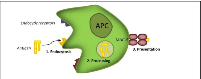
Considered as professional APCs, DCs are able to initiate primary immune responses by activating naïve T cells (Banchereau et al., 2000). Originating from monocytes, the immature DC possess the capacity to endocytose the antigens. Following their migration to the secondary lymphoid organs, DCs mature and express a wide panel of markers, endowing them with the capacity to activate T cells (Figure 6).
Figure 6. FVIII endocytosis and presentation on major histocompatibility class II (MHC II) molecules. The FVIII molecule is endocytosed through receptor(s) present at the surface of the APC. After FVIII internalization in the endosomes, the FVIII-derived peptides associate with MHC II molecules and are addressed at the surface of the APCs.
29
I.2.2.2 Macrophages
Macrophages are often described as scavenger cells, owing to the fact that they express a wide variety of endocytosis receptors on their surface (Taylor et al., 2005). Within the bloodstream, they circulate as monocytic precursors and differentiate into macrophages after their extravasation in the inflamed tissue. A wide variety of sub-populations of macrophages are also present in the spleen. In the spleen, metallophilic and marginal zone macrophages have been described to mediate exogenous FVIII endocytosis in FVIII-deficient mice and to participate in the initiation of the anti-FVIII immune response (Navarrete et al., 2009). Their capacity to activate T cells is lower than that of DCs and the endocytosed antigen have been proposed to mostly go towards the degradation pathway rather than the presentation pathway (Trombetta and Mellman, 2005).
I.2.2.3 B cells
B cells have been described to endocytose circulating antigens through their B cell receptor (BCR) which is a membrane bound immunoglobulin. Thus, it has been demonstrated that memory B cells are key players in secondary immune responses. The initiation of primary immune responses by B cells has been documented; however their role in initiating the anti-FVIII immune response in the context of hemophilia A has been poorly studied.
I.2.3 The spleen
The spleen comprises three distinct regions: the red pulp, the white pulp and the marginal zone. Blood borne antigens enter the spleen through the splenic artery, and are filtered by the macrophage-rich red pulp and the marginal zone. The marginal zone contains B cells, several macrophages sub-populations and DCs. To initiate an immune response, the antigen-loaded APC reach the white pulp where they activate T cells (Nolte et al., 2000). Thus, the spleen has a tremendous role in both the capture and presentation of blood borne antigens (Mebius and Kraal, 2005). This is in line with the facts that, in mice, the administered FVIII was found to
30
accumulate in the spleen and that splenectomy prior to the administration of FVIII reduces the anti-FVIII immune response (Navarrete et al., 2009).
I.2.4 The anti-FVIII immune response in hemophilia A patients I.2.4.1 T-cell activation
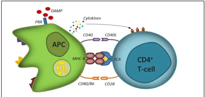
After its internalization by an appropriate APC, the FVIII molecule will be proteolysed in the endosomes. The peptides that are generated associate with the MHC class II and are presented at the cell surface. Within the secondary lymphoid organs, CD4 positive T cells “sense” the presence of their specific antigen in association with the MHC class II through their T cell receptor (TCR). This constitutes the first activation signal for T cells. If no other activation signal is provided, the T cells are anergized and may acquire a regulatory phenotype (Quaratino et al., 2000). Thus, co-stimulatory molecules have to be expressed on APCs to activate T cells. These co-stimulation markers are expressed only if the APCs receive a “danger signal”. Once such a signal is provided, the APC matures, leading to the overexpression of surface molecules such as CD80, CD86 and CD40 (Figure 6). The expression of these molecules contributes to the activation of T cells. Thus, the CD80 and CD86 molecules expressed on APCs interact with CD28 on T cells, and CD40 interacts with the CD40L that is expressed on activated CD4+ T cells. Such a cross-talk contributes to T-cell activation but also strengthen the stimulation of APCs. It has been shown that the in vivo invalidation of CD80 or CD86, but also the blockade of CD40, abrogates the anti-FVIII immune response (Qian et al., 2000). To achieve the activation of T cells, a third signal is required. Indeed, activated APCs secrete different types of cytokines, depending on the danger signal that they initially received. The released cytokines polarize the differentiation of T cells into T helper 1, 2 or 17 cells (Figure 7). In inhibitor-positive patients with hemophilia A, all three subsets of T cells were found (Ettinger et al., 2009). However, patients with low inhibitory titres seem to have a Th1 profile whereas patients with high inhibitory titres present
31
with a Th2 profile, as evaluated by analysis of the dominant IgG subclasses (Reding et al., 2002).
Figure 7. CD4+ T-cell activation by APC. Initially, the TCR recognizes the antigenic peptide in association
with the MHC class II. The presence of a danger signal (danger associated molecular pattern, DAMP) leads to the expression of co-stimulation markers on the APC (CD80:86 and CD40). These markers engage the CD28 and CD40L expressed on T-cells. This second signal leads to the secretion of cytokines by the APC which finalizes the activation of the T cell.
I.2.4.2 B-cell activation
As explained previously, B cells also have the capacity to recognize and endocytose circulating antigens through their BCR. As in the case of dendritic cells, the endocytosed antigen is cleaved and presented at the B-cell surface in association with the MHC class II. At this step, the B cells are screened by the TCR of activated antigen-specific T cells. When T-cells encounter FVIII-presenting B T-cells, the interaction of the T-cell CD40L and CD28 with the B-cell CD40 and CD80/86 completes the activation of the B cells (De Groot and Scott, 2007). The activated B cells then differentiate into plasmocytes or memory B cells. The plasmocytes migrate to the bone marrow and produce antigen-specific immunoglobulins.
32
Figure 8. B-cell activation. After the internalization of a soluble antigen through their BCR, the B cells process and present the antigenic peptide on MHC II molecules. The recognition of the complex peptide/MHC II by an activated antigen-specific T cell leads to the maturation and differentiation of the B cell into memory B cells and plasmocytes. Plasmocytes are producing antigen-specific immunoglobulins.
I.3 Risk factors
As presented earlier; the initiation and amplification of an immune response requires the participation of inflammatory mediators. This phenomenon is inherent to the danger theory, initially proposed by P. Matzinger. Thus, several genetic and non-genetic risk factors have been identified or proposed to participate in the initiation of the anti-FVIII immune response.
I.3.1 The danger signal theory
The concept of danger signal is derived from the danger theory proposed in 1994 by P. Matzinger (Matzinger, 1994). According to this, APCs are activated by danger signals. These signals can originate from invading pathogens, which express several structures able to trigger the immune response. These structures are called “pathogen-associated molecular patterns” (PAMP). It can also originate from self-cells, which secrete or expose several endogenous molecules in response to cellular stress. In 2002, P. Matzinger merged these two kinds of molecules (i.e., endogenous and exogenous PAMPs) under the same terminology, i.e., “damage-associated molecular patterns” (DAMP).
33
I.3.1.1 Exogenous danger signals
PAMPs correspond to molecular components found only in microorganisms such as bacteria, viruses or fungi. These microorganisms possess specific structures such as carbohydrates or glycolipids that are not present in the mammalian host. These components may derive from the microorganism wall such as lipopolysaccharide (LPS) and peptidoglycan for bacteria, or carbohydrates for viruses. They can also originate from the cytosolic compartment of the microorganism, such as nucleic acids, that are exposed upon pathogen lysis or phagocytosis (Sirisinha, 2011).
I.3.1.2 Endogenous danger signals
The first identified danger signals were the CD40 ligand (CD40L), the tumor necrosis factor-alpha (TNF-α) and the interleukine-1 beta (IL1-β). These molecules are able to directly induce the maturation of APC (Koide et al., 1987; Santiago-Schwarz et al., 1993; Caux et al., 1994). Under physiological conditions, these factors are stored inside the cells and are not in contact with their specific receptors on the immune cells. In case of cellular stress or cellular damage, these mediators are released in the extracellular microenvironment. The general example is provided by necrotic cells, which release several molecules able to activate the immune system such as heat shock proteins (HSP) (Quintana and Cohen, 2005), the chromatin associated high mobility group box 1 (HMGB1) (Scaffidi et al., 2002), and some purine-metabolism derived molecules such as ATP or uric acid (Bours et al., 2006).
I.3.2 Genetic risk factors
Several immune-related genes have been under investigation to assess their role in facilitating the anti-FVIII immune response. Amongst them are found the type of FVIII mutation, the HLA haplotype, and polymorphisms in a wide variety of immune-related genes. The type of FVIII mutation is particularly important because, as explained previously, it dictates the establishment of a regulatory response to FVIII during the ontogeny of the immune system.
34
The HLA haplotype is also of particular interest because it is a key component in antigen presentation. Eventually, the identification of immune-related gene polymorphisms may allow the identification of patient at higher risk of FVIII inhibitors development.
I.3.2.1 FVIII mutations
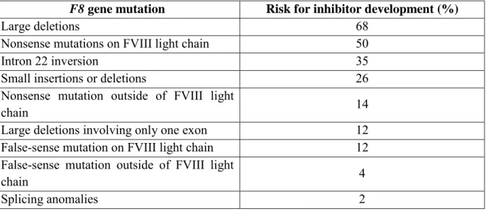
As detailed in section I.1.1.3, several types of mutations are affecting the F8 gene. Although the common feature of mutations in severe hemophilia A patients is the complete abolition of FVIII activity, the different mutations may have different effects on the production, function and structure of FVIII. Thus, the presence of a non-functional FVIII in the circulation imparts the patient with a lower risk for inhibitor formation compared to a patient in whom the FVIII molecule is not synthesized or secreted. Thus, the risk for inhibitor development has been linked to the type of mutation as presented in Table 1.
Table 1. F8 gene mutations and risk for inhibitor development
F8 gene mutation Risk for inhibitor development (%)
Large deletions 68
Nonsense mutations on FVIII light chain 50
Intron 22 inversion 35
Small insertions or deletions 26 Nonsense mutation outside of FVIII light
chain 14
Large deletions involving only one exon 12 False-sense mutation on FVIII light chain 12 False-sense mutation outside of FVIII light
chain 4
Splicing anomalies 2
Table adapted from the 2006 AFSSAPS report Surprisingly, in patients with the Inv22 mutation, the incidence of FVIII inhibitors is lower than in the case of patients with large deletions. In Inv22 patients, since no circulating FVIII antigen is detected, one would expect the absence of thymic education towards FVIII, and the ensuing failure to eliminate FVIII-specific CD4+ T cells and to generate regulatory T cells.
35
However, as explained in section I.1.1.3, the work from Pandey et al. has suggested that, in patients with the Inv22 mutation, FVIII is synthesized as two polypeptides that remain inside the cells. In particular, Pandey et al. claimed to have detected FVIII antigen in the peripheral blood mononuclear cells (PBMCs) of healthy donors and patients. These data lead the authors to propose that immunological tolerance to FVIII might be triggered at a better level in Inv22 patients as compared to patients where the F8 gene mutation abrogates the production of the protein. These data are however contradicted by recent publications documenting the preponderant role of endothelial cells in FVIII synthesis and absence of detection of the FVIII mRNA in peripheral blood cells(Everett et al., 2014).
I.3.2.2 HLA haplotype
Several studies have emphasized the associating between the HLA haplotypes of the patients with the development of FVIII inhibitors. Thus, it appears that the HLA class II alleles DRB1*15 and DQB1*0602 are associated with a higher risk for FVIII inhibitor development (Hay et al., 1997; Pavlova et al., 2009). Conversely, the HLA class II alleles DRB1*11 and DQB1*03 have recently been associated with a reduced risk for FVIII-inhibitor development (Pergantou et al., 2013). It is important to keep in mind however that haemophilia A is a rare disease and that it is extremely challenging to constitute large enough cohorts of patients to accurately determine the importance of respective HLA haplotypes.
I.3.2.3 Polymorphisms in immune-related genes
The analysis of single nucleotide polymorphisms (SNPs) in different immune-related genes resulted in the identifications of variants in cytokine genes that were associated with the development of FVIII inhibitors. Thus, SNPs in the promoter of the genes encoding IL-10, TNF-α and CTLA-4 were associated with a higher incidence of FVIII inhibitor in patients with hemophilia A (Astermark et al., 2006a; Gouw and van den Berg, 2009; Pavlova et al., 2009). Indeed, the presence of a 134-bp-long variant of a CA repeat microsatellite in the
36
promoter region of the IL-10 gene, previously associated with high levels of autoantibodies in several auto-immune diseases, was also associated with the presence of FVIII inhibitors in patients with severe hemophilia A (Astermark et al., 2006b). Similarly, within the TNF-α promoter, four SNPs were identified (-308 G>A, -827 C>T -238 G>A and -670 A>G) that affect the transcription of the TNF-α encoding gene. Subsequently, patients with severe hemophilia A possessing the combination of alleles -308AA -827CC -238GG and -670AA were found to be more susceptible to develop an inhibitor (Astermark et al., 2006a). Similarly, carriage of the G allele at position +49 of the CTLA-4 gene is associated with a decreased expression of CTLA-4, leading to an increased proliferation of activated T-cell, was associated with a higher incidence of inhibitor in patients with severe hemophilia A (Astermark et al., 2007). Recently, the Fc gamma receptor IIa (FcγRIIa or CD32) R131H polymorphism, that increases the binding affinity of the FcγRIIa to IgG1 and IgG2 isotypes was associated with inhibitor development in patients with severe hemophilia A (Eckhardt et al., 2014). Moreover, a genome-wide association study (GWAS) was conducted using a combined cohort involving 833 patients with severe hemophilia A from three independent cohorts (HIGS, MIBS and HGDS). This GWAS analysis evaluated the association of 13,331 SNPs from 1,081 genes with the presence of FVIII inhibitor in patients with severe hemophilia A. The authors identified 53 SNPs as significant predictors of inhibitor status. More precisely, within the 13 SNPs that were concordant in the three cohorts analyzed, 5 were associated with an increased risk for the development of FVIII-inhibitor (MAPK9, DOCK2, CD44, IQGAP2 and CSF1R) and 8 were found to be protective (PDGFRB, PCGF2, HSP90B1, F13A1, IGSF2, ALOX5AP, MAP2K4 and PTPRN2) (Astermark et al., 2013).
37 I.3.3 Non-genetic risk factors
The idea that the anti-FVIII immune response is not only dictated by patients‟ intrinsic risk factors originates from the observation that homozygous hemophilic twins do not respond equally to FVIII infusion from an immunological point of view: some might develop inhibitors while other might not (Astermark, 2006). Thus, the age of the patient at the start of the treatment, the mode and intensity of FVIII administration at the first bleeding episode and the state of the immune system at the time of FVIII administration, are among the most debated non-genetic risk factors.
I.3.3.1 Age at the start of treatment
Lorenzo et al reported in 2001 a higher incidence of inhibitor formation in Spanish patients where FVIII infusion was started before the age of 6 month (Lorenzo et al., 2001). The same conclusion was reached in another cohort from the Hemophilia Center in Utrech where a time-dependence between the age at first FVIII administration was correlated with inhibitor formation (van der Bom et al., 2003). However, this was not confirmed in the Milanese cohort where no association was found (Santagostino et al., 2005). It has since been proposed that early replacement therapy in young patients reflects a more severe clinical conditions that require more intensive treatments, thus suggesting the participation of other risk factors, such as treatment intensity.
I.3.3.2 Mode and intensity of FVIII administration
Different patients with hemophilia A do not display similar bleeding patterns. Thus, the treatment that they received differs depending on their bleeding phenotype. In line with this observation, the question of the repercussion of the treatment schedule and FVIII dosage on the development of the anti-FVIII immune response has been raised. To answer this question, the RODIN study evaluated the incidence of the mode of administration of FVIII and intensity of treatment on FVIII inhibitor formation. This was done using a large cohort of 606
38
patients involving 29 hemophilia treatment centres. The results from this study indicate an increased risk for inhibitor development after intensive FVIII treatment and a decreased risk when prophylaxis was used. However, the risk for inhibitor development was the same during the first 20 exposure days, irrespectively of the infusion regimen. As a conclusion, the authors suggest that patients may be divided into three groups: the first group of patients in whom inhibitors will never develop independently of the treatment regimen, patients in whom inhibitors will develop depending on the treatment, and patients who will develop inhibitors in all cases (Gouw et al., 2013).
I.3.3.3 State of the immune system at the time of FVIII infusion
Infectious disease, vaccination, severe bleeds or surgeries are events associated with the activation of the pro-inflammatory machinery, and hence of the immune system. Thus, it appears plausible to hypothesize that the concomitant infusion of FVIII along with one of these events would result in a higher risk for inhibitor development. This concept is supported by the observation that, in some inhibitor-negative hemophilia A patients, an anti-FVIII immune response was observed after a surgical procedure (Sharathkumar et al., 2003). This was however not confirmed in the case-control study by Santagostino et al, where no association between the immune status at start of the treatment and inhibitor development was found.
Deciphering the participation of some of these factors represents the main goal of my PhD work.
39 I.4 Purpose of the PhD work
The global aim of my PhD work was to decipher the bleeding-associated risk factors in the development of FVIII inhibitors. This general question will be addressed in four parts. The first part focuses on the association of a polymorphism in a gene expressed in response to bleeding events with the development of FVIII inhibitors in patients with severe haemophilia A. The second part exploits a mouse model of severe haemophilia A in order to evaluate the impact of severe bleedings in the anti-FVIII immune response. The third chapter of the result section concentrates on whether modulation of the oxidative state may affect the anti-FVIII immune response in vivo. Lastly, I assess whether oxidative modifications of the FVIII molecule enhance its immunogenicity. For a better understanding of these objectives, these four objectives will be introduced separately.