HAL Id: hal-01849243
https://hal.archives-ouvertes.fr/hal-01849243
Submitted on 5 Jun 2019
HAL is a multi-disciplinary open access
archive for the deposit and dissemination of
sci-entific research documents, whether they are
pub-lished or not. The documents may come from
teaching and research institutions in France or
abroad, or from public or private research centers.
L’archive ouverte pluridisciplinaire HAL, est
destinée au dépôt et à la diffusion de documents
scientifiques de niveau recherche, publiés ou non,
émanant des établissements d’enseignement et de
recherche français ou étrangers, des laboratoires
publics ou privés.
Dendrimeric coating of glass slides for sensitive DNA
microarrays analysis.
Véronique Le Berre, Emmanuelle Trévisiol, Adilia Dagkessamanskaia, Serguei
Sokol, Anne-Marie Caminade, Jean Majoral, Bernard Meunier, Jean François
To cite this version:
Véronique Le Berre, Emmanuelle Trévisiol, Adilia Dagkessamanskaia, Serguei Sokol, Anne-Marie
Cam-inade, et al.. Dendrimeric coating of glass slides for sensitive DNA microarrays analysis.. Nucleic Acids
Research, Oxford University Press, 2003, 31 (16), pp.e88. �10.1093/nar/gng088�. �hal-01849243�
Dendrimeric coating of glass slides for sensitive
DNA microarrays analysis
VeÂronique Le Berre
1,2, Emmanuelle TreÂvisiol
1,2,3, Adilia Dagkessamanskaia
1,
Serguei Sokol
1, Anne-Marie Caminade
3, Jean Pierre Majoral
3, Bernard Meunier
3and
Jean FrancËois
1,2,*
1Transcriptome-Biochips Platform of Genopole Toulouse Midi-PyreÂneÂes,2Centre de BioingeÂnierie Gilbert Durand
UMR-CNRS 5504, UMR-INRA 792, Institut National des Sciences AppliqueÂes, F-31077 Toulouse cedex 04, France and3Laboratoire de Chimie de Coordination du CNRS, 205 Route de Narbonne, F-31077, Toulouse cedex
04, France
Received April 1, 2003; Revised and Accepted June 3, 2003
ABSTRACT
Successful use and reliability of microarray technol-ogy is highly dependent on several factors, includ-ing surface chemistry parameters and accessibility of cDNA targets to the DNA probes ®xed onto the surface. Here, we show that functionalisation of glass slides with homemade dendrimers allow pro-duction of more sensitive and reliable DNA micro-arrays. The dendrimers are nanometric structures of size-controlled diameter with aldehyde function at their periphery. Covalent attachment of these spher-ical reactive chemspher-ical structures on amino-silanised glass slides generates a reactive ~100 AÊ layer onto which amino-modi®ed DNA probes are covalently bound. This new grafting chemistry leads to the for-mation of uniform and homogenous spots. More-over, probe concentration before spotting could be reduced from 0.2 to 0.02 mg/ml with PCR products and from 20 to 5 mM with 70mer oligonucleotides without affecting signal intensities after hybridisa-tion with Cy3- and Cy5-labelled targets. More interestingly, while the binding capacity of captured probes on dendrimer-activated glass surface (named dendrislides) is roughly similar to other functional-ised glass slides from commercial sources, detec-tion sensitivity was 2-fold higher than with other available DNA microarrays. This detection limit was estimated to 0.1 pM of cDNA targets. Altogether, these features make dendrimer-activated slides ideal for manufacturing cost-effective DNA arrays applicable for gene expression and detection of mutations.
INTRODUCTION
DNA microarrays are produced by in situ synthesis of oligonucleotides (1) or by mechanical deposition of
pre-fabricated nucleotides onto chemically reactive support by means of ink jetting or spotting robot devices (2,3). The activated glass plates (e.g. microscope slides) are readily available, easy to handle and commonly used to deposit DNA, peptides, antibodies, low-molecular-weight molecules, etc. (4,5), because of their great chemical resistance against solvents, their mechanical stability and low intrinsic ¯uores-cence properties. The performance of functionalised glass surface, e.g. signal intensity, signal-to-noise ratio, spot homogeneity, is directly in¯uenced by the amount of DNA probes attached to the surface, by the probe length and by the accessibility of the labelled targets to the probes. Taking into account these requirements, an ideal support should have an excellent surface chemistry allowing covalent binding of DNA with assurance that this attachment is stable, a high binding capacity obtained by increasing the number of reactive sites to which the probes can be covalently bound, and a low background. Furthermore, probes should not be spread on the support, as this could induce problems of accessibility during the hybridisation step. Shchepinov et al. (6) suggested that an optimal spacer should possess at least 40 atoms in length, to not contain positive or negative charges, in order to space away the probe from the surface.
To try to ful®l these needs, two major achievements have been reported in recent years, which have progressively replaced the low-ef®ciency non-covalent binding of DNA on poly-L-lysine-coated glass slides (7,8). The ®rst progress was
the covalent binding of DNA probes onto silanised glass slides via a spacer of four to 12 carbon units, with an epoxy, aldehyde or isothiocyanate reactive function at its extremity (9±11). The attachment of the DNA probes occurs by their 5¢-termini, which must be amino or thiol-modi®ed, depending on the chemical reactive group of the linkers. While good quality, accuracy and reliability of these microarrays were demon-strated (11), the linear structure of the spacer still leaves a planar surface structure on the glass-slide, which neither increases by very much the accessibility of the targets to the probes nor the loading capacity of the support. To overcome in part these failures, a second major advancement has been to tentatively build up a pseudo-three-dimensional structure using the so-called `dendrimeric' linker system. A signi®cant
contribution to this achievement was provided by the work of Beier and Hoheisel (12) who generated a pseudo-dendritic linker by direct chemical reaction on the glass slide or on polypropylene surfaces. With this structure, they could show an increased binding capacity of the activated-support for the probe beyond that of the glass slide and a high stability of the bonding of the dendrimer to the support. However, a major disadvantage of this product is that the linker system must be done in situ by eight time-consuming chemical steps, precluding any quality control of the linkers during the process, and limiting strongly a potential industrial develop-ment of this methodology. An alternative chemical process was the p-aminophenyltrimethoxysilane (ATMS)/diazotisa-tion chemistry of the glass slides (13). However, major drawbacks of this grafting technology are that the ATMS-activated glass plates must be kept at 4°C throughout the process and that the covalent binding of DNA cannot be controlled as it could occur by any base of the probe. Another work described the use of prefabricated polyamino-functionalised `PAMAM starbust' macromolecules for the manufacture of biochips, which includes an activation of the dendrimer layer surface prior to coupling with the DNA probes (14,15). Though this technology likely provides the best quality for DNA chips in terms of spots homogeneity, high sensitivity, low background and possible reutilisation, the chemical procedure to functionalise glass slides is time-consuming and leads to low thermal stability of the PAMAM dendrimers. Moreover, the support must be grafted with probes immediately upon activation, which limits the large-scale development of PAMAM-activated slides.
In the past 10 years, we have developed in our laboratory the production of dendrimers (16±18) based on an iterative reaction sequence that originates from a central core of hexachlorocyclo-triphosphazene (N3P3Cl6) linked to external
rami®cations to give rise to various generations (Table 1). The dendrimers are nanometric spherical structures whose diam-eter depends on the number of rami®cations and which possess at their periphery a reactive function that can be aldehyde, thiol, epoxy, etc. In this work, we used dendrimers of generation 4 with aldehyde function to generate on amino-silanised glass surface a stable layer onto which NH2-modi®ed
DNA probes are covalently ®xed by their 5¢-ends. Advantages of this dendrimer-based technology and corresponding DNA microarrays over commercially available activated surface and DNA chips are illustrated, as well as their validation in biological applications.
MATERIALS AND METHODS Materials
All chemical and solvents were purchased from Fluka/Sigma (Gillingham, UK) and were of the highest analytical grade. Untreated microscope glass slide (Gold Seal Microslides) and Sigma Screen APS were purchased from Sigma/Aldrich (Gillingham, UK), QMT Amino-slide from Quantifoil Micro Tools GmbH (Jena, Germany), SAL-1 and SA-1 GENORAMAÔ Microarray slides from Asper Biotech (Tartu, Estonia), SuperAldehyde and SuperAmine from ArrayltÔ (Telechem, USA), Ez-rays amino-slide from Apogent Discoveries (Hudson, USA) CMT-Gap, CMT-Gap2
Ultragap from Corning (Kentucky, USA), EasySpotâ Oligo
Microarray slide from U-Vision Biotech (Taipei, Taiwan) and 3D-LinkÔ from Surmodics/Amersham Biosciences (Minnesota, USA). The 70mer oligonucleotides package corresponding to the 6200 yeast ORFs was purchased from Qiagen/Operon (Germany). PCR products that corresponded to yeast ORFS (see Figures 4 and 5 for details) were ampli®ed using the commercially available set of primers (GenePair, Invitrogen Corp.).
Production of the dendrimer-activated glass slides Step 1: glass cleaning. Microscope glass slides were cleaned by immersion for 2 h in 60% ethanol containing NaOH 100 g/l (19). The slides were rinsed three times with MilliQ-®ltered water and were then submerged in 95% ethanol.
Step 2: silanisation of the slides. Cleaned microscope slides were immersed in a solution consisting of 3-aminopropyl-triethoxysilane 10% made in 95% ethanol for 12±15 h at room temperature under constant agitation. The slides were washed twice in 95% ethanol, twice in MilliQ-®ltered water, dried under a stream of nitrogen and ®nally baked for 3 h at 120°C. Step 3: bonding of dendrimers on the silanised slides. The silanised glass slides were immerged in an aqueous solution of KOH (8%) for 5 min. They were rinsed three times in MilliQ-®ltered water and dried by centrifugation (8 min at 500 r.p.m.) or under a nitrogen stream. These activated slides were then incubated in a solution containing phosphorus dendrimers [0.1%, w/v, generation 4 (17)] in dichloromethane for 7 h at room temperature under gentle agitation. The slides were then washed twice with dichloromethane then with 95% ethanol and ®nally with absolute ethanol. They were dried by centrifugation or under a nitrogen stream. The dendrislides were kept at room temperature in plastic bags before use. Spotting DNA probes onto the activated support surfaces
Unless otherwise stated, a 35mer 5¢-labelled Cy5-oligonucleo-tide-3¢NH2
(5¢Cy5-TTAGCGCATTTTGGCATATTTGGGC-GGACAACTT-NH2-3¢) or the PCR products corresponding to
the yeast ORFs were dissolved in the phosphate buffer 0.3 M, pH 9. All capture probes were synthesised with a modi®ed primer bearing an amine at their 5¢- or 3¢-end. The probes were printed on glass slides by mechanical spotting with a robot (Eurogridder from Virtek/Bio-Rad). The print head on the arrayer uses `quill' pins, whose contact on the surface delivers
Table 1. Size of dendrimers and number of aldehyde reactive functions according to the dendrimer generation
Generation Number of reactive
CHO functions Size (AÊ)
1 12 30 2 24 45 3 48 60 4 96 75 5 192 90 6 384 105 7 768 120
~2 nl of DNA solution per spot, with a medium size diameter of ~80 mm under this condition. The quality of the printing was optimal at 45% relative humidity and a constant temperature of 22°C. After spotting, the slides were allowed to dry overnight at room temperature. The reduction of the imines function formed between probes and dendrimer was carried out by immersion of the slides into a solution containing NaBH4 at 3.5 mg/ml for 3 h at room temperature under
agitation. The DNA slides were washed in boiling water for 2 min, twice in room-temperature water and then dried under a stream of N2. The DNA arrays were then stored in a plastic
container at 4°C until use. Preparation of labelled targets
In experiments shown in Figures 2B and 5, the target was a 15mer 5¢-labelled Cy5 oligonucleotide (Cy5-AATGGTAT-CGGAGTA) that is complementary to the 35mer probes (5¢NH2
-GTGATCGTTGTATCGAGGAATACTCCG-ATACCATT). Other targets were prepared from total yeast RNA as a template by incorporation of ¯uorescent-labelled Cy5 or Cy3-deoxyribonucleotides during ®rst-strand cDNA synthesis or by using the post-labelling (amino-allyl) method (20). The reaction was carried out with 15 mg total RNA using the CyScribeÔ First Strand cDNA Labelling Kit or CyScribe Post-Labelling Kit (Amersham Bioscience) and puri®ed on a GFX column (Amersham Bioscience).
Hybridisation to the DNA-arrays, image detection and stripping procedures
Prehybridisation of the printing area was carried out with 20 ml solution containing 18 ml Dig Easy buffer (Roche Diagnostic) and 2 ml of denaturated salmon sperm DNA over 1 h at 42°C. The slide was washed with 23 SSC and then hybridised at 42°C for 12±14 h in a similar solution to which was added labelled cDNA targets that was boiled for 5 min prior to addition. In initial experiments, the hybridisation was carried out in a coverslip hybridisation cassette (Corning Inc), which was kept humid by addition of 20 ml water in the chamber. Experiments reported in Figures 4B and 6 were carried out with an automatic hybridisation chamber (Discovery from Ventana Medical System, Inc). Prehybridisation was carried out with a freshly prepared solution of 1% BSA, 23 SSC, 0.2% SDS over 30 min at 42°C. After automatic washing according to manufacturer's instructions, the slides were hybridised for 10 h in 200 ml of ChipHybeÔ buffer (Ventana Medical System, Inc) containing 10 ml of labelled cDNA puri®ed on the GFX column. After hybridisation, the slides were washed for 2 min in 23 SSC/0.2% (v/v) SDS, for 2 min in 0.23 SSC/0.2% (v/v) SDS and for 2 min in 0.23 SSC, and then dried under a stream of nitrogen. Detection of the ¯uorescence signals was captured with a laser scanner (GenePix 4000A from Axon Instrument, CA) and analysed with the GenePix 3.01 software. Data were further processed by statistical methods using our homemade BioPlot Software (S.Sokol, C.Declercq and J.FrancËois, unpublished data, see http://bio71.gba.insa-tlse.fr/). For re-use, DNA-chips were incubated in a stripping solution [2.5 mM Na2HPO4and 0.1%
(v/v) SDS] for 10 min at 95°C. They were then washed with ddH2O, dried under a stream of N2and checked for complete
loss of ¯uorescence by scanning with the laser scanner
(GenePix 4000A from Axon Inc) at appropriate sensitivity levels of photomultiplier (PMT).
RESULTS AND DISCUSSION
Properties of the dendrimer-activated support
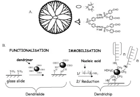
We have developed a chemical process for the covalent immobilisation of DNA probes (either PCR fragments or oligonucleotides) to microscope slides using a poly-functional polymer called a dendrimer. The dendrimers are nanometric spherical structures that originate from a central core from which emerge external rami®cations (16), giving rise to different generations of dendrimers, according to the number of rami®cations. They harbour at their outer sphere a speci®c reactive function (i.e. aldehyde, epoxide or thiol, etc.), the number of which is proportional to the rami®cation units (Table 1). The synthesis of these dendrimers has been described in detail by Launay et al. and Slomkowski et al. (16±18), and the chemical process for their covalent binding on the glass-slide has been patented (21). Figure 1 brie¯y outlines the protocol of manufacturing dendrimer-activated glass slides (dendrislide) and the covalent attachment of DNA probes on these dendrislides (dendrichip). This procedure to yield dendrimer-activated glass slides is very simple, effective and much faster than a previous method using `PAMAM starburst' macromolecules (14). Moreover, the slides can be stored for months at room temperature before use which provides a serious technological advantage over PAMAM-dendrimer-activated glass surface. As the dendrimers differ in size and number of rami®cations, different generations from G1 to G7 (Table 1) have been evaluated for their ability to reduce the signal-to-background after hybridisation of a 15mer Cy5-labelled complementary strand to a 35mer DNA probe. Since the same values for signal-to-background ratio were found with dendrimers G4±G7 (data not shown), we decided to use generation 4 merely because the next gener-ation is dependent on the previous one due to the itergener-ation process for dendrimer synthesis (16). Dendrimer G4 has a diameter size of about 75 AÊ, and displays 96 aldehyde functions at its periphery. We also veri®ed that the binding of DNA probes on the aldehyde function of dendrimers occurs speci®cally via the 5¢-amino modi®ed terminal of the probes because no ¯uorescent signal was recorded after processing of the dendrimer-activated glass surface with an unmodi®ed 15mer Cy3-labelled DNA probe (data not shown).
Choice of spotting conditions
As suggested by Diehl et al. (22), the buffer into which DNA probes are dissolved prior to spotting must be adequate for the chemical function of the activated-glass surface. It was also shown that the quality of the spotting on the nylon membrane was improved when the nucleic acids are dissolved in 50% dimethylsulfoxide (DMSO) (23). The advantage of this organic solvent is its denaturing property and its effect of strongly reducing evaporation. Both properties improve the homogeneity of the spots: they remain circular without a `doughnut' effect. We also successfully applied this protocol for the spotting of DNA probes on amino-silanised glass slides purchased from different companies (amino-silane surface CMT-GAPÔ from Corning; Telechem SuperAmine; Asper
Biotech amino-silane SA, data not shown). However, this protocol did not work appropriately with dendrimer-functionalised glass slides. As shown in Figure 2, the spot's size increases as the concentration of DMSO in the DNA probes increases, and reached ~200 mm in the presence of 50% DMSO. Under this condition, the ¯uorescent signal of hybridisation with a mixture of Cy5-/Cy3-labelled targets obtained from total yeast RNA was 2- to 4-fold lower. This result suggests that DMSO may either reduce the actual amount of DNA ®xed on the dendrimeric-activated support, affect the interaction between probes and targets or may even partially modify the backbone of the dendrimer [likewise a transformation of the P(S) groups into P(O) groups, see Fig. 1]. To circumvent this problem and to ®nd a solvent capable of reducing evaporation during the spotting process, we tried
the methodology of Diehl et al. (22) who showed that the addition of 1.5 M betaine to DNA solution prior to spotting on amino-silanised glass slides increased binding ef®ciency and ¯uorescence signal, and reduced evaporation. In agreement with this, we also found that betaine offered the same advantages with the dendrimer-activated glass slide, and increased the signal intensities after hybridisation by 2±3 times (Fig. 3A). However, we noticed a precipitation of the DNA probes dissolved in 1.5 M betaine after storage at ±20°C. Thus, it is recommended to use the same DNA preparation for spotting within a short period of time.
Immobilisation ef®ciency
The binding capacity of the activated support is an important criterion for high density coupling of DNA probes, which can
Figure 1. Scheme of dendrimer `generation 4' (A) and description of surface activation to ®x the dendrimers and immobilisation method for covalent binding of nucleic acids (DNA probes) on the activated glass slide surface (B).
Figure 2. Images of hybridisation of a DNA probe spotted on activated glass slides with dendrimer generation 4 in the absence or in the presence of DMSO. (A) PCR products from two yeast ORFs (CDC19/YAL038w and CLN3/YAL040c) were resuspended in Na2phosphate buffer 0.3 M, pH 9 in the absence or
in the presence of DMS0 50%. (B) A 5¢-NH235mer oligonucleotide was prepared in the same buffer at two different concentrations (5 and 10 mM) in the
absence or presence of increasing concentration of DMSO. DNAs were spotted in triplicate on the activated dendrislides and hybridisation was carried out with yeast labelled cDNA (A) or with a complementary Cy5-labelled 15mer (5¢-Cy5-AATGGTATCGGAGTA-3¢) (B).
increase the signal-to-noise ratio and lower the detection limit upon hybridisation analysis. However, high surface densities and high hybridisation signal might not necessarily be related, since steric interferences between immobilised probes could hinder access to target DNA strand and reduce the hybridisa-tion ef®ciency (15,24,25). Therefore, we compared the
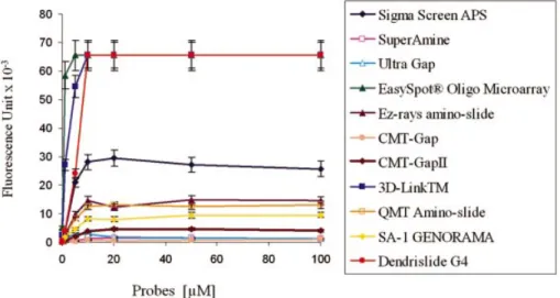
immobilisation capacity of G4-dendrimer slides with 10 conventional/commercial available activated glass slides (Materials and Methods). This was performed by spotting a 35mer oligonucleotide (Cy5-labelled at its 5¢-end and con-taining a NH2 group at its 3¢-end for covalent binding to
supports) four times at concentrations ranging from 0.01 to
Figure 3. Image capture from hybridisation of arrays manufactured with dendrislide bearing DNA probes spotted at decreasing concentrations. (A) PCR from yeast ORF (ORF1: MET14/YKL001c, ORF2: SPC42/YKL042w, ORF3: MRP17/YKL003c) was spotted in duplicate (column) in phosphate buffer 0.3 M, pH 9 at 200 and 20 mg/ml (rows) in the absence or in the presence of 1.5 M betaine. (B) A 70mer oligonucleotide corresponding to the gene SSA1/YAL005c was spotted in quadruplet in the same buffer at decreasing concentration ranging from 20 to 1 mM. The hybridisation was carried out with Cy3-labelled cDNA (A) or a mix of Cy3-/Cy5-labelled cDNA (B) obtained from 10 mg of yeast total RNA. In (A), the value of ¯uorescence intensity (¯uorescent signal ± background) measured at 532 nm is given on the top of the corresponding spots. The ¯uorescence intensity was measured with the Genepix scanner at 500 PMT.
Figure 4. Immobilisation ef®ciency of G4-dendrislides compared with other commercially available activated-glass slides. Single-stranded 35mer oligonucleotide carrying a 3¢-amino function and a 5¢-Cy5 ¯uorophore (5¢-Cy5-TTAGCGCATTTTGGCATATTTGG GCGGACAACTT-NH2-3¢) was spotted
at concentrations from 0.01 to 100 mM, on the different functionalised-glass slides. The value of ¯uorescence intensity (¯uorescent signal ± background) was measured at 635 nm with the Genepix scanner at 500 PMT.
100 mM, which correspond to probe densities ranging from 0.02 to 250 fmol/mm2. The ¯uorescence signal, which
estimates the immobilisation ef®ciency, was recorded with a laser scanner (GenePix 4000A) at PMT 600, and the mean value for the four data points was calculated for each
concentration of spotted DNA probes. Using our dendrislides, the ¯uorescence intensity reached saturation at 10 mM DNA probes (Fig. 4). Apparently, this chemistry did not offer a better binding capacity since saturation was already attained at this concentration with two other commercial arrays that are functionalised for covalent attachment of the probes (EasySpotâ Oligo Microarray slide from U-Vision Biotech
and 3D Links from Surmodics/Amersham Biosciences). Interestingly, this mode of attachment resulted in a 2- to 3-fold higher ¯uorescent intensity than that from amino-silanised slides (as for instance the very commonly used ultra Gap slides from Corning or Sigma screen APS, see Fig. 4). This result suggests that the dendrimer-modi®ed surface has a higher surface coverage and reactivity in comparison to amino-silanised glass slides. The lower reactivity of these latter glass slides might be due to steric constraints or by the fact that the probes are spread over the surface, therefore reducing accessibility of other molecules for binding.
We carried out two other experiments by lowering the concentration of probes for spotting. In the ®rst experiment, unlabelled PCR products ranging from 400 to 600 bp from three yeast ORFs were spotted in duplicate at 0.2 mg/ml and at a 10-fold lower concentration. The binding capacity was indirectly estimated by hybridisation of these DNA probes with Cy5-labelled cDNA from total yeast RNA. Figure 3A shows that the hybridisation signal was the same at both concentrations of the spotted probes, and as indicated above, the signal intensities were 2-fold higher when the probes were prepared in the presence of 1.5 M betaine prior to spotting. The second experiment was carried out with 70mer oligo-nucleotides from a set of 96 yeast genes kindly provided by Operon (Array-Ready Oligo SetÔ Qiagen/Operon). They were spotted in quadruplets at decreasing concentrations from 20 to 1 mM. The arrays were hybridised as described above but using a mixture of Cy5-/Cy3-labelled cDNA targets from
Figure 5. Detection sensitivity of dendrislide compared with commercially available activated glass slides. A single-stranded 35mer amino-oligonucleotide (5¢-NH2-GTGATCGTTGTATCGAGGAATACTCCGATACCATT-3¢) at a concentration of 10 mM was spotted on the different functionalised glass slides.
The target Cy5-labelled 15mer oligonucleotides (5¢-Cy5-AATGGTATCGGAGTA-3¢) were hybridised at concentrations ranging from 0.001 to 100 nM. Image capture was measured at 635 nm with GenePix 4000A at 500 PMT. The value of ¯uorescence intensity reported is the ¯uorescent signal ± background.
Figure 6. Use of dendrislides to detect base mutations. Thirteen 20mer oligonucleotides were spotted in triplicate at 10 mM in Pi buffer 0.3 M, pH 9.0. These oligonucleotides corresponded to a part of the yeast HSP12 gene, and varied from each other by a single or two mutations proximal to the 5¢- or 3¢-end or in the middle of the sequence. The 20mer sequences from HSP12 were as follows: 1: NH2 5¢-AATATGTTTCCGGTCGTGTC-3¢; 2: NH2 5¢-AATATGTTTCGGGTCGTGTC-5¢-AATATGTTTCCGGTCGTGTC-3¢; 3: NH2 5¢-AATATGTT-TCAGGTCGTGTC-3¢; 4: NH2 5¢-AATATGTTTCtGGTCGTGTC-3¢; 5: NH2 5¢-AATATGTTTCCGGTCGTGTA-3¢; 6: NH2 5¢-AATATGTTTCC-GGTCGTGTG-3¢; 7: NH2 5¢-AATATGTTTCCGGTCGTGTT-3¢; 8: NH2 5¢-AATATGATTCCGGaCGTGTC-3¢; 9: NH2 AATATGGTTCCGG-GCGTGTC-3¢; 10: NH2 AATATGCTTCCGGcCGTGTC-3¢; 11: NH2 5¢-AATAAGTTTCCGGTCGTGTC-3¢; 12: NH2 5¢-AATAGGTTTCCGGT-CGTGTC-3¢; 13: NH2 5¢-AATACGTTTCCGGTCGTGTC-3¢. Hybridisation was carried out with Cy5-labelled cDNA from yeast total RNA. At the top of the corresponding spots is given the mean value of ¯uorescent signal (intensity at 635 nm ± background).
total yeast RNA (Fig. 3B). The hybridisation image of the 96 oligonucleotides microarrayed on the dendrimer-activated slides shows that signal intensities were the same from 20 to 5 mM, and dropped by three times at 1 mM DNA probe (see enlarged area of Fig. 3B). This suggests that at concentrations lower than 5 mM, the DNA probe was no longer in excess over the target DNA. From this experiment, we could estimate a minimal concentration of oligonucleotides probes of ~5 mM for ef®cient target/probe hybridisation on our dendrichips. As a consequence, the high surface coverage together with an ef®cient target capture at low oligonucleotide probe concen-tration offer several advantages for the dendrislides in both technological and economical terms.
Comparison of sensitivity and detection limit of dendrichips with other microarrays
The experiments reported above have already indicated that glass slides functionalised with spherical dendrimeric struc-ture not only have higher immobilisation ef®ciency, but also confer an excellent accessibility to the target DNA. To quantify the target/probe hybridisation sensitivity, arrays from dendrislides, as well as from 11 available activated slides from commercial sources, were manufactured with a ®xed amount of a 35mer oligonucleotide (spotted at 10 mM solution) and were hybridised with increasing concentration of a Cy5-labelled 15mer oligonucleotide complementary to the probe from 0.001 to 100 nM. As shown in Figure 5, the ¯uorescent intensity as recorded with the laser scanner set at PMT 600 reached a plateau at a concentration of 10 nM target DNA for almost all manufactured arrays. However, the logarithmic expression of signal intensities versus concentration of targets illustrated a clear advantage of the dendrimer-activated slides over all other microarrays at target concentrations <1 nM. A ¯uorescent signal, which corresponded to total ¯uorescence backgound of 1500 6 30, was measured at a concentration of 0.01 nM of cDNA target, which is about 10-times higher than for other commercially available slides, and at 0.001 nM of DNA target, a signal (¯uorescence background) was still easily quanti®able only using dendrichips. This result indicates a detection sensitivity of dendrichips that is ~10-to 100-fold higher than arrays made with most of other functionalised glass slides currently available on the market (13,19,22,23). The high hybridisation sensitivity is particu-larly interesting when measuring low abundance transcripts in cells and for studies involving very low amounts of biological material, such as biopsies. As an indication of this high sensitivity, we produced Cy3-labelled cDNA from 10 mg total yeast RNA by reverse transcription, and used these labelled cDNA targets at 1- to 100-fold dilution to hybridise dendrichips made with 70mer oligonucleotides from a set of randomly chosen 96 yeast genes provided by Operon. A quanti®able ¯uorescent signal on almost all spots could be recorded even with the lowest concentration of cDNA targets (data not shown).
Caution about re-usability of DNA microarrays
The possibility to re-use DNA arrays could be an advantage in transcriptomic studies, as it allows reproducibility of experi-ments on the same technical support. It also reduces the cost, while improving accuracy of the genomic data. Thus, we investigated the re-usability of the DNA arrays generated from
dendrimer-activated support. The ®rst approach that has been reported in several previous papers (12,13,20,22) was to spot a few sets of PCR or oligonucleotide probes. In our case, we spotted a 35mer oligonucleotide several times on dendrislides, and hybridised this prototype of microarrays with a Cy5-labelled target complementary to the probe. After reading the ¯uorescence intensity, the slides were regenerated by `stripping' using phosphate buffer (pH 7.5) containing SDS (0.1%, w/v) for 5 min at 95°C. After each stripping the slides were scanned to guarantee the complete removal of the target and subjected to rehybridisation. By application of this strategy, the arrays manufactured with dendrislides were re-used at least 10 times without signi®cant loss of the signal intensity. The same advantages were claimed with many other activated-glass slides that allow covalent linkage of the probes (12,15,20). We therefore wanted to verify this performance, using arrays bearing the whole yeast genome spotted as PCR products (300±600 bp length) or 70mer oligonucleotides (purchased from Qiagen/Operon) (see our website http:// bio71.gba.insa-tlse.fr for details on yeast microarrays con-struction). Contrary to expectation, the stripping procedure for these arrays was much less effective; leaving patches of faint colour on the slides that could not be removed even after two or three successive strippings. Similar data were obtained with yeast and rat oligo microarrays from commercial sources (not shown). This lack of complete removal of the targets from the arrays may be due to several reasons, including the heteroduplex interactions which involve secondary structures of the nucleic acids (26), as well as residual unspeci®c binding of dyes on the glass slides.
Biological application to mutations detection
Search for a single mutation in a given gene (SNPs) is a research ®eld where the DNA chip technology should bring a revolution (27±30). Although methods to identify SNPs already exist and most of them rely on the Affymetrix technology (1,31), the development of an alternative, inex-pensive, cost-effective high throughput method to identify point mutations in DNA sequences is still awaited in the context of medical diagnosis. Search of mutations in a given gene must use dedicated chips that contain all possible sequences of the DNA in which one wishes to seek these mutations. We examined the potential application of our dendrimer-activated support in such an endeavour. To this end, we spotted on dendrislides 13 20mer oligonucleotides from HSP12 encoding a protein chaperone in yeast (30), which differed from each other by a single or a double base mutation at positions proximal to 5¢, 3¢ or in the middle of the sequence. These DNA probes were then hybridised with Cy5-labelled cDNA obtained from reverse transcription of yeast total RNA. Figure 6 shows the image capture of this experiment. As expected, the highest ¯uorescent signal was recorded with probe 1, which exhibits the perfect comple-mentary DNA sequence of the yeast HSP12. Interestingly, a lower but measurable ¯uorescence intensity was recorded with oligonucleotides bearing mismatch mutation at the 3¢-end (i.e. probes 5±7). This result is an indirect indication that the probes attached to a dendrislide are highly accessible to cDNA targets.
CONCLUSION
The explosion of genome sequences from microbes, plants, animals and humans forces academic and industrial labora-tories to investigate functional genomics with the use of DNA chips in a massive way. So far, these tools have remained too expensive for most of the academic laboratories, and hence, efforts must be carried out to propose DNA arrays that are more reliable, cheaper and easy to use for these customers. To this end, we have generated a very effective chemically activated glass slide based on the use of homemade neutral phosphorus dendrimers. These G4-dendrimer-coated slides display most of the suitable criteria that one should expect for accurate, reliable and cost-effective DNA arrays. Since the production of dendrimer and the chemistry for activation of the surface are relatively simple and inexpensive, this process could be reproduced at an industrial scale. Moreover, we found that dendrislides were still active after 3±5 months of storage at room temperature. Since other functions, for example thiol, epoxy, etc., can be introduced at the periphery of the dendrimer structure, this renders these polymers particularly attractive for the immobilisation of other macro-molecules such as peptides, proteins, antibodies, lipids, etc. ACKNOWLEDGEMENTS
We are grateful to Drs C. Bergaud (LAAS-CNRS) and G. Pratviel (LCC-CNRS) for stimulating discussion during this work. This work was supported in part by grants from ReÂgion Midi-PyreÂneÂes (01002664) and GeÂnopole Toulouse Midi-PyreÂneÂes.
REFERENCES
1. Lipshutz,R.J., Fodor,S.P.A., Gingeras,T.R. and Lockhart,D.J. (1999) High density synthetic oligonucleotide arrays. Nature Genet., 14, 1675±1680.
2. Hughes,T.R., Mao,L.M., Jones,A.R., Burchard,J., Marton,M.J., Shannon,K.W. et al. (2001) Expression pro®ling using microarrays fabricated by an ink-jet oligonucleotide synthesizer. Nat. Biotechnol., 19, 342±347.
3. Cheung,V.G., Morley,M., Auilar,F., Massimi,A., Kucherlapati,R. and Childs,G. (1999) Making and reading microarrays. Nature Genet., 21, (suppl.), 15±19.
4. MacBeath,G., Koehler,A.N. and Schreiber,S.L.(1999) Printing small molecules as microarrays and detecting protein-ligand interactions en masse. J. Am. Chem. Soc., 121, 7967±7968.
5. Mac Beath,G. and Schreiber,S.L. (2000) Printing proteins as microarrays for high-throughput function determination. Science, 289, 1760±1762. 6. Shchepinov,M.S., Case-Green,S.C. and Southern,E.M. (1997) Steric
factors in¯uencing hybridisation of nucleic acids to oligonucleotide arrays. Nucleic Acids Res., 25, 1155±1161.
7. Schena,M., Shalon,D., Davis,R.W. and Brown,P.O. (1995) Quantitative monitoring of gene expression patterns with a complementary DNA microarray. Science, 270, 467±470.
8. De Risi,J.L., Iyer,V.R. and Brown,P.O. (1997) Exploring the metabolic and genetic control of gene expression on a genomic scale. Science, 278, 680±686.
9. Joos,B., Kuster,H. and Cone,R. (1997) Covalent attachment of hybridisable oligonucleotides to glass supports. Anal. Biochem., 247, 96±101.
10. Rogers,Y.-H., Jianf-Baucon,P., Huang,Z.-J., Bogdanov,V., Anderson,S. and Boyce-Jacino,M.T. (1999) Immobilization of oligonucleotides onto a
glass support via disul®de bonds: a method for preparation of DNA microarrays. Anal. Biochem., 266, 23±30.
11. Guo,Z., Gatterman,M.S., Hood,L., Hansen,J.A. and Petersdorf E.W. (2002) Oligonucleotide arrays for high throughput SNPs detection in the MHC class I genes: HLA-B as a model system. Genome Res., 12, 447±457.
12. Beier,M. and Hoheisel,J.D. (1999) Versatile derivatisation of solid support media for covalent bonding on DNA-microchips. Nucleic Acids Res., 27, 1970±1977.
13. Dolan,P.L., Wu,Y., Ista,L.K., Metzenberg,R.L., Nelson,M.A. and Lopez,G.A. (2001) Robust and ef®cient synthetic method for forming DNA microarrays. Nucleic Acids Res., 29, e107.
14. Benters,R., Niemeyer,C.M. and Wohrle,D. (2001) Dendrimer-activated solid supports for nucleic acid and protein microarrays. Chem. Biochem., 2, 686±694.
15. Benters,R., Niemeyer,C.M., Drutschmann,D., Blohm,D. and Wohrle,D. (2002) DNA microarrays with PAMAM dendritic linker systems. Nucleic Acids Res., 30, e10.
16. Launay,N., Caminade,A.M., Lahana,R. and Majoral,J.P (1994) A general synthetic strategy for neutral phosphorus-containing dendrimers. Int. Ed. Engl., 33, 1589±1592.
17. Launay,N., Caminade,A.M. and Majoral,J.P (1997) Synthesis of bowl-shaped dendrimers from generation 1 to generation 8. J. Organomet. Chem., 529, 51±53.
18. Slomkowski,S., Miksa,B., Chehimi,M.M., Delamar,M., Cabet-Deliry,M., Majoral,J.P. and Caminade,A.-M. (1999) Inorganic-organic systems with tailored properties controlled on molecular, macromolecular and microscopic level. Reactive Functional Polymers, 41, 45±57. 19. Zammatteo,N., Jeanmart,L., Hamels,S., Courtois,S., Louette,P.,
Hevesi,L. and Remacle,J. (2000) Comparison between different strategies of covalent attachment of DNA to glass surfaces to build DNA microarrays. Anal. Biochem., 280, 143±150.
20. Zhao,X., Nampalli,S., Serino,A.J. and Kumar,S. (2001) Immobilization of oligonucleotides with multiple anchors to microchips. Nucleic Acids Res., 29, 955±959.
21. TreÂvisiol,E., Leclaire,J., Pratviel,G., Caminade,A.M., Majoral,J.P., FrancËois,J. and Meunier,B. (2002) Supports solides fonctionaliseÂs par des dendrimeÁres phosphoreÂs, leurs proceÂdeÂs de preÂparation et applications. French patent no. 0205049 (April 23nd).
22. Diehl,F., Grahlmann,S., Beier,M. and Hoheisel,J.D. (2001)
Manufacturing DNA microarrays of high spot homogeneity and reduced background signal. Nucleic Acids Res., 29, e38.
23. Hegde,P., Qi,R., Abernathy,K., Gay,C., Dharap,S., Gaspard,R., Hughes,J.E., Snesrud,E., Lee,N. and Quackenbush,J. (2000) A concise guide to cDNA microarray analysis. Biotechniques, 29, 548±562. 24. Eisen,M.B. and Brown,P.O. (1999) DNA arrays for analysis of gene
expression. Methods Enzymol., 303, 179±205.
25. Henke,W., Herdel,K., Jung,K. Schnorr,D. and Loening,S.A. (1997) Betaine improves the PCR ampli®cation of GC-rich DNA sequences. Nucleic Acids Res., 25, 3957±3958.
26. Peterson,A.W., Heaton,R.J. and Georgiadis,R.M. (2001) The effect of surface probe density on DNA hybridisation. Nucleic Acids Res., 29, 5163±5168.
27. Hacia,J. (1999) Resequencing and mutational analysis using oligonucleotide microarrays. Nature Genet., 21, 42±47. 28. Hacia,J.G. and Collins,F.S. (1999) Mutational analysis using
oligonucleotide microarrays. J. Med. Genet., 36, 730±736. 29. Pastinen,T., Raitio,M., Lindroos,K., Tainola,P., Peltonen,L. and
Syvanen,A.C. (2000) A system for speci®c, high-throughput genotyping by allele-speci®c primer extension on microarrays. Genome Res., 10, 1031±1042.
30. Praekelt,U.M. and Meacock,P.A. (1990) HSP12, a new small heat shock gene of Saccharomyces cerevisiae: analysis of structure, regulation and function. Mol. Gen. Genet., 223, 97±106.
31. Maskos,U. and Southern,E.M. (1992) Parallel analysis of
oligodeoxyribonucleotide (oligonucleotide) interactions. I. Analysis of factors in¯uencing oligonucleotide duplex formation. Nucleic Acids Res., 20, 1675±1678.