9870
Rotavirus in various animal species in Ouagadougou,
Burkina Faso: detection of genotype G9.
Nafissatou Ouédraogo1, Stéphanie MT. Ngangas1, Aïssata Tiendrébeogo1, Alfred Sababénédjo
Traoré1, Isidore Juste O. Bonkoungou1,2, Nicolas Barro1
1Laboratoire de Biologie Moléculaire, d’Epidémiologie et de Surveillance des Bactéries et Virus Transmis par les Aliments, Centre de Recherche en Sciences Biologiques Alimentaires et Nutritionnelles (CRSBAN), Université de Ouagadougou, Ouagadougou, Burkina Faso.
2 Laboratoire National de Santé Publique, Direction de la Biologie médicale (DBM), Ouagadougou, Burkina Faso Corresponding author email: [email protected]
Original submitted in on 23rd June 2016. Published online at www.m.elewa.org on 31st July 2016 http://dx.doi.org/10.4314/jab.v103i1.8
ABSTRACT
Objectives: Rotaviruses have a wide host range, infecting many animal species as well as humans. The segmented nature of the genome suggests that rotaviruses are able to form new strains by a mechanism of reassortment. Animal rotaviruses are regarded as a potential reservoir for genetic diversity of human rotaviruses. The aim of this study was to determine the incidence and molecular characteristics of rotavirus in various healthy animals in Ouagadougou, Burkina Faso.
Methodology and results: A total of 618 faeces samples from various animal species with different living environments were collected between June 2009 and August 2011, and analyzed for rotavirus group A antigen detection by immunochromatographic test (SD Bioline Rota/Adeno®; Standard diagnostics, Inc., Korea). A second sample collection between February and March 2015 involved only farm animals (n= 138) and analyzed for rotavirus group A antigen detection by ELISA test (Ridascreen®, R-Biopharm AG, Darmstadt Germany). The rotaviruses antigen-positives samples for ELISA were further confirmed and characterized by reverse-transcription (RT-PCR). For immunochromatographic detection, the prevalence of rotavirus A and adenovirus antigens were found in 7.4% of pig, 31% of poultry, 33.4% of pigeon, 35.7% of rabbit, 46-58% in bovine, 13.8% of shrimps, 14.8% of snails and 28.6% of captain (Lates niloticus). The detection of rotavirus antigen by ELISA reported rates of 7.4% in pigs, 4.1% in cattle and 14.3% in poultry and no case of rotavirus was detected in sheep. The molecular characterization of the strains established that they belong to the G9 genotype (3/ 7; 42.9%).
Conclusion and application of results: This study provides evidence asymptomatic hosts of rotavirus. This study report for the first time rotaviruses detection and presence of the emerged genotype G9 in farms animals in Burkina Faso. These results justify the need to monitoring animals’ rotaviruses in Burkina Faso.
Keywords: Rotavirus group A, Animals, molecular characterization, Burkina Faso.
Journal of Applied Biosciences 103:9870 – 9876
9871 INTRODUCTION
Rotaviruses have a wide host range, infecting many animal species as well as humans (Cook et al., 2004; Estes, 2007; Zhou et al., 2016). Rotaviruses belong to the family Reoviridae and are non-enveloped viruses, 75 nm in diameter. Their genome consists of 11 segments of dsRNA and encodes six structural (VP1–VP4, VP6, VP7) and six non-structural (NSP1–NSP6) proteins (Estes, 2007; Mihalov-Kovacs et al., 2015). Rotaviruses can be distinguished as seven serogroups (A–I), based on the major capsid protein VP6 (Mihalov-Kovacs et al., 2015). The most prevalent rotavirus group in humans and animals is group A (Parashar et al., 1998; Steyer et al., 2008; Zhou et al., 2016) and rotavirus VP7 genotypes G1–G4 are the most frequently identified strains. However, a fifth rotavirus genotype (G9) of which the VP7 gene is presumably derived from a porcine rotavirus, was identified as increasingly important and has emerged across the world in recent years (Clark et al., 2004; Yang et al., 2007; Page et al., 2010). The segmented nature of the genome suggests that rotaviruses are able to form new strains by a mechanism of reassortment. Reassortment can occur when two rotaviruses of two different strains infect the same cell, and during
replication and packaging, they exchange genome segments (Ramig, 1997). Therefore, animal rotaviruses are regarded as a potential reservoir for genetic diversity of human rotaviruses. The introduction of a new human–animal reassortant rotavirus strain into the human population could have a huge impact on the spread of rotavirus disease and on prevention measures (Page et al., 2010). In Burkina Faso, molecular epidemiology of rotavirus in humans is documented (Steele et al., 2010; Bonkoungou et al., 2011; Nitiema et al., 2011; Nordgren et al., 2012) and revealed high diversity and existence of unusual genotype. Rotateq® vaccine was introduced in national immunization program in October 2013 and monitoring was instituted by WHO (World Health Organization) in Burkina Faso. However, the efficiency of rotavirus vaccines on the market has to be followed with particular attention to new, emerging rotavirus genotypes and strains with unusual genotype combinations susceptible to come from various animals. The aim of this study was to determine the incidence and molecular characteristics of rotavirus in various healthy animals in Ouagadougou, Burkina Faso.
MATERIAL AND METHODS
Study design: This study was conducted in Ouagadougou (capital of Burkina Faso), where practices such as animal husbandry at home, using animal faeces to fertilize the soil in gardening and non-compliance with hygiene rule could encourage transmitting of rotavirus. Period and sampling: The collection of samples was conducted in two phases:
(i) A first collection from June 2009 to August 2011, which involves several animal species with different living environments. A total of 618 samples of bovine (n= 43), sheep (n=26), rabbit (n=14), pigs (n=27), poultry (n=180), pigeon (n=10), Guinea-fowl (n =50), Donkey (n= 18), Horse (n= 12), dogs (n=15), fish (n = 59), Shrimps (n = 29), Snails (n = 14), Frogs (n = 40), insects (n = 81). Aquatic animals such as frogs were captured from dirty water in Ouagadougou. Fish and shrimps were from barrages, lakes and rivers. Snails were captured from the environment during wet season. Then, entire guts were transfer to our laboratory for viral investigation in faecal
matter. For pet animals such as horse, donkey their breeding sites were visited in city side for the faecal matter collection. Young and adult dog faeces were obtained through and local system of information of presence of young dogs’ in house enclosure where households live. Animals such as cattle, calves, lambs, young rabbits were visited at sheepfolds located in Ouagadougou city side for faecal matters collection. The intestine of guinea fowls, poultry and pigeons slaughtered during the visiting day were collected and placed in sterile plastic bags and transported in a cool box to the laboratory, where samples were processed for cloacal faeces collection. Two species of fly (Musca domestica and Lucilia caesar) commonly find near houses and wastes, were captured from different places, transported to laboratory, dissected and the entire gut was collected by dissection using the magnifying glass.
(ii) A second collection between February and March 2015 involved only farm animals. A total of 138 intestinal
9872 contents from 49 cattle, 27 pigs, 41 sheep and 21 poultry were collected in the abattoir of “Kossodo” and “Saaba”. All animals were apparently healthy at the time of slaughtering. One faeces sample was collected from each animal in sterile box and stored at -20°C until analysis. Detection of rotavirus group A antigen by immunochromatographic test: The presence of rotavirus group A in the faecal matter of the first sampling was determined by immunochromatographic test (SD Bioline Rota/Adeno®; Standard diagnostics, Inc., Korea). To do this, 0.5 g of the faecal matters of animals were homogenised in 2 ml of diluent (SD Biolone Assay diluent) and centrifuged at 1000 g for 5 min. The supernatant was used for the detection of group A rotavirus and Adenovirus serotypes 40/41 by Immunochromatographic test (SD Bioline Rota/Adeno®; Standard diagnostics, Inc., Korea) following the manufacturer’s instructions. The positive control was human rotavirus strains isolated during early study on enteric virus (Bonkoungou et al., 2011).
Detection of rotavirus group A antigen by ELISA: All faecal specimens from second sampling were tested for the presence of group A rotavirus by enzyme-linked immunosorbent assay (ELISA) (Ridascreen®, R-Biopharm AG, Darmstadt Germany), according to the manufacturer's instructions. Briefly, 10% stool suspension was added to the well containing the solid phase immobilized monoclonal antibody. Simultaneously 100 µl of a monoclonal antibody conjugated to horseradish peroxidase was added to the well and incubated for 60 min. After washing, 100 µl of substrate was added and incubated for 10 min at the room temperature. The enzymatic reaction that converts the colourless substrate to a blue colour was stopped with sulfuric acid (H2SO4) (1N). The optical density (OD) of the solution was read at 450 nm and specimens having OD values above the cut
off value (0.2 + OD of the negative control) were considered positive for rotavirus antigen.
G and P typing: The RVA antigen-positive samples for ELISA were further confirmed and characterized by reverse-transcription (RT-PCR). So, viral RNA was extracted from 10% stool suspensions by using the QIAamp Viral RNA Mini Kit (Qiagen, Hilden, Germany) according to the manufacturer’s instructions. Twenty-eight µL of dsRNA was mixed with 2.5 µg of random hexadeoxynucleotides (pd [N] 6 primer; GE Healthcare, Uppsala, Sweden), denatured at 97°C for 5 min and chilled on ice for 2 min. The suspension was then added to 1 RT-PCR bead (GE Healthcare) with RNase-free water to a final volume of 50 µL. The RT reaction was performed for 30 min at 42°C for cDNA synthesis. G typing was performed by using a multiplex PCR method as described by Gouvea et al. (1990) and adapted by Iturriza-Gomara et al. (2004) and Banerjee et al. (2007). Briefly, G typing used the G-type specific primer RVG9, and primers aBT1, aCT2, aET3, aDT4, aAT8, aFT9, G10, and G12, which are specific for G types 1, 2, 3, 4, 8, 9, 10, and 12, respectively. For P typing, a similar method using a multiplex PCR adapted from the method described by Gentsch et al. [1992] was used. P typing used the P-type specific primer Con3 and primers 1T-1, 2T-1, 3T-1, 4T-1, and 5T-1, which are specific for types P [8], P [4], P [6], P [9] and P [10], respectively. The following thermal cycling conditions were used: an initial denaturation step at 95°C for 5 min followed by 30 cycles of amplification (1 min at 94°C, 2 min at 42°C, and 3 min at 72°C), with a final extension of 5 min at 72°C. The PCR products were later visually examined on a 2% agarose gel stained with ethidium bromide and observed in ultraviolet light. The G and P types were determined by the specific sizes of the amplicons on agarose gels.
RESULTS
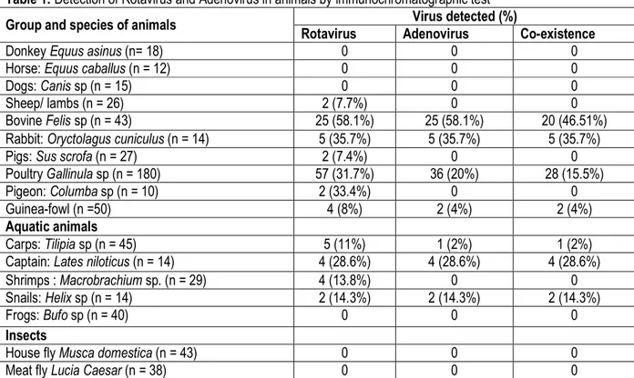
Rotavirus detection by Immunochromatographic: Table 1 shows the results of rotavirus and adenovirus antigens detection in faecal matters of various animals. The tested animals were classified into four groups: pets, farm animals, aquatic animals and insects. Rotavirus and/or adenovirus positive samples were recorded for all the abattoir animals, and 4 species of aquatic animals. None rotavirus and adenovirus positive samples were observed among the pets and insects tested. The detection of group A rotavirus and enteric adenovirus antigens in this study highlight the reservoir role of several animals living close to human. A prevalence of
31-35% and 58% were observed from poultry, pigeons, rabbit and bovine respectively. Low prevalence (7-8%) was recorded for sheep/lambs, pigs and guinea fowls. In addition, co-existence of rotavirus and adenovirus were found from bovine (46.5%), rabbit (35.7%), chickens (15.5%) and guinea fowl (4%). Concerning aquatic animals, 28.6% of fish commonly called captain and 11% of carps, 13.8% of shrimps and 14.3% of snails were tested positive for rotavirus. Co-infections by rotavirus and adenovirus (2 to 28.6%) were found from all aquatic animals except in shrimps and frogs.
9873
Table 1: Detection of Rotavirus and Adenovirus in animals by immunochromatographic test
Group and species of animals Virus detected (%)
Rotavirus Adenovirus Co-existence
Donkey Equus asinus (n= 18) 0 0 0
Horse: Equus caballus (n = 12) 0 0 0
Dogs: Canis sp (n = 15) 0 0 0
Sheep/ lambs (n = 26) 2 (7.7%) 0 0
Bovine Felis sp (n = 43) 25 (58.1%) 25 (58.1%) 20 (46.51%)
Rabbit: Oryctolagus cuniculus (n = 14) 5 (35.7%) 5 (35.7%) 5 (35.7%)
Pigs: Sus scrofa (n = 27) 2 (7.4%) 0 0
Poultry Gallinula sp (n = 180) 57 (31.7%) 36 (20%) 28 (15.5%)
Pigeon: Columba sp (n = 10) 2 (33.4%) 0 0
Guinea-fowl (n =50) 4 (8%) 2 (4%) 2 (4%)
Aquatic animals
Carps: Tilipia sp (n = 45) 5 (11%) 1 (2%) 1 (2%)
Captain: Lates niloticus (n = 14) 4 (28.6%) 4 (28.6%) 4 (28.6%)
Shrimps : Macrobrachium sp. (n = 29) 4 (13.8%) 0 0
Snails: Helix sp (n = 14) 2 (14.3%) 2 (14.3%) 2 (14.3%)
Frogs: Bufo sp (n = 40) 0 0 0
Insects
House fly Musca domestica (n = 43) 0 0 0
Meat fly Lucia Caesar (n = 38) 0 0 0
Rotavirus detection by ELISA and genotyping: Table 2 shows the results of rotavirus antigens detection in animals by ELISA. The detection of rotavirus antigen in animals reported rates of 7.4% in pigs, 4.1% in cattle and
14.3% in poultry. During this study, no case of portage of rotavirus was detected in sheep. The molecular characterization of the strains established that they belong to the G9 genotype (3/ 7; 42.9%).
Table 2: Detection of Rotavirus and Adenovirus in animals by ELISA
Species of animals Rotavirus detected (%)
Pigs Sus scrofa (n= 27) 2 (7.4%)
Poultry Gallinula sp (n= 21) 3 (14.3%)
Sheep (n= 41) 0
Bovine Felis sp (n= 49) 2 (4.1%)
DISCUSSION
Rotaviruses are enteric pathogens causing acute watery dehydrating diarrhoea in various host species, including birds and mammals. Rotaviruses (RVs), an important cause of gastroenteritis in various animal species, cause approximately 0.5 million deaths (under the age group of five years) out of 0.8 million total diarrhoea associated child deaths annually (Kotloff et al., 2013). Several studies are bound this situation to socioeconomic and hygienic level of countries (Estes and Kapikian, 2007; Barro et al., 2008; Bonkoungou et al., 2011; Jain et al., 2014). Epidemiological information related to the prevalence and genotype specificities of rotavirus group A
in animals (in particular in cattle and pigs) are beneficial for the development of effective vaccines. The animal constituted by birds (poultry, pigeons, and guinea fowls), aquatic animals (shrimp, fish and snail) and rabbit were found to be the main reservoirs of rotavirus group A antigen. Aquatic animals were contaminated by streaming water polluted by infectious particles from farm animals and human faecal matter. The land animals-water-aquatic animal - land animals cycle is also possible and argued by several authors (Estes and Kapikian, 2007; Barro et al., 2008; Ferreira et al., 2009). Pigs, cattle and poultry rotaviruses were reported in many countries (Steele et al.,
9874 2003, Abe et al., 2009, Oyetunde and Amubieya, 2010; Zhou et al., 2016). The zoonotic potential of rotavirus was analysed by Cook et al. (2004) supporting our hypothesis that transmission of porcine, bovine and avian rotavirus strains can be higher in developing countries with farms under intensive or extensive management. In many developing countries, there is close contact between humans and domestic livestock. Exposure of humans to animal rotaviruses strains may also be promoted through contact with domestic pets, particularly cats and dogs (Nakagomi et al., 1990; Saif et al., 1994; Desselberger et al., 2001; Tsugawa and Hoshino, 2008). In Ouagadougou, as well as Burkina Faso in general, farm animals and humans live in close proximity, thus increasing the possibility of rotavirus transmission between animals and humans. Otherwise, the faeces of farm animals were used as fertilizer for soil in gardening in Ouagadougou. The pathways of rotavirus strains spread include environment and faecal-oral routes of transmission to animals and human. Faecal matter is in the centre of rotavirus spread mechanism in environment. Defecation of faeces can lead to direct contamination of animals and/or human, and water sources. The second important particle spreading factor is water. Virus particles in the faeces left in the environment, can contaminated water sources and aquatic animals through the streaming water. Then, it exist a secondary pathways
of virus propagation such as contaminated surface, foods and aerosol. The dried faecal matter, when containing viral particles can be suspended in dust and spread through aerosols. The excreta from infected cattle, pigs and sheep contain large numbers of infectious rotavirus particles and are a potential source of contamination in various ways such as water-borne and food-borne diseases (Cook et al., 2004, Zhou et al., 2016). Therefore, environment can be considered as ‘box’ containing rotavirus strains from human and animals and play an important role in their mixing to generate new strains and their spread. The molecular characterization of the strains established that they belong to the G9 genotype (3/ 7; 42.9%). Serotype G9 strains have been detected sporadically and in localized outbreaks in humans of various African countries, including Burkina Faso, South Africa, Botswana, Malawi, Kenya, Cameroon, Nigeria, Ghana, Guinea-Bissau, Libya, and Mauritius (Armah et al., 2003, Page et al, 2010, Bonkoungou et al., 2011) . African G9 strains were associated with both DS-1-like characteristics and Wa-like characteristics, indicating the predisposition of G9 strains to frequently reassort (Page et al., 2010). A low viral load and/or the untypable strain have accumulated point of mutations may partially explain the place of untypable strain in this study.
CONCLUSION AND APPLICATION OF RESULTS This study was limited by the fact it was only asymptomatic animals and no characterization of RVA antigen-positive samples for immunochromatographic. However, this study report for the first time rotaviruses
detection and presence of the emerged genotype G9 in farms animals in Burkina Faso. These results justify the need to monitoring animals’ rotaviruses in Burkina Faso.
ACKNOWLEDGMENTS
This work was supported by International Foundation of Science (Grant N° E/5392-1). We thank Laboratory of Food borne Pathogen Epidemiology and Surveillance of
CRSBAN, University of Ouagadougou, Burkina Faso and National Laboratory of public Health of Burkina Faso for the technical assistance.
REFERENCES
Abe M, Ito N, Morikawa S, Takasu M, Murase T, Kawashima T, Kawai Y, Kohara J, Sugiyama M (2009): Molecular epidemiology of rotaviruses among healthy calves in Japan: Isolation of a novel bovine rotavirus bearing new P and G genotypes. Virus Research 144: 250–257. Armah GE, Steele AD, Binka FN, Esona MD, Asmah RH,
Anto F, Brown D, Green J, Cutts F, Hall A. (2003): Changing patterns of rotavirus genotypes in Ghana: Emergence of human
rotavirus G9 as a major cause of diarrhoea in children. J Clin Microbiol 41:2317–2322. Banerjee I, Ramani S, Primrose B, Iturriza-Gomara M,
Gray JJ, Brown DW, Kang G. (2007). Modification of rotavirus multiplex RT-PCR for the detection of G12 strains based on characterization of emerging G12 rotavirus strains from South India. J Med Virol 79:1413– 1421.
9875 Barro, N., Sangaré, L., Tahita, M.C., Traoré, O., De
Souza, C.A., Traoré, A.S., (2008). Risk associated with practices, procedures and processes of street-vended foods that lead to foodborne viral contamination: critical review. In: Anderson P.L., Lachan J.P. (Eds), Hygiene and it Role in Health nova Science Publisher Inc New York, pp. 129-153.
Bonkoungou IJ, Damanka S, Sanou I, Tiendrebeogo F, Coulibaly SO, Bon F, Haukka K, Traore AS . Genotype diversity of group A rotavirus strains in children with acute diarrhoea in urban Burkina Faso, 2008-2010. J Med Virol 2011. 83:1485-1490.
Clark HF, Lawley DA, Schaffer A, Patacsil JM, Marcello AE, Glass RI, Jain V and Gentsch J. (2004): Assessment of the Epidemic Potential of a New Strain of Rotavirus Associated with the Novel G9 Serotype Which Caused an Outbreak in the United States for the First Time in the 1995-1996 Season. J.Clin Microbiol. 42 (4):1434– 1438, DOI: 10.1128/JCM.42.4.1434–1438.2004. Cook, N., Bridger, J., Kendall, K., Iturriza-Gómara, M.,
El-Attarb, L., Gray, J., (2004). The zoonotic potential of rotavirus. J. Infect. 48, 289-302. Desselberger, U., Iturriza-Gomara, M., Gray, J.J., (2001).
Rotavirus epidemiology and surveillance. In: Chadwick, D, Goode, J.A., (Eds), Gastroenteritis viruses. New York: Wiley, p. 82-100.
Estes, M.K., Kapikian, A.Z., (2007). Rotaviruses In: Knipe, D.M., Howley P.M., Griffin D.E., Lamb, R.A., Martin, M.A., Roizman B., Straus S.E., (Eds.), Fields virology, 5th ed. Philadelphia: Kluwer/Lippincott, Williams and Wilkins, pp. 1917-1974.
Ferreira, F.F., Guimaraes, F.R., Fumian, T.M., Victoria, M., Vieira, C.B., Luz, S., Shubo, T., Leite, J.P., Miagostovich, M.P., (2009). Environmental dissemination of group A rotavirus: P-type, G-type and subgroup characterization. Water Sci. Technol. 60, 633-642.
Gentsch JR, Glass RI, Woods P, Gouvea V, Gorziglia M, Flores J, Das BK, Bhan MK. (1992). Identification of group A rotavirus gene 4 types by polymerase chain reaction. J Clin Microbiol 30:1365–1373.
Gouvea V, Glass RI, Woods P, Taniguchi K, Clark HF, Forrester B, Fang ZY. (1990). Polymerase chain reaction amplification and typing of rotavirus
nucleic acid from stool specimens. J Clin Microbiol 28:276–282.
Iturriza-Gomara M, Isherwood B, Desselberger U, Gray J. (2004). Characterization of G10P [11] rotaviruses causing acute gastroenteritis in neonates and infants in Vellore. Ind J Clin Microbiol 42:2541–2547.
Jain S, Vashistt J, Changotra Harish. (2014). Rotaviruses: Is their surveillance needed? Vaccine. 32:3367–3378.
Kotloff KL, Nataro JP, Blackwelder WC, Nasrin D, Farag TH, Panchalingam S, Burden and aetiology of diarrhoeal disease in infants and young children in developing countries (the Global Enteric Multicenter Study – GEMS): a prospective, case–control study (2013). Lancet; 382:209–22. Mihalov-Kovacs E, Gellert A, Marton S, Farkas SL, Fehér E, Oldal M, Jakab F, Martella V, Bányai K. Candidate new rotavirus species in sheltered dogs, Hungary (2015). Emerg Infect Dis.; 21(4):660-3. doi: 10.3201/eid2104.141370. Nakagomi, O., Ohshima, A., Aboudy, Y., Shif, I.,
Mochizuki, M., Nakagomi, T., Gotlieb-Stematsky, T., (1990). Molecular Identification by RNA–RNA hybridization of a human rotavirus that is closely related to rotaviruses of feline and canine origin. J. Clin. Microbiol. 28, 1198-1203. Nitiema LW, Nordgren J, Ouermi D, Dianou Di, Traore
AS, Svensson L. (2011): Burden of rotavirus and other enteropathogens among children with diarrhoea in Burkina Faso. I J Infect Dis.; 15: e646–e652.
Nordgren J, Nitiema LW, Sharma S, Ouermi D, Traore AS, Simpore J, Svensson L. Emergence of Unusual G6P[6] Rotaviruses in Children, Burkina Faso, 2009–2010 (2012). Emerg Infect Diseases.; 18( 4): 589-597.
Oyetunde OO and Amubieya AO (2010): Genetic evidence of rotavirus in chicken in Nigeria. Bulletin of animal Heath and production in Africa.58(3).
Page N, Esona M, Armah G, Nyangao J, Mwenda J, Sebunya T, Basu G, Pyndiah N, Potgieter N, Geyer A, and Steele AD (2010): Emergence and Characterization of Serotype G9 Rotavirus Strains from Africa. Jf Infect Dis. 2010; 202(S1):S55–S63.
Parashar, U.D., Bresee, J.S., Gentsch, J.R., Glass, R.I., (1998). Rotaviruses. Emerg. Infect. Dis 4, 561-570.
9876 Ramig RF (1997). Genetics of the rotaviruses. Ann Rev
Microbiol; 51:225—255.
Saif, L.J., Rosen, B., Parwani, A., (1994). Animal Rotaviruses. In: Kapikian, A. (Ed.), Viral Infections of the Gastrointestinal Tract. Marcel Dekker, Inc, New York, pp. 279-367.
Steyer, A., Poljšak-Prijatelj, M., Barlič-Maganja D., and Marin, J., (2008). Human, porcine and bovine rotaviruses in Slovenia: evidence of interspecies transmission and genome reassortment. J. Med. Virol. 79, 626-632.
Steele, A.D., Geyer, A., Gerdes, G.H., (2004). Rotavirus infections. In: Coetzer J.A.W., Tustin R.C. (Eds) Infectious Diseases of Livestock, 2nd ed, Cape Town: Oxford University Press. pp. 1256-1264. Steele AD, Page N, de Beer M, Sawadogo S. (2010): Antigenic and molecular characterization of unusual rotavirus strains in Burkina Faso in 1999. J Infect Dis 202:S225–S230.
Tsugawa, T., Hoshino, Y., (2008). Whole genome sequence and phylogenetic analyses reveal human rotavirus G3P [3] strains Ro1845 and HCR3A are examples of direct virion transmission of canine/feline rotaviruses to humans. Virol. 380, 344-353.
Yang, J., Wang, T., Wang, Y., Lu, B., Bai, X., Zhang, L., Wang, M.,Wang, H., (2007). Emergence of human rotavirus group A genotype G9 strains, Wuhan, China. Emerg. Infect. Dis. 13, 1587– 1589.
Zhou W, Ullman K, Chowdry V, Reining M, Benyeda Z, Baule C, Juremalm M, Wallgren P, Schwarz L, Zhou E, Pedrero SP, Hennig-Pauka I, Segales J, Liu L. Molecular investigations on the prevalence and viral load of enteric viruses in pigs from five European countries (2016). Vet
Microbiol; 182:75-81. doi:
10.1016/j.vetmic.2015.10.019.Epub 2015 Oct 21.