HAL Id: inserm-01807415
https://www.hal.inserm.fr/inserm-01807415
Submitted on 4 Jun 2018
HAL is a multi-disciplinary open access
archive for the deposit and dissemination of sci-entific research documents, whether they are pub-lished or not. The documents may come from teaching and research institutions in France or abroad, or from public or private research centers.
L’archive ouverte pluridisciplinaire HAL, est destinée au dépôt et à la diffusion de documents scientifiques de niveau recherche, publiés ou non, émanant des établissements d’enseignement et de recherche français ou étrangers, des laboratoires publics ou privés.
Decrease in Diversity of Propionibacterium acnes
Phylotypes in Patients with Severe Acne on the Back
Marie-Ange Dagnelie, Stéphane Corvec, Mélanie Saint-Jean, Valérie Bourdès,
Jean-Michel Nguyen, Amir Khammari, Brigitte Dréno
To cite this version:
Marie-Ange Dagnelie, Stéphane Corvec, Mélanie Saint-Jean, Valérie Bourdès, Jean-Michel Nguyen, et al.. Decrease in Diversity of Propionibacterium acnes Phylotypes in Patients with Severe Acne on the Back. Acta Dermato-Venereologica, Society for Publication of Acta Dermato-Venereologica, 2018, 98 (2), pp.262 - 267. �10.2340/00015555-2847�. �inserm-01807415�
A
cta
DV
A
cta
DV
A
dvances in dermatology and venereology
A
ctaD
ermato-V
enereologicaActa Derm Venereol 2018; 98: XX–XX
This is an open access article under the CC BY-NC license. www.medicaljournals.se/acta doi: 10.2340/00015555-2847 Propionibacterium acnes, a major member of normal
skin microbiota, is subdivided into 6 phylotypes: IA1, IA2, IB, IC, II and III. This study investigated P. acnes subgroups on the face and back in patients with se-vere acne and in healthy controls. In 71.4% of patients with severe acne, P. acnes phylotypes were identical on the face and back, whereas this was the case in only 45.5% of healthy controls. The healthy group car-ried phylotypes IA1 (39.1%) and II (43.4%), whereas the acne group carried a high predominance of IA1 (84.4%), especially on the back (95.6%). In addi-tion, the single-locus sequence typing (SLST) method revealed A1 to be the predominant type on the back of patients with acne, compared with a wide diversity in the healthy group. We report here that severity of acne on the back is associated with loss of diversity of P. acnes phylotype, with a major predominance of phylotype IA1. The change in balance of cutaneous P.
acnes subgroups might be an inducing factor in the
ac-tivation of P. acnes, which could trigger inflammation.
Key words: acne, P. acnes phylotypes; clonal complexes; SLST-types.
Accepted Nov 13, 2017; Epub ahead of print Nov 14, 2017 Acta Derm Venereol 2018; 98: XX–XX.
Corr: Brigitte Dréno, Department of Dermatology, Nantes University Hos-pital, 1 Place Alexis Ricordeau, FR-44035 Nantes Cedex 01, France. E-mail: brigitte.dreno@wanadoo.fr
A
cne is one of the most common skin diseases world-wide, affecting up to 85% of the population (1). At the pathophysiological level, 2 factors play a crucial role: the sebaceous gland and Propionibacterium acnes. P.ac-nes is a commensal anaerobic Gram-positive bacterium of
the healthy skin. This bacterial genus has high specificity, from face to feet areas (2). This study showed a predo-minance of Propionibacterium genus on the face and back. This microorganism diversity on the human body also depends on several factors, such as host factors (e.g. age, sex, hair follicle density) and environmental factors (e.g. occupation, clothing choice, antibiotic use) (3, 4).
P. acnes plays an important role in the maintenance
of normal cutaneous microbiota, by inhibiting the deve-lopment of some pathogenic bacteria, such as
Staphylo-coccus aureus and StreptoStaphylo-coccus pyogenes. It produces
propionic acid and, thus, can maintain acidic pH in the
pilo-sebaceous follicles. In addition to acne, P. acnes has also been implicated in deep infections and orthopaedic ab-scesses (5–7), lung abab-scesses (8), prostate cancer (9), and sarcoidosis (10). Its development in deep infections may be related to the secretion of a biofilm that increases both its adherence to surfaces and antibiotic resistance (11).
In the skin, P. acnes, as an anaerobic bacterium, is located more specifically in the pilo-sebaceous follic-les. A number of studies have shown that P. acnes can activate innate immunity, mainly via Toll-like receptors (TLRs) expressed by keratinocytes and monocytes (12, 13). P. acnes also stimulates the secretion of interleukins (IL)-12, IL-8, IL-1β and IL-17 cytokines by monocytes (14). Thus, it plays a crucial role in the development of inflammatory lesions in acne.
P. acnes population strains are divided into 6 main
phylotypes: IA1, IA2, IB, IC, II and III. Recent genomic studies have also highlighted the presence of subgroups among phylotypes, according to genome analysis called multi-locus sequence typing (MLST) (15) and single-locus sequence typing (SLST). These cluster differentia-tions are called clonal complexes (CCs). Some P. acnes phylotypes are associated with skin disease conditions, such as phylotype III and progressive macular hypome-lanosis (16). Moreover, different P. acnes phylotypes are known to induce distinct immune responses in the context of acne (12, 17). The aim of this study was to determine and compare the different P. acnes phylotypes, CCs and SLST types on the face and back of patients with severe acne of the back vs. a healthy population.
MATERIALS AND METHODS
Recruitment of healthy volunteers and patients
At the first visit, patients with acne who fulfilled the inclusion criteria were selected. The inclusion criteria were: age (16–35 years), at least 2 nodules on the back, complying with wash-out periods for acne drugs including systemic antibiotics (1 month), oral retinoids (6 months) and topical treatments (2 weeks). Healthy volunteers were selected according to inclusion criteria, including the total absence of acne on the face and back, and the absence of other history or current dermatological pathologies. All patients provided signed informed consent and the study was approved by the health authorities (Agence Nationale de Sécurité du Médi-cament et des Produits de Santé (ANSM); number 151141B-42) and ethics committee (Comité de Protection des Personnes (CPP); number 21–15).
Decrease in Diversity of Propionibacterium acnes Phylotypes in
Patients with Severe Acne on the Back
Marie-Ange DAGNELIE1, Stéphane CORVEC1,3, Mélanie SAINT-JEAN1,2, Valérie BOURDÈS5, Jean-Michel NGUYEN1,4, Amir
KHAMMARI1,2 and Brigitte DRÉNO1,2
1CIC, Inserm U1232, 2Department of Dermatology, 3Bacteriology and Hygiene Unit, Biology Institute, 4Biostatistic Department, PIMES,
A
cta
DV
A
cta
DV
A
dvances in dermatology and venereology
A
ctaD
ermato-V
enereologicaM-A. Dagnelie et al.
2
www.medicaljournals.se/acta Clinical data
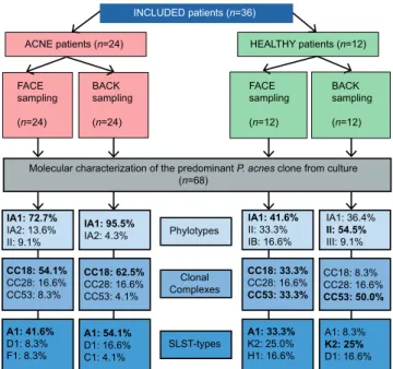
For each patient with acne, face and back acne scores were esta-blished by a dermatologist. These scores were estimated from 0 to 5 according to the type of acne (mild to severe), for both areas. All scores are shown in Table I. Scores for back acne were established according to the Echelle de Cotation des Lésions d’Acné (ECLA) scale (18). Scores for face acne were established according to the Groupe Expert Acné (GEA) scale (19). The experimental scheme of the study is detailed in Fig. 1.
Bacteriological sampling and microbiology study
For all included patients, swab samples were taken from the
face and the back at the same visit, from a surface area of 1 cm2
surrounding an inflammatory lesion (papule), by rubbing with the swab for 45 s. Samples were collected in both groups from
the back and face zones, using a cotton swab, and separately discharged into a brain heart infusion medium and delivered to the Bacteriology Department of Nantes University Hospital within 30 min. Each sample was cultured anaerobically at 37°C for 7–10 days. Colonies with the macroscopic morphology of P. acnes were picked from each plate to recover the predominant strain of P.
acnes. As reported recently (20), this picking method allows the
predominant clone of P. acnes present in the cutaneous skin flora to be recovered. Indeed, it has been shown that the representative isolate constitutes 85–100% of the sample.
All isolates were identified by matrix-assisted laser desorption/
ionization time-of-flight mass spectrometry with a VitekMS®
mass spectrometer (MALDI-TOF MS) (bioMérieux SA, Marcy-l’Etoile, France). All strains were identified accurately with a value > 99.9%.
DNA extraction and phylotype determination
Total DNA from P. acnes isolates was extracted using the Insta-Gene Matrix method (Bio-Rad Laboratories, Hercules, CA, USA) according to the manufacturer’s instructions.
The 6 main phylotypes for all isolates were determined as de-scribed previously (21).
Multi- and single-locus sequence type determination and sequencing protocol
MLST is a method to determine to which clonal complex a P.
acnes strain belongs. Recently, 2 main MLST schemes have been
developed (22, 23), but we decided to use the method developed by Lomholt & Kilian (23) according to its best discriminant power. This scheme is based on partial sequencing of 9 housekeeping genes, comprising a total of 4,287 nucleotides, and is available at http://pacnes.mlst.net/.
SLST is a molecular typing method based on the analysis of a single locus of the P. acnes genome. The portion of amplified DNA enables the identification of P. acnes SLST-types (A1, A5 etc.). SLST was carried out on all isolates, as described previously (24). Reference sequences for alignment and trimming are described in the web-interface typing tool at http://medbac.dk/slst/pacnes
Sequencing was performed on a 3130xl-1 Hitachi (Applied Biosystems, Foster City, USA). At the end of the sequencing, each sequence for each sample was reviewed and analysed using Seqscape software v2.5 (Applied Biosystems) before comparison with online databases.
Statistical analysis
Statistical analyses were performed using Fisher exact test (R soft-ware (R Foundation for Statistical Computing, Vienna, Austria)).
RESULTS
Clinical isolates and cultivable skin microbiota
A total of 24 patients with acne and 12 healthy volun-teers were recruited. For each patient with acne, GEA and ECLA scores were established by a dermatologist for the face and the back, respectively. Concerning the back zone, 15 of the 24 patients scored 3, 7 scored 4, and 2 scored 5. In addition, concerning the face zone, 14 scored 1, 8 scored 2 and 2 scored 3. The clinical data are shown in Table I.
Positive culture was obtained for all patients and con-trols (100%). Cultures identified P. acnes, but also other
Fig. 1. Experimental scheme of the study and summary of the main results obtained. CC: clonal complex; SLST: single-locus sequence typing.
INCLUDED patients (n=36) ACNE patients (n=24) FACE sampling (n=24) BACK sampling (n=24) HEALTHY patients (n=12) FACE sampling (n=12) BACK sampling (n=12)
Molecular characterization of the predominant P. acnes clone from culture (n=68) IA1: 72.7% IA2: 13.6% II: 9.1% IA1: 95.5% IA2: 4.3% IA1: 41.6% II: 33.3% IB: 16.6% IA1: 36.4% II: 54.5% III: 9.1% Phylotypes CC18: 54.1% CC28: 16.6% CC53: 8.3% CC18: 62.5% CC28: 16.6% CC53: 4.1% Clonal Complexes CC18: 33.3% CC28: 16.6% CC53: 33.3% CC18: 8.3% CC28: 16.6% CC53: 50.0% A1: 41.6% D1: 8.3% F1: 8.3% A1: 54.1% D1: 16.6% C1: 4.1% SLST-types A1: 33.3%K2: 25.0% H1: 16.6% A1: 8.3% K2: 25% D1: 16.6%
Table I. Clinical data for acne patients
Pat. No. Back score (ECLA) Sex Age, years Face score (GEA)
9008 5 F 21 2 9020 5 M 23 1 9001 4 M 24 1 9002 4 M 31 2 9004 4 M 22 2 9006 4 M 33 1 9014 4 M 21 1 9017 4 M 34 1 9021 4 F 24 2 9003 3 M 22 2 9005 3 M 29 1 9007 3 M 28 2 9009 3 M 33 1 9010 3 M 21 2 9011 3 M 22 2 9012 3 M 18 3 9013 3 F 19 3 9015 3 M 31 1 9016 3 M 20 1 9018 3 M 29 1 9019 3 M 21 1
A
cta
DV
A
cta
DV
A
dvances in dermatology and venereology
A
ctaD
ermato-V
enereologicabacterial species, including Staphylococcus aureus (acne group 2/48; 4.2% vs. healthy group 2/24; 8.3%), and
Staphylococcus epidermidis (acne group 33/48; 68.8%
vs. healthy group 14/24; 58.3%). P. acnes was syste-matically recovered from all collected samples (n = 72) in culture, except for 4 samples. Instead of P. acnes, other species were discovered, including P. avidum in the healthy group (2/24; 8.3%), P. granulosum in the acne group (1/48; 2.1%) and P. namnetense in the acne group (3/48; 6.3%). However, no significant difference in bacteria diversity was noted on the back between acne and healthy individuals (p = 0.820). In addition, no significant difference in bacteria diversity was noted between the face samples from subjects with acne and healthy subjects (p = 0.690).
P. acnes phylotype diversity on face vs. back
The P. acnes phylotypes found both on the back and on the face in acne vs. healthy groups were different.
In healthy group samples (face and back), 5 of the 6 main phylotypes were present, showing a large diversity: IA1, IA2, IB, II and III (Fig. 2). The most predominant ones were phylotypes IA1 and II (respectively, 39.1% and 43.5% of isolates), 8.7% were phylotype IB, 4.3% phylotype III and 4.3% phylotype IA2. On the face, the most predominant phylotypes found were phylotype IA1 (41.6%), and II (33.3%). This trend was the same on the back zone for this group, with a majority of phylotypes IA1 and II (36.3% and 54.5%, respectively).
In acne group samples (face and back), 4 out of the 6 main phylotypes were identified: IA1, IA2, IC, and II (Fig. 2). Phylotype IA1 represented 84.4% of typed P.
acnes. The other phylotypes represented only 6.6% for
phylotypes II, 2.1% for IC, and 6.6% for IA2 (6.6%) (Fig. 2). No phylotype III was identified. Concerning the back zone, a large predominance of phylotype IA1 was
found (95.6%) with a significant association (p < 0.001), phylotype II represented only 4.3% of the P. acnes strains, no other phylotype was detected on this body site. On the face zone, phylotype IA1 was the most predominant phylotype (72.7%), compared with 13.6% for phylo-type IA2, 9.1% phylophylo-type II. Moreover, a rare P. acnes phylotype was also found in the acne group on the face: phylotype IC (1/48; 2.1%). For both zones, no significant association was found between acne severity (GEA and/ or ECLA scores) and phylotypes (p = 0.649 for the face and p = 0.391 for the back). The main results concerning phylotypes identification are shown in Fig. 1.
In conclusion, there was a higher diversity of P. acnes phylotypes on the face vs. the back zone, in both groups, and phylotype IA1 was largely represented in the acne group, especially in the back zone.
Analysis of multi-locus and single-locus sequence type determination
To go deeper in the analysis of P. acnes lineages involved in acne lesions, 2 additional molecular typing methods were performed: MLST and SLST. These additional methods offer more precise data about the phylogenetic clades distribution of P. acnes isolates. SLST-type A1 was predominant in the acne group (Fig. 3). A higher diversity of SLST-types associated with known phylotypes (IA1 and II) with new SLST-types never described before was also noted: K16 (phylotype II) and L7 (phylotype III) in the healthy group; A27, A28 (phylotype IA1), F11 (phy-lotype IA2), and K17 (phy(phy-lotype II) in the acne group. Analysis of the distribution of clonal complexes (CC) among both healthy and acne populations in the back/ face zones revealed that CC18 and CC53 were more
Fig. 3. Propionibacterium acnes single-locus sequence typing (SLST) type distribution according to 2 body sites on healthy volunteers and patients with acne (n = 12 and n = 24, respectively). Categories “Healthy”
and “Acne” represent the percentages of the different SLST types found on back and on face in either the healthy group or the acne group. “ND” category represents the other species found instead of P. acnes, where typing was not possible.
Fig. 2. Propionibacterium acnes phylotype distribution on 2 sites on healthy volunteers and acne patients (n = 12 and n = 24, respectively).
Categories “Healthy” and “Acne” represent the percentages of phylotypes found on the back and the face, respectively.
A
cta
DV
A
cta
DV
A
dvances in dermatology and venereology
A
ctaD
ermato-V
enereologicaM-A. Dagnelie et al.
4
www.medicaljournals.se/acta
associated with the acne group and the healthy group, respectively (Fig. 4). The main results concerning CC identification are shown in Fig. 1.
In healthy group samples, 41.6% belonged to the CC53 subgroup (phylotype II and K SLST-types), 20.8% of the P. acnes strains belonged to the CC18 subgroup and 16.6% to the CC28 subgroup belonging to phylotype IA1 including SLST-types A1, C1 and D1 (Figs 3 and 4). Taking into account the location (face or back), 50.0% of the back samples were CC53 (phylotype II including K1, K2, and K8 SLST-types), 16.6% were CC28 (phylotype IA1 including D1 SLST-type), 8.3% were CC18 (phylo-type IA1 and A1 SLST-(phylo-type), 8.3% were CC3 (phylo(phylo-type IA1 and C1 SLST-type) and, finally, 8.3% were CC43 (phylotype III and new SLST-type named “L7”) (Figs 3 and 4). Concerning the face zone, 33.3% of the strains were CC18 (phylotype IA1 and A1 SLST-type) and 33.3% were CC53 (phylotype II including K1 and a new SLST-type named “K16”), whereas 16.6% were CC28 (phylotypes IA1 and IA2 corresponding to D1 and F4 SLST-types) and 16.6% were CC36 (phylotype IB and H1 SLST-types) (Figs 3 and 4).
Interestingly, the only P. acnes phylotype III found in this study was recovered in the healthy group, associa-ted with CC43 and a new SLST-type L7 not previously described.
In acne group samples, 58.3% of the P. acnes strains belonged to CC18 and 16.6% belonged to CC28, including SLST types A1, A5, C1, D1 and E3 (Fig. 3, 4). Taking into account the location, 62.5% of the back samples were CC18, demonstrating a significant association of this CC with back acne skin-condition
(p < 0.001). Moreover, 16.6% of the back samples were CC28, corresponding to phylotype IA1 (total absence of phylotype II), whereas 54.1% of the face samples were CC18, 16.6% were CC28, and 8.3% were CC53 (phy-lotype II corresponding to K SLST-types). In addition, a significant association was found between SLST-type A1 and back acne skin-condition (p = 0.002).
All new SLST-types described for the first time in this study are as listed: A27 (acne back), A28 (acne face), F11 (acne face), K17 (acne back), K16 (healthy face and heal-thy back), and L7 (healheal-thy back). These new SLST-types have been incremented in the online database, based on Christian F. P. Scholz’s genetic system (http://medbac.dk/ slst/pacnes). The main results concerning identification of CC and SLST-types are summarized in Fig. 1.
DISCUSSION
In summary, this study reveals that inflammatory severe acne of both face and back is associated with diversity loss of P. acnes phylotypes, and a high predominance of phylotype IA1, both on the face (72.7%) and the back (95.6%), which has not been described previously. In healthy individuals, 2 main phylotypes have been found: IA1 (39.1%) and II (43.4%). In addition, this study identifies A1 SLST-type as the predominant SLST-type recovered from nodular acne of the back.
Distribution of phylotypes and CCs on the human body in the context of acne has been studied previously. Lomholt & Kilian analysed the distribution of CCs in 2 patients with acne on the face/back (25), and showed the presence of several CCs, including CC3 and CC18 on face, but a lower diversity of CCs on the back. Moreover, the results of the current study confirm those of Lomholt et al. (20) regarding the predominance of IA1 phylotype (CC18) in patients with acne, but this study did not per-form determination of SLST-types. In our study, among the large diversity of SLST-types found, we describe the significant association between A1 SLST-type and severe back acne. Overall, despite a highly conserved genome of P. acnes (26), we show that acne lesions are associated with the development of a specific subpopula-tion of P. acnes.
From our 72 isolates, the A1 SLST-type was signifi-cantly associated with acne skin condition. In addition, we found 6 new SLST-types that had not been described previously (i.e. F11, L7, K16, K17, A28, and A27), which have been incremented in the online database (24) (http:// medbac.dk/slst/pacnes). Currently, the impact of the different SLST-types on skin disease is not known; this field represents an interesting avenue for future clinical investigations. To our knowledge, 2 studies currently describe P. acnes SLST-types found in acne vulgaris (27, 28). In Nakase et al., the SLST-type predominantly found in severe acne is A5. In our data, we found the SLST-type A1 as the predominant type in severe acne of the back.
Fig. 4. Propionibacterium acnes clonal complex (CC) distribution on 2 sites on healthy volunteers and acne patients (n = 12 and n = 24,
respectively). Categories “Healthy” and “Acne” represent the percentages of the different CCs found on the back and face in either the healthy or the acne group. “ND” category represents the other species found instead of P. acnes, where the typing was not possible.
A
cta
DV
A
cta
DV
A
dvances in dermatology and venereology
A
ctaD
ermato-V
enereologicaThis difference can be due to ethnicity, skin care (i.e. use of cleansers, moisturizers, etc.), climate, and acne therapeutic care, which differs from country to country, notably with regard to use of antibiotics, isotretinoin and benzoyl peroxide. Nevertheless, in both cases we found clade A to be predominant in severe acne.
In contrast to SLST-types, association of CCs with some diseases is starting to be well-documented, such as: CC36 and CC53/60 in prostate cancer and absence of CC18 (29), and CC18 association with acne-skin condi-tion (20). Previous works have reported a high diversity of CCs in sarcoidosis: CC36, CC28, CC53, CC18 and singletons (30). In the same manner, our data reveal a diversity of CC, found on both the back and face zones, in the healthy group. For example, phylotype IA1 was associated with CC18, CC28 and CC3. In addition, our study confirms the association between CC18 and acne, and between CC53 and healthy skin.
Our results show a decrease in diversity of P. acnes phylotypes in severe back acne (all patients had a mini-mum of 2 nodules on the back and 40% of patients were scored 4–5). This loss of diversity could be the result of hyperseborrhoea, which is associated with both qualita-tive and quantitaqualita-tive sebum modifications inducing alte-ration of the skin barrier and, subsequently, microbiota changes. Interestingly, it has been shown that there is a direct link between hyperseborrhoea, proliferation of P.
acnes and onset of acne lesions (31). As the microbiota
modulates the innate immunity of the skin (32, 33), this loss of diversity could activate innate immunity, trig-gering the development of inflammatory acne lesions.
We also report differences in the pattern of P. acnes phylotypes between the back and face. We hypothesize that these differences might be related to several factors that impact differently on face/back skin bacterial po-pulations, such as cleansers and physical agents (wind, sun, etc.), although this has not been investigated for P.
acnes phylotypes (3, 4).
In culture we identified P. namnetense in 3 patients (2 minor acne face score 1; 1 severe acne back score 3), which has not been described previously in acne and could lead to further investigations about the link bet-ween this bacterium and specific clinical forms of acne. All patients and controls were positive for P. acnes or related species with our sampling method, which is an important point at the methodological level, compared with previous studies in which culture positivity was found in approximately 70–84% of patient samples (34, 35). Concerning the association between S. epidermidis and P. acnes, we identified S. epidermidis in 50% of acne back samples (41.6% in healthy back), and 75% of acne face samples (75% in healthy face) (data not shown), respectively. Interestingly, these data favour the hypo-thesis of a symbiotic association between P. acnes and
S. epidermidis, as a crucial element for skin microbiota
balance (3, 36).
At the bacteriological level this study sheds light on an important question: Do all CCs that belong to a same cluster (i.e. IA1) stimulate innate immunity effectors in the same way? Seeing how different the P. acnes CCs and SLST-types are from one body area to another in both acne/healthy context, the elucidation of this ques-tion will permit better understand of the role of certain
P. acnes subgroups (i.e. SLST-types and CCs), and
determine whether some of them are associated with pro-inflammatory reactions in human skin.
The results of this study suggest that acne could be associated with the proliferation of one specific phylo-type. These results can be related to the study from Tax et al. (37), who found a difference in growth properties and propionic acid production between the different P.
acnes phylotypes. Taken together, these data provide a
potential explanation for the role of the different P. acnes phylotypes in acne physiopathology.
This study opens up new areas of research into inno-vative alternative treatments for acne. In 2015, Yu et al. reviewed P. acnes molecular typing methods, and pointed out the importance of investigating P. acnes populations in acne vulgaris (38). The microbiological results presented here may be crucial for the elaboration of innovative therapies, such as probiotic treatments, as suggested previously (38). For instance, future topical treatments could restore the phylotype diversity, through re-introduction of P. acnes subgroup CC53. In addi-tion, our data give a precise lineage description of the dominant P. acnes clones found in the context of severe acne, identifying for the first time the A1 SLST-type as the predominant SLST-type found in this disease. This information is currently the most precise genetic level of subgroup identification concerning P. acnes bacterium (24). It suggests the possibility of setting up a vaccine targeting A1 SLST-type, especially for children with a family history of severe acne, and with previous isotreti-noin treatment in parents or brothers and sisters. Indeed, as acne occurs earlier and is more severe in patients with a positive family history, there is a need to investigate ways to prevent acne development, such as vaccination. Such treatments might prevent the irreversible scars as-sociated with severe acne, which is a major problem in this disease.
ACKNOWLEDGEMENTS
This study was supported by a research grant from Galderma R&D - CUTIS (Sophia Antipolis). The authors would like to thank Dr Knol, Elodie Belliot, Nathalie Defossé and Julie Marraillac for technical support and assistance, and Aurélie Boisrobert for assistance with clinical sampling.
Conflicts of interest: SC, MS-J. and JMN have no conflicts of
interest to declare. BD, AK and MAD declare a potential conflict of interest, as the study was funded by Galderma R&D - CUTIS (Sophia Antipolis, France). VB is an employee of Galderma R&D - CUTIS (Sophia Antipolis, France).
A
cta
DV
A
cta
DV
A
dvances in dermatology and venereology
A
ctaD
ermato-V
enereologicaM-A. Dagnelie et al.
6
www.medicaljournals.se/acta
REFERENCES
1. Dawson AL, Dellavalle RP. Acne vulgaris. BMJ 2013; 346: f2634.
2. Bouslimani A, Porto C, Rath CM, Wang M, Guo Y, Gonzalez A, et al. Molecular cartography of the human skin surface in 3D. Proc Natl Acad Sci U S A 2015; 112: E2120–2129. 3. Dréno B, Araviiskaia E, Berardesca E, Gontijo G, Sanchez
Viera M, Xiang LF, et al. Microbiome in healthy skin, update for dermatologists. J Eur Acad Dermatol Venereol 2016; 30: 2038–2047.
4. Grice EA, Segre JA. The skin microbiome. Nat Rev Microbiol 2011; 9: 244–253.
5. Aubin GG, Portillo ME, Trampuz A, Corvec S. Propionibacte-rium acnes, an emerging pathogen: From acne to implant-infections, from phylotype to resistance. Médecine Mal Infect 2014; 44: 241–250.
6. Daguzé J, Frénard C, Saint-Jean M, Dumont R, Touchais S, Corvec S, et al. Two cases of non-prosthetic bone and joint infection due to Propionibacterium acnes. J Eur Acad Der-matol Venereol 2016; 30: e136–e137.
7. Khassebaf J, Hellmark B, Davidsson S, Unemo M, Nilsdotter-Augustinsson Å, Söderquist B. Antibiotic susceptibility of Propionibacterium acnes isolated from orthopaedic implant-associated infections. Anaerobe 2015; 32: 57–62. 8. Veitch D, Abioye A, Morris-Jones S, McGregor A.
Propionibac-terium acnes as a cause of lung abscess in a cardiac transplant recipient. BMJ Case Rep 2015; 2015. pii: bcr2015212431. 9. Davidsson S, Mölling P, Rider JR, Unemo M, Karlsson MG,
Carlsson J, et al. Frequency and typing of Propionibacterium acnes in prostate tissue obtained from men with and without prostate cancer. Infect Agent Cancer 2016; 11: 26. 10. de Brouwer B, Veltkamp M, Wauters CA, Grutters JC,
Jans-sen R. Propionibacterium acnes isolated from lymph nodes of patients with sarcoidosis. Sarcoidosis Vasc Diffuse Lung Dis Off J WASOG 2015; 32: 271–274.
11. Bayston R, Ashraf W, Barker-Davies R, Tucker E, Clement R, Clayton J, et al. Biofilm formation by Propionibacterium acnes on biomaterials in vitro and in vivo: impact on diagnosis and treatment. J Biomed Mater Res A 2007; 81: 705–709. 12. Jasson F, Nagy I, Knol AC, Zuliani T, Khammari A, Dréno
B. Different strains of Propionibacterium acnes modulate differently the cutaneous innate immunity. Exp Dermatol 2013; 22: 587–592.
13. Nagy I, Pivarcsi A, Koreck A, Széll M, Urbán E, Kemény L. Distinct strains of Propionibacterium acnes induce selective human beta-defensin-2 and interleukin-8 expression in human keratinocytes through toll-like receptors. J Invest Dermatol 2005; 124: 931–938.
14. Agak GW, Qin M, Nobe J, Kim M-H, Krutzik SR, Tristan GR, et al. Propionibacterium acnes induces an IL-17 response in acne vulgaris that is regulated by vitamin A and vitamin D. J Invest Dermatol 2014; 134: 366–373.
15. Kilian M, Scholz C, Lomholt HB. multilocus sequence typing and phylogenetic analysis of propionibacterium acnes. J Clin Microbiol 2012; 50: 1158–1165.
16. Barnard E, Liu J, Yankova E, Cavalcanti SM, Magalhães M, Li H, et al. Strains of the Propionibacterium acnes type III lineage are associated with the skin condition progressive macular hypomelanosis. Sci Rep 2016; 6: 31968.
17. Yu Y, Champer J, Agak GW, Kao S, Modlin RL, Kim J. Different Propionibacterium acnes phylotypes induce distinct immune responses and express unique surface and secreted proteo-mes. J Invest Dermatol 2016; 136: 2221–2228.
18. Dreno B, Bodokh I, Chivot M, Daniel F, Humbert P, Poli F, et al. La grille ECLA : un système de cotation de l’acné pour la pratique quotidienne du dermatologue. Ann Dermatol Venereol 1999; 126: 136–141.
19. Dréno B, Poli F, Pawin H, Beylot C, Faure M, Chivot M, et al. Development and evaluation of a Global Acne Severity Scale (GEA Scale) suitable for France and Europe. J Eur Acad Dermatol Venereol 2011; 25: 43–48.
20. Lomholt HB, Scholz CFP, Brüggemann H, Tettelin H, Kilian M.
A comparative study of Cutibacterium (Propionibacterium) acnes clones from acne patients and healthy controls. Anae-robe 2017; 47: 57–63.
21. Barnard E, Nagy I, Hunyadkürti J, Patrick S, McDowell A. Multiplex touchdown PCR for rapid typing of the opportunistic pathogen Propionibacterium acnes. J Clin Microbiol 2015; 53: 1149–1155.
22. McDowell A, Gao A, Barnard E, Fink C, Murray PI, Dowson CG, et al. A novel multilocus sequence typing scheme for the opportunistic pathogen Propionibacterium acnes and characterization of type I cell surface-associated antigens. Microbiol Read Engl 2011; 157: 1990–2003.
23. Lomholt HB, Kilian M. Population genetic analysis of Propio-nibacterium acnes identifies a subpopulation and epidemic clones associated with acne. PLoS One 2010; 5: e12277. 24. Scholz CFP, Jensen A, Lomholt HB, Brüggemann H, Kilian M.
A novel high-resolution single locus sequence typing scheme for mixed populations of Propionibacterium acnes in vivo. PloS One 2014; 9: e104199.
25. Lomholt HB, Kilian M. Clonality and anatomic distribution on the skin of antibiotic resistant and sensitive Propionibacte-rium acnes. Acta Derm Venereol 2014; 94: 534–538. 26. Scholz CFP, Brüggemann H, Lomholt HB, Tettelin H, Kilian
M. Genome stability of Propionibacterium acnes: a compre-hensive study of indels and homopolymeric tracts. Sci Rep 2016; 6: 20662.
27. Paugam C, Corvec S, Saint-Jean M, Le Moigne M, Khammari A, Boisrobert A, et al. Propionibacterium acnes phylotypes and acne severity: an observational prospective study. J Eur Acad Dermatol Venereol 2017; 31: e398-e399.
28. Nakase K, Hayashi N, Akiyama Y, Aoki S, Noguchi N. Anti-microbial susceptibility and phylogenetic analysis of Pro-pionibacterium acnes isolated from acne patients in Japan between 2013 and 2015. J Dermatol 2017; 44: 1248–1254. 29. Mak TN, Yu S-H, De Marzo AM, Brüggemann H, Sfanos KS.
Multilocus sequence typing (MLST) analysis of Propionibac-terium acnes isolates from radical prostatectomy specimens. The Prostate 2013; 73: 770–777.
30. Minegishi K, Watanabe T, Furukawa A, Uchida K, Suzuki Y, Akashi T, et al. Genetic profiles of Propionibacterium acnes and identification of a unique transposon with novel inser-tion sequences in sarcoid and non-sarcoid isolates. Sci Rep 2015; 5: 9832.
31. Mourelatos K, Eady EA, Cunliffe WJ, Clark SM, Cove JH. Temporal changes in sebum excretion and propionibacterial colonization in preadolescent children with and without acne. Br J Dermatol 2007; 156: 22–31.
32. Gallo RL, Nakatsuji T. Microbial symbiosis with the innate immune defense system of the skin. J Invest Dermatol 2011; 131: 1974–1980.
33. Lai Y, Di Nardo A, Nakatsuji T, Leichtle A, Yang Y, Cogen AL, et al. Commensal bacteria regulate Toll-like receptor 3-dependent inflammation after skin injury. Nat Med 2009; 15: 1377–1382.
34. Giannopoulos L, Papaparaskevas J, Refene E, Daikos G, Stav-rianeas N, Tsakris A. MLST typing of antimicrobial-resistant Propionibacterium acnes isolates from patients with moderate to severe acne vulgaris. Anaerobe 2015; 31: 50–54. 35. Ma Y, Zhang N, Wu S, Huang H, Cao Y. Antimicrobial activity
of topical agents against Propionibacterium acnes: an in vitro study of clinical isolates from a hospital in Shanghai, China. Front Med 2016; 10: 517–521.
36. Christensen GJM, Scholz CFP, Enghild J, Rohde H, Kilian M, Thürmer A, et al. Antagonism between Staphylococcus epidermidis and Propionibacterium acnes and its genomic basis. BMC Genomics 2016; 17: 152.
37. Tax G, Urbán E, Palotás Z, Puskás R, Kónya Z, Bíró T, et al. Propionic acid produced by propionibacterium acnes strains contributes to their pathogenicity. Acta Derm Venereol 2016; 96: 43–49.
38. Yu Y, Champer J, Garbán H, Kim J. Typing of Propionibacte-rium acnes: a review of methods and comparative analysis. Br J Dermatol 2015; 172: 1204–1209.