HAL Id: inserm-02909255
https://www.hal.inserm.fr/inserm-02909255
Submitted on 30 Jul 2020
HAL is a multi-disciplinary open access
archive for the deposit and dissemination of
sci-entific research documents, whether they are
pub-lished or not. The documents may come from
teaching and research institutions in France or
abroad, or from public or private research centers.
L’archive ouverte pluridisciplinaire HAL, est
destinée au dépôt et à la diffusion de documents
scientifiques de niveau recherche, publiés ou non,
émanant des établissements d’enseignement et de
recherche français ou étrangers, des laboratoires
publics ou privés.
sperm programming for embryonic transcription
Mami Oikawa, Angela Simeone, Eva Hormanseder, Marta Teperek, Vincent
Gaggioli, Alan O’Doherty, Emma Falk, Matthieu Sporniak, Clive d’Santos,
Valar Nila Roamio Franklin, et al.
To cite this version:
Mami Oikawa, Angela Simeone, Eva Hormanseder, Marta Teperek, Vincent Gaggioli, et al.. Epigenetic
homogeneity in histone methylation underlies sperm programming for embryonic transcription. Nature
Communications, Nature Publishing Group, 2020, 11 (1), pp.3491. �10.1038/s41467-020-17238-w�.
�inserm-02909255�
Epigenetic homogeneity in histone methylation
underlies sperm programming for embryonic
transcription
Mami Oikawa
1,2,9
, Angela Simeone
1,2,9
, Eva Hormanseder
1,2
, Marta Teperek
1,2
, Vincent Gaggioli
1,2
,
Alan O
’Doherty
3
, Emma Falk
4
, Matthieu Sporniak
4
, Clive D
’Santos
5
, Valar Nila Roamio Franklin
5
,
Kamal Kishore
5
, Charles R. Bradshaw
1,2
, Declan Keane
6
, Thomas Freour
7
, Laurent David
4
,
Adrian T. Grzybowski
8
, Alexander J. Ruthenburg
8
, John Gurdon
1,2
& Jerome Jullien
1,2,4
✉
Sperm contributes genetic and epigenetic information to the embryo to ef
ficiently support
development. However, the mechanism underlying such developmental competence remains
elusive. Here, we investigated whether all sperm cells have a common epigenetic con
fig-uration that primes transcriptional program for embryonic development. Using calibrated
ChIP-seq, we show that remodelling of histones during spermiogenesis results in the
retention of methylated histone H3 at the same genomic location in most sperm cell. This
homogeneously methylated fraction of histone H3 in the sperm genome is maintained during
early embryonic replication. Such methylated histone fraction resisting post-fertilisation
reprogramming marks developmental genes whose expression is perturbed upon
experi-mental reduction of histone methylation. A similar homogeneously methylated histone H3
fraction is detected in human sperm. Altogether, we uncover a conserved mechanism of
paternal epigenetic information transmission to the embryo through the homogeneous
retention of methylated histone in a sperm cells population.
https://doi.org/10.1038/s41467-020-17238-w
OPEN
1Wellcome Trust/Cancer Research UK Gurdon Institute, University of Cambridge, Tennis Court Road, Cambridge CB2 1QN, UK.2Department of Zoology,
University of Cambridge, Downing Street, Cambridge CB2 3EJ, UK.3UCD School of Agriculture and Food Science, University College Dublin, Dublin 4 D04
V1W8, Ireland.4CRTI, INSERM, UNIV Nantes, Nantes, France.5Cancer Research UK Cambridge Institute, University of Cambridge, Robinson Way,
Cambridge CB2 0RE, UK.6ReproMed Ireland, Rockfield Medical Campus, Northblock, Dundrum, Dublin 16 D16 W7W3, Ireland.7Service de Biologie de la
Reproduction, CHU Nantes, Nantes, France.8Department of Molecular Genetics and Cell Biology, The University of Chicago, 920 East 58th Street, Chicago,
IL 60637, USA.9These authors contributed equally: Mami Oikawa, Angela Simeone. ✉email:jerome.jullien@inserm.fr
123456789
F
ertilisation of eggs by sperm produces embryos with much
higher efficiency than embryos generated by other methods,
such as nuclear transfer
1. What is the basis for such a high
developmental potential of sperm? Previously we and others have
shown that spermatids, the immediate precursors of sperm, with
the same haploid genetic content but with a different chromatin
structure, have a reduced ability to support embryonic
develop-ment
2–4. Hence, beside the delivery of the paternal genetic material,
sperm also deliver epigenetic cues that are necessary for embryonic
development. Such epigenetic features from the sperm have been
proposed to participate in normal embryonic development through
the regulation of gene expression in early embryos. This hypothesis
is based on the observation that in mammals, zebrafish and frogs,
developmentally important genes are marked by modified histones
in sperm, a feature that correlates with their expression in the early
embryos
5–8. Importantly, global interference with sperm histone
methylation during either spermiogenesis or at fertilisation appears
to alter embryonic gene expression and development
2,9. This
sug-gests that modified histones in sperm chromatin are required for
embryonic development.
In order to understand the importance of the epigenetic
con-tribution of sperm to embryos, it is crucial to evaluate the
homogeneity of chromatin composition between single-sperm
cells. Indeed, intra cytoplasmic sperm injection to eggs
demon-strated that almost every sperm cell, regardless of its ability to
fertilise an egg, is competent to support development
10. This
implies that sperm-derived epigenetic features required for
proper embryonic gene regulation must be present in every sperm
cell of a population (i.e., homogeneous). The question of
homo-geneity in sperm chromatin composition is particularly important
with regards to modified histones. Indeed, spermiogenesis is
associated with a massive rearrangement of chromatin that entails
loss of histones and deposition of protamines
11. In mammals in
particular, only 0.3–10% of histones found in somatic cells are
retained in mature sperm
5,12,13. Because of this loss, it has not
been possible to clearly distinguish whether some histones are
always retained at the same position in the genome of most sperm
cells and hence could transmit epigenetic information, or if they
are randomly distributed and are therefore unlikely to carry
important epigenetic information
14,15. Addressing this question
of epigenetic homogeneity in sperm is therefore crucial, as it will
establish whether modified histones have the required attributes
to participate in a faithful transmission of epigenetic information
from the sperm to the embryo.
In Xenopus laevis, the deposition of protamines during
sper-miogenesis is associated with only a partial loss of some of the
core histones
16,17. This feature places this vertebrate in an
intermediate position between the situation found in mammals,
where the majority of histones are replaced, and zebrafish in
which sperm do not lose any histones
5,7. Taking advantage of this
unique feature of Xenopus laevis spermiogenesis in combination
with a recently developed quantitative ChIP technology
18, we are
able to provide a detailed analysis of histone and modified histone
distribution on chromatin in a sperm population. We show that
during spermiogenesis, the programming of sperm genes for
embryonic development is associated with the formation of
chromatin regions of homogeneous epigenetic constitution within
a sperm population. Finally, we provide evidence of
homo-geneous histone methylation in human sperm, suggesting a
conservation of sperm epigenetic programming mechanisms
between vertebrates.
Results
Packaging of
Xenopus sperm chromatin by histones. Sperm core
histones and their associated post-translational modifications are
potential carriers of epigenetic information instructive for
orches-trating embryonic gene expression. A prerequisite for such core
histone involvement in the epigenetic programming of sperm for
development is their presence at the same genomic location in most
sperm cells. We therefore evaluated how and where core histones
package the chromatin in sperm. We
first analysed sperm
chro-matin core histone composition. Using quantitative western
blot-ting, we found that the content of histone H3 and H4 in X. laevis
sperm is comparable to that of somatic cell, whereas that of histone
H2A and H2B decreases by ~60–70% (Fig.
1
a) as previously
reported
17. This raises the question of how core histones are
associated with sperm DNA in X. laevis. Indeed, the stoichiometry
of core histones retained in frog sperm implies that a large fraction
of histone H3/H4 cannot be associated with DNA as nucleosomes.
To reveal how DNA is packaged with the core histones retained in
frog sperm, we performed a micrococcal nuclease (MNase)
diges-tion assay, as described before
2.
Similar to what is observed in somatic cells, MNase treatment of
sperm chromatin generated 150 bp fragments corresponding to
nucleosomes (Fig.
1
b). Two additional DNA fragments with a size
of ~70 and ~110 bp appear specifically after digestion of sperm
chromatin. Such shorter DNA fragments could potentially
correspond to MNase getting access to the DNA wrapped around
nucleosomes and leading to an internal cut by the nuclease
(Fig.
1
c). Alternatively, shorter fragments could arise from
protection of the DNA by different nucleoprotein complexes
altogether. To distinguish between these two possibilities, we used
sucrose gradient centrifugation to separate DNA-protein
com-plexes (hereafter named particles) according to their size. The 70,
110, and 150 bp DNA fragments are recovered at gradually lower
position on the gradient, clearly indicating that these DNA
fragments are present in nucleoprotein complex of increasing size
(Fig.
1
d). To elucidate the composition of these particles we
performed quantitative mass spectrometry (tandem mass tag,
TMT) on proteins collected from the sucrose gradient fractions
containing the 150 or 70 bp DNA fragments, in four biological
replicates. The 110 bp particles were excluded from the analysis as
we could not isolate a fraction containing only these types of DNA
fragments. Using this approach, we identified 840 proteins
associated with the purified chromatin particles (Supplementary
Data 1). Relative abundance of proteins associated with the 150 bp
versus 70b bp DNA was evaluated after normalisation to histone
H4 signal. Both DNA complexes show similar association with
core histones H3, H3.3 as well as linker histones and HMGN1-3
proteins (|logFC (150/70)| < 1.5 and FDR > 0.05). Interestingly,
compared with the 70 bp DNA, the 150 bp DNA is enriched for all
known variants of core histone H2A and H2B (logFC (150/70) >
1.5 and FDR < 0.05). By contrast, the 70 bp particles show
increased association with testis-specific histone H1FX
19and
protamine (sperm basic protein 5
20) as well as numerous proteins
involved in chromatin structure such as CBX3 (also known as
HP1gamma), HMGB1-3, HMGA2, WDR5 and WDR16 (logFC
(150/70) <
−1.5 and FDR < 0.05). We confirmed by western blot
analysis that the 70 bp fragments contain a lower ratio of H2A and
H2B to H3 and H4 as that found in the nucleosome particles (150
bp fragments), as well as a higher ratio of HMGB1 to H4, and a
similar ratio of HMGN2 to H4 (Fig.
1
e and Supplementary Fig. 1).
These data show that the 110 and 70 bp fragments do not
correspond to nucleosomes formed of core histone octamers, but
rather correspond to DNA fragments protected from MNase
digestion by core histone complexes depleted of H2A and H2B
possibly as (H3/H4)
2tetramers (70 bp) or (H3/H4)
2(H2A/H2B)
hexamers (110 bp) (Fig.
1
f and refs.
21,22).
From these analyses, we conclude that Xenopus laevis sperm
DNA has a similar density of histone H3 and H4 as a somatic cell.
However, in sperm, these two core histones are packaging DNA
f
Sperm XL177 150 bp 110 bp 70 bp Different particles Nucleosome cut internally ord
e
a
b
c
Centrifuge 5% 15% MNase digested sperm chromatin Linear sucrose gradient 150 bp NucleosomeH3–H4 tetramer + 2 H2A–H2B dimers 110 bp Sub-nucleosome
H3–H4 tetramer + H2A–H2B dimer 70 bp Sub-nucleosome
H3–H4 tetramer
Top Sucrose gradient Bottom
Quantitative proteomic analysis Nucleosome H2A, H2B H4, H3 HMGN Sub-nucleosome 100 bp 50 bp 25 bp 200 bp Sperm (whole extract) ES (whole extract) Purified particles 70 bp 110 bp 150 bp H3 H4 H2B H4 150 bp 110 bp 70 bp 200 bp 100 bp 300 bp 400 bp Collect fractions > DNA length or protein composition analysis 150 bp 110 bp 70 bp HMGB1-3, Hp1g H1Fx, Protamine 5 Ratio of signal intensity 0.0 0.5 1.0 1.5 2.0 2.5 0.0 0.1 0.2 0.3 0.4 0.5 Ratio of signal intensity H2B/H4 H3/H4 Sperm extract Sperm particles ES extract Proposed model for chromatin organizaion in frog sperm
0.0 0.2 0.4 0.6 0.8 1.0 1.2 H3 H4 H2A H2B XL177 Sperm Fluorescence intensity (arbitary unit) Histone proteins
Fig. 1 Somatic level of histone H3/H4 is retained as nuclesosomes and subnucleosomes in sperm chromatin. aXenopus laevis sperm core histones
content relative to that found in a somatic cell (XL-177) as measured by quantitative WB (H2A, H2B, H3n = 2, H4 n = 3, biologically independent samples
error bar on H4 shows standard deviation).b DNA fragments generated by MNase digestion ofXenopus laevis sperm and somatic cell. c Schematic
representation of the possible origin of subnucleosomal sized fragments generated by MNase treatment of sperm chromatin.d Nucleoproteic particles
generated by MNase treatment of sperm are centrifugated on a sucrose gradient. Subsequently, particles isolated along the gradient are analysed for
associated DNA fragment length (electrophoresis) and for associated proteins (mass spectrometry).e WB analysis confirms mass spectrometry analysis.
Similar ratio of H3 to H4, and decreased level H2B to H4 are detected in subnucleosomes compared with nucleosomes.Xenopus sperm and mESCs are
shown as control. Graphs below show the quantification of WB data (n = 2, biologically independent samples). f Model of core histone composition of
either as nucleosomes or as protamine-associated
subnucleo-somes. We next sought to estimate how the different histone
H3-containing particles are distributed in the sperm genome.
Homogeneous H3 packaging of gene regulatory regions. To
identify genomic sites where nucleosome retention or
remodel-ling events are occurring in most sperm cells, we used MNase-seq.
Indeed, because of the somatic cell-like H3 occupancy on the frog
sperm genome, MNase-seq can be used to identify genomic sites
where nucleosome retention or remodelling events are occurring
in most sperm cells, an analysis that cannot be achieved in species
where an extensive loss of core histone occurs
14. Therefore, to
reveal how the three types of H3/H4-containing particles are
distributed along sperm DNA, we performed paired-end
sequencing of DNA fragments generated by MNase digestion of
sperm chromatin (~7 × genome coverage, Supplementary Fig. 2).
We observed that ~50%, ~14% and ~36% of mapped reads
cor-respond to the 150, 110 and 70 bp length DNA fragments,
respectively, in good agreement with the relative intensity
observed by electrophoresis for these fragments (Figs.
1
b and
2
a).
When taking into account the length of DNA protected by each
type of particles, this indicates that the majority of the sperm
genome (66%) is protected by nucleosomes, whereas 21% and
13% is in complex with the 70 and 110 bp particles, respectively.
In order to assess if spermiogenesis leads to a non-random
dis-tribution of these particles on the sperm chromatin, we applied a
hidden Markov model to identify genomic regions enriched for a
particular particle type (see
‘Methods’ for details). We observe
that ~72% of the genome is heterogeneous with no enrichment
for any type of particle, indicating that at these genomic locations
different sperm cells retain different types of particles. By
con-trast, the remaining ~28% of the genome is homogeneous with
regard to particle enrichment (Fig.
2
b). Interestingly, the
remo-delling of nucleosomes into subnucleosomes is globally as
fre-quent as nucleosome retention (Fig.
2
a, left), but happens less
heterogeneously (19% (17
+ 2) versus 9%, Fig.
2
b). Furthermore,
genomic regions showing homogeneous nucleosome retention in
sperm are enriched for gene regulatory regions (promoter and
enhancer), supporting a possible role for sperm-derived
nucleo-somes in embryonic gene regulation (Fig.
2
c).
To better understand how homogeneous particle composition
relates to gene regulatory regions, we performed gene clustering
analysis according to the extent to which the region surrounding
their TSS
+/−2 kb (thereafter named promoter) retains
nucleo-somes and/or subnucleonucleo-somes. We observe that the majority of
promoters have a heterogeneous particle composition in a sperm
population (Fig.
2
d, cluster 6, ~59% of genes). However, in the
remaining 41% of gene promoters, the particle composition is
homogeneous in a sperm population with ~7% of genes retaining
nucleosomes (cluster 5) and ~30% retaining subnucleosomes
(cluster 2, 3 and 4). Large domain spanning most of
+/−2 kb
intervals surrounding the TSS appear to be homogeneous for
nucleosome retention (cluster 5) or remodelling (cluster 2).
Interestingly, an enrichment for function related to spermatogenesis
is observed for cluster 2 where the highest level of nucleosome to
subnucleosome remodelling is observed (Supplementary Data 2).
Here, nucleosome remodelling of spermatogenesis-related genes
regulatory regions might contribute to the programming of sperm
for development by resetting chromatin structure on genes
transcribed during the previous developmental phase.
We conclude from these analyses that spermiogenesis is
associated with homogeneous retention or remodelling of
nucleosomes on gene regulatory regions in a sperm population
(Fig.
2
e).
H3K4 and H3K27 are always methylated on a sperm genes
subset. Our results so far indicate that some sperm histone
H3-containing particles have the required attributes to act as carrier
of epigenetic information to the next generation as they are found
to be retained at the same genomic location within a sperm cell
population. To further characterise histone H3-containing
parti-cles in sperm chromatin, we investigated H3 trimethylation on
lysine 4 and lysine 27, two well-studied modifications associated
with an active and repressed state of gene expression, respectively.
We used internal standard calibrated chromatin
immunopreci-pitation sequencing (ICeChIP-seq
18) to estimate the percentage
of histone H3 methylation at a given locus in a sperm population
(apparent histone methylation density, HMD) (Supplementary
Fig. 3 and
‘Methods’). We observed that most of the genomic loci
with methylated histones have low levels of methylation (Fig.
3
a,
0% < HMD < 80%). At these genomic sites histone methylation is
therefore heterogeneous within the sperm population. However,
~0.4% and 6% of the sperm genome show an HMD > 80% for
H3K4 and H3K27, respectively. These sites have histone
methy-lation in most sperm and cover a fraction of the genome similar
to that found in an ESC population
18, suggesting that modified
histones might have functional relevance in sperm as is the case
for ESC (Fig.
3
a). When focusing on peaks of histone marks, we
observed that 20% and 70% of peaks have an HMD > 80% for
H3K4 and H3K27, respectively (Fig.
3
b). Given the high level of
retained histone H3 in Xenopus sperm chromatin (Fig.
1
a), we
conclude that, at these genomic sites, most sperm harbour a
methylated H3. Such genomic sites are enriched for gene
reg-ulatory regions (TSSs and enhancers (Fig.
3
c)), and appear
enriched for binding motifs recognised by transcription factors
implicated in early embryonic development (i.e., NFY-A/Dux
23;
Ascl1
24, ZFP281
25; Fig.
3
d). High methylation density for H3K4
is localised in the immediate vicinity of the TSSs whereas high
level of H3K27 methylation is observed on most of the
+/−2 kb
interval surrounding the TSSs (Fig.
3
e). Several gene clusters
show co-occurrence of high degree of methylation density for
both H3K4 and H3K27 (clusters 2, 4 and 5) and are associated
with GO categories related to development, especially when
H3K4 methylation cover a broader domain around the TSS
(cluster 4) (Supplementary Data 3).
To conclude, specific regulatory regions near developmental
genes are homogeneously methylated on H3K4 and/or H3K27 in
most sperm of a population.
Homogeneous bivalent H3 marking on sperm developmental
genes. So far, we have identified parts of the sperm genome that
are homogeneous in the way histone H3 package chromatin (as
nucleosomes or subnucleosomes, Fig.
2
) and those that are
homogeneous for the methylation of histone H3 (on Lys4 or
Lys27, Fig.
3
). We next investigated if homogeneity for these two
types of histone H3 epigenetic features occurs at the same
genomic location.
We
first ask if the chromatin sites with homogeneous histone
methylations (peaks of H3K4 or H3K27 with HMD > 80)
are occurring at locations that always retain H3 within a
nucleosome, always remodel H3 into subnucleosomes or have
either of these particles in a sperm population (Fig.
4
a, b).
Interestingly, we observe an enrichment for homogeneous histone
H3 methylation at the genomic locations where nucleosomes are
always retained, and to a lesser extend at genomic locations where
nucleosomes are always remodelled into subnucleosomes. In
particular there is a tendency, albeit weak, for homogeneous
retention of H3K4me3 in the context of a nucleosome.
As they could represent instructions for future embryonic gene
expression we further characterised chromatin sites homogeneous
for H3 particle type and for H3 methylation, focusing on gene
regulatory regions (Fig.
4
c and Supplementary Data 4). To that
end, we clustered all gene TSSs according to homogeneity for the
four H3 epigenetic parameters evaluated in this study
(methyla-tion of H3K4, methyla(methyla-tion of H3K27, nucleosome reten(methyla-tion and
nucleosome remodelling). The
+/−2 kb region around the TSS is
divided in 200 bp bins. For each bin, the four epigenetic
parameters are classified as either homogeneous or
heteroge-neous. Specifically, histone methylation is considered
homoge-neous if the bin has an HMD > 80%, and heterogehomoge-neous
otherwise. In addition, a bin is classified as homogeneous for
the presence of a nucleosome or for subnucleosome if an
enrichment is detected (two-layered inference strategy, see
M&M) and heterogeneous otherwise. In that way we obtain a
Nucleosome Subucleosome(110)Subucleosome (70) 0 10 20 30 9 2 17b
50.8% 13.5% 35.5% 66% 21% 13% x DNA lengtha
Proportion of sequenced particles type Proportion of sequenced genome by particle typesc
d
lntrons Exons
lntergenic
TSS
Gene body Enhancer
Nucleosome (150 bp) Fold enrich. 0.0 0.2 0.4 0.6 0.8 1.0 1.2 1.4 lntrons Exons lntergenic TSS
Gene body Enhancer
Subnucleosome (70 and 110 bp) Fold enrich. 0.0 0.2 0.4 0.6 0.8 1.0 1.2 1.4 Particle enrichment:
No specific particle enriched Nucleosome
Subnucleosomes Nucleosome and one type of subnucleosome
6 5 4 3 12 Nucleosome (150 bp) Subnucleosome (110 bp) Subnucleosome (70 bp)
e
Spermatid SpermGene regulatory regions Homogeneous particles composition in most sperm
2 kb –2 kb TSS
Proposed model for homogeneous nucleosome retention during spermiogenesis 34373 genes PAM clusters Fraction of genome (%) *** *** *** *** *** *** *** *** *** *** *** ***
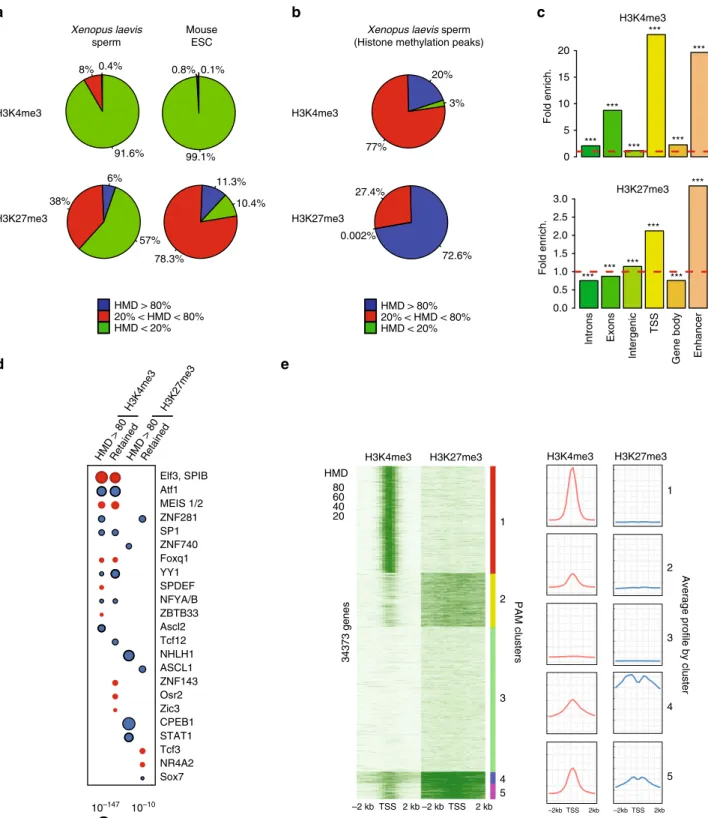
Fig. 2 Nucleosome loss/retention associated with spermiogenesis occurs non-randomly in a large fraction of the genome. a Relative abundance (left) and genome coverage (right) of nucleosomes and subnucleosomes in the sperm chromatin. The left pie chart reports the fraction of DNA fragments from Xenopus Laevis sperm corresponding to each type of particle; the right pie chart reports the fraction of genome covered by each type of particle. b Fraction of the genome with homogeneous particles composition. The bar graph indicates the percentage of the genome that possess nucleosomal or
subnucleosomal structure across most sperm of the population sequenced (genome binned in 50 bp windows).c Fold enrichment (observed/random)
over 1000 randomisations for homogeneous nucleosomes (left) or subnucleosomes (right) composition at the indicated genomic features; ***: empirical p value < 1e−3. Input data from two independent replicates were pooled. d PAM (partitioning around medoids) clustering of promoter (TSS +/−2 kb)
according to enrichment for nucleosomes or subnucleosomes.e Model of nucleosomes and subnucleosomes distribution in sperm and spermatid. Source
Mouse ESC HMD > 80% 20% < HMD < 80% HMD < 20% Xenopus laevis sperm
a
b
Xenopus laevis sperm (Histone methylation peaks)
HMD > 80% 20% < HMD < 80% HMD < 20% 0.4% 8% 91.6% 6% 38% 57% 0.1% 0.8% 99.1% 11.3% 78.3% 10.4% H3K4me3 H3K27me3 20% 77% 3% 72.6% 27.4% 0.002% H3K4me3 H3K27me3 lntrons Exons lntergenic TSS
Gene body Enhancer
Fold enrich. 0 5 10 15 20
c
Fold enrich. 0.0 0.5 1.0 1.5 2.0 2.5 3.0 H3K27me3 H3K4me3e
20 40 60 80 HMD H3K4me3 H3K27me3 0 25 50 75 0 100 200 300 400 500 0 25 50 75 0 100 200 300 400 500 0 25 50 75 0 100 200 300 400 500 0 25 50 75 0 100 200 300 400 500 0 25 50 75 0 100 200 300 400 500 H3K4me3 H3K27me3 5 4 3 1 2 2 kb –2 kb TSS –2 kb TSS 2 kb –2kb TSS 2kb –2kb TSS 2kbAverage profile by cluster
PAM clusters 5 4 3 1 2 34373 genes
d
HMD > 80RetainedHMD > 80Retained Elf3, SPIB Atf1 MEIS 1/2 ZNF281 SP1 ZNF740 Foxq1 YY1 SPDEF NFYA/B ZBTB33 Ascl2 Tcf12 NHLH1 ASCL1 ZNF143 Osr2 Zic3 CPEB1 STAT1 Tcf3 NR4A2 Sox7 10–10 10–147 p value H3K4me3 H3K27me3 *** *** *** *** *** *** *** *** *** *** *** ***Fig. 3 A fraction of the genome harbours methylated H3K4 and/or H3K27 at the same location in most sperm of a population. a Percentage of the
genome with different range of apparent histone H3 methylation density (HMD) on Lysine 4 and 27 inXenopus Laevis sperm and mouse ESC. b Percentage
of H3K4me3 and H3K27me3 peaks with different range of HMD inXenopus Laevis sperm. c Fold enrichment (observed/random) over 1000 randomisations
of peaks with homogeneous histone methylation (HMD > 80) at the indicated genomic features; ***: empiricalp value < 1e−3. ICe-ChIP data from two
independent replicates were pooled.d Dot matrix showing transcription factors with enriched motifs (y-axis) in the different histone methylation
categories (x-axis). Circle size represents –log10 (p value) of the motif enrichment; and the circle colour indicates whether evidences exist indicating that
the corresponding transcription factors is present maternally (blue) or not (red). Retained HMD > 80 peaks correspond to sperm histone methylation
peaks maintained after extract treatment as in Fig.5.e Heat map after PAM clustering of promoters (TSS+/−2 kb) according to histone H3-methylation
density on Lysine 4 and 27. The plots on the left show the average HMD profile for each cluster. Source data related to a, b, d, and e are provided as Source
view of the sperm epigenetic landscape at TSS that reveals
features conserved within the sperm population.
As expected, homogeneous H3K4me3 is mostly found in the
vicinity of the TSSs where homogeneous nucleosome retention is
also observed. However, this analysis also revealed that, as a
whole, TSSs with the less heterogeneous histone methylation
(H3K4me3 cluster 1, and H3K27me3 cluster 6) differ from TSSs
characterised by homogenous block of nucleosome and/or
subnucleosome (clusters 2, 4 and 5). In particular, genes with
stretch of homogeneous nucleosome retention (cluster 4) or
remodelling (cluster 2) surrounding the TSS are associated with
reduced H3K4me3 density when compared with genes with
heterogeneous particles composition (clusters 1 and 6). We also
identified a set of genes with homogeneous methylation on both
H3K27me3 and H3K4me3 (cluster 6). This set of genes show
co-occurrence of K4 and K27 methylation in most sperm. These
bivalent genes include many transcription factors involved in
early embryogenesis (i.e., members of the Hox, Fox, Sox, Gata,
Tbx and Pax transcription factor families). Moreover, globally,
these bivalent genes are associated with GO terms related to
development (Supplementary Data 4).
We conclude that, in general, homogeneity for histone
methyla-tion and histone particle composimethyla-tion is occurring on different
group of genes. Importantly, this analysis reveals the existence of
bivalent genes marking of developmental genes in all sperm.
Sperm-methylated histones are maintained during DNA
replication. Because the epigenetic features investigated in this
study are present at the same genomic location in most sperm
cells, they could represent necessary information delivered by the
sperm at fertilisation to support embryonic development. Such
necessary epigenetic cues would need to be transmitted to the
cells of the developing embryo to exert their action. To test this
hypothesis, we
first incubated permeabilised sperm in egg extract
to mimic the chromatin assembly and replication steps associated
with the
first embryonic cycle
26(Fig.
5
a and Supplementary
Fig. 4). H3K4me3 and H3K27me3 ChIP-seq analysis indicated
that 75% of sperm H3K4me3 peaks and 24% of H3K27me3 peaks
are retained after egg extract treatment (Fig.
5
b). In sperm, prior
to treatment, these retained peaks had higher methylation density
(i.e., less heterogeneous in the sperm population) and were larger
in size than lost peaks (Fig.
5
c, d). We also observed that retention
of peaks is favoured over gene regulatory regions (Fig.
5
e).
However, the context within which high histone H3 methylation
occurs (with or without enrichment for a given H3 particle) is not
generally predictive of the fate of the methylated histone post
replication (Supplementary Fig. 5). Finally, sperm incubation
with extracts containing the DNA replication inhibitor
gemi-nin
27,28suggests that most histone methylation peak loss
fol-lowing treatment is associated with chromatin assembly on sperm
DNA rather than with DNA replication (Supplementary Fig. 6).
Focusing on the embryonically abundant H3K4me3, we
evaluated the fate of sperm-methylated histones in vivo after
several replication cycles. For that purpose, we performed
ChIP-seq analysis using formaldehyde-fixed early blastula embryos
(after ~8 embryonic cell cycles but before the activation of zygotic
transcription) (Fig.
5
a). We observed that almost all H3K4me3
peaks detected in blastulae were already present in the sperm
chromatin (Fig.
5
f). Similar to peak retention after egg extract
treatment, we observed that H3K4me3 peaks retained in blastula
a
b
c
Epigenetic feature: Heterogeneous HomogeneousH3K4me3 H3K27me3 Nucleosome Subnucleosme
5 4 3 1 2 6 2 kb –2 kb TSS 2 kb –2 kb TSS –2 kb TSS 2 kb –2 kb TSS 2 kb Nucleosome SubnucleosomeHeterogeneous H3K4me3 peaks Fold enrich. 0.0 0.2 0.4 0.6 0.8 1.0 1.2 H3K27me3 peaks Fold enrich. 0.0 0.2 0.4 0.6 0.8 1.0 Nucleosome SubnucleosomeHeterogeneous 34373 genes PAM clusters
***
***
***
***
***
***
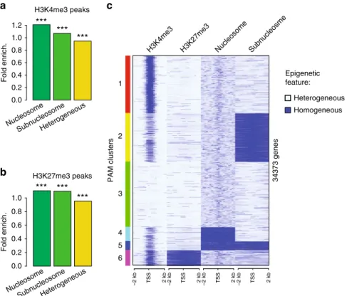
Fig. 4 Homogeneous bivalent marking of histone H3 on sperm developmental genes. a Fold enrichment (observed/random) over 1000 randomisations of peaks with homogeneous H3K4 methylation (HMD > 80) in regions homogeneous for nucleosomes, homogeneous for subnucleosomes or with
heterogeneous particle composition in a sperm population; ***: empiricalp value < 1e−3. b Same as in a for H3K27 methylation. c PAM clustering of
promoter (TSS+/−2 kb) according to homogeneity for methylation of histone H3 (HMD > 80) on Lysine 4, on Lysine 27, enrichment for nucleosomes
and enrichment for subnucleosomes. Source data related toc is provided as a Source Datafile. ICe-ChIP data from two independent replicates were
embryos corresponds to gene regulatory sites that harbour
stretches of H3K4me3 in most sperm cells (Fig.
5
g–i).
These observations show that the epigenetic information
present at the same genomic location in most sperm cells can
be faithfully transmitted to the mitotic progeny in early frog
embryos.
Sperm-methylated
histone
programmes
embryonic
gene
expression. We then thought to evaluate if the homogeneous
epigenetic fraction of the sperm genome that is transmitted to the
embryo contributes to the regulation of embryonic gene
expres-sion. We
first compared early embryonic expression of genes with
heterogenous methylation in sperm to that of genes that have a
Gem Chromatin assembly and 1× replication Chromatin assembly only ~8× replication Egg extract Fertilisation ChIP-seq H3K4me3/H3K27me3a
Spermb
Replicated sperm H3K4me3 peaks Sperm Replicated sperm H3K27me3 peaksc
d
e
lntrons Exons lntergenic TSSGene body Enhancer
Ratio fold enrich.
replicated sperm/sperm 0 2 4 6 H3K4me3
f
g
h
H3K4me3 peaks Sperm Blastulai
H3K4me3 Sperm >Blastula Replicated sperm >Blastula H3K27me3 0.0 0.5 1.0 1.5 2.0 lntrons Exons lntergenic TSSGene body Enhancer
Ratio fold enrich.
replicated sperm/sperm 0.0 0.5 1.0 1.5 2.0 H3K4me3
Ratio fold enrich. blastula/sperm
lntrons Exons
lntergenic
TSS
Gene body Enhancer
2089 8720 27,300 42,640 13,214 702 581 23,175 12,845 H3K4me3 *** *** 0 50 100 150 Sperm Retained Lost HMD *** *** 0 500 1000 1500 Sperm Retained Lost Peak size (bp) *** *** 0 100 200 300 400 500 Sperm Retained Lost HMD *** *** 0 2500 5000 7500 10,000 12,500 Sperm Retained Lost Peak size (bp) *** *** *** *** 0 500 1000 1500 Sperm Retained_B Lost_B Retained_RB Lost_RB Peak size (bp) *** *** *** *** 0 50 100 150 Sperm Retained_B Lost_B Retained_RB Lost_RB HMD H3K4me3 H3K27me3 H3K4me3 H3K27me3 Sperm >Blastula Replicated sperm >Blastula
homogeneous methylation in sperm and that retain this
methy-lation after replication (Supplementary Data 5). We
find that the
set of genes associated with homogenous methylation in sperm is
enriched for genes expressed at zygotic gene activation (ZGA)
(Fig.
6
a). Specifically, homogeneous methylation of H3K27 in
sperm is associated with ZGA genes involved in development
whereas homogeneous methylation of H3K4 in sperm is
asso-ciated with ZGA genes involved in housekeeping function.
Importantly, genes with heterogeneous methylation in the sperm
population do not show such enrichment for these ZGA gene
categories. Therefore, the epigenetically homogeneous fraction of
the sperm genome that is maintained after replication is strongly
associated with early embryonic gene transcription.
To further test the idea that homogeneous marking in sperm is
functionally linked to embryonic gene expression, we asked
whether interference with H3K4 or H3K27 methylation at
fertilisation, as reported in our previous work
2, preferentially
affect genes with an homogeneous methylation in a sperm
population. In that study we identified genes that were
misregulated in gastrulae generated from egg expressing a
demethylase targeting either H3K4 (Kdm5b) or H3K27 (Kdm6b)
methylation prior to sperm injection. The set of genes that are
misregulated upon demethylation of H3K27me3 (Kdm6b
sensi-tive) is enriched for genes with homogeneous methylation of
H3K27me3 in sperm (Fig.
6
b). In addition, a significant
proportion of genes sensitive to both demethylation of H3K27
and depletion for the maternally provided factor Ascl1 harbour
homogeneously methylated H3K27 and the binding motif for this
transcription factor (Supplementary Fig. 7). This suggests that
sperm-provided modified histones might regulate maternal factor
activity.
Unexpectedly, however, we observe that the set of genes
misregulated upon H3K4me3 demethylation (Kdm5b sensitive) is
also enriched for gene with homogeneous methylation of
H3K27me3 (Fig.
6
b). This could be explained if demethylation
of sperm H3K4me3 affects subsequent embryonic gene
expres-sion only when it co-occurs with methylation of H3K27 (i.e.,
sperm bivalent genes). To test this hypothesis we
first investigated
the fate of the fraction of sperm genome homogeneously
methylated on H3K4me3 only, H3K27me3 only or both residues
(bivalent) (Fig.
6
c). We
find that peaks of homogeneous
H3K4me3 in sperm are very well conserved after replication
(>80%) whereas peaks of homogeneous H3K27me3 are poorly
retained (<25%). Interestingly when co-occuring with H3K4me3,
H3K27me3 peaks are better retained after replication (~50%).
When focusing on the set of genes associated with these retained
bivalent peaks, we observed an enrichment for genes misregulated
upon H3K4 or H3K27 methylation (Fig.
6
d).
We conclude that the homogenous fraction of sperm-modified
histone that is propagated in early embryos prime developmental
genes for embryonic expression. We next sought to evaluate if
such epigenetic regulatory principle apply in human.
Homogeneous histone methylation in a human sperm
popu-lation. We
first applied ICe-ChIP to quantify the level of
methylation of the fraction of histone H3 retained in mature
human sperm. To that end human sperm from a fertile individual
were treated with an MNase concentration that yields
nucleosome-sized fragments as well as smaller fragments
(Sup-plementary Fig. 8). Digested chromatin was used for H3K4me3
and H3K27me3 ICe-ChIP-Seq analysis. Interestingly, in human,
H3K4me3 and H3K27me3 ChIP recovers only nucleosome-sized
fragment (Supplementary Fig. 8). So unlike in Xenopus, in
human-sperm-modified histone H3 are not found associated with
small DNA fragments generated by MNase digestion. We
observed that part of the human sperm genome harbours a high
density of H3K4me3 or H3K27me3, albeit to a lower extent than
observed in Xenopus and in accordance with the lower histone
retention in human sperm (Fig.
7
a, left). Clustering promoters
according to H3K4me3 and H3K27me3 density highlighted
fur-ther differences between human and Xenopus (Fig.
7
b and
Sup-plementary Data 6). In human, H3K4me3 density is not
particularly higher at the TSSs as observed in frog (compare
Figs.
3
e and
7
b). Instead high H3K4me3 HMD is found on broad
domains around TSSs of a limited number of genes (cluster 4,
1570 genes). This suggests that human spermiogenesis is
asso-ciated with a removal of the majority of H3K4me3 that is usually
associated with a large proportion of genes TSSs in somatic cells.
To focus on the fraction of the human sperm genome that is
homogeneously methylated on histone H3, we then identified
peaks of histone modifications and computed their HMD.
Overall, the peaks identified are well conserved between our
study and previous work
5indicating that peak calling is
consistent across a range of MNase treatments (Supplementary
Data 7). Interestingly, however, our analysis shows that in
human, a very large fraction of histone methylation peaks
identified by conventional ChIP-seq corresponds to low HMD
and are therefore heterogeneous in a sperm population (Fig.
7
a,
right). Nonetheless, some human sperm genes have peaks of high
density of histone H3K4me3 (130 genes) or H3K27me3 (320
genes) around their TSSs (i.e., BMI1, TBX3, Fig.
7
c and
Supplementary Data 8). Genes associated with peaks of
H3K4me3 or H3K27me3 methylation in sperm, irrespective of
HMD level, show a significant overlap between human and
Xenopus and these gene sets are enriched for genes expressed at
ZGA in both species (Fig.
7
d)
29. By contrast, no conservation is
observed between Xenopus and human orthologous genes
associated with the homogeneous-methylated fractions (peaks
with HMD > 80) of the sperm genome (Fig.
7
d). However, in both
species the set of genes with high H3K27me3 HMD is related to
Fig. 5 Homogeneously methylated sperm histones are maintained during early embryonic replication. a Experimental setup to monitor the fate of
sperm-methylated histone peaks after replication.b Overlap between peaks of H3K4me3 and H3K27me3 before and after replication of sperm chromatin
in egg extract.c Boxplots of HMD for all sperm peaks and for peaks that are lost or retained after replication. d Boxplots of the size of all sperm peaks,
and of peaks that are lost or retained after replication. Data inc and d are obtained from N. sperm peaks H3K4me3: 36020; N. sperm retained H3K4me3:
27300; N sperm lost H3K4me3: 8715. N. sperm peaks H3K27me3: 55854; N. sperm retained H3K27me3: 13214; N sperm lost H3K27me3: 42635.
***p value < 1e−3 (two-sample Kolmogorov–Smirnov test). e Ratio of fold enrichment of peaks retained after replication over those lost after replication
at indicated genomic features. Fold enrichments (observed/random) were obtained from 1000 randomisations and all instances showed an empirical p value < 1e−3. f Overlap between peaks of H3K4me3 in sperm and in blastula embryos. g Boxplots indicating HMD for all sperm peaks and for peaks that
are lost or retained in blastula compared with sperm, and lost or retained in blastula compared with replicated sperm.h Boxplots of peak sizes for all sperm
peaks and for peaks lost and retained in blastula compared with sperm, and in blastula compared with replicated sperm. Ing and h ***p value < 1e−3 and
are obtained by the two-sample Kolmogorov–Smirnov test. i Ratio of fold enrichment of peaks retained in blastula versus those in sperm at indicated
genomic features. This ratio has been obtained as ine. HMD is from data pooled from two independent replicates. Peaks retention/lost are consensus from
developmental functions (Supplementary Tables 3 and 7). In
addition, an enrichment of high HMD peaks for ZFP281 binding
sites, a transcription factor implicated in early mammalian
embryonic development, is found in both human and Xenopus.
These observations raise the possibility that the fraction of the
sperm genome with homogenous histone methylation could
contribute to early embryonic development in human as it is the
case in Xenopus, but through the regulation of a different set
of genes.
We observe that certain genomic sites, when retaining histones,
always retain them in their methylated form. However, unlike in
Xenopus, we cannot rule out that histone retention happens only
in a subset of sperm cells in humans. DNA methylation and
histone H3K27 methylation have been shown to be mutually
0 2 4 6 8 10 12 14 16 –Log10 (p value)
Bivalent retained
Developmental ZGA House keeping ZGA All genes ZGA KDM5 sens KDM6 sens
d
a
0 10 20 30 40 50 Developmental ZGAHouse keeping ZGA
All genes ZGA
–Log10 (p value)
K27 heterogeneous K27 homogeneous & retained K4 heterogeneous
K4 homogeneous & retained
b
K27 heterogeneous K27 homogeneous & retained K4 heterogeneous
K4 homogeneous & retained
0 10 20 30 40 50 60 –Log10 (p value) Kdm5 sensitive Kdm6 sensitive 0.89 0 0.08 0.25 0.18 0.62 0.44 0.35 0.53 0.71 0.80 Bivalent Bivalent H3K4me3 H3K27me3 H3K4me3 H3K27me3
Replicated sperm chromatin
Sperm chromatin
c
Fraction of overlap
exclusive on chromatin
30. Therefore, we would expect that if
peaks of high HMD methylation on H3K27 reflected the situation
in most sperm cells, they would be associated with genomic
regions that are uniformly DNA hypomethylated in a human
sperm population. We checked this proposition using published
bisulfite sequencing data from single human sperm cells
31. This
dataset allowed us to identify genomic region that harbour
unmethylated DNA in all sperm. We found that high HMD peaks
(HMD > 80) are indeed more likely to be associated with such
unmethylated sperm DNA than low HMD peaks (0 < HMD < 80)
(Fig.
7
e). The observed correlation between lack of DNA
methylation in all sperm and high HMD peak of H3K27
fit with
the hypothesis that high HMD peaks represent methylated
histones present in most human sperm cells. In addition, the set
of genes with high HMD on H3K27 in sperm is enriched
for genes that have a closed TSS configuration in most cells
of the human embryos undergoing ZGA, as shown by
ATAC-seq
31(Fig.
7
f). This suggests that methylated histones on sperm
gene TSSs could regulate chromatin accessibility in embryos
at ZGA.
We conclude that in human sperm, histones near some
developmental genes are always retained in their methylated
form. We provide indirect evidence that methylated histones
might also be homogeneously distributed in a human sperm
population as in Xenopus and could contribute to the regulation
of embryonic chromatin status at ZGA.
Discussion
Multiple lines of evidence point towards the transmission of
epigenetic information from parents to offspring
32. However, the
molecular basis of this epigenetic information is unclear. In
particular the notion that sperm-derived histones are randomly
retained along the genome has gained support, leading to the
proposal that modified histones cannot be the basis for the
epi-genetic information required for embryo development
14. Here we
provide evidence to the contrary. The particular core histone
remodelling event occurring during Xenopus laevis
spermiogen-esis enabled us to refine epigenetic maps of the sperm by
iden-tifying genomic sites where most sperm of a population harbours
a given epigenetic feature. This approach uncovers a subset of
genes in the genome that retains modified histones in all sperm
(Fig.
7
g). Compared with genes with heterogeneous epigenetic
composition, these genes are characterised by their expression at
ZGA, their persistent association with modified histones after
replication and their sensitivity to histone demethylation at
fer-tilisation. Our analysis demonstrates that sperm homogenously
modified histones across a cell population contribute to the
transmission of epigenetic information necessary for the
regula-tion of embryonic gene expression in Xenopus laevis. Moreover,
we identify genomic sites where histones are always retained as
methylated in human sperm. Although we cannot assume that all
sperm retain histones at these locations in human, these genomic
sites represent potential candidate regions for the epigenetic
programming of human sperm for embryonic expression. This
suggests that a modified histone-based mechanism of
inter-generational epigenetic transmission might be conserved between
species.
How is the epigenetic information encoded in sperm-modified
histone then transmitted in the developing embryos? In Zebrafish
and Xenopus, 10–12 cell division occur before the major wave of
zygotic genome activation. In order to affect embryonic gene
expression, sperm-derived epigenetic information therefore needs
to be maintained through multiple cell division. In Zebrafish,
previous work indicated that modified histones were not detected
at early embryonic stage, ruling out direct transmission of
sperm-derived modified histones during early embryogenesis
33,34.
However more recent work indicated that sperm-derived histone
variant H2A.Z acts as a placeholder in early embryos where it
regulates DNA methylation required for proper transcription and
development
35. In the case of Xenopus laevis sperm a direct
transmission mechanism of H3K27me3 is also unlikely. Indeed,
we observe that a much larger fraction of H3K27 methylation
than that of H3K4 methylation is lost after replication (Fig.
5
b), in
agreement with H3K27me3 ChIP-seq data obtained in Xenopus
tropicalis that detected very low level of this mark in gastrulae
36.
We hypothetize that a relay mechanism (placeholder) or other
histone marks associated with H3K27me3 carry the information
into the embryo. By contrast, we observe that most of the H3K4
methylation peaks present in Xenopus laevis embryos just before
ZGA could be traced back to the homogeneous fraction of H3K4
methylation identified in sperm. In human a recent report also
indicates that H3K4me3 persists from fertilisation to post-ZGA
stage embryos
37. Altogether this suggests that sperm-derived
modified histones contribute to the establishment of the
embryonic epigenome prior to ZGA.
We
find that the homogenous fraction of methylated histones
in a Xenopus sperm population is transmitted through the
first
embryonic cell cycle and potentially through cell divisions of the
developing embryos to regulate early embryonic gene expression.
Defect in these epigenetic programming of sperm for embryonic
development could be at the origin of case of idiopathic male
infertility. In addition, the natural mechanisms of epigenetic
information transmission from the sperm to the developing
embryos might also be responsible for the maintenance of an
epigenetic memory of somatic cell identity that characterise
embryos generated by nuclear transfer
2,38. By getting a better
understanding of the processes underlying the transmission of
paternal epigenetic information, we will be able to devise better
strategies to erase somatic cell identity and improve cloning
efficiency.
Fig. 6 The homogeneously methylated histone fraction in frog sperm chromatin regulates embryonic gene expression. a Barplot of enrichmentp values
(−log10) in set of genes expressed at zygotic gene activation (all-, developmental- and house keeping-ZGA genes) for the presence of different type of
H3K4me3 or H3K27me3 sperm peaks. pink and light green bars= enrichment p value for heterogenous (HMD < 80) peaks in promoter; red and green
bars= enrichment p value for homogeneous (HMD > 80) peaks in promoter that are also retained after replication. P values determined by the χ2
proportion test evaluating if the proportion of gene of interest is higher (alternative= ‘greater’) than the genome-wide proportion (i.e., expected
proportion).b Barplot of enrichmentp values (−log10) in set of genes whose embryonic expression is sensitive to the presence of histone H3K4
demethylase (Kdm5b) or histone H3K27 demethylase (Kdm6b) at fertilisation2for the presence of the aforementioned sperm peaks categories.P values
are obtained as ina. c Pie chart indicating the percentage of the homogeneously methylated histone present in sperm that are retained after egg
extract-mediated replication. Three type of sperm peaks are considered: homogeneous for H3K27me3 only, homogeneous for H3K4me3 only and homogeneous for H3K27me3 and H3K4me3 (bivalent). The area of the coloured sector (together with the colour) indicates the fraction of the overlap between each
pair-wise comparison.d Barplot of enrichmentp values (−log10) in various set of genes for the presence of a homogeneous bivalent H3K4me3 and H3K27me3
retained after replication.P values are obtained as in a. HMD are from data pooled from two independent replicates. Peaks retention/lost are consensus
Methods
Experimental model and subject details. Mature Xenopus laevis males were obtained from Nasco (901 Janesville Avenue, PO Box 901, Fort Atkinson, WI
53538-0901;https://www.enasco.com/xenopus). Our work with Xenopus complied
with all relevant ethical regulations for animal testing and research. This work is
covered under the Home Office Project License PPL 70/8591 and frog husbandry
and all experiments were performed according to the relevant regulatory standards.
Animals were kept in a Marine Biotech recirculating system at a density of one
adult/3l, with 10% water change per day. Water was sequentiallyfiltered with
mechanical pad sumpfilter, nitrifying bacteria filter, mechanical canister filter,
carbonfilter and UV sterilised. Water quality parameters were as follow:
con-ductivity 1500 uS; temperature 17–22 °C; PH 6–8. Photoperiod was set to 12 h ON/
12 h OFF. Frogs are fed twice per week with Royal Horizon 4.5 mm pellets
(skretting,https://www.skrettingfishfeeds.co.uk/). Unconsumed food was removed
H3K27me3 Peaks HMD > 80% 20% < HMD < 80% HMD < 20%
d
a
20 40 60 80 HMD H3K4me3 H3K27me3 5 4 3 1 2b
c
H3K4me3 H3K27me3 All peaks HMD > 80 peaks 72.1%* 58.8%* 32% 8% %Orthologues marked in both speciese
H3K4me3 98.5% 1.3% 0.2% 3.3% 0.2% 96.5% 2.3% 45.4% 52.3% 0.4% 23.6% 76% Genome wide Xenopus Homogeneous H3 methylation in most sperm Humang
Gene regulatory regions
0 5 10 Open Open Divergent Divergent Closed Closed H3K4 HMD > 80 H3K27 HMD > 80 –log10 (p value) Underrepresented Overrepressented
f
HMD HMD H3K4me3 H3K27me3 H3K4me3 H3K27me3 100% 100% 100% 100% TBX3 20 kb 20 kb BMI1 SPAG6Proposed model for chromatin organization and homogeneous nucleosome reetention in sperm
–2 kb TSS2 kb–2 bkTSS2 kb PAM clusters 0.0 2.5 5.0 7.5 10.0 HMD0_20HMD20_40HMD40_60HMD60_80HMD80
Average % meC meth. in peak 0 20 40 60
HMD0_20HMD20_40HMD40_60HMD60_80HMD80
10 min after the start of feeding. The researchers and the staff of the Gurdon Institute animal husbandry facility are trained in these experiments, and veter-inarians monitor the health status of the animals.
Xenopus sperm collection. Xenopus sperm collection was performed as described
before2. For each round of sperm purification, testes from six adult Xenopus laevis
males were isolated and manually cleaned from blood vessels and fat bodies in 1×
Marc’s modified ringers (MMR, 100 mM NaCl, 2 mM KCl, 1 mM MgSO4, 2 mM
CaCl2, 3 mM HEPES, pH 7.4) using forceps and paper tissues. It is crucial to clean
the testes well from any non-testicular tissues, as otherwise the cells released from
the tissues may negatively affect thefinal purity of isolated cells. Subsequently,
testes were torn into small pieces with forceps and homogenised with 2–3 strokes of a dounce homogeniser (tissue from one testis at a time). The cell suspension was
thenfiltered to remove tissue debris and cell clumps (CellTrics, cat. 04-0042-2317)
and spun down at 800 rcf, 4 °C, for 20 min. Supernatant was discarded and the cell pellet was resuspended in 12 mL of 1× MMR. If any red blood cells were visible at the bottom of the pellet (a result of incomplete removal of blood vessels), only the uncontaminated part of the pellet was recovered, taking extreme care not to disturb the red blood cells. Subsequently, step gradients of iodixanol (Optiprep; Sigma,
D1556; 60% iodixanol in water) in 1× MMRfinal were manually prepared in
pre-chilled 14 mL ultra-clear centrifuge tubes (Beckman Coulter, #344060) in the fol-lowing order from the bottom to the top of the tube: 4 mL of 30% iodixanol, 1 mL of 20% iodixanol, 5 mL of 12% iodixanol (all in 1× MMR) and 2 mL of cell suspension in 1× MMR on top. Gradients were spun down in a pre-chilled SW40Ti rotor at 7500 rpm (10,000 g), 4 °C, for 15 min, deceleration without brake (Beck-man Coulter Ultra-centrifuge, Optima L-100XP). The pelleted fraction, containing mature sperm, was collected. Collected fractions were diluted six times with 1×
MMR and collected by spinningfirst at 805 rcf, 4 °C, for 20 min and respinning at
3220 rcf, 4 °C, for 20 min to pellet remaining cells. Pelleted cells were subsequently
permeabilized with Digitonin (10 mg/mL as afinal concentration, Sigma, D141) for
5 min at room temperature (RT) as detailed before39.
Human-sperm collection. All human sperm samples were processed in
accor-dance to ReproMed Ireland’s standard procedures and ethical approval was
obtained from the University College Dublin Human Ethics Committee (HREC) (protocol number LS-16-53-ODoherty-Fair). The approval process entailed inde-pendent peer review along with approval from the HREC. Human-sperm samples were donated to the research project by informed consent. Written informed consent was obtained from male partners of expectant couples/couples that recently had a baby (within 6 months of sample donation) that donated fresh sperm samples following 3 days of abstinence. Before giving consent, donors were provided with all of the necessary information about the research project and contact information for the project lead. Specifically, patients signed a consent form authorising the use of their sperm samples for future research purposes, including molecular and epigenetic analyses, and for the results of these studies to be pub-lished in scientific journals. No financial inducements were offered for donation. Chromatin was prepared from fresh ejaculates donated from men with proven fertility. Men were deemed fertile based on the following parameters; female partners are currently pregnant or have given birth within 6 months of sample production. Semen samples were collected from men aged between 30 and 35 years old. Ejaculates were maintained at 37 °C for 10 min immediately following production. A 10 µl aliquot of each fresh ejaculate was subjected to routine semen analysis prior to processing and all samples were in accordance with the World Health Organization guidelines (World Health Organization. WHO laboratory manual for the examination and processing of human semen (5th ed.), WHO Press (2010)) for normal semen samples (sperm density, total number, motility, morphology and semen volume). The recorded parameters for sperm preparations
used in this study were within the following range: progressive motility (PR)=
74–81%; non-progressive motility (NP) = 10–13%; immotile (IM) = 9–11%; total
motility (PR+ NP) = 87–91%.
Motile spermatozoa were purified on a discontinuous Percoll gradient
(Pharmacia, Uppsala, Sweden), as outlined previously40. Gradient-purified
spermatozoa were quantified, resuspended in Hepes-buffered Tyrode’s medium
(Sigma). Permeabilization of sperm nuclei was performed with Digitonin following the same procedure as for Xenopus samples.
Xenopus cell culture. Cell line XL-177 was derived from tadpole epithelium of X.
laevis41. Cultured cells were maintained in medium containing L-15 (SIGMA
L1518), sterile water and FBS (6:3:1vol/vol) and supplemented with 100μl
peni-cillin/streptomycin. Cells were grown at 23 °C in gelatin-coated dish that were
sealed. Confluent culture of cells were split 1:3 to 1:6 following trypsin digestion.
Interphase egg extract preparation. Eggs were collected in 1× MMR, de-jellied with 0.2× MBS (Modified Barth’s Saline) including 2% cysteine (Sigma, W326305) (pH was adjusted to 7.8 using 10 N NaOH) and washed with 0.2× MMR. Subse-quently, eggs were activated for 3 min at RT with 0.2× MMR supplemented with
0.2μg/mL calcium ionophore (Sigma, C7522). Eggs were rinsed with 0.2× MMR
and subsequently all abnormal or not-activated eggs were removed. Eggs were
washed with 50 mL of ice-cold extraction buffer (EB) (5 mM KCl, 0.5 mM MgCl2,
0.2 mM DTT, 5 mM Hepes pH 7.5) supplemented with protease inhibitors (PIs) (Roche, 11873580001), transferred into centrifugation tube (Thinwall.
Ultra-ClearTM, 5 mL, 13 × 51 mm tubes, Beckman Coulter Inc., UK; 344057) and
sup-plemented with 1 mL of EB buffer with PI and 100μg/mL of cytochalasin B (Sigma,
C2743) and placed on ice for 10 min. Subsequently, eggs were spun briefly at 350 x g for 1 min at 4 C (SW55Ti rotor, Beckman Coulter Ultra-centrifuge, Optima L-100XP) and excess buffer was discarded. Eggs were then spun at 18,000 x g for 10 min at 1 C, the extract was collected with a needle, transferred to a fresh,
pre-chilled tube, supplemented with PI and 10μg/mL of cytochalasin B, and respun
using the same conditions. Extract was collected with a needle and used fresh for the replication assay.
Expression and purification of Delta-Geminin. pET28 Delta-Geminin were
transformed into E.coli and transformants were cultured overnight in 5 ml of LB
medium, containing 50μg/ml kanamycin and 30 μg/ml chloramphenicol.
Over-night cultures were transferred into 250 ml of LB medium without glucose and
antibiotics and incubated for 2–2.5 h at 37. One hundred microlitres of 1 M
iso-propyl 1-thio-beta-D-galactopyranoside was added into culture to induce recom-binant protein expression. After 2 h induction, cells were collected by
centrifugation. Cell pellets were suspended into 30–40 ml of MilliQ water and
transferred into 50 ml tubes. After centrifugation, supernatant was removed and
pellets were stored at−80 °C. Frozen pellets were suspended into 5 ml of Dicis
buffer (300 mM NaCl, 150 mM KoAC, 20 mM Tris, pH7.5, 2 mM MgCl2, 10%
glycerol, 0.01% NP40), with 0.1% NP40 and PIs, on ice. The suspensions were sonicated with Vibra-Cell Ultrasonic Processor (SONICS) for six times 15 s on and off cycles. Sonicated samples were centrifuged at 15,000 rpm for 10 min at 4 °C and supernatants were collected. Ni-NTA agarose (Qiagen, 30210) was washed with 5 ml of Dicis buffer for three times in open column. Equilibrated Ni-NTA agarose was transferred into supernatants and rotated for 1 h at 4 °C. Agarose beads were transferred into open column and washed twice with 10 ml of Dicis buffer with 20 mM imidazole. Bound protein was eluted with Dicis buffer with either 100 mM or 200 mM imidazole for three times, respectively. Three fractions with highly con-centrated delta-Geminin were pooled. Fractions were applied to PD10 column (GE Healthcare) equilibrated with buffer containing 10 mM Tris-HCl, pH8.0, 0.5 M NaCl, 5% glycerol and eluted with 3 ml of buffer.
DNA replication in egg extracts. Freshly prepared egg extracts were supple-mented with 0.005 mg/ml creatine kinase (Roche, 10127566001), 0.375 mM crea-tine phosphate (Roche, 10621714001), 0.05 mM ATP (Roche, 10519979001), 0.005
mM EGTA, 1 mM MgCl2. In some experiments, delta-geminin was added (final
concentration 1.5μg/ml) to egg extract to inhibit DNA replication. Permeabilized
sperm cells were added to afinal concentration of 1000 nuclei/μl of extract and
incubated at RT for 2 h with gentle tapping every 10 min. The reaction was stopped
by adding 10 volumes of ice-cold Egg Lysis Buffer—Chromatin Isolation Buffer
(ELB–CIB, 10 mM Hepes pH 7.8, 250 mM sucrose, 2.5 mM MgCl2, 50 mM KCl,
1 mM DTT, 1 mM EDTA, 1 mM spermidine, 1 mM spermine, 0.1% Triton X-100, Fig. 7 Homogeneous retention of methylated nucleosome in a human sperm population. a Percentage of the genome (left) and percentage of peaks
(right) with different levels of H3K4 or H3K27 methylation density in human sperm.b PAM clustering of H3K4 and H3K27 HMD levels at promoter
regions (TSS+/−2 kb) in human sperm. c Genome browser screenshots of TBX3 and BMI1 HMDs in human sperm. d Percentage of gene orthologues with
peaks of histone methylation in both human andXenopus sperm. e Barplots of the average percentage (%) of 5mC methylation at H3K27me3 peaks
stratified by methylation level (left); error bars: sem; barplot of the percentage of H3K27me3 peaks showing absence of 5mC methylation (right) (5mC
methylation: single-sperm bisulfite sequencing data from ref.31).f Barplots indicating−log10 (p value) for enrichment of sperm TSSs with HMD > 80 for
H3K4 or H3K27 in set of genes with TSSs showing different chromatin accessibility level in eight cell embryos (open and closed corresponds to TSSs open
or closed in all cells of a eight cell embryos), whereas divergent corresponds to TSSs either open or closed in different cells (ATAC-seq data from ref.31);
p values determined by χ2proportion test evaluating if the proportion of gene of interest is higher (green) or lower (red) than the genome-wide proportion
(i.e., expected proportion).g Model of epigenetic homogeneity inXenopus and human sperm. The cartoon summarises the observed trends in retention of