HAL Id: hal-02405538
https://hal.archives-ouvertes.fr/hal-02405538
Submitted on 18 Dec 2020
HAL is a multi-disciplinary open access
archive for the deposit and dissemination of
sci-entific research documents, whether they are
pub-lished or not. The documents may come from
teaching and research institutions in France or
abroad, or from public or private research centers.
L’archive ouverte pluridisciplinaire HAL, est
destinée au dépôt et à la diffusion de documents
scientifiques de niveau recherche, publiés ou non,
émanant des établissements d’enseignement et de
recherche français ou étrangers, des laboratoires
publics ou privés.
About monitoring the dynamics of phase transition in
food and biology by microphotonics: detecting
soft-matter process
Lucas Garnier, Rigoberto Castro-Beltran, A. Saint-Jalmes, H. Lhermite,
Anne-Laure Fameau, Véronique Vié, Eric Gicquel, Hervé Cormerais, Bruno
Bêche
To cite this version:
Lucas Garnier, Rigoberto Castro-Beltran, A. Saint-Jalmes, H. Lhermite, Anne-Laure Fameau, et al..
About monitoring the dynamics of phase transition in food and biology by microphotonics: detecting
soft-matter process. Proceedings of SPIE, the International Society for Optical Engineering, SPIE, The
International Society for Optical Engineering, 2020, Integrated Photonics Platforms: Fundamental
Research, Manufacturing and Applications, V11364, pp.113641T.1-8. �10.1117/12.2564645�.
�hal-02405538�
About monitoring the dynamics of phase transition in food and biology
by micro-photonics: detecting soft-matter process
L. Garnier
a, R. Castro-Beltran
b, A. Saint-Jalmes
a, H. Lhermite
c, A.-L. Fameau
d, V. Vié
a,
E. Gicquel
a, H. Cormerais
c,e, B. Bêche*
a,ca
Univ Rennes, CNRS, IPR (Institut de Physique de Rennes) - UMR 6251, F-35000 Rennes, France ;
bUniversidad de Guanajuato, Departamento de Ingeniería Física, División de Ciencias e
Ingenierías-León, Guanajuato, México ;
cUniv Rennes, CNRS, IETR (Institut d'Electronique et de
Télécommunication de Rennes) - UMR 6164, F-35000 Rennes, France ;
dINRA-BIA, Biopolymères
Interactions Assemblages, 44316 Nantes, France ;
eCentrale/Supelec, Campus de Rennes, 35510
Cesson-Sévigné, France.
E-mail :
bruno.beche@univ-rennes1.fr
;
https://ipr.univ-rennes1.fr/interlocuteurs/bruno-beche
ABSTRACT
We have investigated the ability to monitor the dynamics transition phase of various substances by resonant probe light. Such a specific Micro-Total Analysis Systems (µTAS) can be used in food, cosmetic and biology applications. Such lab-on-chip sensors present the possibility of data treatment with an embedded system. The serial of transduced spectra are then acquired with an optical spectrum analyzer linked to a computer on which Matlab software record and process the data in real time. Then specific quantities can be linked to the intrinsic physico-chemical characteristics of the substances. As an example (not exhaustive) the development and the ability of an optical integrated polymeric resonator, acting as a surface light probe, for monitoring temperature-induced supramolecular phase transitions will be presented. The homogeneous detection of the transitions between different self-assembled structures in an aqueous solution of fatty acids (12-hydroxystearic acid, in association with amino-pentanol) was studied by investigating the coupling between the solution and the integrated photonic micro-cavity. Tuning the self-organized assemblies of surfactant is very attractive for many applications, such as cosmetic products, food, drug delivery and medical, and the development of alternative tools - especially those requiring minute amount of solution - to monitor their structural changes are essential. These original studies at temperatures ranging from 17 to 24 °C, based on a statistical treatment of optical resonance spectra, have evidenced the thermoresponsive nature of the optical features, and that different regimes occur with temperature. The optical results were corroborated with the measurement of the solution viscosity as a function of temperature, confirming that we can ascribe the optically-detected regimes to a surfactant assembly shifting reversibly from a tubular shape to a micellar one. The comparison between the optical and the rheological responses showed different accuracies: while the viscosity data exhibited a rather smooth and monotonous transition, the behavior changes were sharper and non-monotonous in terms of optical properties, allowing us to unambiguously identify in intermediate regime between 18.5 and 20°C. These morphological transition experiments represent a unique opportunity to extend the numbers of available techniques studying these systems through integrated optical techniques with potential opportunities of real time detection and working on low sampling volume. Other examples will be developed as the detecting of phase transition of sphingomyelin in biology and health corroborate by differential scanning calorimetry…
1. INTRODUCTION
A micro-encapsulation process is a useful procedure for packing, isolation and protection of biochemical compounds, and self-assembled supramolecular structures - such as micelles, vesicles or lamellar phases - represent an extraordinary opportunity for encapsulation. A structure that could be filled by bio-chemical compounds is a potential prospect for drug delivery in medicine, environment science, cosmetics and food transformation[1-2]. These applications depend on the complete control of the intrinsic self-assembled supramolecular structures, linked to a reorganization at the molecular scal. From the vast family of lipids, fatty acids are predominant and very important for a huge number of biotechnological applications ranging from sources of fuel for animals, cosmetic products to drug delivery carriers[2]. These systems can be modulated in various shapes adapted to the use of fluids and gels or creams and pastes. From these derivative applications, their supramolecular tunable and reversible structures are highly related to their bulk properties such as viscosity and thermal behavior. Various techniques for investigating supramolecular arrangements, like Differential Scanning Calorimetry (DSC), various microscopies, Wide-Angle X-ray Scattering (WAXS), Small-Angle Neutron Scattering (SANS), or rheometry exist. Those are some of the used technological tests to characterize the morphological and mechanical properties of many self-assembled structures and their different phases. These techniques play complementary roles by providing information at different length and time scales. Various bulk and surface optical techniques, based on scattering intensity modulation, have been proposed to detect the phase transition temperatures of products in biology[3]. Looking for another sensitive techniques that present an opportunity for monitoring fine-changes associated to phase transition, microresonators[4-8] based on integrated optical (IO) chip devices and resonant probe light are shown as outstanding photonic platforms for this type of biochemical sensing applications.
In this paper, the interest is focused on the possibility to measure and detect phase transitions in soft matter products with integrated photonic micro-resonators by studying the influence of such soft matter changes and mechanisms on the mode propagating in photonic resonant structures. The integration of such sensors on relevant chips allows designing lab-on-chip sensors, which facilitate both miniaturization and transportability for on-line on-live tests and measurements. This kind of structures also provides very precise measurements, with low limit of detection, and high sensitivity. As miniaturizable and most accurate, photonic micro-resonator sensors come up as very efficient in following small-scale phenomena, but also macroscopic modification of soft matter substance as demonstrated into such paper. These characteristics are most interesting for both environmental monitoring, food industry, cosmetic and biology together with biomedical or biophysics fundamental research. However, with a large majority of scientific works using integrated micro-resonator sensors focusing on static phenomena, most often the transduced signal of the photonic structure is monitored against concentration values of given species or versus temperature. Then, noteworthy variations of the signal
versus time, so as to measure dynamic reactions and phase transitions are rarely investigated with. In this paper,
integrated micro-resonators are used to perform a dynamic tracking of phase transitions mechanisms on the acting surface of the sensor. The fabrication of the structures is detailed. The experimental setup is presented and then relevant results on fatty acid[9] (cosmetic, food) and sphingomyelin[10] (biology) phase transitions are exposed and discussed.
2. RESONANT STRUCTURE FABRICATION
2.1 Polymer materialIntegrated polymeric micro-resonators are fabricated through deep UV (DUV) photolithography procedures and are based on UV210 photoresist[11-12]. This specific DUV-polymer is made of poly p-hydroxystyrene and poly t-butyl acrylate; it is called an amplified chemical photoresist including a photo acid generator so as to increase the sensitivity with energy exposure and to obtain a better photolithography mechanism. Moreover such a chosen material polymer presents a refractive index smaller than the ones of others materials; for an upper cladding composed of air or specific soft matter substances which is often the case for sensing applications, the evanescent probe-wave or light of the propagating mode is then larger for polymer resonators than for other structures ; this difference can imply better sensing
properties for polymer-based-on resonators. Polymers are lower cost than for example semi-conductors and their specific deposition (MOCVD, MBE…), reducing then the fabrication cost and facilitating industrial large-scale production with spin-coating procedures. The specific absorption band at 248 nm of such DUV210 polymer allows to perform photolithography most suited to design sub-micronic gaps[12]. An advantageous substrate for such structures is a SiO2/Si
wafer, with a SiO2 layer 1 μm in thickness obtained by thermal oxidation. This layer stands as the lower cladding for the
waveguides.
2.2 Polymeric micro-resonators and geometrical characteristics
The resonant quantifications met in such physical systems are due to a geometric recirculation of the light. These quantifications emerge thanks to the installation of a cyclic condition (or stationary waves) written as Popt=m.Maxwell,
with Popt the ‘optical’-perimeter of the microresonator and m an integer. This principle can also be seen as the
quantification of the orbital kinetic momentum as a quantified photonic orbit. Beyond the choice of the material, the shape and the geometrical characteristics of the structure are determinant factors with a view to sensing properties. The structure hinges on a mono-mode access waveguide laterally coupled from either side to racetrack shaped micro-resonators as depicted in figure 1. The microresonator is based on a racetrack shape and light is laterally coupled with a straight tapered optical waveguide, Figure 1. A tapered bus optical waveguide ensures single mode operation of the IO micro-device. In comparison with other microcavities, a racetrack shape presents some advantages linked to its elongated rib zone (define by the coupling length Lc). This length Lc, the parallelism between this resonator and the tapered rib
waveguide, contribute to a stronger optical coupling strength of the microresonator. A length Lc = 5 µm, a radius R= 5
µm, a width w= 3 µm and a gap g= 400 nm (a distance from the tapered waveguide and the microcavity) represent the main geometrical features of our micro-racetrack.
Figure 1: Schematic diagram and optical imaging of the photonic resonant structure. The main geometrical parameter that define the optical micro-device are respectively the gap g, the coupling length Lc, the radius R, plus the width of the rib waveguide w.
It is well reported that this process has allowed resolutions <400nm in the size of the IO devices. The whole fabrication procedure and the full quality control of the photonic device is reported in references[11-12]. The small size of these specific resonators makes possible to take strongly localized measurements spatially with very low volume of products. Globally, using lateral coupling allows to fabricate the chip by way of a single deep-UV insolation, reducing then the fabrication costs and allowing to eradicate any misalignment hazard in case of necessary multiple insolation for 2.5D or 3D others dimensions structures.
2.3 Procedure and fabrication process
The structures have been fabricated in a class 100 cleanroom. The UV210 is spread on the wafer by spin coating, before exposing the sample to deep-UV via a mercury lamp. Then, development is ensured with tetramethylammonium hydroxide (TMAH). The whole process details are exposed in table 1.
Table 1: Method and procedure for the fabrication of the photonic resonant structures.
Procedure steps Steps and Procedures
UV210 polymer Parameters Spin-coating (v, a, t,) thickness, roughness 900 rpm, 5000 rpm/s, 30 s 800-850 nm, <3 nm 900 rpm, 5000 rpm/s, 30 s Softbake (t, T) 3min at 140°C Deep UV exposure (=248nm) (OSRAM Hg lamp, dose, t)
E=20mJ/cm2 , 27 s
Post-exposure softbake (t, T) 1min at 120°C
Development (t, product) 30s, Microposit MF CD-26
Final softbake (t, T) 15h at 120 °C
A quality control is then performed on these structures to verify their geometrical characteristics such as the width of the guides as well as the gap separating the access waveguide and the resonators plus the vertical shape of the development. The roughness of the polymer surface does not exceed 2 nm.
3. EXPERIMENTAL SETUP AND OPTICAL CHARACTERIZATION
So as to carry out the measurements, the resonators are placed on an injection bench as depicted in figure 2. The laser source used in this bench is a Gaussian broadband laser diode with a λ0 = 790 nm central wavelength and FWHM = 40
nm.
The relative positions of microscope objectives and the device are controlled by piezoelectric nano-positioners. The light is then TE-polarized before being injected in the access waveguide. The temperature of the integrated photonic chip is controlled by a thermostatic mount on which the micro-chip rests. The latter operation is performed by way of microscope objectives focusing the light within the guide, so as to obtain the single mode propagation into the photonic structure. Any resulting ray of light is then split by a beamsplitter prior respective analysis with a CCD camera and an optical spectrum analyzer (OSA). The width of the optical spectrum produced by the laser allows us to visualize several resonances together with each spectrum acquired by the OSA (figure 3). Finally, the photonic circuit is thermally controlled so as to quantify the influence of temperature on the behavior of this circuit, while a top-view imaging camera allows correlating the steam behavior with the modifications in the transduced optical signal.
Figure 3: Typical resonant spectrum at fixed temperature of the photonic device detected and stored by OSA and
computer; All data from OSA is automatically recorded by computer and statistically analyzed and presented. The pseudo-period or Free Spectral Range (FSR) is calculated by a classical Fast Fourier Transform (FFT) of the resonant spectrum or comb-shaped periodic wavelength and also corroborated by a numerical derivation to localize all the extrema. Then, the Full Width at Half Maximum (FWHM), is also extracted from the raw spectra by our own Matlab computational code. The latter was developed respectively to control the remote spectrum analyzer and record the spectra, then to calculate the FSR and FWHM parameters by a specific signal processing code. Calculations and signal processing are performed at each spectrum acquisition in order to extract and to plot the FSR(T), FWHM(T) and Q(T) parameters over temperature T.
4. RESULTS, OPTICAL DETECTIONS OF PHASE TRANSITIONS
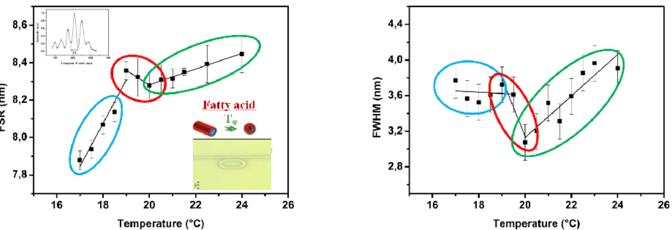
4.1 Fatty acid phase transitionA drop of a few µL of the hybrid C5 (5-amino-1-pentanol) plus 12-HSA (12-hydroxystearic acid) solution is deposited on the surface of the sensor using a µL pipette. An optical spectrum analyzer connected to a computer acquires the output signal of the resonator. The measurements are performed as a function of temperature on the range [17-24]°C, with a step of 0.5°C. For each temperature, 10 spectra are collected in order to reduce the error on the measurements. The thermal evolution results are presented in Figure 3 in terms of FSR and FWHM parameters of the photonic device. Additionally, the thermal dependency of these spectral parameters have been measured for the bare sensor, and subtracted to the results obtained for the phase transition. Indeed, this way, the effects due to the measured phase transition can be discriminated from the intrinsic thermal drift of the bare photonic structure. These results were fully corroborated with measurements of viscosity changes in rheology during the phase transition [9].
Figure 4: Phase transition detection of the fatty acid (C5/12-HSA system) base on the main optical properties of the
resonant photonic device: pseudo-period FSR and FWHM. Measurements were done from 17 to 24°C. A two –step transition ranging between 18.5 and 19.5°C is observable.
The studies demonstrated the high degree of accuracy with respect to the range of temperature where the transition occurs. The comparative study also showed that this photonic device had a higher accuracy to reveal all the details of the transition, while the rheological probe hardly detects these changes. The proof of concept of the sensing platform which allowed continuous real time monitoring was fully demonstrated. The polymeric resonator together with the measurement protocol demonstrated high versatility in their performance to carry out investigations related with phase transition of global thermo-responsive materials and food or cosmetic products presenting micro-structuration when outdated and expired with time.
4.2 Sphingomyelin phase transition
The sphingomyelin is a special class of lipids found in the bio-membranes and is involved on the membrane heterogeneity. The sphingomyelin used in this work, was purified from milk bovine and chosen because the presence of saturated chain is lower than other sources [13, 10]. At the room temperature, the hydrocarbon lipid chains display an all-trans configuration so-called gel phase (figure 5). At a specific temperature (phase all-transition temperature or melting temperature, T), a thermotropic phase transition occurs leading to a fluid phase, as a consequence of trans-gauche chain
isomerizations.
Figure 5: Chemical structure and symbolic representation of a 16:0 sphingomyelin-molecule showing both the
T is affected by the saturation/unsaturation of the hydrocarbon chains; presence of saturated chains increases the T
value. After having implemented the fusion vesicle method deposition that is specific in biology so as to build a multilayer Sphingomyelin-gel structure upon the sensing surface of the device, we may proceed through the specific protocol regarding experimental measurements, described previously with fatty acid, so as to monitor the dynamic evolution of the sphingomyelin lipid phase transition. Figure 6 depict the evolution in temperature of the spectrum-parameters FSR and FWHM. The equilibrium of the regime is clearly broken by the dynamic of the Sphingomyelin and its own phase transition. According to the evolution of the FWHM (Figure 6) the regime grows towards a stronger K-coupling (as FWHM is decreased) before levelling off at the exact beginning of the phase transition of Sphingomyelin or T melting temperature. A change clearly occurs between the two gel/fluid states. Hence, the ability to detect the specific
gel/fluid transition phase of Sphingomyelin lipids and the efficiency to pinpoint the melting temperature at T = 31± 0.5
°C have been precisely demonstrated.
Figure 6: Detection of the gel-liquid phase transition of sphingolipids (myelin). Evolution in temperature (T ranging
[16-42]°C) of the FSR and the FWHM of the photonic structures Si/SiO2/UV210/sphingolipids/air: highlighting clear
breaks of slopes as a way to assess the melting temperature Tof MSM lipids with T [31-32]°C..
This study was corroborated by differential calorimetry measurements, this phase transition being of the first order with a variation in enthalpy H [10]. In conclusion, we have experimentally demonstrated the interest of a new approach to achieve milk Sphingomyelin (MSM) phase transition detection since only one billion times less material than the conventional method called differential scanning calorimetry (DSC) is needed.
5. CONCLUSION
Such a work presents the feasibility of detecting and following dynamic phenomena dedicated to soft matter processes called phase transitions with a photonic resonant signal. It reports the high versatility on the performance of an integrated optical polymeric micro-cavity, based on a resonance probe light, to carry out phase transition detection of self-assembled surfactant system used in cosmetic products, food and sphingolipid (myelin) in biology. Specific micro-resonator structures have been shaped and used. By doing so, this article proposes a new way to design phase transition/inversion sensors based on such integrated devices. These lab-on-chip sensors have the advantage to be producible at industrial scale thanks to the low cost of the polymer materials used, and the single deep-UV photolithography insulation necessary for their fabrication.
Acknowledgments: The authors would like to thank the ‘Direction de l’Innovation et des Relations avec les Entreprises’
(DIRE) of the CNRS, Rennes Metropolis and Fondation Rennes 1 for financially supporting this research.
REFERENCES
[1] Fameau, A.-L., Cousin, F., Navailles, L., Nallet, F., Boué, F. and Douliez, J.-P., "Multiscale Structural Characterizations of Fatty Acid Multilayered Tubes with a Temperature-Tunable Diameter", J. Phys. Chem. B 115, 9033-9039, (2011).
[2] Kulkarni, C.V., "Lipid Self-Assemblies and Nanostructured Emulsions for Cosmetic Formulations", Cosmetics, 3 (37), 1-15, (2016).
[3] Michel, N., Fabiano, A. S., Polidori, A., Jack, R., Pucci B., "Determination of phase transition temperatures of lipids by light scattering", Chem. Phys. Lipids 139 (11), 11-19, (2006).
[4] Rabus, D. G., [Integrated Ring Resonators], Optical Sciences Series, Springer, (2007).
[5] Chremmos, I., Schwelb, O. and Uzunoglu, N., [Photonic Microresonator Research and Applications], Optical Sciences Series, Springer, (2010).
[6] Scheuer, J. and Yariv, A., "Fabrication and characterization of low-loss polymeric waveguides and micro-resonators", J. Eur. Opt. Soc. 1, 06007.1-06007.5, (2006).
[7] Castro-Beltràn, R., Huby, N., Vié, V., Lhermite, H., Camberlein, L., Gaviot, E. and Bêche, B., "A laterally coupled UV210 polymer racetrack micro-resonator for thermal tenability and glucose sensing capability", Adv. Device Mater. 1, 80-87, (2015).
[8] Meziane, F., Raimbault, V., Hallil, H., Joly, S., Conédéra, V., Lachaud, J.L., Béchou, L., Rebière, D. and Dejous, C., "Study of polymer optical microring resonator for hexavalent chromium sensing", Sensors Actuators B 209, 1049-1056, (2015).
[9] Castro-Beltràn, R., Garnier, L., Saint-Jalmes, A., Lhermite., H., Cormerais, H., Fameau, A.-L, Giscquel, E., and Bêche, B., "Microphotonics for monitoring the supramolecular thermoresponsive behavior of fatty acid surfactant solutions", Opt. Comm., to be published (2020).
[10] Li, Q., Vié, V., Lhermite, H., Gaviot, E., Bourlieu, C., Moréac, A., Morineau, D., Dupont, D., Beaufils, S. and Bêche, B., "Polymer resonators sensors for detection of sphingolipid gel/fluid phase transition and melting temperature measurement", Sensors Actuators A 263, 707-717, (2017).
[11] Duval, D., Lhermite, H., Godet, C., Huby, N. and Bêche, B., "Fabrication and optical characterization of sub-micronic waveguide structures on UV210 polymer" J. Opt. 12, 055501/1-6 (2010).
[12] Pluchon, D., Huby, N., Lhermite, H., Duval, D. and Bêche, B., "Investigation of fabrication and resonant optical coupling in various 2D micro-resonator structures in a UV210 polymer", J. Micromech. Microeng. 22, 085016/1-8 (2012).
[13] Brown, D.A., and London, E., "Functions of lipid rafts in biological membranes", Annu.Rev. Cell Dev. Biol. 14, 111-136 (1998).


![Figure 6: Detection of the gel-liquid phase transition of sphingolipids (myelin). Evolution in temperature (T ranging [16-42]°C) of the FSR and the FWHM of the photonic structures Si/SiO 2 /UV210/sphingolipids/air: highlighting clear](https://thumb-eu.123doks.com/thumbv2/123doknet/11483743.292535/8.893.269.616.367.591/detection-transition-sphingolipids-evolution-temperature-structures-sphingolipids-highlighting.webp)
