The apo-structure of the leucine sensor Sestrin2 is still elusive
The MIT Faculty has made this article openly available.
Please share
how this access benefits you. Your story matters.
Citation
Saxton, R. A. et al. “The Apo-Structure of the Leucine Sensor
Sestrin2 Is Still Elusive.” Science Signaling 9.446 (2016): ra92-ra92.
As Published
http://dx.doi.org/10.1126/scisignal.aah4497
Publisher
American Association for the Advancement of Science (AAAS)
Version
Author's final manuscript
Citable link
http://hdl.handle.net/1721.1/106635
Terms of Use
Article is made available in accordance with the publisher's
policy and may be subject to US copyright law. Please refer to the
publisher's site for terms of use.
The apo-structure of the leucine sensor Sestrin2 is still elusive
Robert A. Saxton1,2,3,4,5, Kevin E. Knockenhauer1,6, Thomas U. Schwartz1,*, and David M.
Sabatini1,2,3,4,5,*
1Department of Biology, Massachusetts Institute of Technology (MIT), Cambridge, MA 02139, USA
2Whitehead Institute for Biomedical Research, 9 Cambridge Center, Cambridge, MA 02142, USA 3Howard Hughes Medical Institute, Cambridge, MA 02139, USA
4Koch Institute for Integrative Cancer Research, 77 Massachusetts Avenue, Cambridge, MA 02139, USA
5Broad Institute of Harvard and Massachusetts Institute of Technology, 415 Main Street, Cambridge MA 02142, USA
Abstract
Sestrin2 is a GATOR2 interacting protein that directly binds leucine and is required for the inhibition of mTORC1 under leucine deprivation, indicating that it is a leucine sensor for the mTORC1 pathway. We recently reported the structure of Sestrin2 in complex with leucine (PDB ID: 5DJ4), and demonstrated that mutations in the leucine-binding pocket alter the affinity of Sestrin2 for leucine and result in a corresponding change in the leucine sensitivity of mTORC1 in cells. A lower resolution structure of human Sestrin2 (PDB ID: 5CUF), which was crystallized in the absence of exogenous leucine, showed Sestrin2 to be in a nearly identical conformation as the leucine-bound structure. Based on this observation, it has been argued that leucine binding does not affect the conformation of Sestrin2 and thus that Sestrin2 may not be a sensor for leucine. Here, we show that simple analysis of the reported “apo”-Sestrin2 structure reveals the clear presence of prominent, unmodeled electron density in the leucine-binding pocket that exactly accommodates the leucine observed in the higher resolution structure. Refining the reported “apo”-structure with leucine eliminates the large FO-FC difference density at this position and improves the working and free R-factors of the model. Consistent with this, our own structure of Sestrin2 crystallized in the absence of exogenous leucine also contains electron density that is best explained by leucine. Thus, the structure of apo-Sestrin2 remains elusive.
Introduction
The mechanistic target of rapamycin complex 1 (mTORC1) couples cell growth with the availability of biosynthetic inputs such as amino acids (1,2). Leucine in particular robustly activates mTORC1, and leucine deprivation inhibits mTORC1 signaling in a wide variety of experimental systems (3-5). Recently, we identified Sestrin2 as a key leucine sensor in
HHS Public Access
Author manuscript
Sci Signal
. Author manuscript; available in PMC 2016 October 31.Published in final edited form as:
Sci Signal. ; 9(446): ra92. doi:10.1126/scisignal.aah4497.
A
uthor Man
uscr
ipt
A
uthor Man
uscr
ipt
A
uthor Man
uscr
ipt
A
uthor Man
uscr
ipt
mammalian cells (6,7). Leucine binds directly to Sestrin2 in vitro, and genetic loss of Sestrin2 and its closely related homologs renders mTORC1 signaling resistant to inhibition by leucine starvation. Mechanistically, Sestrin2 inhibits mTORC1 in the absence of leucine by binding to GATOR2, an upstream activator of mTORC1 (7-9). The addition of leucine triggers the dissociation of Sestrin2 from GATOR2 both in vitro and in cells, suggesting that leucine binding induces a conformational change in Sestrin2 that disrupts this interaction (6,7). Consistent with this, leucine substantially increases the thermal stability of purified Sestrin2, as is often observed for ligand-receptor complexes (6,7,10).
We solved the crystal structure of human Sestrin2 in complex with leucine at 2.7 Å resolution, revealing insights into the mechanism of leucine sensing (6). However, Sestrin2 failed to crystallize in the absence of leucine, and the structure of apo-Sestrin2 remained elusive. Subsequently, a 3.5 Å structure of Sestrin2 crystallized without the addition of exogenous leucine was reported, in the same crystal form, showing Sestrin2 in a nearly identical conformation as the leucine-bound structure (11). On the basis of this observation, Lee et al. argued that the similarities between the reported “apo” structure (11) of Sestrin2 and the leucine-bound structure (6) are inconsistent with Sestrin2 being a sensor for leucine (12,13).
To reconcile our structural, biochemical, and cell biological data with the claims made by Lee et al., we reanalyzed their reported “apo”-Sestrin2 structure (PDB ID: 5CUF), revealing the clear presence of a ligand bound in the leucine-binding pocket. In addition, we report our own 3.0 Å structure of Sestrin2 that we obtained without adding exogenous leucine during the purification process, in which leucine is also present.
Results
Using the corresponding structure factors and atomic model deposited in the Protein Data Bank, we calculated the 2FO-FC and FO-FC electron density maps for the protein PDB ID: 5CUF. Simple inspection of the FO-FC difference map, which highlights discrepancies between experimental data and the structural model, revealed the presence of substantial unexplained electron density (positive 7.5σ peak) at the exact location of the leucine-binding pocket observed in the leucine-bound Sestrin2 (PDB ID: 5DJ4, Fig. 1A). This unmodeled density is easily observed in all 5 crystallographically independent copies of Sestrin2 in the asymmetric unit and is the prime location where modeled density and observed density differ. Furthermore, the unexplained electron density in PDB ID: 5CUF resembles the density corresponding to leucine in the higher resolution PDB ID: 5DJ4 structure (Fig. 1B). To test whether this density may represent leucine, we refined the 5CUF model either with or without leucine built into it. As with PDB ID: 5DJ4, refining PDB ID: 5CUF with leucine eliminated the FO-FC map peak at the binding site and improved the R-factors of the model (Fig. 1, B, C and E), consistent with the presence of bound leucine in this structure.
We also analyzed the possibility that one of the buffer components (MES, Tris, sodium, chloride, malonate, TCEP) of the reported crystallization condition of PDB ID: 5CUF may be contributing to the large difference density at the leucine-binding site (11). The only ingredient that could possibly be entertained at the modest resolution of 3.5Å as a
leucine-Saxton et al. Page 2
Sci Signal. Author manuscript; available in PMC 2016 October 31.
A
uthor Man
uscr
ipt
A
uthor Man
uscr
ipt
A
uthor Man
uscr
ipt
A
uthor Man
uscr
ipt
substitute is malonate (Fig. 1D). It is the main precipitant at 1.15 M concentration and it could at least mimic the interaction of the carboxyl group of leucine. However, the contacts formed by the hydrophobic leucine sidechain as well as the cationic amine group would still be unexplained entirely. Nonetheless, refining PDB ID: 5CUF with malonate also eliminated the FO-FC map peak and improved the R-factors of the model (Fig. 1, D and E).
In our own efforts to obtain the structure of apo-Sestrin2, we purified Sestrin2 from E. coli and crystallized it without the addition of leucine following bacterial cell lysis. While pre-incubating Sestrin2 with leucine yielded large, high quality crystals within 24-48 hours of setting drops, the crystals that formed in absence of added leucine were smaller, took much longer to crystalize (7-14 days), and formed in a background of a heavy protein precipitate. Nonetheless, we were able to obtain high quality crystals from which we obtained a structure at 3.0 Å resolution (PDB ID: 5T0N, Table 1). Inspection of the FO-FC difference map for this structure again revealed a large positive peak in the location of the leucine-binding pocket that strongly resembles the electron density of leucine (Fig. 2A). As with PDB ID: 5CUF and PDB ID: 5DJ4, refining the structure with leucine eliminated the FO-FC peak and improved the model statistics (Fig. 2, B and D). Furthermore, at this resolution leucine is clearly the best candidate to fit into this density, as malonate does not fully explain the difference density at this position and did not improve the R-factors as substantially as when the structure was refined with leucine (Fig. 2, C and D).
Discussion
The leucine-binding site of Sestrin2 consists of a shallow hydrophobic pocket flanked on each side by opposing positive and negative charges contributed by Arg390 and Glu451, respectively, making it well suited to specifically accommodate zwitterionic α-amino acids with short aliphatic sidechains (6). Among the amino acids, Sestrin2 has the highest affinity for leucine, with approximately 15-fold and 30-fold lower affinity for the closely related amino acids methionine and isoleucine, respectively, and undetectable affinity for the remaining natural amino acids (7). In addition, while it is impossible to definitively conclude whether the density present in the 3.5 Å PDB ID: 5CUF structure corresponds to leucine or another ligand such as malonate, our own 3.0 Å structure clearly favors leucine as the best candidate to explain the observed density. In any case, these data do conclusively
demonstrate that PDB ID: 5CUF represents the ligand-bound form of Sestrin2, not the apo-Sestrin2 conformation as reported by Lee et. al.
Although leucine is highly abundant in bacterial cells (14), it may seem surprising that Sestrin2 can remain stably bound to leucine throughout the purification process given the reported 20 µM affinity of Sestrin2 for leucine (7). One possibility is that the off-rate of the Sestrin2-leucine interaction is substantially lower in the absence of additional protein components or post-translational modifications present only in mammalian cells, such that a small fraction of Sestrin2 remains bound to leucine even following purification.
Crystallization is, after all, a purification method and thus perfectly suited to enrich a small fraction of leucine-bound Sestrin2 in a large background of non-crystallizing apo-Sestrin. Because all three Sestrin2 crystal structures crystallize under highly similar conditions, in the same crystal form, and that the largest and best diffracting crystals grow in the presence
A
uthor Man
uscr
ipt
A
uthor Man
uscr
ipt
A
uthor Man
uscr
ipt
A
uthor Man
uscr
ipt
of leucine, we conclude that all reported structures are best interpreted as being leucine bound.
In summary, we argue that the PDB ID: 5CUF structure depicts Sestrin2 in the ligand-bound conformation, not the apo-conformation as reported by Lee et al (6). The leucine-dependent dissociation of Sestrin2 from GATOR2 represents the critical first step in the activation of mTORC1 by leucine, and understanding how leucine-binding alters the conformation of Sestrin2 to disrupt its interaction with GATOR2 remains a major open question in the field.
Materials and Methods
Protein production and purification
Human Sestrin2 was expressed and purified as described previously (6). Briefly, full-length, codon-optimized human Sestrin2 was N-terminally fused with a human rhinovirus 3C protease–cleavable His10-Arg8-ScSUMO tag and cloned into a PET-Duet-1 bacterial expression vector. This vector was transformed into Escherichia coli LOBSTR (DE3) cells (Kerafast, 15). Cells were grown at 37°C to 0.6 OD, then protein production was induced with 0.2 mM IPTG at 18 °C for 12–14 h. Cells were collected by centrifugation at 6,000g, resuspended in lysis buffer (50 mM potassium phosphate, pH 8.0, 500 mM NaCl, 30 mM imidazole, 3 mM β-mercaptoethanol (βME) and 1 mM PMSF) and lysed with a cell disruptor (Constant Systems). The lysate was cleared by centrifugation at 10,000g for 20 min. The soluble fraction was incubated with Ni-Sepharose 6 Fast Flow beads (GE Healthcare) for 30 min on ice. After washing of the beads with lysis buffer, the protein was eluted in 250 mM imidazole, pH 8.0, 150 mM NaCl and 3 mM βME. The Ni eluate was diluted 1:1 with 10 mM potassium phosphate, pH 8.0, 0.1 mM EDTA and 1 mM
dithiothreitol (DTT), and was subjected to cation-exchange chromatography on a 5 ml SP sepharose fast flow column (GE Healthcare) with a linear NaCl gradient. The eluted Sestrin2 was then incubated with 3C protease and dialyzed overnight at 4 °C into 10 mM potassium phosphate, pH 8.0, 150 mM NaCl, 0.1 mM EDTA and 1 mM DTT, followed by a second cation-exchange chromatography run on an SP sepharose fast flow column (GE Healthcare) with a linear NaCl gradient. The protein was further purified via size-exclusion
chromatography on a Superdex S200 16/60 column (GE Healthcare) equilibrated in running buffer (10 mM Tris-HCl, pH 8.0, 150 mM NaCl, 0.1 mM EDTA and 1 mM DTT).
Crystallization
Crystals were grown at 18 °C by hanging-drop vapor diffusion with 1 µl of Sestrin2 at 5 mg/ml mixed with an equal volume of reservoir solution containing 100 mM MES, pH 6.0, 1.2 M disodium malonate, and 1% (v/v) Jeffamine ED 2001. Drops were microseeded to obtain well diffracting crystals. Microseeds were obtained as described previously (16). Briefly, a single 2 µl drop containing crystals of Sestrin2 was diluted into a 50 µl solution of mother liquor (100 mM MES, pH 6.0, 1.2 M disodium malonate, and 1% (v/v) Jeffamine ED 2001), vortexed for 5 minutes in the presence of a polystyrene bead, then diluted again into 500 µl of mother liquor. This solution was then diluted at a ratio of 1:1,000 to obtain the final microseed stock. 0.2 µl of microseed stock was then added to the 2 µl hanging drop
Saxton et al. Page 4
Sci Signal. Author manuscript; available in PMC 2016 October 31.
A
uthor Man
uscr
ipt
A
uthor Man
uscr
ipt
A
uthor Man
uscr
ipt
A
uthor Man
uscr
ipt
described above (for a final total dilution of 1:2,500,000). Crystals were cryoprotected in mother liquor supplemented with 18% (v/v) glycerol.
Data collection and structure determination—Data collection was performed at the Advanced Photon Source end station 24-IDC at Argonne National Lab. A complete native dataset was collected to 3.0 Å. All data-processing steps were carried out with programs provided through SBgrid (17). Data reduction was performed with HKL2000 (18). The phase problem was solved by molecular replacement using our published Sestrin2 structure (PDB ID: 5DJ4) as the search model in the Phaser-MR program, run as part of the Phenix Autosol program (19) (space group I23, 5 molecules per asymmetric unit). To reduce potential model bias, we used a search model lacking the bound leucine and surrounding protein residues. An interpretable 3.0-Å electron density map was obtained and model building was carried out in Coot (19). Subsequent refinement was carried out using phenix.refine.
Structure and electron density map analysis
For analysis of published structures 5DJ4 and 5CUF, the FO-FC and 2FO-FC maps were calculated from the models and structure factors deposited in the Protein Data Bank using phenix.maps, and visualized in Coot. Refinement of the models either with or without bound-leucine built in was performed using phenix.refine. XYZ-coordinates and individual B-factors were refined over three cycles. All structure figures were made in PyMol (20).
References
1. Dibble CC, Manning BD. Signal integration by mTORC1 coordinates nutrient input with biosynthetic output. Nature Cell Biology. 2013; 15:555–564. [PubMed: 23728461]
2. Laplante M, Sabatini DM. mTOR signaling in growth control and disease. Cell. 2012; 149:274. [PubMed: 22500797]
3. Lynch CJ, Fox HL, Vary TC, Jefferson LS, Kimball SR. Regulation of amino acid-sensitive TOR signaling by leucine analogues in adipocytes. Journal of cellular biochemistry. 2000; 77:234–251. [PubMed: 10723090]
4. Anthony JC, et al. Leucine stimulates translation initiation in skeletal muscle of postabsorptive rats via a rapamycin-sensitive pathway. J. Nutr. 2000; 130(10):2413–2419. [PubMed: 11015466] 5. Kim D-H, et al. mTOR interacts with raptor to form a nutrient-sensitive complex that signals to the
growth machinery. Cell. 2002; 110:163–175. [PubMed: 12150925]
6. Saxton RA, et al. Structural basis for leucine sensing by the Sestrin2-mTORC1 pathway. Science. 2016; 351:53. [PubMed: 26586190]
7. Wolfson RL, et al. Sestrin2 is a leucine sensor for the mTORC1 pathway. Science. 2016; 351:43. [PubMed: 26449471]
8. Chantranupong L, et al. The Sestrins Interact with GATOR2 to Negatively Regulate the Amino-Acid-Sensing Pathway Upstream of mTORC1. Cell Reports. 2014; 9:1–8. [PubMed: 25263562] 9. Parmigiani A, et al. Sestrins Inhibit mTORC1 Kinase Activation through the GATOR Complex. Cell
Reports. 2014; 9:1281–1291. [PubMed: 25457612]
10. Niesen FH, et al. The use of differential scanning fluorimetry to detect ligand interactions that promote protein stability. Nature Protocols. 2007; 2(9):2212–2221. [PubMed: 17853878] 11. Kim H, et al. Janus-faced Sestrin2 controls ROS and mTOR signalling through two separate
functional domains. Nat. Commun. 2015; 6:10025. [PubMed: 26612684]
12. Lee JH, et al. Sestrin regulation of TORC1: Is Sestrin a leucine sensor? Sci Signal. 2016; 9(431) 13. Ho A, et al. Biochemical Basis of Sestrin Physiological Activities. Trends Biochem Sci. 2016
A
uthor Man
uscr
ipt
A
uthor Man
uscr
ipt
A
uthor Man
uscr
ipt
A
uthor Man
uscr
ipt
14. Sajed T, et al. ECMDB 2.0: A richer resource for understanding the biochemistry of E. coli. Nucleic Acids Res. 2015 p.gkv1060.
15. Andersen KR, Leksa NC, Schwartz TU. Optimized E. coli expression strain LOBSTR eliminates common contaminants from His-tag purification. Proteins. 2013; 81:1857–61. [PubMed: 23852738]
16. Luft JR, DeTitta GT. A method to produce microseed stock for use in the crystallization of biological macromolecules. Acta Cryst. 1999; D55:988–993.
17. Morin A, et al. Collaboration gets the most out of software. eLife. 2013; 2:e01456. [PubMed: 24040512]
18. Otwinowski Z, Minor W. Processing of X-ray diffraction data collected in oscillation mode. Methods Enzymol. 1997; 276:307–326. [PubMed: 27754618]
19. Adams PD, et al. PHENIX: a comprehensive Python-based system for macromolecular structure solution. Acta Crystallogr. D Biol. Crystallogr. 2010; 66:213–221. [PubMed: 20124702]
20. Emsley P, Lohkamp B, Scott WG, Cowtan K. Features and development of Coot. Acta Crystallogr. D Biol. Crystallogr. 2010; 66:486–501. [PubMed: 20383002]
21. Schrodinger, LLC. The PyMOL Molecular Graphics System, Version 1.3r1. 2010
Saxton et al. Page 6
Sci Signal. Author manuscript; available in PMC 2016 October 31.
A
uthor Man
uscr
ipt
A
uthor Man
uscr
ipt
A
uthor Man
uscr
ipt
A
uthor Man
uscr
ipt
Figure 1. Identification of ligand in the “apo”-Sestrin2 structure reported by Lee et al.
1A: Ribbon diagrams of the reported “apo”-Sestrin2 structure (PDB ID: 5CUF, pink, left)
and the leucine-bound structure (PDB ID: 5DJ4, light blue, right). The largest peak in the 3.5 Å 5CUF FO-FC difference map, contoured at 3σ, is shown as a green mesh in the 5CUF structure. The bound leucine built into the 5DJ4 structure is shown in orange.
1B: Close up view of the leucine-binding pocket of Sestrin2 in the 3.5 Å 5CUF (left) and 2.7
Å 5DJ4 structures (right), refined without leucine built into the model. The FO-FC (green mesh) and 2FO-FC (blue mesh) electron density maps were contoured at 3σ and 1σ, respectively, and shown in the location of the bound leucine.
1C: The PDB ID: 5CUF and PDB ID: 5DJ4 structures were refined with leucine built into
the model and shown in the same view as in 1B. The FO-FC and 2FO-FC electron density maps are depicted as in 1B. Hydrogen bonds and electrostatic interactions are shown as black dashed lines.
1D: The PDB ID: 5CUF structure was refined with malonate built into the model and shown
in the same view as in 1B. The FO-FC and 2FO-FC electron density maps are depicted as in 1B.
1E: The Rw/Rf(%) for the PDB ID: 5CUF structure refined with no ligand, leucine, or malonate modeled.
A
uthor Man
uscr
ipt
A
uthor Man
uscr
ipt
A
uthor Man
uscr
ipt
A
uthor Man
uscr
ipt
Figure 2. Sestrin2 crystalizes in complex with leucine without the addition of exogenous leucine
2A: Close up view of the leucine-binding pocket in Sestrin2 (PDB ID: 5T0N, this study)
refined without leucine built into the model. The FO-FC (green mesh) and 2FO-FC (blue mesh) electron density maps, contoured at 3σ and 1σ, respectively, are shown as in 1B.
2B: Close up view of the leucine-binding pocket in Sestrin2 refined with leucine built into
the model. The FO-FC and 2FO-FC electron density maps are depicted as in 2A. Hydrogen bonds and electrostatic interactions are shown as black dashed lines.
2C: Close up view of the leucine-binding pocket in Sestrin2 refined with malonate built into
the model. The FO-FC and 2FO-FC electron density maps are depicted as in 2A. Hydrogen bonds and electrostatic interactions are shown as black dashed lines.
2D: The Rw/Rf(%) for the PDB ID: 5T0N structure (this study) refined with no ligand, leucine, or malonate modeled.
Saxton et al. Page 8
Sci Signal. Author manuscript; available in PMC 2016 October 31.
A
uthor Man
uscr
ipt
A
uthor Man
uscr
ipt
A
uthor Man
uscr
ipt
A
uthor Man
uscr
ipt
A
uthor Man
uscr
ipt
A
uthor Man
uscr
ipt
A
uthor Man
uscr
ipt
A
uthor Man
uscr
ipt
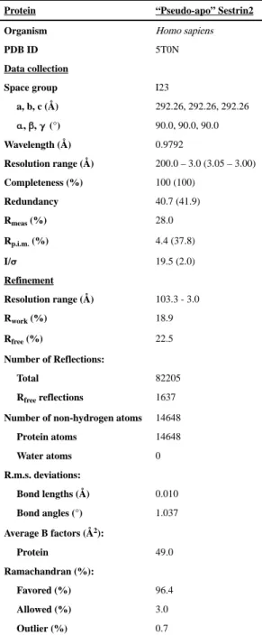
Table 1 Data collection and refinement statisticsProtein “Pseudo-apo” Sestrin2
Organism Homo sapiens
PDB ID 5T0N
Data collection
Space group I23
a, b, c (Å) 292.26, 292.26, 292.26 α, β, γ (°) 90.0, 90.0, 90.0 Wavelength (Å) 0.9792 Resolution range (Å) 200.0 – 3.0 (3.05 – 3.00) Completeness (%) 100 (100) Redundancy 40.7 (41.9) Rmeas (%) 28.0 Rp.i.m. (%) 4.4 (37.8) I/σ 19.5 (2.0) Refinement Resolution range (Å) 103.3 - 3.0 Rwork (%) 18.9 Rfree (%) 22.5 Number of Reflections: Total 82205 Rfree reflections 1637
Number of non-hydrogen atoms 14648 Protein atoms 14648 Water atoms 0 R.m.s. deviations: Bond lengths (Å) 0.010 Bond angles (°) 1.037 Average B factors (Å2): Protein 49.0 Ramachandran (%): Favored (%) 96.4 Allowed (%) 3.0 Outlier (%) 0.7