Analysis of Transcriptional Regulatory Circuitry by
Nicola J. Rinaldi
B.A., Chemistry
Johns Hopkins University, 1994
SUBMITTED TO THE DEPARTMENT OF BIOLOGY IN PARTIAL FULFILLMENT OF THE REQUIREMENTS
FOR THE DEGREE OF
DOCTOR OF PHILOSOPHY
at the
MASSACHUSETTS INSTITUTE OF TECHNOLOGY
September 2004
©Nicola J. Rinaldi, 2004. All rights reserved.
The author hereby grants to MIT permission to reproduce and to distribute Publicly copies of this thesis document in whole or in part.
Signature of Author' Department of Biology July 26, 2004 Certified by c<-Dr. Richard A. Young Professor of Biology Thesis Supervisor Accepted by
'v~~
`7-
~~~~SteveBell
Professor of Biology and Co-Chairperson, Biology Graduate Committee
MASSACHUSETTS INSTM't
OF TECHNOLOGY
SEP 2 2 2004 ARCHIVES
I
i '---Dedication
Acknowledgements
I owe thanks to many people for their help and support during my graduate career. Rick Young and David Gifford for providing an environment where I could pursue my interest in computational biology, and teaching me how to think about the problems and issues that arise with these large scale data. My thesis committee, consisting of David Page, Eric Lander, David Housman and
Martha Bulyk for taking the time to offer suggestions on my work through the years, including on this thesis. In particular I'd like to thank Martha Bulyk for
providing me with access to a manuscript pre-publication. All the people that have been in the Young lab with me over the years have been wonderful,
providing both intellectual stimulation and help with scientific issues, as well as being really fun to hang out with! In particular I'd like to thank Tony Lee, Duncan Odom, Ann Schlesinger and Francois Robert for all their help and mentoring through my four years in the lab. Francois and Sarah Johnstone have also been
super bench-mates! I owe many thanks to Elizabeth (Bizzy) Herbolsheimer without whose computational assistance I would never have been able to finish
my thesis. I would also like to thank my other labmates who have made the
Young lab so entertaining through the years - Craig Thompson, Jo Terragni, Nancy Hannett, Chris Harbison, Dmitry Pokholok, Jean-Bosco Tagne, Jane Yoo, Liz McReynolds, Ezra Jennings, Matt Guenther, David Reynolds, Heather
Murray, Jerry Nau, Megan Cole, Richard Jenner, Julia Zeitlinger, Joerg Schreiber, Nora Zizlsperger, Lea Medeiros, Rhonda Harrison, John Barnett, Itamar Simon, Brett Chevalier, Laurie Boyer, Jen Love, Bruno Chazaro and Tom
Volkert. Kathleen Blackett, Kercine Elie and Carolyn Carpenter were incredibly helpful with all kinds of administrative issues. I also would not have survived without advice and commiseration from fellow graduate students, particularly
Julie Wallace and Natalia Comella. Finally, I would not have made it without my family. Particularly my father, who passed away in 1995, whom I had told I would
be getting my PhD - nothing like that for motivation! Also, my husband Mark has been my biggest supporter through the years. My mom, sister, their husbands and my in-laws have also been such great sources of strength and
Analysis of Transcriptional Regulatory Circuitry
by
Nicola J. Rinaldi
Submitted to the Department of Biology on July 26, 2004
in partial fulfillment of the requirementsJ!for the Degree of
Doctor of Philosophy in Biology
Abstract
The research in this thesis has focused on the analysis of data from two types of microarray technologies with the goal of improving understanding of transcriptional regulatory circuitry in yeast. These microarray technologies, expression analysis and location analysis (ChIP on CHIP), bring unique
challenges for data analysis. I have adapted methods described previously and have developing new algorithms for addressing some of these challenges.
I generated yeast transcription factor location data as part of a team effort to determine how a large set of transcriptional regulators occupy the yeast
genome and to facilitate discovery of regulatory network structures. I also
contributed to another team effort to deduce a portion of the yeast transcriptional regulatory code, the set of genomic DNA sequences recognized by
transcriptional regulators to effect gene expression programs. Finally, I used these experimental and analytical methods to explore changes in gene expression circuitry that occur after exposure to oxidative stress in yeast.
Thesis Supervisor: Dr. Richard A. Young
Title: Professor of Biology
Thesis Supervisor: Dr. David K. Gifford
Table of Contents Title Page ... 1 Dedication ... 2
Acknowledgements
...
... ...
3
Abstract
... ... ...
4
Table of Contents ... 5 Chapter 1: Introduction: Regulation of Transcriptional Activation ... ... 6Chapter 2: Microarray Data Analysis for Biological Insight ... 53
Chapter 3: Transcriptional Regulatory Networks in Saccharomyces cerevisiae ... 98
Chapter 4: Transcriptional Regulatory Code of a Eukaryotic Genome ... 129
Chapter 5: Conclusions: The future of systems biology ... 155
Appendix A: Computational Discovery of Gene Modules and Regulatory Networks ... 167
Chapter 1
Overview
"The discovery of regulator and operator genes, and of repressive regulation of
the activity of structural genes, reveals that the genome contains not only a
series of blue-prints, but a coordinated program of protein synthesis and the
means for controlling its execution"
Jacob and Monod, 1961
The ability of a cell to respond to its environment is based on exquisite
control of gene expression, protein synthesis, and enzymatic activity. The simple
step of initiating transcription of genes from DNA into RNA is a carefully
choreographed process, involving hundred of proteins. The RNA polymerase
holoenzyme, associated general transcription factors, transcriptional activators
and repressors, chromatin modifiers and remodelers, kinases, phosphatases,
import and export proteins are all components of this coordinated program. Of
particular interest is how all this machinery is controlled to regulate the
expression of an individual gene. Transcriptional activators and repressors seem
to provide the requisite specificity by binding to only a select set of DNA
sequences. Combinatorial interactions of these gene-specific factors with other
transcription factors, the chromatin modifying enzymes which control access of
the basic transcriptional machinery to the DNA template, and various
components of the initiation apparatus, determine which genes are transcribed in
response to a particular stimulus. Therefore, this introduction will focus on the
regulation of transcription at the level of these gene-specific transcription factors.
One of the keys to establishing this gene-specific regulation of
transcription is that the transcription factors themselves must be responsive to
varied environmental conditions. Without such changes that affect the DNA
binding or protein-protein interaction abilities of transcription factors, all gene
expression would necessarily be constitutive. Transcription factors have been
shown to be regulated in many different ways including modification of the
transcription factor protein, changes in the amount of protein present in the
nucleus, and sequestration from the DNA.
Initial studies of transcription factors and their modifications focused on
the function of a few regulators at the promoters of single genes or reporter
constructs. With the sequencing of the yeast genome and advent of microarray
technology, however, it is now possible to study the regulation of gene
expression globally. Beyond gene expression, microarray technologies have
been adapted to study the direct effects of transcription factors by finding the
promoters to which the factors are bound in vivo. These data, in combination
with additional information such as sequence conservation between species, are
leading to a much deeper understanding of global mechanisms for controlling the
cellular program, as suggested by Jacob and Monod in 1961.
This introduction will focus, in two sections, on the regulation of gene
expression in Saccharomyces cerevisiae at the level of transcriptional activators.
First, I will discuss the components involved in transcription and activation of
expression, how those components are controlled by interactions with the
themselves are modulated. Following that, I will focus on new microarray
technologies available for the study of transcriptional regulation and how use of
these technologies has impacted our knowledge of the regulatory network of the
cell. Later chapters will discuss methods for analyzing the data obtained from
microarray experiments as well as my contributions to the data analysis arena,
followed by examples of how these analyses have been used to further our
understanding of transcriptional regulation in Saccharomyces cerevisiae.
Transcriptional machinery components
The current understanding of transcription regulation is that gene-specific
transcription factors, activators and repressors, control the expression of
individual genes or sets of genes. This regulation is based upon the specificity
provided by two distinct domains in the transcription factor protein. The
archetypal transcription factor consists of a DNA-binding domain, which contacts
the specific DNA sequence, and an activation ( or repression) domain that
recruits specific pieces of the basal transcription machinery (Ptashne 1988). The
crystal structures of a number of these domains have been solved. Activation
domains tend to be highly unstructured (Donaldson and Capone 1992; Van Hoy
et al. 1993; Schmitz et al. 1994; Cho et al. 1996), and make contacts with other
proteins in a manner that depends on general properties of the activation domain
rather than specific protein-protein contacts. For example, the strength of the
interaction between Gal4 and the basal transcriptional machinery depends on the
specific amino acids (Wu et aL. 1996). Crystal structures of many transcription
factor DNA-binding domains complexed with DNA have also been solved,
including those for the most well-studied activators Gcn4, Gal4 and Hsfl
(Ellenberger et a. 1992; Marmorstein et al. 1992; Harrison et a. 1994). In
contrast to the activation domains, the DNA binding domains are highly
structured and make conserved contacts with specific DNA sequences. These
sequences vary from 4 to approximately 17 base pairs in length, with some
degeneracy allowed (Maniatis et al. 1975; Pelham 1982; Stormo 2000; Bulyk et
al. 2001). Because of the relatively short length of these recognition sequences,
they are found throughout the genome. A four base pair site will arise about
every 256 base pairs by random chance. The Gal4 binding site occurs 236 times
in the S. cerevisiae genome, of which 186 instances are positioned within open
reading frames. However, these sites appear to affect transcription only when
located within promoters (Li and Johnston 2001; Topalidou and Thireos 2003).
This is likely because of the additional elements such as the TATA-box that are
contained in promoters, that help to stabilize the binding interaction between the
transcription factor and DNA (Lee and Struhl 1995; Vashee and Kodadek 1995).
A typical promoter in yeast consists of three elements: the upstream
activating sequences (UAS), at which these transcription factors bind, the
TATA-box (consensus sequence TATAAA), which nucleates the assembly of the
apparatus that actually performs the transcription, and the initiator element (Inr),
sequences, bound by transcriptional repressors, which act to turn off gene
expression.
For the most part, these promoters are not readily accessible to the
transcriptional machinery in the cell. Most cellular DNA is contained in
chromatin, an ordered structure consisting of repeats of approximately 146 base
pairs of DNA wrapped around a complex comprised of histone proteins, into a
structure called a nucleosome. The nucleosomes serve to package DNA into a
small volume, and also to repress transcription of genes. In vitro, the basal
transcriptional machinery is able to bind to and transcribe DNA in the absence of
transcription factors, but in vivo this machinery is unable to overcome the
repression by chromatin. TATA-binding protein (TBP), for example, is unable to
bind to a TATA-box wrapped in a nucleosome (Workman and Roeder 1987;
Imbalzano et al. 1994). The transcriptional activators direct a series of additional
enzymes to modify and remodel the chromatin to allow access to the DNA
template by the transcriptional machinery.
The protein components of this transcriptional machinery (Struhl 1995;
Ptashne and Gann 1997; Kornberg 1998; Martinez 2002; Roeder 2003; Hahn
2004) have been discovered and characterized through a series of biochemical
studies. The machinery that transcribes DNA to RNA is the 12 subunit RNA
polymerase II complex (Bushnell and Kornberg 2003). Tethering of polymerase
subunits to DNA by attaching a DNA binding domain results in synthesis of the
complementary messenger RNA molecule (Barberis et al. 1995; Farrell et al.
other factors are required for transcription to take place. These are termed
General Transcription Factors (GTFs), and provide many of the accessory
functions necessary for the initiation of transcription. GTFs and polymerase
alone are sufficient to transcribe DNA to RNA in vitro (Sayre et al. 1992) but in
vivo gene-specific transcription factors are generally required to recruit both the
enzymes to open the chromatin structure and the GTFs and polymerase.
Additionally, a form of the polymerase called the holoenzyme is also required for
activated transcription (Kelleher et al. 1990; Kim et al. 1994; Koleske and Young
1994). This consists of RNA polymerase II and another complex of
approximately 20 proteins called Mediator (Kim et al. 1994).
The recruitment of these complexes by activators begins the assembly of
a large group of proteins, called the pre-initiation complex (PIC). The assembly
of the PIC is well understood in vitro, however, whether the same model is followed in vivo is not known. In vitro this assembly begins with a GTF called
TATA-binding protein (TBP). TBP binds to the TATA box in the promoter with a
defined orientation, establishing the direction in which transcription will occur
(Struhl 1995). Other GTFs that are components of the PIC include TFIIB and
TFIIF, which recruit the polymerase. These three complexes select the start site
of transcription based on their contacts with the Initiator element in the promoter
DNA (Kornberg 1998; Ziegler et al. 2003; Bushnell et al. 2004). Additional GTFs
involved in the initiation super complex include TFIIE, which appears to be
involved promoter melting, clearance, and recruitment of TFIIH (Goodrich and
TFIIH comprises two enzymatic functions: a DNA helicase, which opens the
DNA at the promoter (Goodrich and Tjian 1994), and a kinase, for
phosphorylating the C-terminal domain (CTD) of the large subunit of RNA
polymerase (Feaver et al. 1991; Sakurai and Fukasawa 1998). After this
complex is assembled, gene transcription begins. The transcriptional activator
and some components of the PIC remain at the promoter to form a scaffold for
reinitiation of transcription, while the polymerase traverses the DNA being
transcribed (Zawel et al. 1995; Yudkovsky et al. 2000).
Model for transcriptional activation
As just mentioned, gene specific transcription factors are used by the cell
to recruit most of the apparatus needed to activate gene transcription. In
general, the first barrier to transcription that must be overcome is the packaging
of DNA into chromatin. A few different models for how the changes in chromatin
that allow gene transcription can occur have been described (Morse 2003). First,
some promoters, which tend to be constitutively transcribed, do not complex with
histones. This can be due either to constraints based on sequence, or binding of
general regulatory factors such as Rebl (Workman and Buchman 1993;
Angermayr and Bandlow 2003; Morse 2003). Poly(dA:dT) sequences near a
promoter are another mechanism by which nucleosome formation can be
discouraged (lyer and Struhl 1995).
In cases where the promoter is found in chromatin, activators can bind
and destabilize the interaction between DNA and histones. For example, the
activator Gal4 has been demonstrated to bind to DNA contained in nucleosomes.
That binding alone seems to be sufficient to destabilize the DNA-histone
interaction, as the activation domain is not necessary for the destabilization
(Workman and Kingston 1992; Axelrod et al. 1993; Morse 2003). In fact, some
activators have been demonstrated to have a higher affinity for DNA complexed
with nucleosomes than for naked DNA, perhaps because the bending of the DNA
around the histones exposes the binding sequence (Cirillo and Zaret 1999).
The chromatin structure is dynamic rather than static, with stretches of
DNA being transiently exposed. This can allow for binding of sequence specific
proteins to DNA packaged in nucleosomes. Restriction enzyme sites were
engineered into the ends and middle of a 150bp DNA molecule that was known
to form a positioned nucleosome. The equilibrium constants for binding of the
restriction enzymes to each restriction site were calculated by comparing the rate
of cleavage in the nucleosomal DNA versus free DNA. Contrary to previous
theory, binding and cleavage was seen even at the DNA positioned in the middle
of the nucleosome, indicating that the DNA does occasionally separate from the
histone proteins for long enough to allow for binding of another protein (Polach
and Widom 1995). This also can explain the cooperativity of binding of
transcription factors to nucleosomal DNA even when the factors do not contact
one another (Adams and Workman 1995). The binding of one activator to the
nucleosome exposes the site to which the second factor binds, significantly
reducing the free energy required for binding of the second factor (Polach and
Another way in which activators can interfere with the nucleosomal
structure is illustrated by the binding of the transcription factor Pho4 to the PH05
promoter (Venter et al. 1994; Svaren and Horz 1997). Pho4 binds to a UAS that is located in the free DNA region between two nucleosomes. This binding event
disrupts the adjacent nucleosome, allowing Pho4 binding to a second UAS which
was previously complexed in the nucleosome and inaccessible. This model was
confirmed in two ways. First, mutation of the free UAS so that Pho4 could no
longer bind blocked the nucleosome remodeling (Svaren et al. 1994). Second,
removal of the activation domain of Pho4 also interfered with the restructuring at
the promoter (Svaren et al. 1994). Unlike the situation with Gal4 where the
binding of the factor to the DNA is sufficient to remodel the chromatin, in this
case the activation domain is required for the remodeling.
Finally, the binding of transcriptional activators to promoters can open
chromatin structure through recruitment of additional proteins. Activators have
been demonstrated to recruit various general transcription factors that can
stabilize the interaction of the transcription factor with DNA by tilting the energy
equilibrium. For example, Gal4 binding is enhanced in the presence of a TATA
site, indicating that recruitment of TBP and its subsequent binding to DNA can
stabilize the Gal4 - DNA interaction (Vashee and Kodadek 1995). Other proteins
that can be recruited by the transcription factors are chromatin modifiers and
remodelers. One group of these enzymes, Histone Acetyl-Tranferases (HATs)
is hypothesized to destabilize chromatin by disrupting nucleosome - nucleosome
interactions (Bannister and Miska 2000; Narlikar et al. 2002).
The other class of complexes recruited by transcription factors consists of
ATP dependent chromatin remodeling enzymes, which function to either slide
histones along DNA to expose different stretches (Meersseman et al. 1992;
Whitehouse et a. 1999; Narlikar et al. 2002), or to change the conformation of
the histones to expose the DNA in situ (Lorch et al. 1998; Jaskelioff et al. 2000;
Schnitzler et al. 2001). Interactions of the activation domain of transcription
factors with at least three subunits of the yeast SWI/SNF complex, one of the
complexes effecting chromatin remodeling, has been demonstrated (Neely et al.
1999; Neely et al. 2002).
Based on studies of how these various chromatin remodeling or modifying
factors are recruited at two different promoters, PHO8 and HO, it appears that
the order in which these events occur is promoter dependent (Cosma 2002;
Neely and Workman 2002). Acetylation of the histones at the PHO8 promoter
was measured in strains containing either a deletion or mutation of the catalytic
subunit of the SWI/SNF chromatin remodeling complex, so that no remodeling
could occur. Hyperacetylation of histones at the promoter region was observed
in these strains, in contrast to the wild type strain, where acetylation at the
promoter was somewhat lower than before activation of the gene. No
hyperacetylation was found in a strain deleted for the HAT Gcn5, or in a strain
with no Pho4. Therefore, the model for activation of transcription at this promoter
SAGA complex, which then acetylates the histones in the nucleosomes at the
promoter. This acetylation leads to recruitment of SWI/SNF, which remodels the
chromatin, with acetylation lost during the remodeling (Reinke et al. 2001). A
subsequent study of the PHO5 promoter found a similar order of events, but with
complete loss of the histones. This might explain the loss of acetylation due to
SWI/SNF at the PHO8 promoter also (Reinke and Horz 2003).
The order of recruitment of these complexes was also studied at the HO
promoter (Cosma et al. 1999). In this case, an epitope tag was added to the
transcription factors or remodeling complex subunits thought to be involved.
Chromatin immunoprecipitation was performed at various times throughout the
cell cycle to detect the presence or absence of each tagged factor. The
interdependence of the events was assessed by using deletion mutants of the
various factors. At this promoter the model consists of the initial step of binding
of the transcription factor Swi5, followed by Swi5 dependent recruitment of the
SWI/SNF chromatin remodeling complex. Subsequent recruitment of SAGA to
acetylate the histones is dependent upon the prior presence of SWI/SNF. Only
after each of these events has occurred can the transcription factor complex SBF
bind to the HO promoter and recruit the transcription apparatus. Finally, this
entire chain of events is obviated when the transcription factor Ash1 is expressed
- Ash1 binds to Swi5 and prevents interactions with the SWI/SNF complex.
Subsequent to the recruitment of activators and the release of repressive
chromatin structures, the transcription initiation apparatus must be recruited.
factors. In vitro interactions between activators and many components of the
basal machinery have been demonstrated: TFIIA (Kobayashi et al. 1995), TFIIB
(Lin et al. 1991), TFIID, comprised of TBP (Stringer et al. 1990) and TAFs (Tjian
and Maniatis 1994), TFIIF (Joliot et al. 1995), TFIIH (Xiao et al. 1994) and the
Rpb5 subunit of RNA polymerase II (Lin et al. 1997). As mentioned earlier,
artificial recruitment of many of these factors has also been shown to activate
transcription in vivo, close to the levels seen with wild type transcription factor /
basal machinery interactions. For example, a fusion of TBP to the LexA DNA
binding domain activates transcription from a LexA promoter (Chatterjee and
Struhl 1995; Ryan et al. 2000). Using fusions of a DNA binding domain and TBP
with leucine zippers to promote dimerization and thus artificially recruit TBP to
the promoter also causes activation of transcription (Klages and Strubin 1995).
This artificial recruitment does not, however, circumvent the requirement for the
prior opening of chromatin. A TBP-Gal4 fusion is unable to activate transcription
from a promoter complexed with histones, in contrast to the normal activation
from a less constrained promoter (Ryan et al. 2000). Artificial tethering of TAFs
associated with TBP in TFIID, as well as artificial recruitment of TFIIB also
activate transcription (Gonzalez-Couto et al. 1997).
In vivo, interactions between TFIIA and TBP are required for activation of
genes regulated by Gcn4, Acel or Gal4 (Stargell and Struhl 1995). A mutant
TBP unable to interact with TFIIA was also incapable of activating transcription
from promoters regulated by these three activators. This implies that TFIIA is the
transcription machinery. It is possible that this recruitment is mediated by other
cofactors - in human cells a positive cofactor PC4 was required for an interaction
between the activator VP16 and TFIIA to take place (Ge et al. 1994). Direct
interactions of activation domains with TBP have also been demonstrated in vivo.
Using a construct where the single cysteine in the activation domain of the
human transcription factor E2F-1 was derivatized with
maleimide-4-benzophenone, photocrosslinking demonstrated a specific interaction between
this activator and TBP (Emili and Ingles 1995). Finally, TFIIB is also a bona fide
target of transcriptional activators. A mutant TFIIB with a serine to proline
substitution is unable to activate PH05 transcription under conditions of
phosphate starvation, although the basal level of transcription is unaffected.
GST pull down experiments demonstrated an inability of the activator Pho4 to
interact with this TFIIB S53P mutant. A similar defect in interaction, and thus in
transcription, was seen with the transcription factor Adrl (Wu and Hampsey
1 999).
Regulation of transcription factor activity
In order that genes be expressed only when needed by the cell, the
transcription factors themselves must be regulated. To effect all the
transcriptional programs that are required by the cell upon various stimuli or
environmental changes, a large number of mechanisms are used to control the
transcription factors. These include modulation of the amount of a transcription
factor present in the cell, covalent modification by phosphorylation, ubiquitination,
or acetylation, direct repression by protein-protein interactions, indirect
repression by competition for general transcription factors or chromatin modifiers,
competition for binding sites within the DNA sequence, binding of a small
molecule substrate, and alterations in subcellular protein localization (Struhl
1995; Reece and Platt 1997; Sharrocks 2000; Tansey 2001). These
mechanisms have been studied in detail for a few canonical activators, as
described below.
The most obvious way for a cell to regulate transcription factors is through
controlling the amount of the factor present in the cell. This can be achieved
through increasing or decreasing the amount of transcription of the gene, through
the half-life of the mRNA, through the rate of protein synthesis, and finally
through the control of protein degradation. Examples of transcription factors
controlled in these varied ways include the the cell cycle factors Swi4, Swi5, and
Ace2, as well as the amino acid biosynthesis master regulator, Gcn4 (Struhl
1995). The transcription of the cell cycle factors seems to be controlled by serial
binding of transcriptional activators to the promoters of activators turned on later
in the cycle (Simon et al. 2001). Swi4, for example, is activated by Swi6 at the M/G1 transition, and later shut down by Mcml in combination with Yox1 and Yhpl (Breeden and Mikesell 1991; Foster et al. 1993; Mclnerny et al. 1997;
Pramila et al. 2002).
The concentration of Gcn4 in the cell is controlled by numerous
mechanisms, one of which is alteration in the rate of translation. The eukaryotic
translation initiation factor 2 (elF-2) is phosphorylated upon amino acid
starvation, leading to an increase in the amount of Gcn4 protein synthesized.
Mutation of the phosphorylated elF-2 serine to an alanine ablates this increase in
Gcn4. Conversely, a serine to aspartate mutation to mimic phosphorylation
derepresses Gcn4 regardless of the amino acid content of the growth media
(Dever et al. 1992). This regulation of Gcn4 occurs through small open reading
frames occurring upstream of the start of the major exon, called uORFs
(Hinnebusch 1984; Hinnebusch 1994). Ribosomes initiate translation at the first
uORF. In the presence of unphosphorylated elF-2 the ribosomes that continue
scanning downstream of uORF1 are able to re-initiate translation of the
downstream uORFs, particularly uORF4, which then blocks initiation at the Gcn4
coding region. The phosphorylated elF-2 is unable to re-initiate as quickly.
Therefore it cannot translate uORF4, but rather starts translation of Gcn4. Yap1
and Yap2 are also regulated through uORFs (Vilela et al. 1998). The Yap1
uORF is comparable to the Gcn4 uORF1, allowing leaky re-initiation of
translation as the ribosomes scan through the mRNA. In contrast, the uORFs in
the Yap2 leader sequence post a strong block to translation of the Yap2 ORF like
the Gcn4 uORF4, and also mediate accelerated decay of the mRNA. How stress
conditions increase synthesis of the Yap regulators when needed has not yet
been described, but it seems likely that these uORFs will be involved, as they are
for Gcn4.
Regulation of transcription factors by phosphorylation seems to be one of
the most common mechanisms by which activity is controlled, based upon the
Database, (Costanzo et al. 2000; Csank et aL. 2002)). This is because a change
in phosphorylation state, as well as potentially modifying the DNA-binding
properties of a transcription factor or activity of its activation domain, can also be
the signal for regulation by another mechanism, such as nuclear exclusion or
protein degradation.
The DNA-binding ability of the transcriptional repressor Rgtl, involved in
regulation of glucose transporters, is modulated by the its phosphorylation state,
which is dependent on the carbohydrate source in the growth medium (Kim et al.
2003; Mosley et al. 2003). In high glucose conditions, Rgtl is
hyperphosphorylated and unable to bind to DNA, and the hexose transport genes regulated by Rgt1 are expressed. On the other hand, in low glucose conditions
the phosphates are removed and DNA binding and repression of the hexose
transporters takes place. The observation was strengthened by assays showing
that removal of the serine residues that undergo phosphorylation induces
constitutive DNA binding and repression of the transporters. Other transcription
factors that are regulated in this manner include Crtl, involved in DNA damage
response (Huang et al. 1998), and Mac1, which regulates intracellular copper
levels (Heredia et a. 2001).
Regulation of the level of activity of transcriptional activation domains by
phosphorylation has been proposed to occur by altering the protein-protein
contacts the activation domain is able to participate in. In one example, Pho4 is
unable to bind to its partner Pho2 when a particular serine in the Pho4 activation
domain is phosphorylated (Komeili and O'Shea 1999). Phosphorylation of Pho2
likewise inhibits the Pho4 - Pho2 interaction (Liu et al. 2000). The interaction
between cell cycle regulators Fkh2 and Nddl is similarly affected by activation
domain phosphorylation, altering the ability to Fkh2 activate gene expression
(Darieva et al. 2003). Phosphorylation of activation domains has also been
postulated to help with interactions of transcription factors with the general
transcription machinery by increasing the negative charge / acidity of the
activation domain (Struhl 1995), but no clear examples of this mechanism for
alteration in activation function have been found.
Phosphorylation of transcription factors also affects interactions with
nuclear import and export proteins. Pho4 contains five phosphorylation sites,
one of which, as mentioned above, affects its interaction with Pho2.
Phosphorylation at two of the remaining four sites is required for interaction with
Msn5, the P-importin family member that exports Pho4 from the nucleus.
Dephosphorylation at yet another site is required for interaction with Psel, the
p-importin that imports Pho4 (Komeili and O'Shea 1999). The subcellular
localization of transcription factors is almost always signaled by a change in
phosphorylation state - other examples include Msn2/4, Gat1, Gln3 (Beck and
Hall 1999), Skol (Pascual-Ahuir et al. 2001) and cell cycle factors Ace2 and
Swi5 (Nasmyth et al. 1990; Moll et al. 1991; O'Conallain et al. 1999), to name just
a few.
Gal4 provides an example of a transcription factor regulated by direct
repression through additional proteins. This transcription factor provides a switch
-···----·---- '·~~~~~l ~j~igCu~r- -. . .-.----.- rr-ru·· .~W Cjj1 dki
glucose is present, GAL genes are transcriptionally inert, repressed by Migl
(Nehlin et al. 1991). Under non-inducing, non-repressing conditions, such as
growth in glycerol, Gal4 is expressed and binds in a complex with Gal80 to the
promoters containing its cognate sequence (Leuther and Johnston 1992). GaI80
is thought to block access of the transcriptional machinery to the Gal4 activation
domain. When the cell is grown in galactose, Gal3 binds to Gal80, likely inducing
a conformational change that exposes the Gal4 activation domain, thus allowing
the recruitment of TBP and TFIID, and activated transcription of the GAL genes
(Suzuki-Fujimoto et al. 1996; Wu et al. 1996)
Regulation through the presence or absence of a small molecule was the
first model described for regulation of a transcription factor in bacteria (Jacob and
Monod 1961). Sensing of cellular conditions through binding of an intermediate
in a biosynthetic pathway or binding of another small molecule to a transcription
factor provides a sensitive mechanism for the cell to regulate gene transcription
based on extracellular conditions. One example of the effects of small molecules
is the regulation of DNA binding of Acel and Mac1 by copper ions. This allows
the cell to tightly regulate the amount of copper and other metal ions available
(Furst and Hamer 1989; Heredia et al. 2001). As the amount of heavy metal in
the cell decreases, these factors can bind to DNA, activating transcription of
heavy metal transporters. This increases the intracellular concentration of these
ions, restoring the required balance. Another transcription factor regulated
similarly is Leu3, which is regulated by the presence of a-lsopropylmalate, one of
intermediate is present, Leu3 binds to the promoters of leucine synthesis genes
but is transcriptionally inactive. As the levels of intermediate drop, the
conformation of the activation domain changes such that components of the
general transcription machinery are recruited (Sze et al. 1992). A final example
of regulation through binding of a small molecule is' illustrated by the interaction
of the regulator Hap1 with heme. In the absence of heme Hap1 forms a high
molecular weight complex and is sequestered from DNA through interactions
with other proteins like Hsp90O, showing yet another possible mechanism of
regulation of regulators (Zhang and Guarente 1994; Zhang et al. 1998). The
activation domain of Hap1 is also regulated by the presence of heme, through a heme responsive domain (Zhang and Guarente 1995). In this case, binding of
heme seems to block the binding site for an additional repressive protein,
perhaps Hsp90O (Zhang et al. 1998).
Changes that activate transcription factors to begin recruitment of the
transcriptional machinery must be reversible in order that transcription be shut
down when the stimuls is no longer present. An elegant model for how this is
accomplished has been described (Chi et al. 2001; Tansey 2001; Ansari et al.
2002). Srb10O, a component of the RNA polymerase II holoenzyme
phosphorylates the transcription factor Gcn4 as the activator and holoenzyme
make contacts at the promoter. This phosphorylation then leads to ubiquitination
of the marked Gcn4 molecule, which is then degraded by the 26S proteasome.
Similarly, Msn2 is phosphorylated by Srb10O during their interaction at the
promoter, leading to the subsequent export of Msn2 from the nucleus. Using the
time during which the transcriptional machinery is contacting the transcription
factor to mark that factor for subsequent removal from the cell ensures that
activation of a gene does not outlive the need for the gene product. Ste12 is also
regulated in this manner (Nelson et al. 2003), and Gal4 and Sip4 are likewise
phosphorylated by SrblO, although in these cases the phosphorylation is
associated with activation of the transcription factors rather than degradation or
removal (Sadowski et al. 1996; Hirst et al. 1999; Vincent et al. 2001).
Microarray technologies and the analysis of transcription
Many of the experiments used to elucidate the mechanisms and ideas
described so far were performed on the level of a single gene. In vitro assays
were used to determine the components necessary for transcription, and how
different complexes were interacting with one another. In vivo, reporter
constructs were used to test activation at a single promoter, or expression levels
of individual genes were tested using Northern or Western blots. However, since
the S. cerevisiae genome was fully sequenced (Goffeau et al. 1996), new
technologies have been developed that enable us to ask similar questions, on a
genome-wide scale.
The first technology to be developed was analysis of the mRNA
expression level of essentially all S. cerevisiae genes using DNA microarrays
(Schena et al. 1995; Shalon et al. 1996). These microarrays consist of cDNAs
corresponding to each gene printed in spots on glass slides, or oligonucleotides
Total RNA or purified mRNA is used as a template for reverse transcription with
nucleotides conjugated to a fluorescent moiety, and the resulting labeled DNA is
hybridized to the array. Genes of interest are found by scanning the array with a
laser, using software to find spots and quantitate the level of fluorescence in
each spot, and performing statistical manipulationsiand tests (Bowtell 1999;
Holloway et al. 2002)
This technology has been used to gain many insights into the global
transcriptional regulation in the cell. Initial experiments were focused on the
changes in gene expression during a cellular process, or following an
environmental perturbation. For example, some of the earliest studies used
various methods to synchronize yeast cells in the cell cycle, then examined the
transcriptional readout as the cells progressed through the cycle (Cho et aL.
1998; Spellman et al. 1998). In another series of experiments, effects of various
environmental stresses on the cell, including changes in pH, high salt
concentration, oxidative and osmotic stress, and nutrient deprivation were
examined (Gasch et a. 2000; Causton et al. 2001). These results highlighted the
global changes in gene expression effected by transcription factors. In another
landmark study, the effects on gene expression of over three hundred
perturbations, including knockouts of selected genes such as transcription
factors, were profiled (Hughes et aL. 2000b). In these experiments, as well as
others performed with transcription factor knockouts, a surprisingly small number
of the genes with changes in expression contain a recognizable DNA binding site
secondary effects that occur based on the regulation of additional transcription
factors, as well as other proteins involved in transcriptional regulation. Large
surveys like these are complemented by hundreds of experiments in which
microarrays have been used to elucidate components of individual pathways and
understand the transcriptional regulation of those pathways (Ogawa et al. 2000;
Natarajan et al. 2001; Agarwal et al. 2003; Schuller et al. 2004).
Aside from studying the downstream effects of transcriptional regulation,
microarrays have also been used to analyze the cellular requirements for
components of the general transcriptional apparatus. In a seminal study, the
function of subunits of each of the main complexes involved in transcriptional
initiation was abolished either through a knockout, a point mutation, or a
temperature sensitive mutation (Holstege et al. 1998). The genome-wide
requirement for each complex was then assessed. Many of the observations
made about the function of these complexes at individual genes were confirmed
and extended. For example, because of the equivalent requirement for RNA
polymerase II and the Srb4 component of the Srb/Mediator complex at almost
every gene, previous assertions about the in vivo requirement for the
holoenzyme rather than just the polymerase were confirmed (Holstege et al.
1998). The kinase subunit of the GTF TFIIH was also required at almost every
gene, giving added credence to the hypothesis that phosphorylation of RNA
polymerase is required for transcriptional initiation to proceed to elongation. The
genome-wide nature of these experiments also enables conclusions about
Micoarray analysis of mutants of essential subunits of the chromatin remodeler
Swi/Snf showed that nucleosome perturbations were localized to individual
genes rather than chromosomal domains (Sudarsanam et al. 2000).
A technique that brings together the direct study of the targets of
transcriptional components with DNA microarrays is variously termed
"Chromatin-immunoprecipitation on a Chip", "ChIP-Chip", and "Genome-wide
location analysis" (Ren et al. 2000; lyer et al. 2001). When these experiments
are performed in yeast, typically an epitope tag is introduced into the genomic
locus of the protein of interest (Knop et al. 1999). The cells containing the
epitope tagged factor are grown under a relevant condition, proteins are
crosslinked to DNA using formaldehyde, and sonicated to shear chromatin. The
cellular extract is then subjected to immunoprecipitation using an antibody to the
epitope tag. Alternatively, an antibody raised directly against the protein can be
used. This immunoprecipitation selectively purifies the factor of interest, along
with any DNA to which it may be binding. The DNA is then amplified in the
presence of fluorescently labeled nucleotides, genomic DNA from the same
strain is labeled with a different fluorophore, and the two samples are hybridized
to a microarray containing yeast intergenic regions. Statistical analyses (Hughes
et al. 2000b; Lee et a/. 2002) are used to assign a significance of enrichment to
each spot, and spots with a high significance are likely to be bound by the protein
of interest.
Genome-wide location analysis has led to a better understanding of the
by the complexes SBF, consisting of Swi4 and Swi6, and MBF, comprising Mbpl
and Swi6, were examined in one study (yer et aL 2001). Approximately 200
novel targets for these factors were found, and the genes downstream of these
promoters were enriched for the functions that take place during the G1 phase of
the cell cycle, when these factors are active. Sixteen of the promoters to which
SBF and MBF were binding putatively regulate transcription factors.
Genome-wide location analysis was performed on nine of these additional transcription
factors, and these were found to bind to a number of genes performing additional
functions necessary for the G1 phase (Horak et al. 2002). This idea of
transcription factor cascades was delineated in another study, where the nine
known cell cycle transcription factors were profiled (Simon et al. 2001). The
transcriptional regulation of the entire cell cycle was shown to be cyclical, with
transcription factors from each stage regulating the transcription of regulators
from the next stage, as well as other signaling molecules that stimulate and shut
down the activation function of these regulators. Additional transcriptional
regulators regulating the cell cycle, as well as an increased number of genes
regulated by the cell cycle transcription factors were found by using an algorithm
to combine expression and location data (Lee et aL. 2002; Bar-Joseph et al. 2003).
In an approach highly complementary to location analysis, binding of
transcription factors and other sequence specific DNA binding proteins to
microarrays has been used to analyze the specificity of that binding. Initial
enzymes on DNA immobilized on an array (Bulyk et al. 1999). Subsequently, the
sequence specificities of a number of zinc finger mutants were assessed (Bulyk
et al. 2001). The second zinc finger of a three finger mouse transcription factor
was mutagenized, and displayed on a phage. Binding of this protein to
oligonucleotides containing all 64 variants of the second finger's binding sites
was tested. Interestingly, different mutants showed varying levels of selectivity:
some mutants were only able to bind to one or two of the sixty four
oligonucleotides, while others were able to bind to almost all of the
oligonucleotide variants. Finally, tagged transcription factors were bound directly
to arrays containing most yeast intergenic regions to find all sequences to which
DNA binding by the factors was possible (Mukherjee et al. 2004). A direct
comparison with location data for the same factors (Lee et al. 2002) showed that
while there was significant overlap in the intergenic regions bound for two out of
three factors, there were intergenic regions that were bound only in one of the
two assays (Mukherjee et al. 2004). Promoters at which these differences were
seen could be used to study the different determinants of binding in the in vitro
versus in vivo situations.
Each of these sources of microarray data has been used computationally
to attempt to determine the actual sequence specificity of the gene-specific
transcription factors. Prior to the advent of the microarray technologies, the
sequence motifs to which a few factors bound had been painstakingly identified.
The first motifs were defined in the bacterium E. coli by searching for patterns by
and the -10 promoter element (Pribnow 1975). In S. cerevisiae, the consensus
motif for the transcription factor Hsfl was one of the first to be identified (Pelham
1982). The DNA sequence necessary for upregulation of the heat shock protein
Hsp70 by Hsfl was narrowed down to a 20 base pair stretch by deletion
analysis, then a palindromic sequence within this region was also found
upstream of several other genes regulated by Hsfl. It was quickly realized that
unlike restriction enzymes, the sequence specificity of transcription factors was
much less stringent (Pribnow 1975). This led to the use of Position Weight
Matrices (PWMs) to account for the variable affinity of transcription factor
proteins for variations in their binding sites (Stormo 2000).
Microarray data were initially used to find consensus sequences or PWMs
by clustering genes with similar expression patterns then searching for sequence
motifs, based on the assumption that the similar expression patterns were
effected by the same regulators (DeRisi et al. 1997; Brazma et al. 1998; Roth et
al. 1998; Spellman et al. 1998; van Helden et al. 1998). Since then, many other
methods have been developed or used to take advantage of these data, using
varied statistical sampling to determine sensitivity and specificity. These include
expectation-maximization (EM) (Lawrence and Reilly 1990; Spellman et al.
1998), and Gibbs sampling (Hughes et al. 2000a; Liu et al. 2001). The inclusion
of additional data sources, such as location data, has been used to further
narrow the set of input sequences to raise the signal to noise ratio of the site
specific sequence within the background random DNA (Ren et al. 2000; lyer et
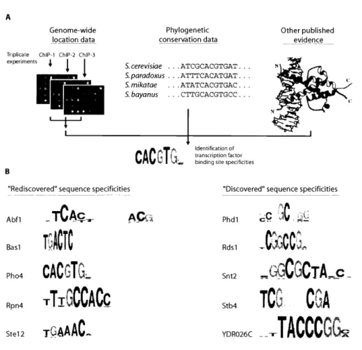
Harbison et al. 2004). The sequencing of multiple similar species has added an
additional constraint, making motif finding easier with the assumption that the
nucleotides important for transcription factor binding will be more conserved
between species than nucleotides without constraint on mutation (Cliften et al.
2003; Kellis et al. 2003; Harbison et al. 2004).
The knowledge of the sequence specificity of transcription factors allows
prediction of genes regulated by the factor, and provides an important
component of any network model of the cell. While large leaps in the ability to do
this have been achieved over the past decade as described above, we are still
far from a complete understanding of the phenomenon. For example,
transcriptional activators seem to bind to and regulate genes where no sequence
motif for the factor can be found (Lieb et al. 2001; Lee et al. 2002). Conversely,
there are hundreds of copies of factor specific motifs throughout the genome,
many of which are not bound by the site's cognate transcription factor (Lieb et a/.
2001; Harbison et al. 2004). And for a large percentage of additional genes, no
site specific regulator can be found (Harbison et a/. 2004).
The holy grail in the study of transcriptional regulation is to determine the
complete network of interactions that drives gene expression in the living cell.
Each of the microarray technologies described provides information towards
attaining this goal. Expression analysis provides a functional readout of the
results of the transcriptional regulation, but with expression data one cannot
deconvolute direct effects of transcription factor binding from indirect effects.
about which genes are likely to be the direct targets of a transcription factor. This
method has also been used to examine the regulation of transcription at other
levels. Histone density (D. Pokholok, unpublished data), chromatin modifications
(Kurdistani et al. 2002; Ng et al. 2002; Robyr et al. 2002; Wang et al. 2002; Ng et
al. 2003), and the promoters at which other components of the complete
transcription apparatus are acting ((Odom et aL. 2004), D. Pokholok, unpublished
data) have all been assayed using location analysis. Binding of transcription
factors directly to arrays, particularly arrays containing intergenic regions, allows
determination of every possible genomic sequence to which a particular factor
can bind. The derived sequence specificity of these factors based on these data
will also play a part in the construction of a complete network model, along with
additional high throughput data sources. The in vivo subcellular localization of
most yeast proteins (Huh et al. 2003), systematic analysis of synthetic lethality
between genes (Tong et al. 2004), and high-throughput analysis of protein
complexes using mass spectrometry (Krogan et al. 2004) all provide a wealth of
additional data towards the goal.
My contributions to this work
When I started my graduate studies, I was interested in doing both
experimental and computational work. At that time, microarray analysis of gene
expression was in full swing, opening exciting avenues to explore many aspects
of biology. The number of experiments using expression microarrays was
developed for publication in Ren et al. 2000. Both of these experimental
approaches generate vast amounts of data, but in 1999 computational support
and analytical methods were in their infancy and still required development and
refinement. I joined the Young lab because their studies with microarrays
afforded me the opportunity to work both at the bench and on the computer. My initial contribution to the lab came in the computational arena, when I
set up a public website for Itamar Simon's paper on the cell-cycle. This website
was among the first to make data from genome-wide studies publicly available
and searchable. Through this open approach, I set up a forum to not only share
our data, but also to address the need to standardize microarray data and the
analytical approaches in this burgeoning field.
My first summer, I started performing location analysis experiments with
the aim of elucidating networks of interactions between transcription factors. I
was interested in several fundamental metabolic systems; my first experiments
were performed with factors involved in glucose metabolism, Rgtl, Mthl and
Migl and I followed up by investigating factors from the nitrogen and phosphate
metabolic pathways. These experiments yielded a wealth of data which led me
to believe that the approach which would most benefit the yeast community
consisted of profiling as many factors as possible. I decided to work as a team
with several other investigators in the Young lab, synergizing our efforts to
perform hundreds of chips worth of location-analysis experiments.
I also focused on improving the analysis of the data we were acquiring.
determine which intergenic regions were bound by the profiled transcription
factor, and then meta-analyses to gain a deeper understanding of the overall
biology. The framework of the analysis for each experiment had been laid out in
Ren et al. (2001), based on an error model described for gene expression data in
Hughes TR et al. (2000). I expanded significantly on this model. I put in place a
series of templates for use by researchers for analyzing individual experiments,
making them as versatile and user-friendly as possible. In addition, I wrote
scripts to perform batch analyses for making comparisons between data sets, a
key step to integrate data from different experiments and expand comparative
analyses. I also took advantage of the large number of data sets that we had
generated to help with noise reduction (see discussion in chapter 2). Another of
my computational contributions consisted of a number of meta-analyses, which
ranged from determining various statistics about the data in general to
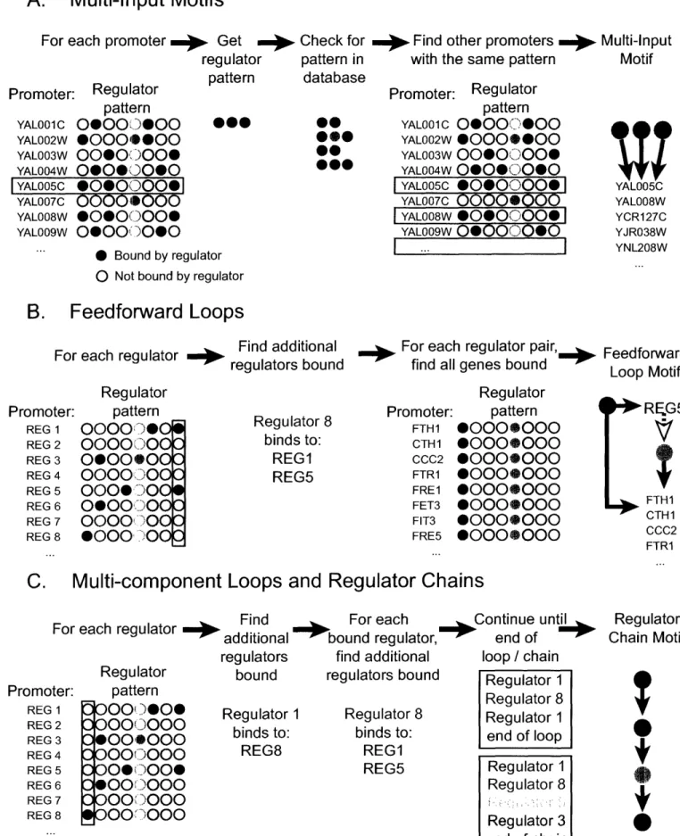
collaborating with Tony Lee to determine the definitions of the network motifs.
Based on these analyses, I developed computational tools such as a series of
scripts for finding network motifs. The results from the genome-wide location
analysis, interpreted using the tools I developed, was published (Lee, Rinaldi,
Robert et al. 2002), and is included in chapter 3.
I next extended our results, by addressing one of the weaknesses of our
approach. Looking at transcription under a single condition, in most cases the
rich medium Yeast extract - Peptone - Dextrose, affords a snapshot of the cell's
function. It does not, however, help to understand the most important role of the
constantly need to adjust the amount of metabolic enzymes based on the
available nutrients, to respond to cellular damage caused by external forces, and
to remove toxic elements from the cell. The work we published in Lee et al. did
not investigate cellular adaptation. We were able to find transcription factors
interacting with the promoters of only approximately 30% of the genome.
Additionally, although we did use expression data from conditions in which the
external environment was manipulated, without the matching binding data it was
difficult to assess which promoter-transcription factor interactions were leading to
productive regulation. We described regulatory network motifs based on the
binding data in rich media, but could not determine which of the motifs were
essential to transcription of the genes involved, as opposed to those that might
be used in another condition. To remedy this, location analysis under 12
additional conditions on a number of factors was performed. I focused mainly on
the experiments in hydrogen peroxide, and performed the data analysis for the
>400 experiments we had accumulated. These data and analyses were
published (Harbison et al. 2004) and are included in chapter 4.
In order to further improve the analysis of microarray data, I collaborated
with Ron Dror and Jon Murnick on a project that originated in the "Computational
Functional Genomics" course taught by Rick Young, David Gifford and Tommi
Jaakkola. We were concerned with the practice that was common at the time of
"flooring" Affymetrix intensity results to an arbitrary small number, and of
averaging repeated measurements without regard for their reliability. Instead, we
introduced throughout the experiments, and used a Bayesian estimation method
to provide a principled way of dealing with negative results, combining repeated
measurements, and determining differentially expressed genes. This work was
published as Dror et al. 2003. 1 also took part in the development of the Genetic
RegulAtory Modules algorithm, performing the large scale analysis of location
data that was required, collecting and curating over 500 expression data sets
from publicly available sources, and participating in a series of discussions during
the development. This work was published (Bar-Joseph et al. 2003), and can be
found in Appendix A.
Through my research and reading I have come to understand many of the
nuances involved in the various steps necessary for thorough analysis of
genome-wide data. My experience with both bench work and computational
analysis has allowed me a well-rounded view of the field of genomics. I have
included, in Chapter 2 of this thesis, many of my findings and observations in this
References
Adams CC and Workman JL (1995). "Binding of disparate transcriptional
activators to nucleosomal DNA is inherently cooperative." Mol Cell Biol 15(3): 1405-21.
Agarwal AK, Rogers PD, Baerson SR, Jacob MR, Barker KS, Cleary JD, Walker LA, Nagle DG and Clark AM (2003). "Genome-wide expression profiling of the
response to polyene, pyrimidine, azole, and echinocandin antifungal agents in
Saccharomyces cerevisiae." J Biol Chem 278(37): 34998-5015.
Allfrey VG, Faulkner R and Mirsky AE (1964). "Acetylation and Methylation of Histones and Their Possible Role in the Regulation of Rna Synthesis." Proc Natl Acad Sci U S A 51: 786-94.
Angermayr M and Bandlow W (2003). "Permanent nucleosome exclusion from the Gal4p-inducible yeast GCY1 promoter." J Biol Chem 278(13): 11026-31. Ansari AZ, Koh SS, Zaman Z, Bongards C, Lehming N, Young RA and Ptashne
M (2002). "Transcriptional activating regions target a cyclin-dependent kinase."
Proc Natl Acad Sci U S A 99(23): 14706-9.
Axelrod JD, Reagan MS and Majors J (1993). "GAL4 disrupts a repressing
nucleosome during activation of GAL1 transcription in vivo." Genes Dev 7(5): 857-69.
Bannister AJ and Miska EA (2000). "Regulation of gene expression by transcription factor acetylation." Cell Mol Life Sci 57(8-9): 1184-92.
Barberis A, Pearlberg J, Simkovich N, Farrell S, Reinagel P, Bamdad C, Sigal G and Ptashne M (1995). "Contact with a component of the polymerase II
holoenzyme suffices for gene activation." Cell 81(3): 359-68.
Bar-Joseph Z, Gerber GK, Lee TI, Rinaldi NJ, et al. (2003). "Computational
discovery of gene modules and regulatory networks." Nat Biotechnol 21(11): 1337-42.
Beck T and Hall MN (1999). "The TOR signalling pathway controls nuclear
localization of nutrient-regulated transcription factors." Nature 402(6762): 689-92.
Bowtell DD (1999). "Options available--from start to finish--for obtaining
expression data by microarray." Nat Genet 21(1 Suppl): 25-32.
Brazma A, Jonassen I, Vilo J and Ukkonen E (1998). "Predicting gene regulatory elements in silico on a genomic scale." Genome Res 8(11): 1202-15.
Breeden L and Mikesell GE (1991). "Cell cycle-specific expression of the SWI4
transcription factor is required for the cell cycle regulation of HO transcription."
Genes Dev 5(7): 1183-90.
Bulyk ML, Gentalen E, Lockhart DJ and Church GM (1999). "Quantifying DNA-protein interactions by double-stranded DNA arrays." Nat Biotechnol 17(6): 573-7.
Bulyk ML, Huang X, Choo Y and Church GM (2001). "Exploring the DNA-binding
specificities of zinc fingers with DNA microarrays." Proc Natl Acad Sci U S A 98(13): 7158-63.
Bushnell DA and Kornberg RD (2003). "Complete, 12-subunit RNA polymerase II
at 4.1-A resolution: implications for the initiation of transcription." Proc Natl Acad
Sci U S A 100(12): 6969-73.
Bushnell DA, Westover KD, Davis RE and Kornberg RD (2004). "Structural basis
of transcription: an RNA polymerase II-TFIIB cocrystal at 4.5 Angstroms."
Science 303(5660): 983-8.
Causton HC, Ren B, Koh SS, Harbison CT, et al. (2001). "Remodeling of yeast genome expression in response to environmental changes." Mol Biol Cell 12(2):
323-37.
Chatterjee S and Struhl K (1995). "Connecting a promoter-bound protein to TBP
bypasses the need for a transcriptional activation domain." Nature 374(6525): 820-2.
Chi Y, Huddleston MJ, Zhang X, Young RA, Annan RS, Carr SA and Deshaies
RJ (2001). "Negative regulation of Gcn4 and Msn2 transcription factors by Srb10
cyclin-dependent kinase." Genes Dev 15(9): 1078-92.
Cho HS, Liu CW, Damberger FF, Pelton JG, Nelson HC and Wemmer DE
(1996). "Yeast heat shock transcription factor N-terminal activation domains are
unstructured as probed by heteronuclear NMR spectroscopy." Protein Sci 5(2):
262-9.
Cho RJ, Campbell MJ, Winzeler EA, Steinmetz L, et al. (1998). "A genome-wide
transcriptional analysis of the mitotic cell cycle." Mol Cell 2(1): 65-73.
Cirillo LA and Zaret KS (1999). "An early developmental transcription factor complex that is more stable on nucleosome core particles than on free DNA." Mol
Cell 4(6): 961-9.
Cliften P, Sudarsanam P, Desikan A, Fulton L, Fulton B, Majors J, Waterston R, Cohen BA and Johnston M (2003). "Finding functional features in
Saccharomyces genomes by phylogenetic footprinting." Science 301(5629):
Cosma MP (2002). "Ordered recruitment: gene-specific mechanism of
transcription activation." Mol Cell 10(2): 227-36.
Cosma MP, Tanaka T and Nasmyth K (1999). "Ordered recruitment of
transcription and chromatin remodeling factors to a cell cycle- and developmentally regulated promoter." Cell 97(3): 299-311.
Costanzo MC, Hogan JD, Cusick ME, Davis BP, et al. (2000). "The yeast
proteome database (YPD) and Caenorhabditis elegans proteome database (WormPD): comprehensive resources for the organization and comparison of model organism protein information." Nucleic Acids Res 28(1): 73-6.
Csank C, Costanzo MC, Hirschman J, Hodges P, et al. (2002). "Three yeast
proteome databases: YPD, PombePD, and CalPD (MycoPathPD)." Methods Enzymol 350: 347-73.
Darieva Z, Pic-Taylor A, Boros J, Spanos A, Geymonat M, Reece RJ, Sedgwick SG, Sharrocks AD and Morgan BA (2003). "Cell cycle-regulated transcription
through the FHA domain of Fkh2p and the coactivator Nddlp." Curr Biol 13(19):
1740-5.
DeRisi JL, lyer VR and Brown PO (1997). "Exploring the metabolic and genetic
control of gene expression on a genomic scale." Science 278(5338): 680-6.
Dever TE, Feng L, Wek RC, Cigan AM, Donahue TF and Hinnebusch AG (1992).
"Phosphorylation of initiation factor 2 alpha by protein kinase GCN2 mediates
gene-specific translational control of GCN4 in yeast." Cell 68(3): 585-96.
Donaldson L and Capone JP (1992). "Purification and characterization of the carboxyl-terminal transactivation domain of Vmw65 from herpes simplex virus
type 1." J Biol Chem 267(3): 1411-4.
Ellenberger TE, Brandl CJ, Struhl K and Harrison SC (1992). "The GCN4 basic
region leucine zipper binds DNA as a dimer of uninterrupted alpha helices: crystal structure of the protein-DNA complex." Cell 71(7): 1223-37.
Emili A and Ingles CJ (1995). "Promoter-dependent photocross-linking of the
acidic transcriptional activator E2F-1 to the TATA-binding protein." J Biol Chem
270(23): 13674-80.
Farrell S, Simkovich N, Wu Y, Barberis A and Ptashne M (1996). "Gene
activation by recruitment of the RNA polymerase II holoenzyme." Genes Dev
10(18): 2359-67.
Feaver WJ, Gileadi O, Li Y and Kornberg RD (1991). "CTD kinase associated