Nicotinamide
mononucleotide (NMN) supplementation
promotes
anti-aging miRNA expression profile in the aorta
of
aged mice, predicting epigenetic rejuvenation
and
anti-atherogenic effects
Tamas
Kiss
&Cory
B. Giles
&Stefano
Tarantini
&Andriy
Yabluchanskiy
&Priya
Balasubramanian
&Tripti
Gautam
&Tamas
Csipo
&Ádám
Nyúl-Tóth
&Agnes Lipecz
&Csaba Szabo
&Eszter Farkas
&Jonathan D. Wren
&Anna Csiszar
&Zoltan
Ungvari
Abstract Understanding molecular mechanisms
in-volved in vascular aging is essential to develop novel
interventional strategies for treatment and prevention of
age-related vascular pathologies. Recent studies provide
critical evidence that vascular aging is characterized by
NAD+ depletion. Importantly, in aged mice, restoration of
Tamas Kiss, Cory B. Giles and Stefano Tarantini contributed equally to this work.
T. Kiss
:
C. B. Giles:
S. Tarantini:
A. Yabluchanskiy:
P. Balasubramanian:
T. Gautam:
T. Csipo:
Á. Nyúl-Tóth:
A. Lipecz:
J. D. Wren:
A. Csiszar:
Z. Ungvari (*) Vascular Cognitive Impairment and Neurodegeneration Program, Reynolds Oklahoma Center on Aging/Department of Geriatric Medicine, University of Oklahoma Health Sciences Center, 975 NE 10th Street, BRC 1311, Oklahoma City, OK 73104, USA e-mail: [email protected]T. Kiss
:
T. Csipo:
A. Lipecz:
E. Farkas:
A. Csiszar:
Z. UngvariDepartment of Medical Physics and Informatics / Theoretical Medicine Doctoral School, University of Szeged, Szeged, Hungary
C. B. Giles
:
J. D. WrenOklahoma Medical Research Foundation, Genes & Human Disease Research Program, Oklahoma City, OK and Department of Biochemistry and Molecular Biology, University of Oklahoma Health Science Center, Oklahoma City, OK, USA
S. Tarantini
:
A. Yabluchanskiy:
A. Csiszar:
Z. Ungvari Translational Geroscience Laboratory, Department of Geriatric Medicine, University of Oklahoma Health Sciences Center, Oklahoma City, OK, USAA. Yabluchanskiy
:
A. Csiszar:
Z. UngvariThe Peggy and Charles Stephenson Cancer Center, University of Oklahoma Health Sciences Center, Oklahoma City, OK 73104, USA
T. Csipo
:
A. Lipecz:
Z. UngvariDepartment of Public Health / Doctoral School of Basic and Translational Medicine, Semmelweis University, Budapest, Hungary
Á. Nyúl-Tóth
Institute of Biophysics, Biological Research Centre / Theoretical Medicine Doctoral School, Hungarian Academy of Sciences, Szeged, Hungary
C. Szabo
Chair of Pharmacology, Department of Medicine, University of Fribourg, Fribourg, Switzerland
A. Csiszar
Institute of Human Physiology and Clinical Experimental Research, Semmelweis University, Budapest, Hungary Z. Ungvari
Department of Health Promotion Sciences, College of Public Health, University of Oklahoma Health Sciences Center, Oklahoma City, OK, USA
http://doc.rero.ch
Published in "GeroScience 41(4): 419–439, 2019"
which should be cited to refer to this work.
cellular NAD+ levels by treatment with the NAD+ booster
nicotinamide mononucleotide (NMN) exerts significant
vasoprotective effects, improving endothelium-dependent
vasodilation, attenuating oxidative stress, and rescuing
age-related changes in gene expression. Strong
experimen-tal evidence shows that dysregulation of microRNAs
(miRNAs) has a role in vascular aging. The present study
was designed to test the hypothesis that age-related NAD+
depletion is causally linked to dysregulation of vascular
miRNA expression. A corollary hypothesis is that
func-tional vascular rejuvenation in NMN-treated aged mice is
also associated with restoration of a youthful vascular
miRNA expression profile. To test these hypotheses, aged
(24-month-old) mice were treated with NMN for 2 weeks
and miRNA signatures in the aortas were compared to
those in aortas obtained from untreated young and aged
control mice. We found that protective effects of NMN
treatment on vascular function are associated with
anti-aging changes in the miRNA expression profile in the aged
mouse aorta. The predicted regulatory effects of
NMN-induced differentially expressed miRNAs in aged vessels
include anti-atherogenic effects and epigenetic
rejuvena-tion. Future studies will uncover the mechanistic role of
miRNA gene expression regulatory networks in the
anti-aging effects of NAD+ booster treatments and determine
the links between miRNAs regulated by NMN and sirtuin
activators and miRNAs known to act in the conserved
pathways of aging and major aging-related vascular
diseases.
Keywords Senescence . Atherosclerosis . Vascular
cognitive impairment . Epigenetics . Vascular aging .
Endothelial dysfunction . Oxidative stress
Introduction
Age-related diseases of the cardiovascular system are a
leading cause of morbidity and mortality in the elderly
(Abdellatif et al.
2018
; Minamino and Komuro
2007
;
Wang and Bennett
2012
; Alfaras et al.
2016
; Ungvari
et al.
2018
). Vascular aging is associated with stiffening
of the large arteries, endothelial dysfunction, oxidative
stress, and inflammation, promoting the development of
atherosclerotic vascular diseases (ischemic heart diseases,
stroke, peripheral artery disease) and aorta aneurysm
(Wang and Bennett
2012
; Ungvari et al.
2018
).
Microvas-cular aging is also a major contributing factor to the
pathogenesis of vascular cognitive impairment (VCI),
Alzheimer’s disease, cerebral microhemorrhages,
sarcopenia, heart failure, chronic kidney disease and
(Ungvari et al.
2018
; Mullins et al.
2014
; Ungvari et al.
2017a
; Toth et al.
2017
; Tarantini et al.
2017a
; Tarantini
et al.
2016a
; Sagare et al.
2013
; Sweeney et al.
2018
;
Montagne et al.
2017
; Kisler et al.
2017
; Payne
2006
;
Hoenig et al.
2008
; Long et al.
2012
). Understanding
molecular mechanisms involved in vascular aging is
es-sential to develop novel interventional strategies for
treat-ment and prevention of age-related vascular pathologies.
MicroRNAs (miRNA) are short, endogenous,
non-coding transcripts that repress gene expression at the
post-transcriptional level in both physiological and
patho-logical conditions. Strong experimental evidence suggest
that miRNAs have a role in regulation of lifespan in model
organisms (Boehm and Slack
2005
; Grillari and
Grillari-Voglauer
n.d.
; Ibanez-Ventoso et al.
2006
) and that
alter-ations in cellular miRNA expression profile also play a role
in mammalian aging (Bates et al.
n.d.
; Maes et al.
2008
;
Inukai et al.
2012
; Inukai and Slack
2013
; Ito et al.
2010
;
Mercken et al.
2013
; Smith-Vikos and Slack
2012
;
Ungvari et al.
2013a
; Zhang et al.
2012
; Zovoilis et al.
2011
; Smith-Vikos et al.
2016
; ElSharawy et al.
2012
).
Importantly, miRNAs were also reported to regulate
sev-eral important aspects of endothelial biology and vascular
function (Bonauer et al.
2009
; Doebele et al.
n.d.
;
Kuehbacher et al.
2007
; Chen et al.
2015a
; Hergenreider
et al.
2012
; Kim et al.
2014
; Leung et al.
2013
; Lovren
et al.
2012
; O’Rourke and Olson
2011
; Rotllan et al.
2013
;
Stellos and Dimmeler
2014
; Weber et al.
2014
; Zampetaki
et al.
2014
). Several studies have demonstrated that
age-related miRNA dysregulation importantly contributes to
the development of vascular aging phenotypes (Ito et al.
2010
; Ungvari et al.
2013a
,
b
; Menghini et al.
2014
; Badi
et al.
2018
; Guo et al.
2017
; Hazra et al.
2016
; Regina et al.
2016
; Boon et al.
2013
; Csiszar et al.
2014
) and promotes
the pathogenesis of atherosclerotic diseases (Ono et al.
2011
) encompassing every step from sterile vascular
in-flammation, plaque formation to plaque destabilization and
rupture (Hartmann et al.
2016
; Lu et al.
2018
; Zhang et al.
2018
). Dysregulation of miRNA expression has also been
linked to microvascular aging phenotypes, including
im-paired angiogenesis (Ungvari et al.
2013b
; Csiszar et al.
2014
; Che et al.
2014
; Jansen et al.
2015
). Experimental
interventions that both extend lifespan and prevent/delay
age-related vascular dysfunction in rodents, including
ca-loric restriction (Csiszar et al.
2014
) and induction of
early-life IGF-1 deficiency (Tarantini et al.
2016b
), were shown
to reverse aging-induced alterations in vascular miRNA
expression. Despite these advances, fundamental cellular
and molecular processes of aging that are responsible for
dysregulation of vascular miRNA expression have not
been elucidated.
NAD
+is a rate-limiting co-substrate for sirtuin
en-zymes, which are key regulators of pro-survival pathways
in the vasculature (Das et al.
2018
; Csiszar et al.
2009a
;
Csiszar et al.
2009b
; Csiszar et al.
2008
). Aging is
asso-ciated with cellular NAD
+depletion (Gomes et al.
2013
;
Massudi et al.
2012
), which has been proposed to be a
critical driving force of aging processes. In support of this
theory, it was demonstrated that enhancing NAD
+bio-synthesis extends lifespan in lower organisms (Anderson
et al.
2002
) and improves health-span in mouse models of
aging (Mitchell et al.
2018
). Recent studies provide
crit-ical evidence that vascular aging is also characterized by
NAD+ depletion (Tarantini et al.
2019
; Csiszar et al.
2019
; Kiss et al.
2019
). Importantly, we 69 and other
laboratories demonstrated (Das et al.
2018
; de Picciotto
et al.
2016
) that in aged mice restoration of cellular NAD
+levels by treatment with the NAD+ precursor
nicotin-amide mononucleotide (NMN) (Yoshino et al.
2018
)
confers potent anti-aging vascular effects, reversing
en-dothelial dysfunction, improving mitochondrial function,
and attenuating oxidative stress.
The present study was designed to test the hypothesis
that age-related NAD+ depletion is causally linked to
dysregulation of vascular miRNA expression. A corollary
hypothesis is that functional vascular rejuvenation in
NMN-treated aged mice is also associated with restoration
of a youthful vascular miRNA expression profile. To test
these hypotheses, aged mice were treated with NMN for
2 weeks and miRNA signatures in the aortas were
com-pared to those in aortas obtained from untreated young and
aged control mice.
Methods
Animals, NMN supplementation
Young (3-month-old) and aged (24-month-old) male
C57BL/6 mice were purchased from the aging colony
maintained by the National Institute on Aging at
Charles River Laboratories (Wilmington, MA). The
biological age of 24-month-old mice corresponds to
that of ~ 60-year-old humans. Mice were housed
un-der specific pathogen-free barrier conditions in the
Rodent Barrier Facility at University of Oklahoma
Health Sciences Center under a controlled
photope-riod (12 h light; 12 h dark) with unlimited access to
water and were fed a standard AIN-93G diet (ad
libitum). Mice in the aged cohort were assigned to
two groups. One group of the aged mice was injected
daily with NMN (i.p. injections of 500 mg NMN/kg
body weight per day) or the equivalent volume of
PBS for 14 consecutive days at 6 PM and 8 AM on
day 14 and were sacrificed 4 h after last injection.
Similar dosages of NMN have been shown to exert
potent anti-aging effects on mouse health span (de
Picciotto et al.
2016
). All procedures were approved
by the Institutional Animal Use and Care Committees
of the University of Oklahoma Health Sciences
Cen-ter. All animal experiments complied with the
AR-RIVE guidelines and were carried out in accordance
with the National Institutes of Health guide for the care
and use of Laboratory animals (NIH Publications No.
8023, revised 1978). The effects of NMN treatment on
cognitive function, cerebromicrovascular responses,
and aorta endothelial function in the same cohort of
mice have been recently reported (Tarantini et al.
2019
).
Quantitative real-time RT-PCR and miRNA expression
profiling
A quantitative real time RT-PCR technique was used to
analyze miRNA expression profiles in the aorta of mice
from each experimental group as reported (Ungvari et al.
2013b
; Csiszar et al.
2014
; Tarantini et al.
2016b
). In
brief, total RNA was isolated with a mirVana™ miRNA
Isolation Kit (ThermoFisher Scientific) and was reverse
transcribed using TaqMan® MicroRNA Reverse
Tran-scription Kit as described previously (Ungvari et al.
2013b
; Csiszar et al.
2014
; Tarantini et al.
2016b
). The
expression profile of mouse miRNAs in aortas derived
from young and aged control mice and aged
NMN-treated mice was analyzed using the TaqMan Array
Ro-dent MicroRNA A+B Cards Set v3.0 (ThermoFisher
Scientific). The qPCR data were quantified using the
ΔΔCt method (Livak and Schmittgen
2001
). Predicted
and experimentally validated microRNA targets were
obtained from the TargetScan database (Agarwal et al.
2015
), and Gene Ontology enrichment analysis was
per-formed on differentially expressed microRNA targets
using Fisher’s exact test between TargetScan targets and
annotations from the Gene Ontology database (Harris
et al.
2004
). To identify relationships between miRNA
targets and terms in the biomedical literature, we utilized
the IRIDESCENT system (Wren and Garner
2004
).
IR-IDESCENT uses a statistical model to determine whether
each target gene co-occurs with a term of interest more
frequently than would be expected by chance, and
quan-tifies this in terms of the mutual information measure.
Results
Changes in vascular miRNA expression profile in mice
associated with aging and with NMN treatment
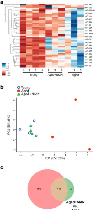
We assessed changes in miRNA expression in the
mouse aorta associated with aging and with NMN
treat-ment. Hierarchical clustering (Fig.
1a
) and principal
component analysis (Fig.
1b
) of miRNA expression
showed a clear separation between the young and aged
groups. Aged control mice and aged NMN-treated mice
were also separated in the principal component analysis
and hierarchical clustering. In contrast, miRNA
expres-sion in young mice and NMN-treated aged mice was
similar and these groups did not separate well in the
principal component analysis and hierarchical
cluster-ing. The Venn diagram in Fig.
1c
shows that expression
of several miRNAs, which are differentially expressed
in the aortas of young and aged mice, was restored to
youthful levels in aortas of NMN-treated aged mice.
These data suggest that NAD
+depletion has a critical
role in age-related dysregulation of vascular miRNA
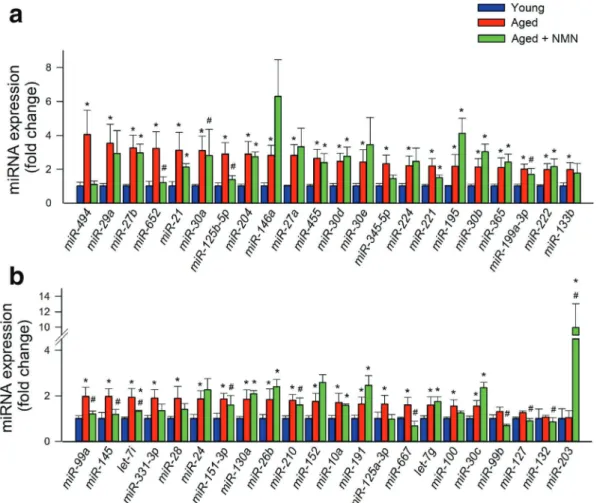
expression. Figure
2
shows changes in expressions of
individual miRNAs in the mouse aorta associated with
age and NMN treatment.
Since the discovery of miRNA regulation of genes,
several studies have been focused on predicting the
biolog-ically relevant target genes for miRNAs. We have used
TargetScan database to predict putative biological targets
of miRNAs differentially expressed with age whose
expres-sion is restored to youthful levels in aortas of aged mice by
NMN supplementation (Table
1
). GO terms enriched
among miRNAs differentially expressed with age whose
expression is restored to youthful levels in aortas of aged
mice by NMN supplementation are shown in Table
2
.
Analysis of the differentially expressed miRNAs indicated
that a statistically significant number of them had target sites
within genes associated with pathways regulating the
intra-cellular signaling, protein homeostasis, and inflammation
(Table
2
). The results are consistent with the predicted
anti-aging effects of NMN treatment.
Fig. 1 NMN treatment reverses age-related changes in miRNA ex-pression profile in the mouse aorta. a The heat map is a graphic representation of normalized miRNA expression values in aortas de-rived from young (3-month-old), aged (24-month-old), and NMN-treated aged mice. Hierarchical clustering analysis revealed the similar-ities on miRNA expression profiles of aortas from young and NMN-treated aged mice. b Principal component analysis (PCA) plot of miRNA expression profiles from aortas derived from young, aged control, and NMN-treated aged mice. The profiles from aged mice (red dots) cluster separately to clusters representative of young mice (blue circles) and NMN-treated aged mice (green triangles). PC1 and PC2: Principal components 1 and 2, respectively. c Venn diagrams showing the differentially expressed miRNAs in each group, which are significantly up- or down-regulated in aortas from aged mice compared to those from young mice or aged NMN-treated mice
We also attempted to predict the biological effects of
the differentially expressed miRNAs by identifying
re-lationships between miRNA targets and terms in the
biomedical literature utilizing the IRIDESCENT system
(Wren and Garner
2004
). The results of this analysis
suggest that NMN supplementation likely promotes
epi-genetic rejuvenation and confers anti-atherogenic
ef-fects (Table
3
).
Discussion
Our study demonstrates that protective effects of NMN
treatment on vascular function is associated with
anti-aging changes in the miRNA expression profile in the
aorta in a mouse model of aging that recapitulates
vascular alterations and deficits present in elderly
humans at risk for cardiovascular and cerebrovascular
diseases.
Age-related changes in vascular miRNA
expres-sion likely play important pathogenic roles targeting
critical signaling pathways, inflammatory processes,
and cellular mechanisms involved in protein
homeo-stasis and thereby impairing the structural and
func-tional integrity of the vasculature (Fig.
3
). Among
others, miR-29a (Huang et al.
2016
), miR-27b
(Signorelli et al.
2016
), miR-652 (Pilbrow et al.
2014
), miR-221 (Wei et al.
2013
), miR-28 (Wang
et al.
2017
), miR-21 (Urbich et al.
2008
),
miR-125b-5p (Ohukainen et al.
2015
) , miR-494 (Wezel et al.
2015
), and miR-145 (Faccini et al.
2017
), which are
up-regulated in aging, have been implicated in
vas-cular inflammation and atherogenesis.
Fig. 2 Effects of aging and NMN treatment on miRNA sion in the mouse aorta. a, b qPCR data showing miRNA expres-sion in aortas isolated from young (3-month-old), aged
(24-month-old), and NMN-treated aged mice. Data are mean ± S.E.M. (n = 3– 4 for each data point). *P < 0.05 vs. young; #P < 0.05 vs. aged
Ta b le 1 S elected genes , whose expression ch anges w ith age and are predicted to be tar geted by NMN-dependent dif ferentially ex pres sed miRNAs. Shown are (1) the n u mber of m iR N A s ta rg et in g the ge ne , w ho se ex p re ssi on is si g n if ic an tl y ch an g ed m y N M N treat ment ; (2) relat ive age-depen dent chang es in gene expr essi on, pred icte d b y the AgeAtlas software; and (3) cellular function of the protein encod ed by the g ene and its putative role in vascular pathologies Gene symbol Gene fu ll name NMN-induced significant miRNAs AgeA tla s chan ge Cellular functi on Role in vascular pathology Sec62 SEC62 homo log 4 − 0.0089 Component of the protein translocation apparatus Single nucleotide polym orphis m is asso ciated with vulnerable p laque (de Boer et al. 2018 ) Nb eal 1 n eu robe achi n like 1 3 − 0.81 Plays a role in vesicle traf ficking, membrane dynamics, receptor si gnaling, pre-mRNA proces sing, signal trans duction and cyto-skeleton assembly Single nucleotide polym orphis m is asso ciated with early atherogenesis (Hixson et al. 2017 ) and developmen t o f ischemic white matter hyperintensities in stroke patients (T raylor et al. 2016 ) Fyn Fyn proto-oncogene 2 − 0.31 Kinase Genome-wide analysis of DNA methylation showed association with aortic athe rosc le rosi s (Y ama da et al . 2014 ); in v itro overexpresse d in activated smooth musc le ce ll s (Sin gh et al. 201 7 ); Mef2 a m yocyte enhan cer factor 2A 2 − 0.094 T ranscrip tion facto r In v itro increased expres sion in senescence endothelial cell; in creased plasma level in coronary arte ry dis eas e p ati ents (Li u et al. 2019 ) T et2 tet methylcytosine dioxygenase 2 2 − 0.45 Ep igenetic regulator Contributes to the dev elopment of atherosclerosi s b y epigenetic modification (Aavik et al. 2019 ;P en ge ta l. 2016 ;L iu et al . 2013 ) Pt ch1 pat che d 1 2 − 0.0072 Hedgehog signaling pathway Overexpress ed in athero sclerotic plaque in mouse carotid ar te ry (Ali et al . 2013 ) Ad ra2b adrener gic receptor , alph a 2 b 2 − 0.061 Seven-pass trans membrane protein role in hypertens ion (Kintsurash vili et al. 2009 ) Ab cg4 A T P -binding cassette, sub-family G (W HI TE ), m em b er 4 2 − 0.0038 ABC -trans porter Cholesterol transporter , strongly linked to atherosclerosi s and other cardiovascular disease (Schumacher and Benndorf 2017 ; W ester te rp et al . 2014 ) Epha6 Eph receptor A6 2 − 0.59 Ep hrin receptor GenS alt and MES A studies: SNP varian t associated with hypertens ion (Li et al. 2016 ; Kim et al. 2 017 ); in vit ro ac tiva ted in cells relevant for atherogenesis (Sakamoto et al . 201 1 )
http://doc.rero.ch
To our knowledge, this is the first study to
demonstrate that NMN treatment in aged mice
reverses, at least in part, age-related,
pro-inflam-matory, and pro-atherogenic alterations in miRNA
expression profile in the aorta. These findings raise
the possibility that changes in post-transcriptional
control of expression of genes that encode critical
targets for vascular health contribute to the
bene-ficial effects of treatment with NAD+ boosters on
health span. Demonstration of NMN-induced
changes in miRNA biology in the vasculature is
particularly important as alterations in miRNA
ex-pression profile have been causally linked to the
development of cardiovascular aging phenotypes
(Ungvari et al.
2013a
; Boon et al.
2013
; Csiszar
et al.
2014
) and the pathogenesis of cardiovascular
diseases (Ono et al.
2011
). A single miRNA can
target up to several hundred mRNAs, thus capable
of significantly altering gene expression regulatory
networks. Systematic prediction of target pathways
supports the concept that chronic NMN treatment
may exert significant anti-atherogenic effects via
epigenetic rejuvenation of the vasculature. These
miRNA-mediated vasoprotective effects of NMN
treatment appear to be synergistic with its
endo-thelial protective, anti-aging, and pro-angiogenic
effects demonstrated by recent studies (Tarantini
et al.
2019
; Csiszar et al.
2019
; Kiss et al.
2019
).
The molecular mechanisms contributing to
aging-induced decline in NAD
+in the vasculature
are likely multifaceted and may include
down-r e g u l a t i o n
o f
n i c o t i n a m i d e
phosphoribosyltransferase (NAMPT, also known
as NMN synthase; which catalyzes the rate
limit-ing step in the biosynthesis of NAD
+) (Tarantini
et al.
2019
) and increased utilization of NAD
+by
activated Poly [ADP-ribose] polymerase 1
(PARP-1) (Csiszar et al.
2019
; Pacher et al.
2002
).
Addi-tional studies are warranted to determine the
effi-cacy of combination treatments that simultaneously
increase NAD+ production and inhibit its
degrada-tion (e.g., NMN plus a PARP-1 inhibitor) for the
prevention of age-related vascular pathologies.
Previous studies demonstrate that restoration of
NAD
+levels by NMN treatment exert protective
effects on endothelial vasodilation in aged rodents
by reducing ROS generation and restoring
mito-chondrial function in a sirtuin-dependent manner
(Tarantini et al.
2019
). The mechanisms by which
Ta b le 1 (continu ed) Gene symbol Gene full name NMN-induced significant miRNAs Ag eAt las ch ange C ellular function Rol e in vascular pathology Atf2 act ivat ing transc ript ion fa ctor 2 2 − 0.34 T ran scription factor In mouse models p articipates in foam cells activatio n signaling (Raghavan et al. 2018 ); vas cular smooth cell activation (W u et al . 2014 ) Homer2 home r sca ffol ding prot ein 2 2 − 0.31 Glutamate signaling pathway Bio marker of at heroscleros is (Zhu et al. 2016 ) Kcnb1 potass ium voltage g ated channel, Shab-relat ed subfamily , m ember 1 3 − 0.21 Potas sium channel subunit Changed expression in arteries in rat model of hypertension (Cox et al. 2008 ) Rap1a RAS-re lat ed prot ein -1a 2 − 0.52 R as signaling pathway Potential role in carotid atheros clerosis (Mao et al . 2018 ) Fryl FR Y like tr anscripti on coac tiva tor 2 − 0.71 T ran scription factor; Notch signaling Downregulated in hypertensive mouse aorta (Rippe et al. 2017 )
http://doc.rero.ch
NAD+ boosters regulate miRNA expression are
likely multifaceted and may include both
transcrip-tional and post-transcriptranscrip-tional regulatory
mecha-nisms (Fig.
3
). NMN-induced transcriptional
regula-tion may involve changes in the expression of
miRNA genes due to altered transcription factor
activity, changes in genome accessibility (e.g.,
his-tone modifications), and altered methylation status
of the promoter of the miRNA genes.
Post-transcriptional mechanisms affected by NMN
treat-ment may include rescue of miRNA processing
pathways (Ungvari et al.
2013b
) and miRNA
stabil-ity. Activation of sirtuins by NAD+ boosters, which
has been linked to attenuation of age-related
vascu-lar oxidative stress (Tarantini et al.
2019
; Kiss et al.
2019
), may potentially contribute to both
transcrip-tional and post-transcriptranscrip-tional regulation of miRNA
expression in the vasculature. In particular, future
Table 2 Predicted regulatory effects of miRNAs whose expres-sion is restored to youthful levels in aortas of aged mice by NMN supplementation. Shown are GO terms enriched among miRNAs differentially expressed with age in the aorta whose expression is significantly affected by NMN treatment. N = genes in each GO
category, targeted by miRNAs that are differentially regulated in the aged mouse aorta. Significance was determined by Fisher’s exact test; odds ratio: (observed to expected ratio); SLPV: signed log10 P value
GO term ID Name of biological process/molecular function N Odds Ratio SLPV
6886 Intracellular protein transport 20 3.17 3.26
7218 Neuropeptide signaling pathway 7 7.32 2.54
5198 Structural molecule activity 6 9.40 2.45
51082 Unfolded protein binding 7 5.49 2.20
45778 Positive regulation of ossification 6 6.27 2.07
50839 Cell adhesion molecule binding 10 3.49 1.92
15137 Citrate transmembrane transporter activity 3 inf 1.84
48227 Plasma membrane to endosome transport 3 inf 1.84
8188 Neuropeptide receptor activity 3 inf 1.84
7217 Tachykinin receptor signaling pathway 3 inf 1.84
42594 Response to starvation 3 inf 1.84
70536 Protein K63-linked deubiquitination 6 4.70 1.77
71108 Protein K48-linked deubiquitination 6 4.70 1.77
5102 Receptor binding 27 1.82 1.72
90630 Activation of GTPase activity 10 2.85 1.71
31338 Regulation of vesicle fusion 7 3.66 1.68
1664 G-protein coupled receptor binding 6 3.76 1.52
6631 Fatty acid metabolic process 6 3.76 1.52
45777 Positive regulation of blood pressure 4 6.25 1.47
32924 Activin receptor signaling pathway 4 6.25 1.47
70530 K63-linked polyubiquitin binding 4 6.25 1.47
10863 Positive regulation of phospholipase C activity 4 6.25 1.47
16579 Protein deubiquitination 8 3.13 1.47
18107 Peptidyl-threonine phosphorylation 9 2.57 1.42
48015 Phosphatidylinositol-mediated signaling 5 3.91 1.36
7200 Phospholipase C-activating G-protein coupled receptor signaling pathway 5 3.91 1.36
71837 HMG box domain binding 5 3.91 1.36
61578 Lys63-specific deubiquitinase activity 3 9.37 1.33
33674 Positive regulation of kinase activity 3 9.37 1.33
43122 Regulation of I-kappaB kinase/NF-kappaB signaling 3 9.37 1.33
50995 Negative regulation of lipid catabolic process 3 9.37 1.33
Ta b le 3 Literature commona lities o f the genes tar geted b y miR NAs whose expression is restored to yo uthfu l levels in aortas of aged mice by NMN supplementatio n. The IR ID ESCENT literature-mini ng software wa s used to ident ify commonali ties (e.g. , g enes, dis eas es, phenot ypes, biol ogical p rocesses) of th e g enes predict ed to b e tar geted by the miRNAs. A network of rel ate d o bjec ts wa s esta blishe d b y their co -oc curre nce w ithin MED LINE records, sha re d rel ati onships we re id enti fi ed, and the ir st ati stic al rele v ance was scored b y comparin g observed frequen cies with what would b e expected in a rando m n etwork model. Number of sh ared relationships is the number of genes (out of the to p 100 most signifi cant) co-mentioned with the terms in th e left-hand column. The observed to expected (o bs/ex p) ratio is the enrichment for the te rm. References and not es on how each one relates to v ascular pathophysiology of aging are shown in the rightmost column Lit erature ass ocia tions Remark/full name # sh are d rel ationships Obs /exp Score Biolog ical process/function CTNNB1 Catenin Beta 1 6 3 2.25 139.9 Adhe rent junctions; Wnt/beta-catenin signaling in VS MCs co ntribute to Inti ma l thic keni ng (T saous i et al. 201 1 ) Wnt 56 2.02 1 10.8 Wnt signaling regulates atherogenesis (Z hao et al . 2018 ;B h at t et al . 2012 ) PTEN Phosphatas e and tensin homo log 43 2. 2 9 3 Regulates VSMC p henotype (Moulton et al . 2018 ) epithelial-mesenchymal transition 41 2.07 83.3 Endotheli al to menesn chymal transition contributes to atherogenes is (Bostrom et al. 2016 ;E v ra rde ta l. 2016 ; M oonen et al. 2015 ) SMA RCA4 SWI/S NF rela ted, mat ri x associ ate d, ac tin dependent regulator of chromatin, su bfamily a, member 4 22 3.73 81 Chromatin remodeling; genome wide association study showed its potent ial ro le in at heroscl ero sis (Ma o et al . 2017 ) FGFR1 Fibroblast growth factor receptor 1 2 8 2.63 72.3 Receptor; FGF recepto r signaling reg ulat es at hero genes is (Che et al . 201 1 ) EP300 E1A binding protein p300 32 2.27 71.3 T ranscriptional coactivator; VEGF A triggers chang es in transcriptional activity of endothelial cells via epige net ic re gulat ion w ith the he lp of EP300 (Zhang et al. 2 013 ) EZH2 Enhancer of zeste 2 polycomb repressive complex 2 subunit 25 2.78 68.6 Histone methyltransferase; epigenetic suppressio n o f gene ex pression sumoylation 24 2.56 60.6 Sumolylation reactions play a role in atherogenesis (Heo et al. 2013 ;H eo et al . 2015 ; S te in et al . 2014 ) RNA polymeras e II 2 9 2 .08 58.9 mRNA transcription SOX2 Sex determining region Y -box 2 2 7 2.15 57. 7 T ranscription factor , stem cell functio n; upregulated in aortic endothelial cells
http://doc.rero.ch
Ta b le 3 (continu ed) Lit erature ass ocia tions Remark/full name # sh are d rel ationships Obs /exp Score Biolog ical process/function in athe rosc le roti c m ic e (Bostrom et al . 2016 ). Limiting Sox2 decreases cal cificati on in aort as of Ap oE(-/-) mice (B ostrom et al. 2016 ). chro matin remodelin g 2 9 2 .03 57.3 (Kh yzha et al. 2017 ) CDH2 N-cadherin 27 2.1 1 55.5 Adherent junctions; neo intima forma tion (Jones et al . 2002 ) CDKN1A Cyclin-dependent kinase inhibitor 1 ; p21 27 2.02 53.3 Senescence; regu lates atherog enes is and neoi ntima fromati on (Y ang et al. 1996 ) KMT2D Histone-lysine N-methyltransferase 2D 15 3.6 53 Histone methyltransfe rase; epigenetic reg ulation of gene expression SMAD4 Moth ers agains t d ecapentaplegic homolog 4 2 3 2.26 51 T ranscription factor , m ediates TGF β signaling, regulates pathways involved in atherogenesis (Kintscher et al . 2002 ) KDM1A L ysin e-s pecific h iston e demethylase 1A 16 3.02 47.2 Histone methyltransferase; epigenetic reg ulation of gene expression (Pojoga et al. 201 1 ) FGFR2 Fibroblast growth factor receptor 2 2 3 2.08 47 FGF receptor signaling regulates atherogenesis (Che et al. 201 1 ) BCOR BCL-6 corepressor 12 3.96 46.4 transcription repressor P A X6 Paired box 6 2 0 2.34 46.1 T ranscription factor; DNMT1 DNA met hyltrans feras e (c ytosine -5) 1 2 2 2.05 44.5 Chromatin remodeling; mediate macro phage activation and partic ipat e inflamma tion in athe rosc le roti c les ions (Y u et al. 2016 ) polycomb 19 2.31 42.7 Chromatin remodeling, regulate ABCA1 expr ession in the macro phages and consequently h as an im porta nt role in the de velop men t of the inflammat ion in the athe rosc le roti c lesi on (Lv et al. 2016 ) MECP2 Methyl CpG binding protein 2 1 8 2.25 39.5 Key ep igenetic factor regul ati ng gl obal gene trans criptio n b y g athering the histon e d eacetylase complex to the
http://doc.rero.ch
Ta b le 3 (continu ed) Lit erature ass ocia tions Remark/full name # sh are d rel ationships Obs /exp Score Biolog ical process/function promoter regions of the genes; with polycomb, it regu lates ABCA1 expression in the macrophages (Lv et al . 2016 ) TSC1 TSC complex subunit 1 1 6 2.48 38.9 T umor suppressor gen e; regulates ma mmali an ta rget of rapa myc in complex 1 (mT OR C1) sign aling, thought the mTHOR pathway it plays a cri tica l rol e in the de velop -ment of atherosclerotic lesions (Ku rdi et al . 2016 ) CREB BP CREB binding protein 18 2.18 38.4 Involved in the trans criptio nal coac tiva tio n o f m an y d if fe re nt transcript ion factors ID2 Inhibitor o f DNA binding 2 1 6 2 .42 37.9 T ranscriptional regulator , inhibit the functions of basic helix-loop-helix tra nscript ion fa ctors; vas cula r smooth muscle phenoty pic change in athe rosc le rosi s (Zh u et al. 2015 ) BAP1 BRCA1 associated protein 1 1 2 3.02 35.9 Ubiquitin C-terminal hydrolase, removes u biquitin from proteins TCF4 (alias of TCF7L2) transcription factor 7 like 2 1 7 2.12 35.5 T ranscription factor; epidemiological data shows SNP polymorphism assoc iat ed wi th athe rosc le rosi s (E strada-V el asc o et al. 2013 ; Muendlein et al. 201 1 ) PIK3CA Phosphatidylinositol-4,5-bisphos phate 3-kinase catalytic subunit alpha 16 2.12 33.5 Kinase activity; somatic mu tation causes venous (Limaye et al. 2015 ) and lymphatic malformation (B le singe r et al. 2018 ) ARHGEF 2 R ho/Rac guanine nucleotide exchange factor 2 1 6 2.05 32.2 Participates in the rho-dependent sig-naling pathway P A X8 Paired box 8 1 3 2.56 32.2 T ranscription factor FGF8 Fibroblast growth factor 8 1 4 2.35 31.5 Mit ogeni c and ce ll survival ac ti viti es; reg ulates the cardiovas cular development (B rown et al. 2004 ) BTG2 BTG anti-pro liferation factor 2 1 1 2 .8 7 30.8 Involved in the regulatio n o f the G1/S transition of the cell cycle
http://doc.rero.ch
Ta b le 3 (continu ed) Lit erature ass ocia tions Remark/full name # sh are d rel ationships Obs /exp Score Biolog ical process/function FBXW7 F-bo x and WD repeat domain co ntaining 7 1 2 2.48 29.6 Phosphorylation-dependent protein ubiquitinati on; regulates angiogenesis (Izumi et al. 2012 )a n d the barri er funct ion of end othel ial cel ls (P ronk et al . 2019 ) FMR1 Fra gile X m en tal ret arda tio n 1 14 2. 09 28.9 Controls the proliferation and angiogenesis of endothelial cells via the miR-181a-mediated calmo dulin (C aM)/CaMKII p athway (Zhao et al. 2018 ) MSTN Myost ati n 1 4 2.1 1 28.8 Partic ipa tes in TGF-beta signa ling; in -cre ase d expressi on in at he rosc leroti c lesi ons, espec ial ly in vasc ular smoot h m usc le ce lls (V erzola et al. 2017 ) HDAC3 Histone deacetylase 3 1 4 2.07 28.5 T ranscriptional regulator b y epigen etic modification of DNA; regulats the atherosclerotic phenotype of macro phages (Hoeksema et al. 2014 ) CCNE1 Cyclin E1 12 2.35 27.9 Regulator o f cell cycle SMA RCB1 SWI/S NF rela ted, mat ri x associ ate d, actin dependent regulator of chromatin, subf am il y b , m em be r 1 1 1 2.54 27.5 Chromatin remodeling AXIN2 Axin 2 1 2 2.31 27.3 Participates in G-protein signaling; participate in Wnt signaling which known to regulates atherosclerosis (Z hao et al . 2018 ;B h at t et al . 2012 ; T ia n et al . 2017 ) RASS F1 Ras as sociation domain family member 1 1 2 2.29 26.9 regulator of DNA methylation and DNA repa ir FOXP1 Fork head box P1 1 1 2.48 26.7 T ranscr iption factor; in atheros clerosis downregulation of miR -206 causes the upregulation of F OXP 1 contrib-uting the development of the plaque (Xi n g et al . 2017 ) T O P2A DNA topoisomeras e II alpha 12 2.21 26.1 Controls and alters the topologic states of DNA du ring transcription; T opo II inhibitor tenipos ide reduce
http://doc.rero.ch
studies should determine how NMN treatment and
sirtuin activation affect activity/expression of the
Dicer/TRBP complex (Ungvari et al.
2013b
).
Fur-ther, the anti-aging vascular effects of caloric
restric-tion also have been causally linked to sirtuin
activa-tion (Csiszar et al.
2009a
). Importantly, caloric
re-striction also promotes significant anti-inflammatory
and anti-atherogenic changes in vascular miRNA
expression (Csiszar et al.
2014
). Various humoral
factors (e.g., hormones, cytokines) can also affect
vascular miRNA expression. Additional studies are
needed to determine the indirect effects of
NMN-i n d u c ed c h a n g e s NMN-i n h um or al fa ct or s ( e. g. ,
adipokines) on vascular miRNA expression profile.
The available evidence also supports the concept
that a bi-directional link exists between NAD+
levels and miRNA expression (Choi et al.
2013
).
Recent studies identify the miR-34a/NAMPT
(nic-otinamide phosphoribosyltransferase) regulatory
axis, which regulates SIRT1 activity through
alter-ing NAD+ levels (Choi et al.
2013
). Interestingly,
miR-34a tends to be increased in the aged mouse
aorta (~ 2.9-fold), which associates with a
down-regulation of NAMPT (Tarantini et al.
2019
).
Conclusions
In conclusion, rescue of vascular function and
atten-uation of oxidative stress in the vasculature of
NMN-treated aged mice is accompanied by
anti-aging changes in miRNA expression profile in the
aorta. The predicted regulatory effects of
NMN-induced differentially expressed miRNAs in aged
vessels include anti-atherogenic affects and
epige-netic rejuvenation (Fig.
3
) and are consistent with
the anti-aging functional effects of treatment with
both NMN (Das et al.
2018
; Tarantini et al.
2019
;
Kiss et al.
2019
; de Picciotto et al.
2016
) and sirtuin
activators (Pearson et al.
2008
; Csiszar et al.
2012
;
Mattison et al.
2014
; Toth et al.
2015
; Toth et al.
2014
; Zhang et al.
2009
; Oomen et al.
2009
; Minor
et al.
2011
; Chen et al.
2015b
; Gano et al.
2014
)
observed both in vivo and ex vivo. We hope that our
findings will facilitate future endeavor of uncovering
the mechanistic role of miRNA gene expression
regulatory networks in the anti-aging effects of
NAD+ booster treatments. Future studies should
also investigate the links between miRNAs
Ta b le 3 (continu ed) Lit erature ass ocia tions Remark/full name # sh are d rel ationships Obs /exp Score Biolog ical process/function cal cific ati on of athe rosc le roti c lesi ons (L iu et al . 2018 ) ASCL1 Achaete-s cute family bHLH tran scr iption factor 1 1 1 2.42 26 T ranscription factor; presence of ASCL1 polymorphism correlate with the dev elopment of subclinical athe rosc le rosi s (Lo pez-Me jia s et al . 2016 ) FOXA1 Fork head box A1 1 1 2.21 23.9 T ranscription factor AXIN1 Axin 1 1 1 2.15 23.2 Participate in G-protein signaling SOX10 Sex determining region Y -box 10 1 1 2.08 22.4 T ranscription factor MAP 2K4 Mit ogen-ac tiva ted protei n k in ase ki nase 4 1 1 2 21.6 Protein kin ase; p articipates in VEGF signaling (S ulli van et al. 2019 )
http://doc.rero.ch
regulated by NMN and sirtuin activators and
miRNAs known to act in the conserved pathways
of aging (Ungvari et al.
2018
; Menghini et al.
2014
;
Tarantini et al.
2016b
; Kennedy et al.
2014
; An et al.
2017
; Ashpole et al.
2017
; Bennis et al.
2017
;
Deepa et al.
2017
; Fang et al.
2017
; Fulop et al.
2018
; Lee et al.
2018
; Reglodi et al.
2018
; Menghini
et al.
2009
; Fan et al.
2018
) and major aging-related
diseases (Csiszar et al.
2017
; Meschiari et al.
2017
;
Tarantini et al.
2017b
; Tucsek et al.
2017
; Ungvari
et al.
2017b
; Carlson et al.
2018
; Csipo et al.
2018
;
Tana et al.
2017
; Feinberg and Moore
2016
).
Poten-tially, miRNA-regulated anti-aging mechanisms of
NAD+ booster treatments and sirtuin activators
could be harnessed for development of new
pharma-cological approaches for the prevention and
treat-ment of age-related vascular diseases.
Funding information This work was supported by grants from the American Heart Association (ST), the Oklahoma Center for the
Advancement of Science and Technology (to AC, AY, ZU), the National Institute on Aging (R01-AG047879; R01-AG038747; R01-AG055395), the National Institute of Neurological Disorders and Stroke (NINDS; R01-NS056218 to AC, R01-NS100782 to ZU), the Oklahoma Shared Clinical and Translational Resources (OSCTR) program funded by the National Institute of General Medical Sciences (GM104938, to AY and JW), the Presbyterian Health Foundation (to ZU, AC, AY), the NIA-supported Geroscience Training Program in Oklahoma (T32AG052363), the Oklahoma Nathan Shock Center (P30AG050911), and the Cellular and Molecular GeroScience CoBRE (1P20GM125528, sub#5337). The funding sources had no role in the study design; in the collection, analysis, and interpretation of data; in the writing of the report; and in the decision to submit the article for publication.
References
Aavik E, Babu M, Yla-Herttuala S (2019) DNA methylation processes in atheosclerotic plaque. Atherosclerosis. 281: 168–179
Abdellatif M, Sedej S, Carmona-Gutierrez D, Madeo F, Kroemer G (2018) Autophagy in cardiovascular aging. Circ Res 123: 803–824
Fig. 3 Proposed scheme for the mechanisms by which restoration of NAD+ levels in the aged vasculature by NMN supplementation promotes anti-aging miRNA expression profile, rescues endothe-lial function, and prevents atherogenesis. The model, based on our present and previous findings and earlier data from the literature (Tarantini et al.2019; Csiszar et al.2019), predicts that increased NAD+ activates sirtuin-mediated pathways, restores cellular ener-getics and attenuates mitochondrial ROS (mtROS) production,
which lead to epigenetic changes promoting youthful gene/ miRNA expression, restore Dicer1-mediated miRNA processing, increase NO bioavailability, decrease inflammation, and improve protein homeostasis. All of these effects are predicted to act to decrease large artery stiffness, inhibit atherogenesis, improve va-sodilation, and promote angiogenesis at the level of the microcirculation
Agarwal V, Bell GW, Nam JW, Bartel DP (2015) Predicting effective microRNA target sites in mammalian mRNAs. Elife. 4
Alfaras I, Di Germanio C, Bernier M, Csiszar A, Ungvari Z, Lakatta EG, de Cabo R (2016) Pharmacological strategies to retard cardiovascular aging. Circ Res 118:1626–1642 Ali H, Emoto N, Yagi K, Vignon-Zellweger N, Nakayama K,
Hatakeyama K, Asada Y, Rikitake Y, Hirata K (2013) Localization and characterization of a novel secreted protein, SCUBE2, in the development and progression of atheroscle-rosis. Kobe J Med Sci 59:E122–E131
An JY, Quarles EK, Mekvanich S, Kang A, Liu A, Santos D, Miller RA, Rabinovitch PS, Cox TC and Kaeberlein M. Rapamycin treatment attenuates age-associated periodontitis in mice. Geroscience. 2017; https://doi.org/10.1007/s11357-017-9994-6.
Anderson RM, Bitterman KJ, Wood JG, Medvedik O, Cohen H, Lin SS, Manchester JK, Gordon JI, Sinclair DA (2002) Manipulation of a nuclear NAD+ salvage pathway delays aging without altering steady-state NAD+ levels. J Biol Chem 277:18881–18890
Ashpole NM, Logan S, Yabluchanskiy A, Mitschelen MC, Yan H, Farley JA, Hodges EL, Ungvari Z, Csiszar A, Chen S, Georgescu C, Hubbard GB, Ikeno Y, Sonntag WE (2017) IGF-1 has sexually dimorphic, pleiotropic, and time-dependent effects on healthspan, pathology, and lifespan. Geroscience. 39:129–145
Badi I, Mancinelli L, Polizzotto A, Ferri D, Zeni F, Burba I, Milano G, Brambilla F, Saccu C, Bianchi ME, Pompilio G, Capogrossi MC, Raucci A (2018) miR-34a Promotes vascu-lar smooth muscle cell calcification by downregulating SIRT1 (Sirtuin 1) and Axl (AXL receptor tyrosine kinase). Arterioscler Thromb Vasc Biol 38:2079–2090
Bates DJ, Li N, Liang R, Sarojini H, An J, Masternak MM, Bartke A, Wang E MicroRNA regulation in Ames dwarf mouse liver may contribute to delayed aging. Aging Cell 9:1–18 Bennis MT, Schneider A, Victoria B, Do A, Wiesenborn DS,
Spinel L, Gesing A, Kopchick JJ, Siddiqi SA, Masternak MM (2017) The role of transplanted visceral fat from the long-lived growth hormone receptor knockout mice on insu-lin signainsu-ling. Geroscience. 39:51–59
Bhatt PM, Lewis CJ, House DL, Keller CM, Kohn LD, Silver MJ, McCall KD, Goetz DJ, Malgor R (2012) Increased Wnt5a mRNA expression in advanced atherosclerotic lesions, and oxidized LDL treated human monocyte-derived macro-phages. Open Circ Vasc J 5:1–7
Blesinger H, Kaulfuss S, Aung T, Schwoch S, Prantl L, Rossler J, Wilting J, Becker J (2018) PIK3CA mutations are specifical-ly localized to specifical-lymphatic endothelial cells of specifical-lymphatic malformations. PLoS One 13:e0200343
Boehm M, Slack F (2005) A developmental timing microRNA and its target regulate life span in C. elegans. Science. 310: 1954–1957
de Boer S, Baran Y, Garcia-Garcia HM, Eskin I, Lenzen MJ, Kleber ME, Regar E, de Jaegere PJ, Ligthart JM, van Geuns RJ, Lehtimaki T, Laaksonen R, Boersma E, Marz W, Halperin E, Serruys PW, Koenig W (2018) The European collaborative project on inflammation and vascular wall re-modeling in atherosclerosis - intravascular ultrasound (ATHEROREMO-IVUS) study. EuroIntervention. 14:194– 203
Bonauer A, Carmona G, Iwasaki M, Mione M, Koyanagi M, Fischer A, Burchfield J, Fox H, Doebele C, Ohtani K, Chavakis E, Potente M, Tjwa M, Urbich C, Zeiher AM, Dimmeler S (2009) MicroRNA-92a controls angiogenesis and functional recovery of ischemic tissues in mice. Science. 324:1710–1713
Boon RA, Iekushi K, Lechner S, Seeger T, Fischer A, Heydt S, Kaluza D, Treguer K, Carmona G, Bonauer A, Horrevoets AJ, Didier N, Girmatsion Z, Biliczki P, Ehrlich JR, Katus HA, Muller OJ, Potente M, Zeiher AM, Hermeking H, Dimmeler S (2013) MicroRNA-34a regulates cardiac ageing and function. Nature. 495:107–110
Bostrom KI, Yao J, Guihard PJ, Blazquez-Medela AM, Yao Y (2016) Endothelial-mesenchymal transition in atherosclerotic lesion calcification. Atherosclerosis. 253:124–127
Brown CB, Wenning JM, Lu MM, Epstein DJ, Meyers EN, Epstein JA (2004) Cre-mediated excision of Fgf8 in the Tbx1 expression domain reveals a critical role for Fgf8 in cardiovascular development in the mouse. Dev Biol 267: 190–202
Carlson BW, Craft MA, Carlson JR, Razaq W, Deardeuff KK, Benbrook DM (2018) Accelerated vascular aging and persis-tent cognitive impairment in older female breast cancer sur-vivors. Geroscience. 40:325–336
Che J, Okigaki M, Takahashi T, Katsume A, Adachi Y, Yamaguchi S, Matsunaga S, Takeda M, Matsui A, Kishita E, Ikeda K, Yamada H, Matsubara H (2011) Endothelial FGF receptor signaling accelerates atherosclerosis. Am J Physiol Heart Circ Physiol 300:H154–H161
Che P, Liu J, Shan Z, Wu R, Yao C, Cui J, Zhu X, Wang J, Burnett MS, Wang S, Wang J (2014) miR-125a-5p impairs endothe-lial cell angiogenesis in aging mice via RTEF-1 downregu-lation. Aging Cell 13:926–934
Chen LJ, Chuang L, Huang YH, Zhou J, Lim SH, Lee CI, Lin WW, Lin TE, Wang WL, Chen L, Chien S, Chiu JJ (2015a) MicroRNA mediation of endothelial inflammatory response to smooth muscle cells and its inhibition by atheroprotective shear stress. Circ Res 116:1157–1169
Chen YX, Zhang M, Cai Y, Zhao Q, Dai W (2015b) The Sirt1 activator SRT1720 attenuates angiotensin II-induced athero-sclerosis in apoE(-)/(-) mice through inhibiting vascular in-flammatory response. Biochem Biophys Res Commun 465: 732–738
Choi SE, Fu T, Seok S, Kim DH, Yu E, Lee KW, Kang Y, Li X, Kemper B, Kemper JK (2013) Elevated microRNA-34a in obesity reduces NAD+ levels and SIRT1 activity by directly targeting NAMPT. Aging Cell 12:1062–1072
Cox RH, Fromme SJ, Folander KL, Swanson RJ (2008) Voltage gated K+ channel expression in arteries of Wistar-Kyoto and spontaneously hypertensive rats. Am J Hypertens 21:213– 218
Csipo T, Fulop GA, Lipecz A, Tarantini S, Kiss T, Balasubramanian P, Csiszar A (2018) Ungvari Z and Yabluchanskiy A. Short-term weight loss reverses obesity-induced microvascular endothelial dysfunction, Geroscience Csiszar A, Labinskyy N, Podlutsky A, Kaminski PM, Wolin MS, Zhang C, Mukhopadhyay P, Pacher P, Hu F, de Cabo R, Ballabh P, Ungvari Z (2008) Vasoprotective effects of res-veratrol and SIRT1: attenuation of cigarette smoke-induced oxidative stress and proinflammatory phenotypic alterations. Am J Physiol Heart Circ Physiol 294:H2721–H2735
Csiszar A, Labinskyy N, Jimenez R, Pinto JT, Ballabh P, Losonczy G, Pearson KJ, de Cabo R, Ungvari Z (2009a) Anti-oxidative and anti-inflammatory vasoprotective effects of caloric re-striction in aging: role of circulating factors and SIRT1. Mech Ageing Dev 130:518–527
Csiszar A, Labinskyy N, Pinto JT, Ballabh P, Zhang H, Losonczy G, Pearson KJ, de Cabo R, Pacher P, Zhang C, Ungvari ZI (2009b) Resveratrol induces mitochondrial biogenesis in en-dothelial cells. Am J Physiol Heart Circ Physiol
Csiszar A, Sosnowska D, Wang M, Lakatta EG, Sonntag WE, Ungvari Z (2012) Age-associated proinflammatory secretory phenotype in vascular smooth muscle cells from the non-human primate Macaca mulatta: reversal by resveratrol treat-ment. J Gerontol A Biol Sci Med Sci 67:811–820
Csiszar A, Gautam T, Sosnowska D, Tarantini S, Banki E, Tucsek Z, Toth P, Losonczy G, Koller A, Reglodi D, Giles CB, Wren JD, Sonntag WE, Ungvari Z (2014) Caloric restriction con-fers persistent oxidative, pro-angiogenic, and anti-inflammatory effects and promotes anti-aging miRNA ex-pression profile in cerebromicrovascular endothelial cells of aged rats. Am J Physiol Heart Circ Physiol 307:H292–H306 Csiszar A, Tarantini S, Fulop GA, Kiss T, Valcarcel-Ares MN, Galvan V, Ungvari Z, Yabluchanskiy A (2017) Hypertension impairs neurovascular coupling and promotes microvascular injury: role in exacerbation of Alzheimer’s disease. Geroscience.
Csiszar A, Tarantini S, Yabluchanskiy A, Balasubramanian P, Kiss T, Farkas E, Baur JA and Ungvari ZI (2019) Role of endo-thelial NAD+ deficiency in age-related vascular dysfunction. Am J Physiol Heart Circ Physiol:in press.
Das A, Huang GX, Bonkowski MS, Longchamp A, Li C, Schultz MB, Kim LJ, Osborne B, Joshi S, Lu Y, Trevino-Villarreal JH, Kang MJ, Hung TT, Lee B, Williams EO, Igarashi M, Mitchell JR, Wu LE, Turner N, Arany Z, Guarente L, Sinclair DA (2018) Impairment of an endothelial NAD(+)-H2S sig-naling network is a reversible cause of vascular aging. Cell. 173:74–89 e20
Deepa SS, Bhaskaran S, Espinoza S, Brooks SV, McArdle A, Jackson MJ, Van Remmen H, Richardson A (2017) A new mouse model of frailty: the Cu/Zn superoxide dismutase knockout mouse. Geroscience. 39:187–198
Doebele C, Bonauer A, Fischer A, Scholz A, Reiss Y, Urbich C, Hofmann WK, Zeiher AM, Dimmeler S Members of the microRNA-17-92 cluster exhibit a cell intrinsic anti-angiogenic function in endothelial cells. Blood.
ElSharawy A, Keller A, Flachsbart F, Wendschlag A, Jacobs G, Kefer N, Brefort T, Leidinger P, Backes C, Meese E, Schreiber S, Rosenstiel P, Franke A, Nebel A (2012) Genome-wide miRNA signatures of human longevity. Aging Cell 11:607–616
Estrada-Velasco BI, Cruz M, Madrid-Marina V, Martinez-Nava GA, Gomez-Zamudio J, Burguete-Garcia AI (2013) IRS1, TCF7L2, ADRB1, PPARG, and HHEX polymorphisms as-sociated with atherogenic risk in Mexican population. Biomed Res Int 2013:394523
Evrard SM, Lecce L, Michelis KC, Nomura-Kitabayashi A, Pandey G, Purushothaman KR, d’Escamard V, Li JR, Hadri L, Fujitani K, Moreno PR, Benard L, Rimmele P, Cohain A, Mecham B, Randolph GJ, Nabel EG, Hajjar R, Fuster V, Boehm M, Kovacic JC (2016) Endothelial to mesenchymal
transition is common in atherosclerotic lesions and is associ-ated with plaque instability. Nat Commun 7:11853 Faccini J, Ruidavets JB, Cordelier P, Martins F, Maoret JJ,
Bongard V, Ferrieres J, Roncalli J, Elbaz M, Vindis C (2017) Circulating miR-155, miR-145 and let-7c as diagnos-tic biomarkers of the coronary artery disease. Sci Rep 7: 42916
Fan B, Luk AOY, Chan JCN, Ma RCW (2018) MicroRNA and diabetic complications: a clinical perspective. Antioxid Redox Signal 29:1041–1063
Fang Y, McFadden S, Darcy J, Hill CM, Huber JA, Verhulst S, Kopchick JJ, Miller RA, Sun LY, Bartke A (2017) Differential effects of early-life nutrient restriction in long-lived GHR-KO and normal mice. Geroscience. 39:347–356 Feinberg MW, Moore KJ (2016) MicroRNA regulation of
athero-sclerosis. Circ Res 118:703–720
Fulop GA, Kiss T, Tarantini S, Balasubramanian P, Yabluchanskiy A, Farkas E, Bari F, Ungvari Z, Csiszar A (2018) Nrf2 deficiency in aged mice exacerbates cellular senescence pro-moting cerebrovascular inflammation. Geroscience. 40:513– 521
Gano LB, Donato AJ, Pasha HM, Hearon CM Jr, Sindler AL, Seals DR (2014) The SIRT1 activator SRT1720 reverses vascular endothelial dysfunction, excessive superoxide pro-duction, and inflammation with aging in mice. Am J Physiol Heart Circ Physiol 307:H1754–H1763
Gomes AP, Price NL, Ling AJ, Moslehi JJ, Montgomery MK, Rajman L, White JP, Teodoro JS, Wrann CD, Hubbard BP, Mercken EM, Palmeira CM, de Cabo R, Rolo AP, Turner N, Bell EL, Sinclair DA (2013) Declining NAD(+) induces a pseudohypoxic state disrupting nuclear-mitochondrial com-munication during aging. Cell. 155:1624–1638
Grillari J, Grillari-Voglauer R Novel modulators of senescence, aging, and longevity: small non-coding RNAs enter the stage. Exp Gerontol 45:302–311
Guo Y, Li P, Gao L, Zhang J, Yang Z, Bledsoe G, Chang E, Chao L, Chao J (2017) Kallistatin reduces vascular senescence and aging by regulating microRNA-34a-SIRT1 pathway. Aging Cell 16:837–846
Harris MA, Clark J, Ireland A, Lomax J, Ashburner M, Foulger R, Eilbeck K, Lewis S, Marshall B, Mungall C, Richter J, Rubin GM, Blake JA, Bult C, Dolan M, Drabkin H, Eppig JT, Hill DP, Ni L, Ringwald M, Balakrishnan R, Cherry JM, Christie KR, Costanzo MC, Dwight SS, Engel S, Fisk DG, Hirschman JE, Hong EL, Nash RS, Sethuraman A, Theesfeld CL, Botstein D, Dolinski K, Feierbach B, Berardini T, Mundodi S, Rhee SY, Apweiler R, Barrell D, Camon E, Dimmer E, Lee V, Chisholm R, Gaudet P, Kibbe W, Kishore R, Schwarz EM, Sternberg P, Gwinn M, Hannick L, Wortman J, Berriman M, Wood V, de la Cruz N, Tonellato P, Jaiswal P, Seigfried T, White R, Gene Ontology C (2004) The gene ontology (GO) database and informatics resource. Nucleic Acids Res 32:D258–D261
Hartmann P, Zhou Z, Natarelli L, Wei Y, Nazari-Jahantigh M, Zhu M, Grommes J, Steffens S, Weber C, Schober A (2016) Endothelial Dicer promotes atherosclerosis and vascular in-flammation by miRNA-103-mediated suppression of KLF4. Nat Commun 7:10521
Hazra S, Henson GD, Morgan RG, Breevoort SR, Ives SJ, Richardson RS, Donato AJ, Lesniewski LA (2016)
Experimental reduction of miR-92a mimics arterial aging. Exp Gerontol 83:165–170
Heo KS, Chang E, Le NT, Cushman H, Yeh ET, Fujiwara K, Abe J (2013) De-SUMOylation enzyme of sentrin/SUMO-specific protease 2 regulates disturbed flow-induced SUMOylation of ERK5 and p53 that leads to endothelial dysfunction and atherosclerosis. Circ Res 112:911–923
Heo KS, Le NT, Cushman HJ, Giancursio CJ, Chang E, Woo CH, Sullivan MA, Taunton J, Yeh ET, Fujiwara K, Abe J (2015) Disturbed flow-activated p90RSK kinase accelerates athero-sclerosis by inhibiting SENP2 function. J Clin Invest 125: 1299–1310
Hergenreider E, Heydt S, Treguer K, Boettger T, Horrevoets AJ, Zeiher AM, Scheffer MP, Frangakis AS, Yin X, Mayr M, Braun T, Urbich C, Boon RA, Dimmeler S (2012) Atheroprotective communication between endothelial cells and smooth muscle cells through miRNAs. Nat Cell Biol 14: 249–256
Hixson JE, Jun G, Shimmin LC, Wang Y, Yu G, Mao C, Warren AS, Howard TD, Heide RSV, Van Eyk J, Wang Y, Herrington DM (2017) Whole exome sequencing to identify genetic variants associated with raised atherosclerotic lesions in young persons. Sci Rep 7:4091
Hoeksema MA, Gijbels MJ, Van den Bossche J, van der Velden S, Sijm A, Neele AE, Seijkens T, Stoger JL, Meiler S, Boshuizen MC, Dallinga-Thie GM, Levels JH, Boon L, Mullican SE, Spann NJ, Cleutjens JP, Glass CK, Lazar MA, de Vries CJ, Biessen EA, Daemen MJ, Lutgens E, de Winther MP (2014) Targeting macrophage Histone deacetylase 3 stabilizes atherosclerotic lesions. EMBO Mol Med 6:1124–1132
Hoenig MR, Bianchi C, Rosenzweig A, Sellke FW (2008) The cardiac microvasculature in hypertension, cardiac hypertro-phy and diastolic heart failure. Curr Vasc Pharmacol 6:292– 300
Huang YQ, Cai AP, Chen JY, Huang C, Li J, Feng YQ (2016) The relationship of plasma miR-29a and oxidized low density lipoprotein with atherosclerosis. Cell Physiol Biochem 40: 1521–1528
Ibanez-Ventoso C, Yang M, Guo S, Robins H, Padgett RW, Driscoll M (2006) Modulated microRNA expression during adult lifespan in Caenorhabditis elegans. Aging Cell 5:235– 246
Inukai S, Slack F (2013) MicroRNAs and the genetic network in aging. J Mol Biol
Inukai S, de Lencastre A, Turner M, Slack F (2012) Novel microRNAs differentially expressed during aging in the mouse brain. PLoS One 7:e40028
Ito T, Yagi S, Yamakuchi M (2010) MicroRNA-34a regulation of endothelial senescence. Biochem Biophys Res Commun 398:735–740
Izumi N, Helker C, Ehling M, Behrens A, Herzog W, Adams RH (2012) Fbxw7 controls angiogenesis by regulating endothe-lial Notch activity. PLoS One 7:e41116
Jansen F, Yang X, Nickenig G, Werner N, Vasa-Nicotera M (2015) Role, function and therapeutic potential of microRNAs in vascular aging. Curr Vasc Pharmacol 13:324–330
Jones M, Sabatini PJ, Lee FS, Bendeck MP, Langille BL (2002) N-cadherin upregulation and function in response of smooth muscle cells to arterial injury. Arterioscler Thromb Vasc Biol 22:1972–1977
Kennedy BK, Berger SL, Brunet A, Campisi J, Cuervo AM, Epel ES, Franceschi C, Lithgow GJ, Morimoto RI, Pessin JE, Rando TA, Richardson A, Schadt EE, Wyss-Coray T, Sierra F (2014) Geroscience: linking aging to chronic disease. Cell. 159:709–713
Khyzha N, Alizada A, Wilson MD, Fish JE (2017) Epigenetics of atherosclerosis: emerging mechanisms and methods. Trends Mol Med 23:332–347
Kim CW, Kumar S, Son DJ, Jang IH, Griendling KK, Jo H (2014) Prevention of abdominal aortic aneurysm by anti-microRNA-712 or anti-microRNA-205 in angiotensin II-infused mice. Arterioscler Thromb Vasc Biol 34:1412–1421 Kim M, Yoo HJ, Kim M, Kim J, Baek SH, Song M, Lee JH (2017) EPHA6 rs4857055 C > T polymorphism associates with hypertension through triglyceride and LDL particle size in the Korean population. Lipids Health Dis 16:230
Kintscher U, Lyon C, Wakino S, Bruemmer D, Feng X, Goetze S, Graf K, Moustakas A, Staels B, Fleck E, Hsueh WA, Law RE (2002) PPARalpha inhibits TGF-beta-induced beta5 integrin transcription in vascular smooth muscle cells by interacting with Smad4. Circ Res 91:e35–e44
Kintsurashvili E, Shenouda S, Ona D, Ona L, Ahmad S, Ravid K, Gavras I, Gavras H (2009) Hypertension in transgenic mice with brain-selective overexpression of the alpha(2B)-adrenoceptor. Am J Hypertens 22:41–45
Kisler K, Nelson AR, Montagne A, Zlokovic BV (2017) Cerebral blood flow regulation and neurovascular dysfunction in Alzheimer disease. Nat Rev Neurosci 18:419–434 Kiss T, Balasubramanian P, Valcarcel-Ares MN, Tarantini S,
Yabluchanskiy A, Csipo T, Lipecz A, Reglodi D, Zhang XA, Bari F, Farkas E, Csiszar A and Ungvari Z (2019) Nicotinamide mononucleotide (NMN) treatment attenuates oxidative stress and rescues angiogenic capacity in aged cerebromicrovascular endothelial cells: a potential mecha-nism for prevention of vascular cognitive impairment. GeroScience.:in press.
Kuehbacher A, Urbich C, Zeiher AM, Dimmeler S (2007) Role of Dicer and Drosha for endothelial microRNA expression and angiogenesis. Circ Res 101:59–68
Kurdi A, De Meyer GR, Martinet W (2016) Potential therapeutic effects of mTOR inhibition in atherosclerosis. Br J Clin Pharmacol 82:1267–1279
Lee HJ, Feliers D, Barnes JL, Oh S, Choudhury GG, Diaz V, Galvan V, Strong R, Nelson J, Salmon A, Kevil CG, Kasinath BS (2018) Hydrogen sulfide ameliorates aging-associated changes in the kidney. Geroscience. 40:163–176
Leung A, Trac C, Jin W, Lanting L, Akbany A, Saetrom P, Schones DE, Natarajan R (2013) Novel long noncoding RNAs are regulated by angiotensin II in vascular smooth muscle cells. Circ Res 113:266–278
Li C, He J, Chen J, Zhao J, Gu D, Hixson JE, Rao DC, Jaquish CE, Gu CC, Chen J, Huang J, Chen S, Kelly TN (2016) Genome-wide gene-sodium interaction analyses on blood pressure: the genetic epidemiology network of salt-sensitivity study. Hypertension. 68:348–355
Limaye N, Kangas J, Mendola A, Godfraind C, Schlogel MJ, Helaers R, Eklund L, Boon LM, Vikkula M (2015) Somatic activating PIK3CA mutations cause venous malformation. Am J Hum Genet 97:914–921
Liu R, Jin Y, Tang WH, Qin L, Zhang X, Tellides G, Hwa J, Yu J, Martin KA (2013) Ten-eleven translocation-2 (TET2) is a
master regulator of smooth muscle cell plasticity. Circulation. 128:2047–2057
Liu L, Zeng P, Yang X, Duan Y, Zhang W, Ma C, Zhang X, Yang S, Li X, Yang J, Liang Y, Han H, Zhu Y, Han J, Chen Y (2018) Inhibition of vascular calcification. Arterioscler Thromb Vasc Biol 38:2382–2395
Liu B, Wang L, Jiang W, Xiong Y, Pang L, Zhong Y, Zhang C, Ou W, Tian C, Chen X and Liu SM (2019) Myocyte enhancer factor 2A delays vascular endothelial cell senescence by activating the PI3K/p-Akt/SIRT1 pathway. Aging (Albany NY)..
Livak KJ, Schmittgen TD (2001) Analysis of relative gene expres-sion data using real-time quantitative PCR and the 2(-Delta Delta C(T)) Method. Methods. 25:402–408
Long DA, Norman JT, Fine LG (2012) Restoring the renal micro-vasculature to treat chronic kidney disease. Nat Rev Nephrol 8:244–250
Lopez-Mejias R, Genre F, Remuzgo-Martinez S, Gonzalez-Juanatey C, Robustillo-Villarino M, Llorca J, Corrales A, Vicente E, Miranda-Filloy JA, Magro C, Tejera-Segura B, Ramirez Huaranga MA, Pina T, Blanco R, Alegre-Sancho JJ, Raya E, Mijares V, Ubilla B, Minguez Sanchez MD, Gomez-Vaquero C, Balsa A, Pascual-Salcedo D, Lopez-Longo FJ, Carreira P, Gonzalez-Alvaro I, Rodriguez-Rodriguez L, Fernandez-Gutierrez B, Ferraz-Amaro I, Castaneda S, Martin J, Gonzalez-Gay MA (2016) Influence of elevated-CRP level-related polymorphisms in non-rheumatic Caucasians on the risk of subclinical atherosclerosis and cardiovascular disease in rheumatoid arthritis. Sci Rep 6: 31979
Lovren F, Pan Y, Quan A, Singh KK, Shukla PC, Gupta N, Steer BM, Ingram AJ, Gupta M, Al-Omran M, Teoh H, Marsden PA, Verma S (2012) MicroRNA-145 targeted therapy re-duces atherosclerosis. Circulation. 126:S81–S90
Lu Y, Thavarajah T, Gu W, Cai J, Xu Q (2018) Impact of miRNA in atherosclerosis. Arterioscler Thromb Vasc Biol 38:e159– e170
Lv YC, Tang YY, Zhang P, Wan W, Yao F, He PP, Xie W, Mo ZC, Shi JF, Wu JF, Peng J, Liu D, Cayabyab FS, Zheng XL, Tang XY, Ouyang XP, Tang CK (2016) Histone methyltransferase enhancer of zeste homolog 2-mediated ABCA1 promoter DNA methylation contributes to the progression of athero-sclerosis. PLoS One 11:e0157265
Maes OC, An J, Sarojini H, Wang E (2008) Murine microRNAs implicated in liver functions and aging process. Mech Ageing Dev 129:534–541
Mao C, Howard TD, Sullivan D, Fu Z, Yu G, Parker SJ, Will R, Vander Heide RS, Wang Y, Hixson J, Van Eyk J, Herrington DM (2017) Bioinformatic analysis of coronary disease asso-ciated SNPs and genes to identify proteins potentially in-volved in the pathogenesis of atherosclerosis. J Proteom Genom Res 2:1–12
Mao Z, Wu F, Shan Y (2018) Identification of key genes and miRNAs associated with carotid atherosclerosis based on mRNA-seq data. Medicine (Baltimore) 97:e9832
Massudi H, Grant R, Braidy N, Guest J, Farnsworth B, Guillemin GJ (2012) Age-associated changes in oxidative stress and NAD+ metabolism in human tissue. PLoS One 7:e42357 Mattison JA, Wang M, Bernier M, Zhang J, Park SS, Maudsley S,
An SS, Santhanam L, Martin B, Faulkner S, Morrell C, Baur JA, Peshkin L, Sosnowska D, Csiszar A, Herbert RL,
Tilmont EM, Ungvari Z, Pearson KJ, Lakatta EG, de Cabo R (2014) Resveratrol prevents high fat/sucrose diet-induced central arterial wall inflammation and stiffening in nonhuman primates. Cell Metab 20:183–190
Menghini R, Casagrande V, Cardellini M, Martelli E, Terrinoni A, Amati F, Vasa-Nicotera M, Ippoliti A, Novelli G, Melino G, Lauro R, Federici M (2009) MicroRNA 217 modulates en-dothelial cell senescence via silent information regulator 1. Circulation. 120:1524–1532
Menghini R, Stohr R, Federici M (2014) MicroRNAs in vascular aging and atherosclerosis. Ageing Res Rev 17:68–78 Mercken EM, Majounie E, Ding J, Guo R, Kim J, Bernier M,
Mattison J, Cookson MR, Gorospe M, de Cabo R, Abdelmohsen K (2013) Age-associated miRNA alterations in skeletal muscle from rhesus monkeys reversed by caloric restriction. Aging (Albany NY) 5:692–703
Meschiari CA, Ero OK, Pan H, Finkel T, Lindsey ML (2017) The impact of aging on cardiac extracellular matrix. Geroscience. 39:7–18
Minamino T, Komuro I (2007) Vascular cell senescence: contri-bution to atherosclerosis. Circ Res 100:15–26
Minor RK, Baur JA, Gomes AP, Ward TM, Csiszar A, Mercken EM, Abdelmohsen K, Shin YK, Canto C, Scheibye-Knudsen M, Krawczyk M, Irusta PM, Martin-Montalvo A, Hubbard BP, Zhang Y, Lehrmann E, White AA, Price NL, Swindell WR, Pearson KJ, Becker KG, Bohr VA, Gorospe M, Egan JM, Talan MI, Auwerx J, Westphal CH, Ellis JL, Ungvari Z, Vlasuk GP, Elliott PJ, Sinclair DA, de Cabo R (2011) SRT1720 improves survival and healthspan of obese mice. Sci Rep 1.https://doi.org/10.1038/srep00070
Mitchell SJ, Bernier M, Aon MA, Cortassa S, Kim EY, Fang EF, Palacios HH, Ali A, Navas-Enamorado I, Di Francesco A, Kaiser TA, Waltz TB, Zhang N, Ellis JL, Elliott PJ, Frederick DW, Bohr VA, Schmidt MS, Brenner C, Sinclair DA, Sauve AA, Baur JA, de Cabo R (2018) Nicotinamide improves aspects of healthspan, but not lifespan, in mice. Cell Metab 27:667–676 e4
Montagne A, Zhao Z, Zlokovic BV (2017) Alzheimer’s disease: a matter of blood-brain barrier dysfunction? J Exp Med 214: 3151–3169
Moonen JR, Lee ES, Schmidt M, Maleszewska M, Koerts JA, Brouwer LA, van Kooten TG, van Luyn MJ, Zeebregts CJ, Krenning G, Harmsen MC (2015) Endothelial-to-mesenchymal transition contributes to fibro-proliferative vas-cular disease and is modulated by fluid shear stress. Cardiovasc Res 108:377–386
Moulton KS, Li M, Strand K, Burgett S, McClatchey P, Tucker R, Furgeson SB, Lu S, Kirkpatrick B, Cleveland JC, Nemenoff RA, Ambardekar AV, Weiser-Evans MC (2018) PTEN defi-ciency promotes pathological vascular remodeling of human coronary arteries. JCI Insight 3
Muendlein A, Saely CH, Geller-Rhomberg S, Sonderegger G, Rein P, Winder T, Beer S, Vonbank A, Drexel H (2011) Single nucleotide polymorphisms of TCF7L2 are linked to diabetic coronary atherosclerosis. PLoS One 6:e17978 Mullins RF, Khanna A, Schoo DP, Tucker BA, Sohn EH, Drack
AV, Stone EM (2014) Is age-related macular degeneration a microvascular disease? Adv Exp Med Biol 801:283–289 O’Rourke JR, Olson EN (2011) Modulating the
MicroRNArchitecture of an aging aorta. Circ Res 109: 1098–1099