Transgenic expression of human signal regulatory protein
alpha in Rag2
−/−
γ
c
−/−
mice improves engraftment
of human hematopoietic cells in humanized mice
Till Strowiga,b, Anthony Rongvauxa,b, Chozhavendan Rathinama,b, Hitoshi Takizawab,c,d, Chiara Borsottib,c,d, William Philbricke, Elizabeth E. Eynona,b,f, Markus G. Manzb,c,d,1, and Richard A. Flavella,b,f,1,2
aDepartment of Immunobiology,bBill and Melinda Gates Foundation Grand Challenges in Global Health GC#4 Consortia,eDepartment of Internal Medicine, andfHoward Hughes Medical Institute, Yale University School of Medicine, New Haven, CT 06520;cDivision of Hematology, University Hospital Zurich, 8091 Zurich, Switzerland; anddInstitute for Research in Biomedicine, 6500 Bellinzona, Switzerland
Contributed by Richard A. Flavell, June 17, 2011 (sent for review February 17, 2011) Transplantation of human hematopoietic stem cells into severely immunocompromised newborn mice allows the development of
a human hematopoietic and immune system in vivo. NOD/scid/γc−/−
(NSG) and BALB/c Rag2−/−γc−/− mice are the most commonly
used mouse strains for this purpose and a number of studies have demonstrated the high value of these model systems in areas spanning from basic to translational research. However, limited cross-reactivity of many murine cytokines on human cells and re-sidual host immune function against the xenogeneic grafts results in defective development and maintenance of human cells in vivo. Whereas NSG mice have higher levels of absolute human engraft-ment than similar mice on a BALB/c background, they have a shorter lifespan and NOD ES cells are unsuitable for the complex genetic engineering that is required to improve human hemato-poiesis and immune responses by transgenesis or knockin of hu-man genes. We have generated mice that faithfully express a transgene of human signal regulatory protein alpha (SIRPa), a
re-ceptor that negatively regulates phagocytosis, in Rag2−/−γc−/−
mice on a mixed 129/BALB/c background, which can easily be
ge-netically engineered. These mice allow significantly increased
en-graftment and maintenance of human hematopoietic cells reaching levels comparable to NSG mice. Furthermore, we found improved functionality of the human immune system in these mice. In
sum-mary, hSIRPa-transgenic Rag2−/−γc−/− mice represent a unique
mouse strain supporting high levels of human cell engraftment, which can easily be genetically manipulated.
CD34+cell
|
xenorejectionM
ice play a crucial role as the prime model organism to study many aspects of development and function in hematology and immunology. However, their habitats and pathogens that shape and constantly challenge the immune system have di-verged between species, resulting in the fact that genes related to immunity, together with genes involved in reproduction and ol-faction, are the most divergent between the two species (1). Mice rendered genetically suitable to support human cells and tissues have become a favorite model bridging the gap between mouse models and studies in humans (2–4). Particularly, mice that re-constitute a functional human immune system after engraftment of hematopoietic stem and progenitor cells (HSPCs) are of high interest to study vaccine candidates and the biology of pathogens restricted to humans in vivo. To achieve efficient xenotransplan-tation, mice lacking an adaptive immune system and natural killer (NK) cells have been successfully developed in the last years and the major models differ mainly in the background strains used. The first one employs the BALB/c Rag2−/−γc−/− (DKO) mice, and neonatal intrahepatic HSPC transfer (5, 6). A second model reconstitutes instead NOD/scid/γc−/−(NSG) mice by i.v. or intrahepatic injection of human HSPCs (7–9). After transfer into these mice, human HSPCs can develop into most of the hematopoietic lineages and the human chimerism is main-tained for several months (5, 8). Overall the composition ofengrafted cells is similar in these models but higher human en-graftment levels were obtained in NOD-based models (10). This advantage is thought to be caused at least partially by a poly-morphism in the gene encoding the inhibitory receptor signal regulatory protein alpha (SIRPa) (11).
SIRPa is a transmembrane protein containing three Ig-like domains in its extracellular region and putative tyrosine phos-phorylation sites in its cytoplasmic region (12). SIRPa is strongly expressed in neurons and in macrophages, dendritic cells, and neutrophils. The ligands of SIRPa are CD47 and surfactant A and surfactant D and their binding to the receptor induces the recruitment of phosphatases SHP-1 and SHP-2 to the plasma membrane. In phagocytic cells, this recruitment negatively reg-ulates phagocytosis upon binding to its ligands (13). CD47 is ubiquitously expressed in all cells of the body, including all lin-eages of hematopoietic cells. The inhibitory signaling via CD47-SIRPa ligation has important consequences in vivo because upon transfer into WT mice, CD47−/− cells are rapidly cleared by splenic red pulp macrophages (14). Subsequently it was recog-nized that the regulation of CD47 expression plays important functions in such diverse biological processes as cell migration, the regulation of the erythrocyte life span, and HSC circulation (14–16). Whereas it had been recognized that mouse phagocytes regulate human cell and tissue transplantation into mice (17–19), it has been recently demonstrated that, due to allelic variation, partial engagement of NOD SIRPa but not C57BL6 SIRPa on respective phagocytes by human CD47 leads to decreased phagocytosis of human cells in vitro (11, 20). Given the above discussed additional residual xenogeneic engraftment impair-ment, we hypothesized that expression of hSIRPa on mouse macrophages would lead to decreased phagocytosis of human CD47-expressing cells (14, 21, 22). Thus, to create an improved platform for future generations of humanized mice, we have generated human SIRPa transgenic mice faithfully expressing the receptor using F1 129/BALB/c Rag2+/−γcy/−ES cells, which allow easy and rapid genetic modifications.
Results
hSIRPa Is Faithfully Expressed and Functional in hSIRPa-Trangenic
Mice.A bacterial artificial chromosome encompassing the coding
region of human SIRPa was identified and engineered to contain a eukaryotic selection marker to allow transgenesis in ES cells. We generated hSIRPa-transgenic Rag2−/−γc−/− (hSIRPa-DKO) mice expressing the human transgene under the human
regula-Author contributions: T.S., M.G.M., and R.A.F. designed research; T.S., A.R., C.R., H.T., C.B., and W.P. performed research; W.P. contributed new reagents/analytic tools; T.S., E.E.E., M.G.M., and R.A.F. analyzed data; and T.S., M.G.M., and R.A.F. wrote the paper. The authors declare no conflict of interest.
1M.G.M. and R.A.F. contributed equally to this work.
2To whom correspondence should be addressed. E-mail: richard.fl[email protected].
This article contains supporting information online atwww.pnas.org/lookup/suppl/doi:10. 1073/pnas.1109769108/-/DCSupplemental.
IMM
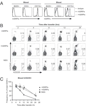
tory elements after targeting an ES cell line (F1 129/BALB/c Rag2+/−γcy/−), which can easily be genetically manipulated (23, 24). Upon obtaining several lines of hSIRPa-expressing mice, the expression levels of hSIRPa were analyzed on mouse CD45+ cells byflow cytometry. hSIRPa was faithfully expressed in BAC-transgenic mice as cells expressing mouse SIRPa expressed also hSIRPa (Fig. 1A andFig. S1A). In contrast, no hSIRPa expres-sion was detected on cells not expressing mouse SIRPa. hSIRPa-DKO mice had similar numbers of leukocytes, including neu-trophils and monocytes, in the blood and did not show any signs of thrombocytopenia and anemia, which is a prominent feature in SIRPa−/−mice (25) (Fig. S1B–D). This indicates that expres-sion of hSIRPa does not interfere with the function of mSIRPa in vivo. Clearance of erythrocytes is regulated in vivo in a CD47-SIRPa–dependent manner (14). To evaluate, if hSIRPa was functional on mouse cells, human erythrocytes that express CD47 were injected into DKO, hSIRPa-DKO, and NSG mice and their clearance was monitored. Similarly to NSG mice, significantly higher numbers of erythrocytes were present in the blood of hSIRPa-DKO mice compared with DKO mice, indicating that hSIRPa is functional in the mouse in vivo, negatively regulating phagocytosis and thus elimination of human CD47-expressing cells (Fig. 1B and C).
Multilineage Engraftment in the Blood of hSIRPa-DKO Mice.To test
whether hSIRPa expression in immunocompromised mice would lead to increased engraftment levels after human CD34+ cell transplantation, we next transplanted newborn irradiated DKO, hSIRPa-DKO, and NSG mice with human fetal liver-derived CD34+cells, a population highly enriched in HSPCs. A total of
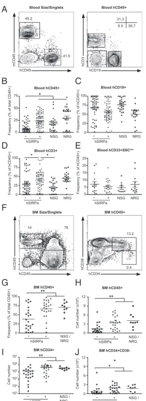
88% of engrafted hSIRPa-DKO mice had greater than 1% hu-man CD45+ cells in the peripheral blood, which is similar to NSG mice, whereas only 52% of DKO surpassed this value (Table S1). Compared with DKO mice, NSG mice had signifi-cantly higher frequencies of human CD45+cells in the blood of engrafted mice, 10–12 wk after transplantation (6.6% compared with 17.6%,P < 0.001) (Fig. 2B). Strikingly, hSIRPa-DKO had increased frequencies of human CD45+ cells compared with DKO mice (23.3% compared with 6.6%, P < 0.001) reaching levels at least similar to NSG mice (Fig. 2B). We furthermore compared DKO and hSIRPa-DKO mice to NRG mice (26). NRG mice had significantly higher engraftment levels compared with DKO (29.6% compared with 6.6%,P < 0.001), and similar high engraftment levels as NSG and hSIRPa-DKO. Hence, no significant differences were found between hSIRPa-DKO mice and NOD-based strains. As previously described, we were able to detect in all models multilineage development of human CD45+ cells in vivo (Fig. 2 C–E andFig. S2). When these mice were analyzed in detail, the overall composition of human CD45+ cells regarding B cells and myeloid cells was similar in all mouse strains (Fig. 2C–E). Notably, an increase in T cell frequency in the blood of hSIRPa-DKO mice compared with DKO and NSG mice was detected, which was not seen when hSIRPa-DKO mice were compared with NRG mice (hSIRPa, 40% compared with DKO, 29.3%; NSG, 19.6%; and NRG, 45%, respectively).
Enhanced Human Cell Engraftment in the Bone Marrow and the
Periphery of hSIRPa Mice.Next we analyzed bone marrow, spleen,
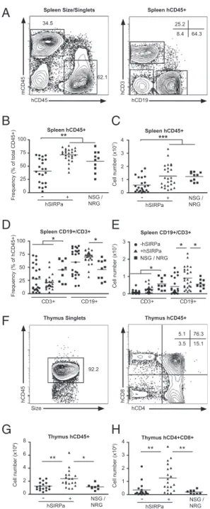
and thymus 12–14 wk posttransplantation to characterize whether similar differences could be found in these hematopoietic organs. In the bone marrow, hSIRPa-DKO and NSG and NRG mice (which were analyzed together in the subsequent experiments) contained significantly higher frequencies and total numbers of hCD45+cells than DKO mice (Fig. 2G and H). As observed in blood, the composition of hCD45+in the bone marrow was not different between the different strains with similar frequencies of CD34+HSPCs, CD19+B cells, CD33+SSClow monocytes/den-dritic cells, and CD33intCD66+SSChigh granulocytes. Neverthe-less, due to increased total numbers of hCD45+ cells, the numbers of all subsets including CD34+and CD34+CD38−cells, a population enriched for human early progenitor cells and HSCs, were significantly increased in hSIRPa-DKO mice (Fig. 2 I and J). Further analysis showed that also frequencies of human CD45+ cells in the spleen were increased in SIRP-DKO mice, reaching levels observed in NSG and NRG mice (Fig. 3A and B). Besides an increase in the frequency of CD3−NKp46+cells, which include predominantly NK cells but also lymphoid tissue inducer cells, in hSIRPa-DKO mice (2.8% vs. 1.3%,P < 0.03), overall composition was not significantly different between DKO and hSIRPa -DKO mice (Fig. 3 D and E andFig. S3). Notably, in contrast to the peripheral blood NSG/NRG mice had slightly higher CD3+ T cell numbers, but lower CD19+B cell numbers com-pared with hSIRPa-DKO mice, whereas overall hCD45+ num-bers were similar. In all groups of mice, the ratio between CD4+ and CD8+ T cells was similar to the ratio found in humans (Fig. S3). In all mouse strains, CD4+T cells consisted of naïve cells (CCR7+CD45RO−) and subsets of memory cells (CCR7+/− CD45RO+) with a significant variability between mice (Fig. S3). In the thymus, hSIRPa-DKO mice had higher numbers of total hCD45+ cells and CD4+CD8+ thymocytes compared with the DKO mice (Fig. 3 G and H). Cell numbers were also higher than we observed in NSG mice, indicating that additional strain-specific factors or even better interaction of human CD47 and human SIRPa compared with human CD47 and NOD-SIRPa might influence T cell development and maintenance in this inbred strain.
Enhanced Human Cell Maintenance in hSIRPa Mice.The duration of
human hematopoiesis after engraftment of human stem and progenitor cells is limited as demonstrated by relatively low recovery of cells that are able to efficiently engraft secondary
mSIRPa hSIRPa B220+ CD3+ CD11b+ B220+ CD3+ CD11b+ -hSIRPa +hSIRPa isotype Blood Blood 75 50 25 0 Frequency (% of total CD45+) 100 0 4 8 12 16 20 24 28
Time after transfer (h) Blood hCD235+ -hSIRPa +hSIRPa NSG -hSIRPa +hSIRPa NSG
B
C
A
2 4 8 12 24Time after transfer (hrs)
hCD235 m/hCD45 0.12 0.08 0.05 0.02 0 0.1 0.08 0.06 0.07 0.05 0.2 0.11 0.09 0.08 0.04 * * *
Fig. 1. hSIRP is faithfully expressed and is functional in BAC-transgenic mice. (A) Expression of mouse and human SIRPa in the blood was analyzed by FACS staining in hSIRPa-transgenic (gray solid line) and control (black solid line) mice. Isotype staining for hSIRP is shown as dashed line. (B and C) Human erythrocytes were transferred in Rag2−/−γc−/− (−hSIRPa), hSIRPa+ Rag2−/−γc−/−mice (+hSIRPa), and NSG mice. Their clearance was monitored by FACS staining for hGPA-expressing cells in the peripheral blood of mice. Data are a summary of two independent experiments with a total of 10 mice per group. *P< 0.01 by one-way ANOVA test.
recipients in vivo (23, 27). Accordingly, we observed a steady decrease of human cell numbers in the bone marrow. At 23–26 wk, numbers of hCD45+were around one-third of the numbers after 12–14 wk in DKO and hSIRPa-DKO mice and NSG/NRG mice (Fig. S4 A and B). Cell numbers further decreased with time and hCD45+cells could be recovered in significant numbers only from hSIRPa-DKO after 35–37 wk (Fig. S4A and B). We were unable to analyze NSG mice at this late time point, because of a high mortality that became apparent beyond 6 mo (Table S2), which has also been reported previously (28). In the spleen, we observed a similar trend with cell numbers declining in all strains of mice from 3 to 6 mo (Fig. S4C and D). Notably, in hSIRPa-DKO, numbers in the spleen did not decline much further between 6 and 9 mo, indicating that differentiated cells can persist in these mice (Fig. S4C and D). At 24 and 36 wk postengraftment, hSIRPa-DKO mice contained on average 3.3-fold and 5.1-3.3-fold more human CD45+ cells in the spleen than DKO mice, respectively. At later time points, the frequency of T cells increased, whereas the frequency of B cells decreased (Fig. S4E–H). In summary, these results indicate that hSIRPa-DKO mice lose the ability for human hematopoiesis in a similar way to DKO mice, likely due to the loss of HSPCs; however, they have a superior capacity to maintain differentiated cells in the periphery.
Increased Antigen-Specific Humoral Immune Responses in
hSIRPa-Transgenic Mice.To test whether the increase of human immune
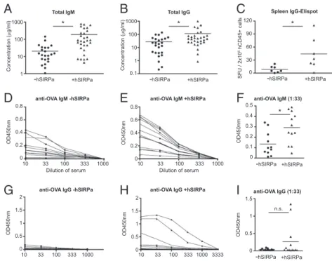
cells translated into quantitative and qualitative changes of the human adaptive immune system in vivo, wefirst analyzed total levels of human immunoglobulins in mice 12–16 wk posten-graftment. Indeed, compared with DKO mice, hSIRPa-DKO mice had increased levels of human IgM (185 ± 55 μg/mL vs. 24± 9 μg/mL, P < 0.03, mean ± SEM) and IgG (113 ± 36 μg/mL vs. 26 ± 6 μg/mL, P < 0.02) in the plasma (Fig. 4 A and B). Accordingly, a higher number of human IgG-producing cells were detected in the spleen of hSIRPa-DKO mice (Fig. 4C). This correlated with an increased frequency of CD27+ memory B cells in the spleen (Fig. S5). Next, DKO and hSIRPa-DKO mice were immunized with a protein antigen (ovalbumin, OVA) mixed with an adjuvant (complete Freund’s adjuvant) to assess the de novo development of antigen-specific immune responses. Because most studies using CD34+cell-engrafted mice reported only low levels of antigen-specific immune responses, we decided to use this potent adjuvant to provide the strongest conditions for priming of de novo immune responses (7, 8, 29). Mice were boosted 2 wk later with OVA/incomplete Freund’s adjuvant and bled 10 d after the boost. Whereas nonimmunized DKO and hSIRPa-DKO did not have significant levels of OVA anti-bodies (IgM, IgG), we detected anti-OVA IgM in 66.6% (12 of 18) of DKO mice and in 88% (21 of 24) of hSIRPa-DKO mice (Table S3). Using endpoint dilution of the sera of immunized mice, antibody titers were determined for IgM and IgG from individual mice. For anti-OVA IgM, there was no significant difference in the frequency of mice responding between the two groups, nevertheless hSIRPa-DKO mice had increased antibody titers compared with DKO mice (Fig. 4 D–F). When antigen-specific IgG were analyzed, a more striking difference was dis-cernible. In only 2 out of 18 (11%) DKO mice could antigen-specific IgG be detected, whereas IgG was detectable in 16 of 24 (66.6%) hSIRPa-DKO mice (Table S3 and Fig. 4G–I). In summary, upon immunization we observed significant difference in the levels of specific antibody responses as evidenced by the higher IgM titers in hSIRPa-DKO mice and a higher frequency of hSIRPa-DKO mice producing antigen-specific IgG. This will 75 50 25 0 Frequency (% of total CD45+) hSIRPa NSG NRG 75 50 25 0 Frequency (% of hCD45+) 100 75 50 25 0 Frequency (% of hCD45+) 100 15 10 5 0 Frequency (% of hCD45+) 20 25 NSG NRG NSG NRG NSG NRG - + hSIRPa - + hSIRPa - + hSIRPa - + Blood hCD45+ Blood hCD19+
Blood hCD3+ Blood hCD33+SSClow
75 50 25 0 Frequency (% of total CD45+) 100 NSG / NRG hSIRPa - + BM hCD45+ 9 6 3 0 Cell number (x10 6) 12 NSG / NRG hSIRPa - + BM hCD45+ 106 105 103 102 Cell number 107 NSG / NRG hSIRPa - + 104 BM hCD34+ 9 6 3 0 Cell number (x10 4) 12 NSG / NRG -hSIRPa + BM
hCD34+CD38-B
C
D
E
G
H
I
J
F
* ** ** * ** * * hCD45 mCD45 hCD19 hCD3 hCD45 mCD45 hCD34 hCD38Blood Size/Singlets Blood hCD45+
BM Size/Singlets BM hCD45+ 45.2 41.5 31.3 59.7 6.5 14 76 13.2 0.4
A
Fig. 2. hSIRPa-transgenic mice have improved hematopoiesis upon CD34+ cell engraftment. Irradiated newborn mice were transplanted with CD34+ cells and engraftment was monitored after 10 to 12 wk by FACS. (A) Rep-resentative staining pattern in the peripheral blood of a hSIRP+Rag2−/−γc−/− mouse. (B–E) Frequencies of hCD45+, hCD3+, CD19+, and CD33+SSClowcells in the blood were compared after engraftment between different immuno-deficient strains. Data are a summary of at least four experiments with a total of 60 DKO mice, 63 hSIRPa-DKO mice, 26 NSG mice, and 29 NRG mice. (F) Representative staining pattern in the bone marrow of a hSIRPa+Rag2−/−γc−/ −mouse. (G and H) Frequency and number of human CD45+cells was com-pared after 12–14 wk between different immunodeficient strains. (I and J) Numbers of human CD34+progenitor cells and CD34+CD38−cells were cal-culated on the basis of total cell number and frequencies determined by FACS. Data are a summary of three experiments with a total of 22 DKO mice,
24 hSIRPa-DKO mice, and 9 NSG/NRG mice. Data were analyzed by one-way ANOVA test and individual P values for posttest are displayed. *P< 0.05, **P< 0.01.
IMM
have important implications for the further development of this platform for human vaccine development.
Discussion
Severely immunocompromised mice lacking T cells, B cells, and NK cells have become widely used hosts for the xenotrans-plantation of human cells due to their diminished rejection of
cells and tissues of human origin (5, 7–9). However, it has been noted that there are additional strain-specific factors that influence engraftment efficiencies as demonstrated by the incapability of C57Bl6 Rag2−/−γc−/−, in contrast to NOD/Rag1−/−γc−/−mice, to support engraftment of human cells. The importance of murine macrophages in xenorejection had been noted more than 10 y ago, but the mechanisms of xenorecognition were only described re-cently (11, 17, 18). It has been established that binding of CD47 on target cells to SIRPa on macrophages sends a “Don’t eat me” signal to the phagocyte, i.e., murine CD47−/− cells are rapidly cleared from WT mice (14). In the context of xenotransplantation, the advantage of NOD/scid mice as hosts for human cells com-pared with CB17/scid or C57Bl6/Rag mice was subsequently suggested to require a specific variant of the polymorphic in-hibitory receptor SIRPa (11). A number of polymorphisms in the extracellular domain of SIRPa enabled SIRPa (NOD) to bind to human CD47, whereas SIRPa (C57Bl6) was unable to bind hu-man CD47 (11). In vitro assays were further used to characterize the direct effect of SIRPa on human hematopoiesis, but it remained formally unconfirmed whether SIRPa is sufficient for the enhanced engraftment in NOD-based strains. Notably, the NOD strain is characterized by a number of well-documented alterations in immune functions such as complement deficiency and impaired dendritic cell maturation (30). We demonstrate in this study that transgenic, faithful expression of human SIRPa in mice is indeed sufficient to strongly decrease rejection of hu-man cells in Rag2−/−γc−/−on a mixed 129/BALB/c background, resulting in increased human cell numbers and an increased functionality of the human adaptive immune system in vivo.
In our initial proof-of-concept experiments to evaluate whether hSIRPa is functional in transgenic mice, human erythrocytes were transferred into mice. This approach was chosen because negative regulation of erythrophagocytosis is highly dependent on the interaction of CD47 and SIRP (14). Human erythrocytes were cleared within hours in DKO mice and the decreased clearance of erythrocytes in hSIRPa-DKO mice compared with DKO mice indicates that hSIRPa is able to negatively regulate phagocytosis by murine macrophages and that human erythrocyte clearance is indeed modulated via CD47–SIRPa interaction. However, not only phagocytosis of erythrocytes is regulated by this interaction, as also murine CD47−/− leukocytes are rapidly cleared upon transfer into WT mice, leading to a failure of CD47−/−cells to repopulate lethally irradiated mice (21). Moreover, in wild-type mice, circulating murine HSCs up-regulate CD47 to avoid phago-cytosis in the spleen, demonstrating a requirement for HSPC survival (15). In line with thesefindings we demonstrated that expression of hSIRPa in 129/BALB/c Rag2−/−γc−/− mice en-hanced the efficiency of engraftment of human hematopoietic stem and progenitor cells at two levels. First, the frequency of mice with detectable human cell engraftment in the peripheral blood was almost doubled, and second, frequencies of human cell engraftment were significantly increased. In comparison with NSG mice, hSIRPa-DKO mice were equally well engrafted, but we observed a slightly increased early mortality (<12 wk) of engrafted NSG mice, which can likely be attributed to increased gamma-irradiation sensitivity of scid strains compared with Rag1/ Rag2-deficient strains (Table S1). As a consequence, fewer en-grafted mice can be used for experiments (DKO, 40%; hSIRPa-DKO, 70%; and NSG, 53%) (Table S1). Although no formal survival analysis was performed, we noted that the difference in survival became larger at later time points (Table S2). In line with a previous report, we found no NSG mice alive after 9 mo, impairing the value of this model for long-term studies (28). Our analysis of hematopoietic organs in the different strains of mice demonstrate increased numbers of human HSPCs in the bone marrow of hSIRPa-DKO mice compared with DKO mice. Striking differences were also visible in the blood and thymus and spleen with two- to threefold increased cell numbers after 3 mo in SIRP-DKO mice compared with DKO mice. Interestingly, the overall composition of the hematopoietic system in the spleen was similar in DKO, hSIRPa-DKO, and NOD-based mice, indicating 75 50 25 Frequency (% of total CD45+) 100 Spleen hCD45+ 0 3 2 1 Cell number (x10 7) 4 Spleen hCD45+ 0 NSG / NRG hSIRPa - + NSG / NRG hSIRPa - + 75 50 25 Frequency (% of hCD45+) 100 Spleen CD19+/CD3+ 0 3 2 1 Cell number (x10 7) Spleen CD19+/CD3+ 0 CD3+ CD19+ CD3+ CD19+ -hSIRPa +hSIRPa NSG / NRG Thymus hCD45+ Thymus hCD4+CD8+ 3 2 1 Cell number (x10 6) 4 0 NSG / NRG hSIRPa - + 6 4 2 Cell number (x10 6) 8 0 NSG / NRG hSIRPa - +
B
C
D
E
G
H
** ** * ** * * * * ** *** hCD45 mCD45 hCD19 hCD3A
Spleen Size/Singlets Spleen hCD45+Size
hCD45
hCD4
hCD8
Thymus Singlets Thymus hCD45+
F
34.5 62.1 25.2 64.3 8.4 92.2 5.1 15.1 3.5 76.3 *Fig. 3. Elevated numbers of human cells in the periphery of hSIRPa-transgenic mice. (A) Representative staining pattern in the spleen of a hSIRPa+Rag2−/−γc−/−mouse. (B and C) Frequencies and numbers of total human CD45+cells in the spleen were determined after 12–14 wk. (D and E) At the same time, frequencies and numbers of CD3+T cells and CD19+B cells in the spleen were determined. (F) Representative staining pattern in the thymus of a hSIRPa+Rag2−/−γc−/−mouse. (G) Enumeration of the number of human thymocytes and of (H) CD4+CD8+thymocytes after 12–14 wk by combination of FACS staining and total cell count. Data are a summary of three experiments with a total of 22 DKO mice, 24 hSIRPa-DKO mice, and 9 NSG/NRG mice. Data were analyzed by one-way ANOVA test and individual P values for posttest are displayed. *P< 0.05, **P < 0.01.
that hSIRPa expression affects the efficiency of initially trans-ferred stem and progenitor cells to seed the bone marrow and subsequently differentiate into various lineages of cells. However, some significant differences were observed, which include in-creased frequencies of CD3−NKp46+ cells in the spleen and significantly increased numbers of CD4+CD8+ double-positive thymocytes. The latter might be a direct result of decreased phagocytic activity in this organ, which contains numerous phago-cytes normally responsible for removing negatively selected thy-mocytes. Alternatively, this might also be a consequence of in-creased CD47 signaling in developing T cells as ligation of CD47 sends costimulatory signals (31–33). Another notable difference was observed when mice were analyzed for the presence of pla-telets and erythrocytes. Whereas hSIRPa-DKO mice had an in-creased number of human platelets compared with DKO mice, they did not reach levels observed in NSG mice. Similarly, fre-quencies of erythrocytes were significantly higher in NSG mice compared with DKO and hSIRPa-DKO mice. This might be the result of additional strain-specific mutations beyond SIRPa that either favor development or persistence of these cell lineages in vivo (30). Longitudinal analysis of engraftment in DKO and hSIRPa-DKO mice revealed that, whereas DKO mostly lost hu-man cells after 9 mo, they were still routinely detectable in hSIRPa-DKO mice. This could be mediated either by prolonged hematopoiesis in the bone marrow or enhanced survival of dif-ferentiated cells in the peripheral organs of hSRIPa-transgenic mice. Importantly, the analysis of older mice (∼9 mo postengraft-ment) revealed one of the shortcomings of current mouse models as human hematopoietic stem and progenitor cells were almost com-pletely lost. Hence, we predict that combinations of hSIRPa with additional human knockins may overcome this limitation.
Recently, several approaches have been used to improve hu-man cell engraftment and the unbalanced lineage differentiation in CD34+cell engrafted mice. These include transient approaches such as hydrodynamic injection of plasmid DNA (34), injections of cytokines, and infections of mice or CD34+cells with lentivi-ruses (35–37). Alternatively, transgenic expression of human MHC molecules has been demonstrated to improve the de-velopment of antigen-specific immune responses in vivo (38–40). Nonetheless, overexpression of cytokines might also have detri-mental side effects due to the unphysiological expression such as in mice transgenic for SCF, GM-CSF, and IL-3 (41). An alter-native approach to provide human growth factors in vivo is to
genetically engineer mice and replace the mouse genes with their human counterparts resulting in their expression in the appro-priate niche at physiological levels. Indeed, faithful replacement of mouse GM-CSF and IL-3 as well as thrombopoietin (TPO) by our group has resulted in improved development of human macrophages in the lung and HSC and HPC maintenance in the bone marrow, respectively (23, 24). Notably, in human TPO knockin mice, despite a highly increased engraftment level of stem and progenitor cells in the bone marrow, no changes were ob-served in the periphery, demonstrating the existence of limiting factors in the periphery such as destruction by phagocytes. With the hSIRPa-DKO mice, we have generated a strain that combines superior engraftment level and the possibility of long-term genetic manipulations to further enhance the murine host.
A highly desired application of mice with functional human immune systems is the development and testing of human vac-cines. However, the induction of immune responses in vivo has been relative inefficient so far (5, 7–9, 29). Several studies have reported pathogen-specific immune responses upon infection. Although it was reported that around 50% of mice produced virus-specific IgM and IgG upon dengue virus infection (42), other studies reported frequencies below 20% of mice producing antigen-specific IgM and IgG after HIV and EBV infection (29, 43). Upon immunization with adjuvant and antigen, class switching of antigen-specific immunoglobulins is similarly in-efficient as only a fraction of immunized animals show antigen-specific IgG responses (5, 7–9, 44, 45). These studies included NSG and BALB/c DKO mice and different adjuvant/antigen combinations. At this point it remains open why antibody pro-duction is limited, but because B cells from humanized mice respond normally in vitro, it indicates that the human immune system provides only inefficient help in vivo (44). In hSIRPa-DKO mice, immunization with a T cell-dependent antigen in-duced stronger immune responses as measured by higher titers of antigen-specific IgM compared with DKO mice. Furthermore, more hSIRPa+mice produced antigen-specific IgG. To provide help for B cells, antigen-specific T cells need to recognize anti-gens presented in the context of MHC molecules. Hence, the increased functionality in SIRP-DKO mice is likely the result of improved selection and differentiation of T cells in vivo due to overall higher numbers of human immune cells. Similarly, HLA-DR4 transgenic mice and humanized mice that are generated by cotransplantation of CD34+cells and human fetal thymus pieces 100 10 1 Concentration ( g/ml) 1000 Total IgG 0.1 100 10 Concentration ( g/ml) 1000 1 Total IgM
anti-OVA IgM +hSIRPa
0.6 0.4
OD450nm
0.8
0
anti-OVA IgM -hSIRPa
0.2 0.6 0.4 OD450nm 0.8 0 0.2
anti-OVA IgG +hSIRPa
1.5 1
OD450nm
2
0
anti-OVA IgG -hSIRPa
0.5 OD450nm 1.5 1 2 0 0.5 anti-OVA IgG (1:33) anti-OVA IgM (1:33) 0.3 0.2 OD450nm 0.5 0 0.1 1 0.5 OD450nm 1.5 0 0.4
-hSIRPa +hSIRPa -hSIRPa +hSIRPa
-hSIRPa +hSIRPa -hSIRPa +hSIRPa
A
B
D
E
F
G
H
I
SFU / 2x10 5 hCD45+ cells Spleen IgG-Elispot -hSIRPa +hSIRPa 90 60 30 120 0 10 33 100 333 1000 10 33 100 333 1000 10 33 100 333 1000 10 33 100 333 1000 3333Dilution of serum Dilution of serum
*
n.s.
* * *
C
Fig. 4. Improved humoral antigen-specific immune responses in hSIRPa-transgenic mice. Characterization of humoral immune responses before (A–C) and after (D–I) immunization. (A and B) Total serum levels of human IgM (A) and IgG (B) were determined by ELISA in Rag2−/−γc−/−(−hSIRPa, n = 28) and hSIRPa-transgenic Rag2−/−γc−/−(+hSIRPa, n = 30) mice. (C) The frequencies of human IgG-producing cells in the spleen were measured using ELISPOT without immunization. (D–I) Mice were immunized with OVA/CFA and boosted 14 d later with OVA/IFA. (D–G) Anti-OVA IgM (D and E) and IgG (G and H) were assayed by ELISA and OD450nm readings are displayed for serial dilution of serum from individual DKO (D and F) or hSIRPa-DKO (G and H) mice. (F and I) OD450nm readings for a serum dilution of 1:33 are shown; each dot represents a mouse from one experiment. Data were analyzed using Mann-Whitney test, *P< 0.05.
IMM
have improved HLA-restricted T cell responses and also improved antigen-specific antibody responses (40, 46). However, further studies will be needed to characterize and quantify antigen-specific T cell responses in hSIRPa-DKO mice.
In summary, we achieved improved frequencies of engrafted mice and increased levels of engraftment of human cells by transgenic expression of hSIRPa in 129/BALB/c Rag2−/−γc−/− mice, resulting in an improved functionality of the human im-mune system in vivo. Supporting our finding of the central function of CD47-SIRPa is a study by Legrand et al. (47). Using lentiviral transduction of human HSPCs with mouse CD47 and breeding of NOD-SIRPa to BALB/c Rag2−/−γc−/− mice, they demonstrate similar quantitative and qualitative improvements of the human immune system in vivo. Genetic engineering in our strain can be used to rapidly generate mice expressing genes of interest and analyze their influence on engraftment of human tissues and cells. On the basis of our successful completion of diverse genetic modifications such as the replacement of
com-plete mouse genes with their human counterparts and expres-sion of human genes using BAC transgenes, we believe that this approach enables targeted modifications to further improve the murine host for transplantation of human tissues and cells. Materials and Methods
Generation of Human SIRPa-Transgenic Mice. hSIRPa-transgenic mice were generated by transgenesis using a BAC containing the entire 45-kb SIRPa gene along with∼51 kb of flanking DNA on the 5′ end and 78 kb on the 3′ end that was manipulated to contain a hygromycin resistance cassette. F1 129/Balbc Rag2+/−γcy/− ES cells were electroporated and selected using hygromycin. Positive colonies were screened by PCR to map whether the full-length BAC had been integrated. Further details can be found inSI Materials and Methods.
ACKNOWLEDGMENTS. We thank J. Alderman, R. Weber, A. Franco, P. Ramney, and A. Hafemann for technical help, and F. Manzo for help with submission of the manuscript.
1. Mestas J, Hughes CC (2004) Of mice and not men: Differences between mouse and human immunology. J Immunol 172:2731–2738.
2. Legrand N, et al. (2009) Humanized mice for modeling human infectious disease: Challenges, progress, and outlook. Cell Host Microbe 6:5–9.
3. Shultz LD, Ishikawa F, Greiner DL (2007) Humanized mice in translational biomedical research. Nat Rev Immunol 7:118–130.
4. Manz MG (2007) Human-hemato-lymphoid-system mice: Opportunities and chal-lenges. Immunity 26:537–541.
5. Traggiai E, et al. (2004) Development of a human adaptive immune system in cord blood cell-transplanted mice. Science 304:104–107.
6. Gimeno R, et al. (2004) Monitoring the effect of gene silencing by RNA interference in human CD34+ cells injected into newborn RAG2-/- gammac-/- mice: Functional in-activation of p53 in developing T cells. Blood 104:3886–3893.
7. Ito M, et al. (2002) NOD/SCID/gamma(c)(null) mouse: An excellent recipient mouse model for engraftment of human cells. Blood 100:3175–3182.
8. Ishikawa F, et al. (2005) Development of functional human blood and immune sys-tems in NOD/SCID/IL2 receptor gamma chain(null) mice. Blood 106:1565–1573. 9. Shultz LD, et al. (2005) Human lymphoid and myeloid cell development in
NOD/LtSz-scid IL2R gamma null mice engrafted with mobilized human hemopoietic stem cells. J Immunol 174:6477–6489.
10. Brehm MA, et al. (2010) Parameters for establishing humanized mouse models to study human immunity: Analysis of human hematopoietic stem cell engraftment in three immunodeficient strains of mice bearing the IL2rgamma(null) mutation. Clin Immunol 135:84–98.
11. Takenaka K, et al. (2007) Polymorphism in Sirpa modulates engraftment of human hematopoietic stem cells. Nat Immunol 8:1313–1323.
12. Matozaki T, Murata Y, Okazawa H, Ohnishi H (2009) Functions and molecular mechanisms of the CD47-SIRPalpha signalling pathway. Trends Cell Biol 19:72–80. 13. Okazawa H, et al. (2005) Negative regulation of phagocytosis in macrophages by the
CD47-SHPS-1 system. J Immunol 174:2004–2011.
14. Oldenborg PA, et al. (2000) Role of CD47 as a marker of self on red blood cells. Science 288:2051–2054.
15. Jaiswal S, et al. (2009) CD47 is upregulated on circulating hematopoietic stem cells and leukemia cells to avoid phagocytosis. Cell 138:271–285.
16. Motegi S, et al. (2003) Role of the CD47-SHPS-1 system in regulation of cell migration. EMBO J 22:2634–2644.
17. Rozemuller H, et al. (2004) Enhanced engraftment of human cells in RAG2/gammac double-knockout mice after treatment with CL2MDP liposomes. Exp Hematol 32: 1118–1125.
18. Terpstra W, et al. (1997) Facilitated engraftment of human hematopoietic cells in severe combined immunodeficient mice following a single injection of Cl2MDP lip-osomes. Leukemia 11:1049–1054.
19. Andres A, et al. (2005) Macrophage depletion prolongs discordant but not concor-dant islet xenograft survival. Transplantation 79:543–549.
20. Takizawa H, Manz MG (2007) Macrophage tolerance: CD47-SIRP-alpha-mediated signals matter. Nat Immunol 8:1287–1289.
21. Blazar BR, et al. (2001) CD47 (integrin-associated protein) engagement of dendritic cell and macrophage counterreceptors is required to prevent the clearance of donor lymphohematopoietic cells. J Exp Med 194:541–549.
22. Wang H, et al. (2007) Attenuation of phagocytosis of xenogeneic cells by manipu-lating CD47. Blood 109:836–842.
23. Rongvaux A, et al. (2011) Human thrombopoietin knockin mice efficiently support human hematopoiesis in vivo. Proc Natl Acad Sci USA 94:5320–5325.
24. Willinger T, et al. (2011) Human IL-3/GM-CSF knock-in mice support human alveolar macrophage development and human immune responses in the lung. Proc Natl Acad Sci USA 108:2390–2395.
25. Yamao T, et al. (2002) Negative regulation of platelet clearance and of the macro-phage phagocytic response by the transmembrane glycoprotein SHPS-1. J Biol Chem 277:39833–39839.
26. Pearson T, et al. (2008) Non-obese diabetic-recombination activating gene-1 (NOD-Rag1 null) interleukin (IL)-2 receptor common gamma chain (IL2r gamma null) null
mice: A radioresistant model for human lymphohaematopoietic engraftment. Clin Exp Immunol 154:270–284.
27. Hogan CJ, et al. (1997) Engraftment and development of human CD34(+)-enriched cells from umbilical cord blood in NOD/LtSz-scid/scid mice. Blood 90:85–96. 28. Watanabe S, et al. (2007) Humanized NOD/SCID/IL2Rgamma(null) mice transplanted
with hematopoietic stem cells under nonmyeloablative conditions show prolonged life spans and allow detailed analysis of human immunodeficiency virus type 1 pathogenesis. J Virol 81:13259–13264.
29. Baenziger S, et al. (2006) Disseminated and sustained HIV infection in CD34+ cord blood cell-transplanted Rag2-/-gamma c-/- mice. Proc Natl Acad Sci USA 103:15951– 15956.
30. Shultz LD, et al. (1995) Multiple defects in innate and adaptive immunologic function in NOD/LtSz-scid mice. J Immunol 154:180–191.
31. Reinhold MI, Lindberg FP, Kersh GJ, Allen PM, Brown EJ (1997) Costimulation of T cell activation by integrin-associated protein (CD47) is an adhesion-dependent, CD28-independent signaling pathway. J Exp Med 185:1–11.
32. Ticchioni M, et al. (1997) Integrin-associated protein (CD47) is a comitogenic molecule on CD3-activated human T cells. J Immunol 158:677–684.
33. Latour S, et al. (2001) Bidirectional negative regulation of human T and dendritic cells by CD47 and its cognate receptor signal-regulator protein-alpha: Down-regulation of IL-12 responsiveness and inhibition of dendritic cell activation. J Immunol 167:2547– 2554.
34. Chen Q, Khoury M, Chen J (2009) Expression of human cytokines dramatically im-proves reconstitution of specific human-blood lineage cells in humanized mice. Proc Natl Acad Sci USA 106:21783–21788.
35. O’Connell RM, et al. (2010) Lentiviral vector delivery of human interleukin-7 (hIL-7) to human immune system (HIS) mice expands T lymphocyte populations. PLoS ONE 5: e12009.
36. Huntington ND, et al. (2009) IL-15 trans-presentation promotes human NK cell de-velopment and differentiation in vivo. J Exp Med 206:25–34.
37. van Lent AU, et al. (2009) IL-7 enhances thymic human T cell development in“human immune system” Rag2-/-IL-2Rgammac-/- mice without affecting peripheral T cell ho-meostasis. J Immunol 183:7645–7655.
38. Jaiswal S, et al. (2009) Dengue virus infection and virus-specific HLA-A2 restricted immune responses in humanized NOD-scid IL2rgammanull mice. PLoS ONE 4:e7251. 39. Strowig T, et al. (2009) Priming of protective T cell responses against virus-induced
tumors in mice with human immune system components. J Exp Med 206:1423–1434. 40. Danner R, et al. (2011) Expression of HLA class II molecules in humanized NOD. Rag1KO.IL2RgcKO mice is critical for development and function of human T and B cells. PLoS ONE 6:e19826.
41. Nicolini FE, Cashman JD, Hogge DE, Humphries RK, Eaves CJ (2004) NOD/SCID mice engineered to express human IL-3, GM-CSF and Steel factor constitutively mobilize engrafted human progenitors and compromise human stem cell regeneration. Leu-kemia 18:341–347.
42. Kuruvilla JG, Troyer RM, Devi S, Akkina R (2007) Dengue virus infection and im-mune response in humanized RAG2(-/-)gamma(c)(-/-) (RAG-hu) mice. Virology 369: 143–152.
43. Yajima M, et al. (2008) A new humanized mouse model of Epstein-Barr virus infection that reproduces persistent infection, lymphoproliferative disorder, and cell-mediated and humoral immune responses. J Infect Dis 198:673–682.
44. Watanabe Y, et al. (2009) The analysis of the functions of human B and T cells in humanized NOD/shi-scid/gammac(null) (NOG) mice (hu-HSC NOG mice). Int Immunol 21:843–858.
45. Becker PD, et al. (2010) Generation of human antigen-specific monoclonal IgM an-tibodies using vaccinated“human immune system” mice. PLoS ONE 5.
46. Brainard DM, et al. (2009) Induction of robust cellular and humoral virus-specific adaptive immune responses in human immunodeficiency virus-infected humanized BLT mice. J Virol 83:7305–7321.
47. Legrand N, et al. (2011) Functional CD47/SIRPα interaction is required for optimal human T and NK cell homeostasis in vivo. Proc Natl Acad Sci USA, 10.1073/ pnas.1101398108.