Case Rep. Perinat. Med. 2016; 5(2): 105–108
*Corresponding author: Ioannis Dedes, MD, University Hospital of Zurich, Department of Gynecology and Obstetrics, Zurich, Switzerland, E-mail: ioannis.dedes@usz.ch
Franziska Krähenmann, Gian-Piero Ghisu, Esther Birindelli, Roland Zimmermann and Nicole Ochsenbein: University Hospital of Zurich, Department of Gynecology and Obstetrics, Zurich, Switzerland
Ioannis Dedes*, Franziska Krähenmann, Gian-Piero Ghisu, Esther Birindelli, Roland
Zimmermann and Nicole Ochsenbein
Negative pressure wound treatment for uterine
incision necrosis following a cesarean section
DOI 10.1515/crpm-2015-0100Received December 7, 2015. Accepted May 4, 2016. Previously published online June 8, 2016.
Abstract: Extended-spectrum b-lactamase sepsis with
concurrent uterine incision dehiscence after cesarean sec-tion with T-shaped incision for chorioamnionitis occurred in a 29-year-old patient after her first delivery. Following an exploratory laparotomy with a diagnosis of necrosis of the anterior uterine wall, a uterine negative-pressure wound treatment (NPWT) was performed, successfully avoiding hysterectomy and enabling secondary suture of the uterine wall.
Keywords: Infectious post-cesarean uterine wall
dehis-cence; negative-pressure wound therapy; uterine inci-sional dehiscence; uterine inciinci-sional necrosis.
Introduction
It is estimated that infectious uterine incisional dehis-cence, a rare and severe complication, occurs in 1:700– 1:2400 cesarean section cases [1]. This condition is potentially lethal, due to either sepsis or uncontrolled secondary bleeding, and hysterectomy is considered the standard treatment [2].
Herein, we report a case of septic uterine incisional dehiscence due to abscess formation in a 29-year-old patient after her first delivery. Following an exploratory laparotomy, a uterine negative-pressure wound treatment (NPWT) was performed, successfully avoiding hysterec-tomy and enabling secondary suture of the uterine wall. This is the first report in the literature of such a treatment being used on a uterus.
Case
A 29-year-old primipara patient underwent a cesarean section at 39.4 weeks of gestation due to arrest of descent in the first stage of labor and chorioamnionitis [38.9°C fever, elevated leucocytes (Lc) 14.1 G/L, C-reactive protein (CRP) 32 mg/L] after induction of labor in the context of intrahepatic cholestasis of pregnancy. A cesarean section was performed through a Pfannenstiel skin incision and a low transverse uterine incision, which had to be extended to a T-shaped incision due to difficult delivery of the impacted fetal head. Ceftriaxone 2 g i.v. was given during surgery.
A female neonate weighing 2800 g, with a 5-min Apgar score of 8 and an umbilical artery pH of 7.30, was delivered. The uterine incision was closed using Polysorb®
sutures (Covidien). Continuous closure of the perito-neum parietale and the fascia were performed with a running Polysorb® 2-0 suture. The subcutaneous tissue
was adapted with interrupted Polysorb® 2-0 sutures, and
staples were used to close the skin. The estimated blood loss was 600 mL.
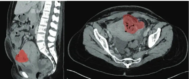
Approximately 48 h postpartum, the patient’s body temperature rose to 41.7°C despite administration of par-acetamol (4 g i.v./day), with elevated infection param-eters (Lc 14.8 G/L; CRP 194 mg/L) despite continuous administration of ceftriaxone (2 g i.v.). Blood cultures revealed Escherichia coli extended spectrum beta lacta-mase (ESBL). Ceftriaxone was stopped and ertapenem (1 g i.v.) was administered on day 3 after surgery. Despite a good clinical condition, intermittent fever spikes and high infection parameters (Lc 16 G/L, CRP 265 mg/L) per-sisted. An abdominal ultrasound examination and com-puted tomography (CT) scan (Figure 1) on day 10 after surgery revealed a collection of fluid at the uterine inci-sion, without other foci of infection. An exploratory re-laparotomy confirmed an abscess at the uterotomy site, with evacuation of 200 mL pus. Meticulous debridement with irrigation of the uterine wall was performed, and two intra-abdominal drains were installed. Swabs taken intraoperatively confirmed E. coli ESBL, and the antibiotic regime with ertapenem was continued.
Brought to you by | Universitaetsbibliothek Basel Authenticated Download Date | 4/29/19 3:36 PM
106 Dedes et al., Negative pressure wound treatment for uterine incision necrosis following cesarean section
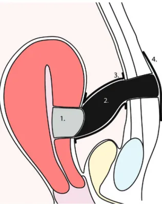
Seven days after the re-laparotomy, a hypoechogenic collection was observed at the uterine wall by follow-up ultrasound and CT scan, highly suggestive of an abscess reformation. A third laparotomy was performed (Figure 2), which showed a partial dehiscence over 3 cm with necro-sis at the uterine incision site. The fascia was unimpaired. An evacuation of the abscess was performed, with careful debridement of the necrotic parts and irrigation. An NPWT was installed at the site of the dehiscence in order to pre-serve the uterus. Kerlix® antimicrobial large roll gauze was
applied to the uterine wound with open-cell foam dressings on top, surrounded by two-layer drainage foil (Suprasorb®
CNP; Lohmann & Rauscher) to protect the intestine and
leading through the abdominal wall throughout the skin, sealing off the dressing. A continuous negative pressure of 40–50 mm Hg was applied. Microbiological samples taken intraoperatively revealed Ureaplasma, Candida albicans, and Bacteroides fragilis, and the antimicrobial regimen was extended to fluconazole 150 mg, azithromycin 1, and metronidazole 2 g/day. The dressings were changed every 2–3 days over a period of nearly 3 weeks, and a secondary closure of the uterotomy was performed with interrupted Vicryl® (Ethicon) 0 sutures on day 35 after cesarean section.
On day 39, the patient redeveloped fever (up to 39.2°C) due to a urinary tract infection with Pseudomonas
aeruginosa, treated with ciprofloxacin 1.5 g/day. Using
Figure 2: Intraoperative situs showing necrosis of the uterine wall: before (left) and after debridement (right).
Figure 1: Contrast-enhanced CT obtained on post-cesarean day 10 demonstrates fluid collection with gas formation in the anterior myome-trium (red), suggesting uterine incisional necrosis and abscess-formation: (Left) transvers sectional plane. (Right) sagittal sectional plane.
Brought to you by | Universitaetsbibliothek Basel Authenticated Download Date | 4/29/19 3:36 PM
Dedes et al., Negative pressure wound treatment for uterine incision necrosis following cesarean section 107
magnetic resonance imaging (MRI), any further surgical site infections or de novo uterine dehiscence were ruled out (Figure 3). The patient was discharged after 50 days. Six weeks later, an ultrasound revealed normal findings
Visualization of the applied NPWT: (1) Kerlix® antimicrobial large
roll gauze (2) open-cell foam dressing (3) two-layer drainage foil (Suprasorb® CNP; Lohmann & Rauscher) (4) adhesive foil sealing off
the dressing.
Figure 3: MRI of pelvis after secondary suture of the uterine incisional dehiscence shows postoperative fluid collection anterior to the uter-otomy. (Left) sagittal sectional plane. (Right) transvers sectional plane.
Figure 4: Transvaginal Ultrasound scan of the uterus in sagittal plane 4 weeks after discharge, showing unimpaired anterior uterine wall and endometrial layer of 4.8 mm.
(Figure 4). An MR scan of the pelvis 18 months after cesar-ean section revealed a physiological scar on the uterus.
Discussion
This is the first report of NPWT applied to the uterus. NPWT is used in surgery to treat a variety of wounds, ranging from superficial ulcers or meshed grafts to deep infected and dehisced wounds, as well as open abdomen treatment [3]. The basic effects of NPWT on wounds is the evacuation of localized wound edema, reduced bacterial
Brought to you by | Universitaetsbibliothek Basel Authenticated Download Date | 4/29/19 3:36 PM
108 Dedes et al., Negative pressure wound treatment for uterine incision necrosis following cesarean section
colonization, increased local blood flow, better oxygena-tion, and stimulation of granulation tissue growth [4].
In suspected metritis, improvement under antibi-otic treatment usually follows within 72 h in nearly 90% of the cases [2]. Persistence of fever after this interval mandates a search for complications of metritis, such as phlegmon or abscess formation. These complica-tions, over the long run, may lead to necrosis and sepa-ration due to extensive cellulitis of the uterine incision. The usual management of uterine incisional necrosis is hysterectomy [2, 5]; uterus-preserving surgery has been reported in only a few cases. In one case, only perito-neal cleansing and antibiotic lavage were performed [6]. In two other cases, re-suture of the uterine inci-sional necrosis was reported [1, 7].
The addition of complementary therapy with NPWT to the traditional treatment with surgery and antimicro-bial regimens might decrease the rate of hysterectomy in infectious post-cesarean uterine incision dehiscence. An MRI examination one and a half years later revealed a physiological scar on the uterus of the patient in this case report, but it is unclear whether a regular uterine cavity was achieved with this treatment.
References
[1] Rivlin ME, Hunt JA, Rubin A. Continual postoperative antibiotic peritoneal lavage in diffuse peritonitis complicating cesarean section. J Reprod Med. 1984;29:173–8.
[2] Cunningham FG GN, Lev- eno KJ, Gilstrap LC III, Hauth JC,
Wenstrom KD, et al. Puerperal infections – clinical course. Williams Obstetrics. 23rd. New York, NY: The McGraw-Hill Group; 2010. p. 717. [3] Bruhin A, Ferreira F, Chariker M, Smith J, Runkel N. Systematic
review and evidence based recommendations for the use of negative pressure wound therapy in the open abdomen. Int J Surg. 2014;12:1105–14.
[4] Desai KK, Hahn E, Pulikkottil B, Lee E. Negative pressure wound therapy: an algorithm. Clin Plast Surg. 2012;39:311–24. [5] Sengupta Dhar R, Misra R. Postpartum uterine wound
dehis-cence leading to secondary PPH: unusual sequelae. Case Rep Obstet Gynecol. 2012;2012:154685.
[6] Rivlin ME, Carroll CS, Morrison JC. Conservative surgery for uter-ine incisional necrosis complicating cesarean delivery. Obstet Gynecol. 2004;103(5 Pt 2):1105–8.
[7] Treszezamsky AD, Feldman D, Sarabanchong VO. Concurrent post-partum uterine and abdominal wall dehiscence and Streptococcus anginosus infection. Obstet Gynecol. 2011;118(2 Pt 2):449–51. The authors stated that there are no conflicts of interest regarding the publication of this article.
Brought to you by | Universitaetsbibliothek Basel Authenticated Download Date | 4/29/19 3:36 PM