Biol. Chem. 2016; 397(9): 857–869
*Corresponding author: Christoph Becker-Pauly, Institute of Biochemistry, University of Kiel, Otto-Hahn-Platz 9, D-24118 Kiel, Germany, e-mail: cbeckerpauly@biochem.uni-kiel.de
Frederike Schmidt, Miryam Müller, Johannes Prox and Dirk Schmidt-Arras: Institute of Biochemistry, University of Kiel, Otto-Hahn-Platz 9, D-24118 Kiel, Germany
Philipp Arnold: Anatomical Institute, University of Kiel, D-24118 Kiel, Germany
Caroline Schönherr and Claus Pietrzik: Institute of
Pathobiochemistry, University Medical Centre of the Johannes Gutenberg University of Mainz, D-55128 Mainz, Germany Claudia Tredup: Pharmazentrum Frankfurt, Institut für Allgemeine Pharmakologie und Toxikologie, Klinikum der Goethe-Universität Frankfurt am Main, D-60590 Frankfurt am Main, Germany
Petra Minder: Department of Molecular and Experimental Medicine, The Scripps Research Institute, La Jolla, CA 92037, USA
Henriette Ebsen and Ottmar Janssen: Institute of Immunology, University of Kiel, University Hospital Schleswig-Holstein Campus Kiel, D-24105 Kiel, Germany
Wim Annaert: Center for Human Genetics, KU Leuven and VIB Center for the Biology of Disease, B-3000 Leuven, Belgium
Erwin E. Sterchi: Institute of Biochemistry and Molecular Medicine, University of Bern, CH-3012 Bern, Switzerland
Frederike Schmidt, Miryam Müller, Johannes Prox, Philipp Arnold, Caroline Schönherr,
Claudia Tredup, Petra Minder, Henriette Ebsen, Ottmar Janssen, Wim Annaert, Claus Pietrzik,
Dirk Schmidt-Arras, Erwin E. Sterchi and Christoph Becker-Pauly*
Tetraspanin 8 is an interactor of the metalloprotease
meprin β within tetraspanin-enriched microdomains
DOI 10.1515/hsz-2016-0126Received January 29, 2016; accepted May 4, 2016; previously published online May 13, 2016
Abstract: Meprin β is a dimeric type I transmembrane
protein and acts as an ectodomain sheddase at the cell
surface. It has been shown that meprin β cleaves the
amy-loid precursor protein (APP), thereby releasing neurotoxic
amyloid β peptides and implicating a role of meprin β in
Alzheimer’s disease. In order to identify non-proteolytic
regulators of meprin β, we performed a split ubiquitin
yeast two-hybrid screen using a small intestinal cDNA
library. In this screen we identified tetraspanin 8 (TSPAN8)
as interaction partner for meprin β. As several members
of the tetraspanin family were described to interact with
metalloproteases thereby affecting their localization
and/or activity, we hypothesized similar functions of
TSPAN8 in the regulation of meprin β. We employed cell
biological methods to confirm direct binding of TSPAN8
to meprin β. Surprisingly, we did not observe an effect of
TSPAN8 on the catalytic activity of meprin β nor on the
specific cleavage of its substrate APP. However, both
pro-teins were identified as present in tetraspanin-enriched
microdomains. Therefore we hypothesize that TSPAN8
might be important for the orchestration of meprin β at
the cell surface with impact on certain proteolytic
pro-cesses that have to be further identified.
Keywords: APP; plasma membrane; protein-protein
inter-action; proteolysis; TEMs.
Introduction
Meprin β belongs to the astacin family of metalloproteases
and is synthesized as a dimeric type I transmembrane
protein (Sterchi et al., 2008). Upon removal of its propeptide
by serine proteases, such as kallikrein-related peptidases,
meprin β gains full catalytic activity (Ohler et al., 2010).
Recently, we identified matriptase-2 as a specific activator of
membrane-bound meprin β (Jäckle et al., 2015). At the cell
surface meprin β acts as an ectodomain sheddase (Arolas
et al., 2012) and processes the amyloid precursor protein
(APP) at the β-secretase site, reminiscent to the β-site
amyloid precursor protein cleaving enzyme 1 (BACE1) (Bien
et al., 2012). Upon additional cleavage of the γ-secretase,
short amyloid β (Aβ) peptides are released prone to form
aggregates, which are assumed to cause neuronal death in
Alzheimer’s patients (Perneczky et al., 2014). However, the
contribution of meprin β in Alzheimer’s disease has to be
further elucidated. Meprin β can also be shed from the cell
surface by a disintegrin and metalloproteinases (ADAMs)
(Hahn et al., 2003; Jefferson et al., 2013) and is able to
cleave soluble extracellular substrates such as mucin 2
(MUC2). MUC2 is important for normal barrier function in
the small intestine and has to be released into the lumen via
proteolysis (Schütte et al., 2014). Meprin β has further been
characterized as procollagen processing enzyme leading to
fibril formation and collagen deposition (Kronenberg et al.,
2010; Broder et al., 2013). In Fra-2 transgenic mice, which
develop cardiac hypertrophy and lung fibrosis, meprin
β was shown to be the most up-regulated gene in fibrotic
lung tissue (Biasin et al., 2014). As up-regulation of meprin
β may play an important role in the onset of certain
pathol-ogies, the regulation of its activity and its shedding has to
be tightly controlled.
In recent years several members of the tetraspanin
family were identified as regulatory proteins for different
proteases (Yanez-Mo et al., 2011). Tetraspanins possess
four membrane helices: the short N- and C-terminal tails
are cytoplasmic whereas a small and a large loop are
formed extracellularly. The latter includes highly
con-served cysteines, forming two to four disulfide bonds,
thereby stabilizing the three-dimensional structure of
this region, which is thought to be involved in tetraspanin
homodimerization and client protein interaction (Charrin
et al., 2014). Tetraspanins form higher-order lipid-protein
membrane complexes termed tetraspanin-enriched
micro-domains (TEMs) that differ from lipid rafts. To build up
these clusters tetraspanins interact not only with other
tet-raspanins, but also with further transmembrane and
cyto-solic proteins (Charrin et al., 2009; Yanez-Mo et al., 2009).
TEMs represent specialized structures providing a
signal-ing platform by which tetraspanins, also called ‘molecular
facilitators’, are involved in a variety of different
biologi-cal processes (Hemler, 2003, 2005). It has been shown that
tetraspanins modulate the cellular localization and
activa-tion of metalloproteases upon binding (Gutierrez-Lopez
et al., 2011; Haining et al., 2012; Prox et al., 2012).
To better understand the mechanisms that control
shedding and proteolytic activity of meprin β, we
per-formed a yeast two-hybrid screen to identify potential
interacting proteins that might represent regulators of
meprin β. We identified the tetraspanin family member
Tetraspanin 8 (TSPAN8) and investigated its impact on
meprin β localization and activity.
Results
TSPAN8 specifically interacts with meprin β
at the cell surface
To identify regulatory non-proteolytic interacting proteins
for human meprin β a split-ubiqutin yeast two-hybrid
screen was performed. As meprin β is strongly expressed
in the intestinal tract in humans, a small intestine cDNA
library was chosen as prey. In this screen TSPAN8 was
identified as putative interaction partner for meprin β
(Figure 1A; complete list of interaction partners is given
in Supplementary Table 1). We further analyzed the
inter-action between TSPAN8 and meprin β, because several
tetraspanins were previously shown to interact with
met-alloproteases, thereby influencing their activity or
locali-zation (Gutierrez-Lopez et al., 2011; Haining et al., 2012;
Prox et al., 2012).
To confirm the protein-protein interaction between
meprin β and TSPAN8 in mammalian cells we performed
a luciferase-based protein complementation assay
(Cas-sonnet et al., 2011). Meprin β and TSPAN8 were each
C-terminally fused to one inactive half of the Gaussia
prin-ceps luciferase (Figure 1B). HEK293T cells were transiently
transfected with these constructs and the corresponding
cell lysates were used for determination of
proximity-mediated luciferase activity reconstitution through
meas-urement of coelenterazine-mediated light emission. The
normalized luciferase ratio (NLR) indicates the extent
of protein-protein interaction and an NLR greater than
3.5 implies specific interaction of two proteins
(Casson-net et al., 2011). The NLR of meprin β and TSPAN8 was
between 50 and 60, thus confirming the interaction
observed in the yeast two-hybrid screen. As meprin β
as well as tetraspanins are known to form homodimers
(Kovalenko et al., 2004; Arolas et al., 2012), we included
this in the complementation assay as positive controls.
Homodimers of meprin β or TSPAN8 showed an NLR of
120–140 (Figure 1B).
To further assess in which cellular compartment the
interaction between meprin β and TSPAN8 takes place, a
red fluorescent protein (RFP) dimerization assay was
per-formed (Figure 1C). This protein complementation
tech-nique is based on the ability of specific RFP monomers
to show a 10-fold increase in fluorescence when
dimer-ization occurs (Alford et al., 2012). HEK293T cells were
transiently transfected with meprin β and TSPAN8, both
C-terminally fused to one RFP monomer each. The RFP
dimerization was evaluated by fluorescence microscopy
48 h post transfection. Red fluorescence was detectable at
the plasma membrane of HEK293T cells expressing both
RFP-fused proteins, corroborating specific interaction of
TSPAN8 and meprin β. Controls that were transfected with
one fusion construct and a RFP monomer alone showed
no red fluorescence. Taken together, direct interaction of
TSPAN8 and meprin β was detected in the yeast-two hybrid
screen as well as in mammalian cells by a luciferase-based
complementation assay and the RFP dimerization assay.
TSPAN8 has no direct influence on the
proteolytic activity of meprin β
To further investigate a potential effect of TSPAN8 on
meprin β activity, we used HEK293T cells expressing
F. Schmidt et al.: Tetraspanin 8 interacts with meprin β within TEMs
859
TSPAN8 in a doxycycline-inducible manner. For further
analysis of the potential interaction site we focused on
the large extracellular loop (LEL) of TSPAN8 since this
region is thought to mediate interaction with client
proteins. Interestingly, in the yeast two-hybrid screen
TSPAN8 was only identified as an interaction partner
for active meprin β and not for its proform. We
there-fore induced two point mutations (E133A and E135A)
in a specific region of the LEL of TSPAN8 that shows
sequence similarities to the propeptide of meprin β
(Supplementary Figure 1A, B and C).
To analyze whether TSPAN8 has a negative impact
on the proteolytic activity of meprin β, a specific meprin
β activity assay was performed (Figure 2A). We used the
quenched fluorogenic peptide (mca)-EDEDED-(K-ε-dnp)
(Broder et al., 2013; Jäckle et al., 2015) to evaluate the
Figure 1: TSPAN8 is a novel interaction partner of the metalloprotease meprin β.
(A) A split-ubiquitin yeast two-hybrid screen was performed using human meprin β (PPhb-mat) as bait and a human small intestinal cDNA library as prey. Several proteins were identified as putative interaction partners for meprin β, among them being tetraspanin 8 (TSPAN8) which was positively sequenced in frame. (B) Gaussia princeps luciferase-based protein complementation assay confirmed interaction between TSPAN8 and meprin β. TSPAN8 and meprin β were each fused to one half of inactive G. princeps luciferase (light blue). Interaction of both proteins led to reconstitution of the luciferase and its activity was analyzed via the conversion of coelenterazine. The extent of protein interaction is indicated by the normalized luciferase ration (NLR). Specific interaction exists when NLR is > 3.5 (red line). n = 3. (C) Red fluorescent protein (RFP) dimerization assay showed interaction of TSPAN8 and meprin β at the cell surface of HEK293T cells. Both proteins were fused to an RFP monomer (RFP A and RFP B) and upon interaction of meprin β and TSPAN8 RFP dimerized leading to a 10-fold increase in fluorescence (upper panel). Controls where only one protein of interest was fused to RFP showed no red fluorescence (middle and lower panel). Scale bar = 20 μm.
activity of meprin β at the surface of HEK293T cells in
presence or absence of TSPAN8. Untransfected cells or
cells expressing only TSPAN8 upon doxycycline
induc-tion showed almost no meprin β activity, whereas a strong
increase in fluorescence was measured in meprin β
trans-fected cells. Interestingly, the relative activity of meprin β
was not altered when coexpressing TSPAN8, indicating
that TSPAN8 has no direct effect on the proteolytic activity
of meprin β (Figure 2B, C).
We then cotransfected the TSPAN8_E133A_E135A
mutant and meprin β into HEK293T cells and again
meas-ured the activity of meprin β at the cell surface. We did not
observe any difference in meprin β activity in the presence
of TSPAN8 or its mutant (Supplementary Figure 1D). This
led us to the conclusion that the two glutamate residues in
the LEL of TSPAN8 are not directly binding the active site
of meprin β and do not influence meprin β activity.
Additionally, we analyzed the culture medium
of HEK293T cells with regard to meprin β activity to
investigate possible alterations in the ectodomain
shed-ding of meprin β from the cell surface. However, we did
not observe a significant difference in meprin β activity in
presence or absence of TSPAN8 (Figure 2D, E).
Processing of APP by meprin β in presence of
TSPAN8
As TSPAN8 has no impact on general meprin β activity,
we wanted to analyze whether TSPAN8 has an impact on
the accessibility of proteinogenic substrates for meprin
β. For this, we chose APP as a well described substrate
readout for meprin β. Previously, meprin β was shown
to cleave APP at the β-secretase site, thereby releasing
short Aβ peptides (Bien et al., 2012) (Figure 3A). Here, we
used secreted Aβ peptides as readout for meprin β
activ-ity. As expected, when coexpressing meprin β and APP
high levels of Aβ peptides were generated (Figure 3B).
150 Cell surface
A
B
C
D
E
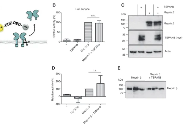
100 300 200 100 -100 0 -TSPAN8 Meprin β Meprin β + TSPAN8 TSPAN8 -Meprin β Meprin β + TSPAN8 n.s. n.s. 50 35 35 55 25 70 100 130 kDa 70 100 130 kDa TSPAN8 Meprin β Meprin β Meprin β + TSPAN8 Meprin β Meprin β TSPAN8 (myc) Actin Relative activity (%) Relative activity (% ) 0Figure 2: Influence of TSPAN8 on meprin β activity.
(A) Proteolytic activity of membrane bound or shed soluble meprin β was analyzed using the specific quenched fluorogenic peptide (mca)-EDEDED-(K-ε-dnp). Intensity of fluorescence was measured at 405 nm with an excitation at 320 nm every 30 s for 2 h at 37°C. dnp: 2,4-dini-trophenol; mca: (7-methyloxycoumarin-4-yl) acetyl. (B) TSPAN8 does not influence the proteolytic activity of meprin β at the plasma mem-brane. HEK293T cells were transfected with meprin β and TSPAN8 expression was induced. Relative activity of meprin β at the cell surface is shown. n = 7. (C) Representative Western blot analysis of meprin β and TSPAN8 expression in cells that were used for the activity assay. Actin served as loading control. (D) TSPAN8 has no effect on the shedding of meprin β from the cell surface. HEK293T cells were transfected with meprin β and TSPAN8 expression was induced. Relative activity of shed meprin β within the cell culture medium is shown. n = 3. (E) Western blot analysis of shed meprin β in the supernatant of transfected HEK293T cells from three independent experiments.
F. Schmidt et al.: Tetraspanin 8 interacts with meprin β within TEMs
861
Correspondingly, only little soluble APPα (sAPPα) was
present in the supernatant of HEK293T cells, since
over-expressed meprin β dominates β-secretase cleavage
which competes with or occurs prior to shedding of APP
by ADAMs at the α-secretase site. Interestingly,
induc-tion of TSPAN8 expression by doxycycline or
anhydrotet-racycline (ATC) treatment resulted in reduced amounts of
Aβ. However, the corresponding level of sAPPα did not
increase. In order to analyze whether the reduction in Aβ
might be an effect of doxycycline or ATC, we performed
a similar experiment but this time transfecting TSPAN8
instead of inducing its expression (Figure 3C). Again,
strong Aβ production was present when coexpressing
APP and meprin β. Of note, the presence of TSPAN8 did
not result in any changes in Aβ generation concluding
that the previous effect was caused by the treatment with
doxycycline or ATC. This experiment shows that the
pres-ence of TSPAN8 does not change the accessibility of the
substrate APP for meprin β, thus not altering the
genera-tion of Aβ peptides.
Figure 3: Processing of APP in the presence of TSPAN8.
(A) The amyloid precursor protein (APP) is a type I transmembrane protein. APP is processed by a β- and subsequently by γ-secretase thereby releasing amyloid β (Aβ) peptides (red). Alternatively, α-secretase cleavage prevents Aβ production via generation of soluble APP (sAPPα). Three different antibodies directed against APP were used in this study (blue Y). (B) Analysis of APP cleavage by meprin β upon induction of TSPAN8 expression. HEK293T cells were transfected with meprin β and APP, TSPAN8 expression was induced with either anhydrotetracycline (ATC) or doxycycline. Meprin β expression yielded in high amounts of Aβ peptides and subsequent decrease of sAPPα levels. In the presence of TSPAN8 Aβ generation was diminished but no increase in sAPPα was detectable. (C) Analysis of APP cleavage by meprin β upon transient transfection of TSPAN8. HEK293T cells were transfected with meprin β, APP and TSPAN8. Again, processing of APP by meprin β was accom-panied by high Aβ levels and reduction in the amount of sAPPα. Under these conditions the presence of TSPAN8 did not alter the generation of Aβ peptides.
TSPAN8 and meprin β colocalize within
tetraspanin-enriched microdomains
As we could exclude a direct effect of TSPAN8 on the
cata-lytic site of meprin β or its shedding from the cell surface,
we decided to further analyze the cell surface localization
of meprin β and TSPAN8. We first analyzed the cell surface
levels of both meprin β and TSPAN8 in the inducible
HEK293T cells by primary amine biotinylation of proteins
(Figure 4A). Indeed, both proteins, meprin β and TSPAN8,
F. Schmidt et al.: Tetraspanin 8 interacts with meprin β within TEMs
863
Figure 4: Distribution of meprin β, TSPAN8, and APP at the cell surface.
(A) HEK293T cells were transiently transfected with meprin β and stable expression of TSPAN8 was induced by doxycycline. Cell surface levels of both proteins were analyzed applying primary amine biotinylation. TSPAN8 as well as meprin β were found at the cell surface of HEK cells. TSPAN8 had no effect on the trafficking of meprin β to the cell surface. Additionally, no change in meprin β maturation was visible in presence of TSPAN8 (upper band: promeprin β, lower band: active meprin β). (B) Isolation of lipid rafts or tetraspanin-enriched microdo-mains (TEMs) via sucrose density gradient centrifugation. Flotillin 2 served as marker protein for lipid rafts (red) whereas the tetraspanin CD9 is located in TEMs (blue). Two different detergents were used for cell lysis: Triton X-100 preserves lipid rafts but destroys TEMs whereas with the milder detergent Brij98 both membrane regions stay intact. Discontinuous sucrose density gradient (0–42.5% sucrose) was pre-pared upon loading the cell lysate at the bottom of the centrifuge tube. After centrifugation TEMs and lipid rafts float within fractions 6–8. (C) HEK293T cells were transfected with meprin β and TSPAN8 expression was induced by doxycycline (dox) (upper part). Cells lysates, either prepared with Triton X-100 or Brij98, were loaded on sucrose density gradient. Marker proteins (Flotillin 2, CD9 and GAPDH) showed that the sucrose density gradient worked. Meprin β is found in lipid rafts but also in TEMs whereas TSPAN8 is predominantly located within TEMs. Endogenous APP was detected to a higher extent in TEMs than lipid rafts. When the expression of TSPAN8 was induced by doxycycline the signal for APP within TEMs was diminished. To verify this we performed the same experiment but this time transfected TSPAN8 instead of inducing its expression via doxycycline (lower part). Again, APP was predominantly found in TEMs but the presence of TSPAN8 did not alter its distribution.
were detectable in the streptavidin bound fractions and
were thus localized at the cell surface. This is in
accord-ance with the observation of the RFP dimerization assay
where TSPAN8-meprin β interaction was mainly
detect-able at the cell surface (Figure 1C). TSPAN8 expression
did not alter the amount of meprin β at the cell surface
as we observed equal protein levels in the induced and
non-induced cells. Moreover the ratio of pro- and mature
meprin β did not change when coexpressing TSPAN8,
suggesting that TSPAN8 does not influence the
matura-tion of meprin β. These data fit nicely to the results from
the peptide cleavage assays (Figure 2) and confirm that
the level and therefore the activity of meprin β at the cell
surface are unchanged and that an altered shedding of
meprin β in the presence of TSPAN8 can be excluded.
We then analyzed the submembranous localization of
TSPAN8 and meprin β by a sucrose density gradient
cen-trifugation of cell lysates (Figure 4B). Tetraspanins were
already described to form lipid raft-like membrane
frac-tions called tetraspanin-enriched microdomains (TEMs)
(Charrin et al., 2009; Yanez-Mo et al., 2009). We used two
different detergents for cell lysis to distinguish between
lipid rafts and TEMs. Triton X-100 destroys TEMs but
pre-serves lipid rafts which float in the sucrose density
gradi-ent whereas the milder deterggradi-ent Brij98 enables floating
of both membrane fractions. The cell lysates were loaded
on discontinuous sucrose gradients and following
cen-trifugation 14 fractions were collected and analyzed by
Western blotting.
We first checked different marker proteins to ensure
that the sucrose density gradient worked properly
(Figure 4C). Flotillin 2 is localized within lipid rafts and
therefore was found in the floating region (fractions 6–8)
when the cells were lysed with Triton X-100. The TEMs,
which are characterized by the presence of the
tetraspa-nin CD9, were also found in the same fractions when
treat-ing the cells with Brij98. GAPDH served as a control as it
is not associated with these membrane regions and
there-fore was only found in the high density fractions 12–14.
We analyzed the localization of meprin β and
demon-strate that meprin β is located in lipid rafts. However, we
did not observe any change in localization within the
mem-brane when inducing the expression of TSPAN8. Usage of
the milder detergent Brij98 revealed that meprin β is also
found in TEMs, as the amount of meprin β increases in
the low density fractions 6–8 and decreases in the high
density fractions 12–14. Under these conditions TSPAN8
was also present in the fractions corresponding to TEMs.
As levels of TSPAN8 are relatively lower in lipid rafts,
this indicates that the interaction between meprin β and
TSPAN8 takes place within the TEMs. Furthermore, we
investigated the distribution of the substrate APP. We could
not detect endogenous APP in lipid rafts in the presence
or absence of TSPAN8. However, APP was found in TEMs
in the absence of TSPAN8. Interestingly, when TSPAN8
expression was induced the amount of endogenous APP
was reduced in these fractions. This is consistent with the
reduced Aβ generation by meprin β when TSPAN8
expres-sion was induced by doxycycline or ATC (Figure 3B). As
we could already show that the effect on Aβ is not visible
any more in the absence of doxycycline, we repeated the
sucrose density gradient centrifugation using cells that
were cotransfected with meprin β and TSPAN8 (Figure 4C
lower part). We confirmed similar distribution of meprin β
within the cell membrane as described above. When
ana-lyzing endogenous APP we could show again that APP
is found only to very little extent in lipid rafts in these
HEK cells. APP is rather located within TEMs, as there is
a strong signal for APP in fractions 7–9 when cells were
lysed with Brij98. However, under these conditions, the
amount of APP in TEMs was not altered in the presence of
TSPAN8, suggesting that doxycycline rather than TSPAN8
caused the previously observed effect on APP. We
con-clude from this experiment that meprin β and TSPAN8 are
both located in TEMs where their interaction might occur.
The cleavage of APP by meprin β is also likely taking place
within the TEMs.
Overall, we identified TSPAN8 as a novel
non-pro-teolytic interaction partner of meprin β. We could show
that this interaction takes place on the cell surface within
TEMs (Figure 5). However, TSPAN8 did not influence the
proteolytic activity of meprin β towards fluorogenic
pep-tides and did not alter the proteolytic processing of APP.
Discussion
Meprin β is important for collagen processing and
depo-sition (Kronenberg et al., 2010; Broder et al., 2013) and
is associated with fibrotic conditions in Fra-2 transgenic
mice (Biasin et al., 2014). As shed protease meprin β is
responsible for the cleavage of MUC2 in the intestine and
subsequent detachment of the mucus layer critical for
proper barrier function (Schütte et al., 2014). Only
mem-brane bound meprin β was shown to cleave APP at the
β-secretase site reminiscent to BACE1 (Bien et al., 2012)
and thus it is likely to play a role in Alzheimer’s disease.
This shows that meprin β could be a promising
phar-macological target for certain pathological conditions.
To identify non-proteolytic interactions with regulatory
Meprin β TSPAN8 TSPAN APP Aβ
γ-secretase Tetraspanin
enriched microdomain
Lipid raft
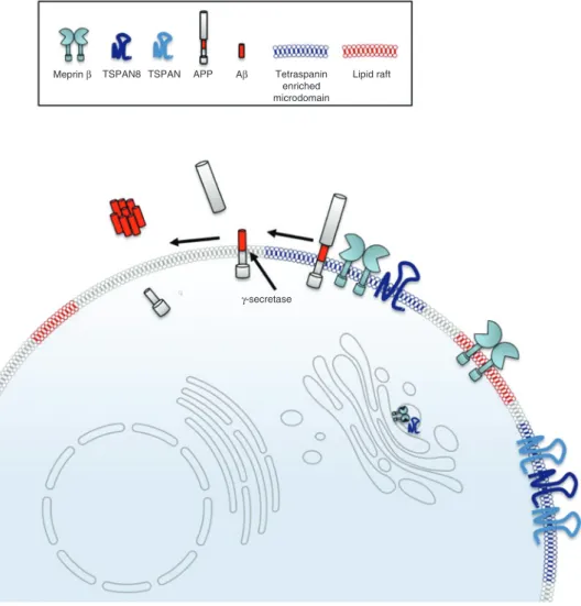
Figure 5: Potential model describing the interaction of meprin β and TSPAN8.
TSPAN8 is a novel non-proteolytic interactor of meprin β. Meprin β is found at the cell surface in both lipid rafts and tetraspanin-enriched microdomains (TEMs) whereas TSPAN8 is predominantly located within TEMs. The interaction of meprin β and TSPAN8 therefore takes place inside the TEMs where cleavage of APP by meprin β most likely occurs. Meprin β acts as β-secretase on APP and upon additional cleavage by the γ-secretase short Aβ peptides are released that are prone to aggregation.
F. Schmidt et al.: Tetraspanin 8 interacts with meprin β within TEMs
865
proteins involved in the activity or distribution of meprin
β we performed a yeast two-hybrid screen using a small
intestinal cDNA library and meprin β as bait. We
identi-fied TSPAN8 as a promising interaction partner in this
screen. Members of the tetraspanin familiy are already
known to interact with different metalloproteases, e.g.
ADAMs (Gutierrez-Lopez et al., 2011; Haining et al.,
2012; Prox et al., 2012). TSPAN8 for instance increases
the cell surface expression of ADAM12, thereby
promot-ing metastasis in esophageal cancer (Zhou et al., 2008).
We confirmed the direct protein-protein interaction in
human cells by a luciferase based protein
complemen-tation assay. Using a RFP dimerization assay, we
local-ized the interaction of meprin β and TSPAN8 to the cell
surface. We then investigated if TSPAN8 has an impact on
the proteolytic activity of meprin β. A quenched
fluoro-genic peptide cleavage assay revealed that the activity
of meprin β at the cell surface is unaltered in the
pres-ence of TSPAN8 in HEK293T cells. This was also the case
when we inserted two point mutations (E1334A, E135A) in
the LEL of TSPAN8, which would eventually fit into the
active site cleft of meprin β as it shows sequence
homol-ogy to the propeptide. Additionally, shedding of meprin β
from the cell surface was not altered as its activity in the
cell culture supernatant did not change when
coexpress-ing TSPAN8. However, it could very well be that TSPAN8
changes the accessibility of meprin β towards
physiologi-cal proteinogenic substrates at the plasma membrane.
For example, ADAM10 activity was shown to be enhanced
by TSPAN12, leading to an increased processing of APP at
the α-secretase site (Xu et al., 2009). We therefore
investi-gated the influence of TSPAN8 on the shedding activity of
meprin β towards APP. However, no difference in Aβ
pro-duction was detectable when TSPAN8 was present,
con-cluding that the accessibility of APP for meprin β is not
changed. We first observed a decrease of Aβ when
induc-ing TSPAN8 expression by doxycycline or ATC. However,
we could not confirm this change in Aβ by transfection of
TSPAN8. Therefore one has to be careful with the
inter-pretation of APP processing when using doxycycline
inducible cells. Furthermore, it is possible that other
sub-strates might be affected in such a system.
It is well known that tetraspanins form TEMs together
with other proteins such as integrins, G protein-coupled
receptors, growth factor receptors, but also cytosolic
pro-teins (Charrin et al., 2009; Yanez-Mo et al., 2009). Thus,
TEMs provide specialized signaling platforms and are
involved in many cellular and biological processes (Hemler,
2003, 2005). Previously, it was shown that ADAM10
inter-acts with several TSPANC8 tetraspanins, an
evolution-ary conserved subgroup of tetraspanins exhibiting eight
cysteines in their LEL, that regulate its maturation and
cellular distribution (Haining et al., 2012). We therefore
investigated the localization of meprin β and TSPAN8
within the cell membrane. First, we performed cell surface
biotinylation and confirmed the expression of both
pro-teins at the cell membrane. However, we did not observe
any differences in the amount of meprin β at the cell
surface as it was described for different ADAM proteases
in the presence of tetraspanins (Zhou et al., 2008; Prox
et al., 2012). Applying sucrose density gradient
centrifug-ation we showed that meprin β is not only found in lipid
rafts but also in TEMs, together with TSPAN8. This
corrob-orates our previous findings and proves that both proteins
are colocalized in the same membrane subdomains. There
was a recent example showing that tetraspanins, namely
TSPAN5 and TSPAN15, have the ability to differentially
regulate the membrane compartmentalization of ADAM10
(Jouannet et al., 2015). Interestingly, TSPAN8 did not alter
the distribution of meprin β within the cell membrane. We
further analyzed whether TSPAN8 might influence the
dis-tribution of meprin β substrates like APP. APP was mainly
found in TEMs providing evidence that the processing of
APP by meprin β takes place in this environment. Similar
observations were described in a recent study dealing with
regulated intramembrane proteolysis (Chen et al., 2015).
They observed that α- and β-secretases are able to form
distinct complexes with γ-secretase and that tetraspanins
were important for this association. This highlights the
importance of protein networks for efficient processing of
transmembrane substrates by different proteases
(Becker-Pauly and Rose-John, 2013). Further investigations will
elucidate the function of meprin β within this
special-ized environment. Potentially, meprin β needs its
interac-tion with TSPAN8 to meet so far unidentified substrates.
Furthermore, detecting the interaction of the
endoge-nous proteins would provide more inform ation about its
importance in certain cells or tissues. This would give a
hint under which conditions this interaction takes place
and what substrates might be affected in consequence.
However, to this stage it is hard to show the interaction
of endogenous meprin β and TSPAN8 due to the lack of
specific antibodies for TSPAN8. Additionally, it is likely
that meprin β and TSPAN8 synergize in the progression
of different cancer types. TSPAN8 was found to be highly
expressed in colon carcinoma, where it also promotes
metastasis (Greco et al., 2010; Richardson et al., 2011; Yue
et al., 2013). As meprin β shows a high expression in the
intestine and might be upregulated in colon cancer as well
(Dietrich et al., 1996; Matters and Bond, 1999), this tumor
type represents a promising condition to further
investi-gate the interplay between meprin β and TSPAN8.
Materials and methods
Chemicals
All chemicals were of analytical grade and obtained from Sigma-Aldrich (St. Louis, MO, USA), Carl Roth (Karlsruhe, Germany), Merck (Darmstadt, Germany), Roche (Basel, Switzerland) and Thermo Fisher Scientific (Waltham, MA, USA).
Split-ubiquitin yeast two-hybrid screen
The split-ubiquitin yeast two-hybrid screen was performed in coopera-tion with Dualsystems Biotech (Schlieren, Switzerland). Shortly, human meprin β cDNA was cloned into the bait vector pBT3-SUC and C-termi-nally fused to Cub-LexA-VP16. N-terminal NubG-tagged human small intestine library in pPR3-N was used as prey. Clones were selected, expanded and prey sequence traces were aligned and grouped. DNA sequences were translated in all three reading frames and searched against the Swissprot database using the BLASTX algorithm.
Gaussia princeps luciferase-based protein
complementation assay
cDNA coding for human meprin β and control, both lacking the stop codon, were cloned into the Gateway© shuttle vector pENTR4
(#17424, Addgene, Cambridge, MA, USA) using NotI/XhoI restriction sites. cDNA coding for human TSPAN8 in pDONR233 was a kind gift from Yves Jacob (Institute Pasteur, Paris, France). The cDNAs were then subcloned by homologous recombination into the destination vectors pSPICA-C1 and C2 (kind gift from Yves Jacob). pSPICA-C1 and C2 contain complementary fragments of Gaussia princeps luciferase located 3′ of the Gateway© recombination sites (Cassonnet et al.,
2011). All coding sequences were verified by DNA sequencing (Gatc Biotech, Konstanz, Germany).
Luciferase complementation assay was performed as described previously (Cassonnet et al., 2011). Briefly, HEK293T cells were seeded at a density of 1 × 10⁵ cells/ml in 500 μl/well of a 24-well plate.
The following day, cells were transfected with plasmid DNA. Cells always received a combination of two plasmids, one coding for the C-terminal part of Gaussia princeps luciferase and one coding for the N-terminal part of Gaussia princeps luciferase, each fused to a protein of interest or as an empty control. On the second day after transfection, cells were washed, lysed in 100 μl passive lysis buffer (#E1941, Promega, Mannheim, Germany) for 15 min at RT, and then transferred, in triplicates of 20 μl each, to a white 96-well biolumi-nescence plate. Coelenterazine was added and biolumibiolumi-nescence was detected using a Glomax plate injector system (Promega). Normal-ized luciferase ratio (NLR) was calculated as described in (Cassonnet et al., 2011). A NLR above 3.5 was considered a specific interaction. All experiments were n = 3.
Red fluorescent protein dimerization assay
Destination vectors with RFP-A and RFP-B located 3′ of the Gateway©
recombination sites were created by deleting the Gaussia princeps
luciferase fragment from pSPICA-C1 using site directed mutagenesis and inserting the restriction enzyme sites for AgeI and PacI. cDNA coding for RFP-A and RFP-B was amplified by PCR from pDDRFP-A1B1-DEVD [#36294, Addgene; original vectors are described in (Alford et al., 2012)] and then cloned into the destination vector using AgeI/PacI restriction sites. Homologous recombination was then per-formed with the entry vectors as described above.
HEK293T cells were seeded at a density of 2.5 × 10⁴ cells/ml in
μ-dishes (Ibidi, Martinsried, Germany). The following day, cells were transfected with plasmid DNA. Cells always received a com-bination of two plasmids, one coding for RFP-A and one coding for RFP-B, each fused to a protein of interest or as an empty control. On the second day after transfection, cells were imaged at an Olympus FluoView 1000 confocal microscope.
Generation of HEK293T cells inducible for TSPAN8
cDNA coding for TSPAN8 with a C-terminal myc tag was amplified by PCR and inserted into pINDUCER10 (Meerbrey et al., 2011) (kind gift from Jörg Müller, Institute of Molecular Cell Biology, Jena, Germany) using the AgeI/MluI restriction sites. The resulting plasmid was veri-fied by sequencing (Gatc Biotech).
HEK293T cells were transfected with the plasmid. Cells were selected for plasmid uptake by culturing them in the presence of 2 μg/ml puromycin. Cells were tested for tetraspanin expression in the presence and absence of 80 μg/ml doxycycline by immunoblotting of cell lysates.
Site-directed mutagenesis for generation of TSPAN8
mutant
Human wild-type cDNA of myc-tagged TSPAN8 in pcDNA3.1 was used as template for the generation of TSPAN8_E133A_E135A. Following primers were designed:
5′-AAA GCT TTT GAG CGC CAC AGG GGC AAG TGC AAA ACA ATT CCA GGA AGC CAT AAT T-3′
3′-AAT TAT GGC TTC CTG GAA TTG TTT TGC ACT TGC CCC TGT GGC GCT CAA AAG CTT T-5′
Mutagenesis was performed using the QuikChange® II XL
Site-Directed Mutagenesis Kit (AgilentTechnologies, Santa Clara, CA, USA) according to the manufacturer’s instructions. Correctness of mutations was proven by sequencing (Gatc Biotech).
Cell culture, transient transfection and induction of
HEK293T cells
TSPAN8 inducible HEK293T cells were cultured in DMEM + Glu-taMAX™ -I (Life Technologies, Carlsbad, CA, USA) supplemented with 10% fetal bovine serum (Invitrogen) and 2 μg/ml puromycin. Transient transfection was performed using polyethylenimine (1 mg/ ml in ddH2O, Polysciences, Eppelheim, Germany) and following
con-structs: pcDNA3.1 (mock), hmeprin β in pSG5, hAPP695 in pCIneo, hTSPAN8 in pcDNA3.1 and hTSPAN8_E133A_E135A in pcDNA3.1. For the induction of TSPAN8 expression, cells were treated with 100 ng/ ml to 80 μg/ml doxycycline or 100 ng/ml anhydrotetracycline (ATC) 24 h upon transfection.
F. Schmidt et al.: Tetraspanin 8 interacts with meprin β within TEMs
867
Cell lysis, SDS-PAGE, and Western blot analysis
Cell lysis, SDS-PAGE and Western blot analysis was performed as described previously (Arnold et al., 2015). Following antibodies were used for detection of protein in lysates: anti-GAPDH (2118; Cell Signaling Technology, Danvers, MA, USA), anti-actin (A2066; Sigma-Aldrich), anti-myc (9B11, 2276; Cell Signaling Technology), anti-meprin β (polyclonal antibody, generated against a peptide of the ectodomain), anti-APP (N) (polyclonal antibody directed against N-terminus of APP; Thermo Fisher Scientific), anti-APP (C) (B63.2, polyclonal antibody directed against the C-terminus of APP), anti-CD9 13118; Santa Cruz, Dallas, TX, USA), anti-Flotillin 2 (SC-28320; Santa Cruz). Supernatants were used for analysis of sAPPα and Aβ (6E10, SIG-39320; Covance, Princeton, NJ, USA).
Meprin β activity assay
Determination of meprin β proteolytic activity was performed using the specific quenched fluorogenic substrate (mca)-EDEDED-(K-ε-dnp) (Broder and Becker-Pauly, 2013; Jäckle et al., 2015). Culture medium and cell suspension with 50 μm substrate were used for this assay. Changes in fluorescence intensity were measured every 30 s for 2 h at 37°C using the fluorescent spectrometer Infinite F200 PRO (Tecan, Maennedorf, Switzerland) with an excitation wavelength of 320 nm and detection of emission at 405 nm.
dnp: 2,4-dinitrophenol; mca: (7-methyloxycoumarin-4-yl) acetyl.
Molecular modeling
The crystal structure of the extracellular domain of meprin β (PDB accession number 4GWN) was used with modeled epidermal growth factor (EGF)-like domain and transmembrane helix fused to it (Arolas et al., 2012). The large extracellular loop (LEL) of TSPAN8 was mod-eled using swiss-modeler (Biasini et al., 2014) where the LEL of CD81 was used as template (PDB accession number 1IV5). Meprin β and TSPAN8 were positioned manually using UCSF Chimera (Pettersen et al., 2004) which was also used for the generation of molecular images.
Cell surface primary amine biotinylation
HEK293T cells were transfected with meprin β and 24 h later TSPAN8 expression was induced. Primary amine biotinylation was performed as described previously (Arnold et al., 2015).
Isolation of detergent resistant membrane fractions by
sucrose density gradient centrifugation
HEK293T cells were transfected with meprin β and 24 h later TSPAN8 expression was induced. Alternatively, cells were transiently trans-fected with both meprin β and TSPAN8. Next day, cells were har-vested and lysed in 1 ml TNE buffer (25 mm Tris, 150 mm NaCl, 5 mm EDTA, complete protease inhibitor) for 30 min at 4°C. For the isolation
of lipid rafts the lysis buffer contained 1% Triton X-100 whereas for preservation of tetraspanin-enriched microdomains (TEMs) 1% Brij98 was added to the lysis buffer. Sucrose gradient preparation, centrifugation and analysis of the corresponding fractions were per-formed as described previously (Ebsen et al., 2015).
Acknowledgments: The authors thank Björn Rabe (Kiel,
Germany) for providing the CD9 antibody, Xavier
Gomis-Rüth (Barcelona, Spain) for the pdb file of
membrane-bound dimeric meprin β and Yves Jacob (Paris, France)
as well as Jörg Müller (Jena, Germany) for providing
plas-mids. This work was supported by the SFB877 ‘Proteolysis
as a Regulatory Event in Pathophysiology’ (project A9),
grant BE 4086/2-1 (to C.B.-P.), grant PI 379/6-1 (to C.U.P.)
and SFB841 (project C1, to D.S.-A.). W.A. is supported by
KULeuven (C16/15/073), VIB, SAO (S#14017) and the
fed-eral government (IAP P7/16).
References
Alford, S.C., Abdelfattah, A.S., Ding, Y., and Campbell, R.E. (2012). A fluorogenic red fluorescent protein heterodimer. Chem. Biol.
19, 353–360.
Arnold, P., Schmidt, F., Prox, J., Zunke, F., Pietrzik, C., Lucius, R., and Becker-Pauly, C. (2015). Calcium negatively regulates meprin beta activity and attenuates substrate cleavage. FASEB J. 29, 3549–3557.
Arolas, J.L., Broder, C., Jefferson, T., Guevara, T., Sterchi, E.E., Bode, W., Stocker, W., Becker-Pauly, C., and Gomis-Ruth, F.X. (2012). Structural basis for the sheddase function of human meprin beta metalloproteinase at the plasma membrane. Proc. Natl. Acad. Sci. USA. 109, 16131–16136.
Becker-Pauly, C. and Rose-John, S. (2013). TNFalpha cleavage beyond TACE/ADAM17: matrix metalloproteinase 13 is a poten-tial therapeutic target in sepsis and colitis. EMBO Mol. Med. 5, 902–904.
Biasin, V., Marsh, L.M., Egemnazarov, B., Wilhelm, J., Ghanim, B., Klepetko, W., Wygrecka, M., Olschewski, H., Eferl, R., Ols-chewski, A., et al. (2014). Meprin beta, a novel mediator of vascular remodelling underlying pulmonary hypertension. J. Pathol. 233, 7–17.
Biasini, M., Bienert, S., Waterhouse, A., Arnold, K., Studer, G., Schmidt, T., Kiefer, F., Cassarino, T.G., Bertoni, M., Bordoli, L., et al. (2014). SWISS-MODEL: modelling protein tertiary and quaternary structure using evolutionary information. Nucleic Acids Res. 42, W252–258.
Bien, J., Jefferson, T., Causevic, M., Jumpertz, T., Munter, L., Multhaup, G., Weggen, S., Becker-Pauly, C., and Pietrzik, C.U. (2012). The metalloprotease meprin β generates amino terminal-truncated amyloid β peptide species. J. Biol. Chem.
287, 33304–33313.
Broder, C. and Becker-Pauly, C. (2013). The metalloproteases meprin alpha and meprin beta: unique enzymes in inflammation, neu-rodegeneration, cancer and fibrosis. Biochem. J. 450, 253–264. Broder, C., Arnold, P., Vadon-Le Goff, S., Konerding, M.A., Bahr,
A., et al. (2013). Metalloproteases meprin α and meprin β are C- and N-procollagen proteinases important for collagen assembly and tensile strength. Proc. Natl. Acad. Sci. USA. 110, 14219–14224.
Cassonnet, P., Rolloy, C., Neveu, G., Vidalain, P.O., Chantier, T., Pel-let, J., Jones, L., Muller, M., Demeret, C., Gaud, G., et al. (2011). Benchmarking a luciferase complementation assay for detect-ing protein complexes. Nat. Methods 8, 990–992.
Charrin, S., Jouannet, S., Boucheix, C., and Rubinstein, E. (2014). Tetraspanins at a glance. J. Cell Sci. 127, 3641–3648. Charrin, S., le Naour, F., Silvie, O., Milhiet, P. E., Boucheix, C., and
Rubinstein, E. (2009). Lateral organization of membrane pro-teins: tetraspanins spin their web. Biochem. J. 420, 133–154. Chen, A.C., Kim, S., Shepardson, N., Patel, S., Hong, S., and Selkoe,
D.J. (2015). Physical and functional interaction between the α- and γ-secretases: a new model of regulated intramembrane proteolysis. J. Cell Biol. 211, 1157–1176.
Dietrich, J.M., Jiang, W., and Bond, J.S. (1996). A novel meprin β′ mRNA in mouse embryonal and human colon carcinoma cells. J. Biol. Chem. 271, 2271–2278.
Ebsen, H., Lettau, M., Kabelitz, D., and Janssen, O. (2015). Subcel-lular localization and activation of ADAM proteases in the context of FasL shedding in T lymphocytes. Mol. Immunol. 65, 416–428.
Greco, C., Bralet, M.P., Ailane, N., Dubart-Kupperschmitt, A., Rubinstein, E., Le Naour, F., and Boucheix, C. (2010). E-cad-herin/p120-catenin and tetraspanin Co-029 cooperate for cell motility control in human colon carcinoma. Cancer Res. 70, 7674–7683.
Gutierrez-Lopez, M.D., Gilsanz, A., Yanez-Mo, M., Ovalle, S., Lafuente, E.M., Dominguez, C., Monk, P.N., Gonzalez-Alvaro, I., Sanchez-Madrid, F., and Cabanas, C. (2011). The sheddase activity of ADAM17/TACE is regulated by the tetraspanin CD9. Cell Mol. Life Sci. 68, 3275–3292.
Hahn, D., Pischitzis, A., Roesmann, S., Hansen, M.K., Leuen-berger, B., Luginbuehl, U., and Sterchi, E.E. (2003). Phorbol 12-myristate 13-acetate-induced ectodomain shedding and phosphorylation of the human meprinbeta metalloprotease. J. Biol. Chem. 278, 42829–42839.
Haining, E.J., Yang, J., Bailey, R.L., Khan, K., Collier, R., Tsai, S., Watson, S.P., Frampton, J., Garcia, P., and Tomlinson, M.G. (2012). The TspanC8 subgroup of tetraspanins interacts with A disintegrin and metalloprotease 10 (ADAM10) and regulates its maturation and cell surface expression. J. Biol. Chem. 287, 39753–39765.
Hemler, M.E. (2003). Tetraspanin proteins mediate cellular penetra-tion, invasion, and fusion events and define a novel type of membrane microdomain. Annu. Rev. Cell Dev. Biol. 19, 397–422.
Hemler, M.E. (2005). Tetraspanin functions and associated microdo-mains. Nat. Rev. Mol. Cell Biol. 6, 801–811.
Jäckle, F., Schmidt, F., Wichert, R., Arnold, P., Prox, J., Mangold, M., Ohler, A., Pietrzik, C.U., Koudelka, T., Tholey, A., et al. (2015). Metalloprotease meprin β is activated by transmembrane ser-ine protease matriptase-2 at the cell surface thereby enhancing APP shedding. Biochem. J. 470, 91–103.
Jefferson, T., Auf dem Keller, U., Bellac, C., Metz, V.V., Broder, C., Hedrich, J., Ohler, A., Maier, W., Magdolen, V., Sterchi, E., et al. (2013). The substrate degradome of meprin metalloproteases
reveals an unexpected proteolytic link between meprin beta and ADAM10. Cell Mol. Life Sci. 70, 309–333.
Jouannet, S., Saint-Pol, J., Fernandez, L., Nguyen, V., Charrin, S., Boucheix, C., Brou, C., Milhiet, P.E., and Rubinstein, E. (2015). TspanC8 tetraspanins differentially regulate the cleavage of ADAM10 substrates, Notch activation and ADAM10 membrane compartmentalization. Cell Mol. Life Sci.
Kovalenko, O.V., Yang, X., Kolesnikova, T.V., and Hemler, M.E. (2004). Evidence for specific tetraspanin homodimers: inhibi-tion of palmitoylainhibi-tion makes cysteine residues available for cross-linking. Biochem. J. 377, 407–417.
Kronenberg, D., Bruns, B.C., Moali, C., Vadon-Le Goff, S., Ster-chi, E.E., Traupe, H., Bohm, M., Hulmes, D.J., Stocker, W., and Becker-Pauly, C. (2010). Processing of procollagen III by meprins: new players in extracellular matrix assembly? J. Invest. Dermatol. 130, 2727–2735.
Matters, G.L. and Bond, J. S. (1999). Expression and regulation of the meprin β gene in human cancer cells. Mol. Carcinog. 25, 169–178.
Meerbrey, K.L., Hu, G., Kessler, J.D., Roarty, K., Li, M.Z., Fang, J.E., Herschkowitz, J.I., Burrows, A.E., Ciccia, A., Sun, T., et al. (2011). The pINDUCER lentiviral toolkit for inducible RNA interference in vitro and in vivo. Proc. Natl. Acad. Sci. USA. 108, 3665–3670.
Ohler, A., Debela, M., Wagner, S., Magdolen, V., and Becker-Pauly, C. (2010). Analyzing the protease web in skin: meprin metal-loproteases are activated specifically by KLK4, 5 and 8 vice
versa leading to processing of proKLK7 thereby triggering its
activation. Biol. Chem. 391, 455–460.
Perneczky, R., Alexopoulos, P., and Kurz, A. (2014). Soluble amyloid precursor proteins and secretases as Alzheimer’s disease biomarkers. Trends Mol. Med. 20, 8–15.
Pettersen, E.F., Goddard, T.D., Huang, C.C., Couch, G.S., Greenblatt, D.M., Meng, E.C., and Ferrin, T.E. (2004). UCSF Chimera – a visualization system for exploratory research and analysis. J. Comput. Chem. 25, 1605–1612.
Prox, J., Willenbrock, M., Weber, S., Lehmann, T., Schmidt-Arras, D., Schwanbeck, R., Saftig, P., and Schwake, M. (2012). Tetraspa-nin15 regulates cellular trafficking and activity of the ectodo-main sheddase ADAM10. Cell Mol. Life Sci. 69, 2919–2932. Richardson, M.M., Jennings, L.K., and Zhang, X.A. (2011). Tetras-panins and tumor progression. Clin. Exp. Metastasis. 28, 261–270.
Schütte, A., Ermund, A., Becker-Pauly, C., Johansson, M.E., Rodri-guez-Pineiro, A.M., Backhed, F., Muller, S., Lottaz, D., Bond, J.S., and Hansson, G.C. (2014). Microbial-induced meprin β cleavage in MUC2 mucin and a functional CFTR channel are required to release anchored small intestinal mucus. Proc. Natl. Acad. Sci. USA. 111, 12396–12401.
Sterchi, E.E., Stocker, W., and Bond, J.S. (2008). Meprins, mem-brane-bound and secreted astacin metalloproteinases. Mol. Aspects Med. 29, 309–328.
Xu, D., Sharma, C., and Hemler, M.E. (2009). Tetraspanin12 regulates ADAM10-dependent cleavage of amyloid precursor protein. FASEB J. 23, 3674–3681.
Yanez-Mo, M., Barreiro, O., Gordon-Alonso, M., Sala-Valdes, M., and Sanchez-Madrid, F. (2009). Tetraspanin-enriched microdo-mains: a functional unit in cell plasma membranes. Trends Cell Biol. 19, 434–446.
F. Schmidt et al.: Tetraspanin 8 interacts with meprin β within TEMs
869
Yanez-Mo, M., Gutierrez-Lopez, M.D., and Cabanas, C. (2011). Func-tional interplay between tetraspanins and proteases. Cell Mol. Life Sci. 68, 3323–3335.
Yue, S., Mu, W., and Zoller, M. (2013). Tspan8 and CD151 pro-mote metastasis by distinct mechanisms. Eur. J. Cancer. 49, 2934–2948.
Zhou, Z., Ran, Y.L., Hu, H., Pan, J., Li, Z.F., Chen, L.Z., Sun, L.C., Peng, L., Zhao, X.L., Yu, L., et al. (2008). TM4SF3 promotes
esopha-geal carcinoma metastasis via upregulating ADAM12m expres-sion. Clin. Exp. Metastasis. 25, 537–548.
Supplemental Material: The online version of this article (DOI: 10.1515/hsz-2016-0126) offers supplementary material, available to authorized users.