HAL Id: hal-02043614
https://hal.archives-ouvertes.fr/hal-02043614
Submitted on 18 May 2020
HAL is a multi-disciplinary open access
archive for the deposit and dissemination of
sci-entific research documents, whether they are
pub-lished or not. The documents may come from
teaching and research institutions in France or
abroad, or from public or private research centers.
L’archive ouverte pluridisciplinaire HAL, est
destinée au dépôt et à la diffusion de documents
scientifiques de niveau recherche, publiés ou non,
émanant des établissements d’enseignement et de
recherche français ou étrangers, des laboratoires
publics ou privés.
Distributed under a Creative Commons Attribution - NonCommercial - NoDerivatives| 4.0
International License
Mass spectrometry analysis of the human endosulfatase
Hsulf-2
Ilham Seffouh, Cédric Przybylski, Amal Seffouh, Rana El Masri, Romain R
Vivès, Florence Gonnet, Régis Daniel
To cite this version:
Ilham Seffouh, Cédric Przybylski, Amal Seffouh, Rana El Masri, Romain R Vivès, et al.. Mass
spectrometry analysis of the human endosulfatase Hsulf-2. Biochemistry and Biophysics Reports,
2019, 18, pp.100617. �10.1016/j.bbrep.2019.01.010�. �hal-02043614�
Mass spectrometry analysis of the human endosulfatase Hsulf-2
Ilham Se
ffouh
a, Cédric Przybylski
a,1, Amal Se
ffouh
b, Rana El Masri
b, Romain R. Vivès
b,
Florence Gonnet
a, Régis Daniel
a,∗aUniversité Paris-Saclay, CNRS, CEA, Univ Evry, LAMBE, 91025, Evry, France bUniv. Grenoble Alpes, CNRS, CEA, IBS, Grenoble, France
A R T I C L E I N F O Keywords: HSulf-2 6-O-Endosulfatase Sulfatase Heparan sulfate Formylglycine Mass spectrometry A B S T R A C T
The human 6-O-endosulfatases HSulf-1 and -2 catalyze the region-selective hydrolysis of the 6-O-sulfate group of the glucosamine residues within sulfated domains of heparan sulfate, thereby ensuring a unique and original post-biosynthetic modification of the cell surface proteoglycans. While numerous studies point out the role of HSulf-2 in crucial physiological processes as well as in pathological conditions particularly in cancer, its structural organization in two chains and its functional properties remain poorly understood. In this study, we report the first characterization by mass spectrometry (MS) of HSulf-2. An average molecular weight of 133,115 Da was determined for the whole enzyme by MALDI-TOF MS, i.e. higher than the naked amino acid backbone (98,170 Da), highlighting a significant contribution of post-translational modifications. The HSulf-2 protein sequence was determined by Nano-LC-MS/MS, leading to 63% coverage and indicating at least four N-glycosylation sites at Asn 108, 147, 174 and 217. These results provide a platform for further structural in-vestigations of the HSulf enzymes, aiming at deciphering the role of each chain in the substrate binding and specificities and in the catalytic activities.
1. Introduction
The human heparan sulfate 6-O-endosulfatases HSulf-1 and HSulf-2 (HSulfs, EC 3.1.6.14) catalyze the regioselective hydrolysis of the 6-O-sulfate groups within heparan 6-O-sulfate (HS) chains present on cell sur-face proteoglycans and extracellular matrix [1,2]. HSulfs exhibit two unique features among human sulfatases, since to date they are the only known sulfatases to be secreted in the extracellular medium, and to be active at neutral pH and at the polymer level [3]. They belong to HS 6-O-endosulfatase family, which wasfirstly discovered in early 2000's in quail embryo [4,5], and then in chick [6], Xenopus laevis [7,8], sea urchin [9], zebrafish [10], Drosophila [11] and mammals [3,12–14]. Sulf enzymes were initially found during quail embryo development, highlighting their roles in modulating the binding of the morphogen Wnt to HS and thereby the Wnt signaling [8,15]. Since then, Sulfs have been shown to be involved in key developmental and tumoral processes through an unprecedented post-biosynthetic editing mechanism of the 6-O-sulfate pattern of HS [16–20]. Being confined to the highly sulfated domain (NS) of HS, HSulf-catalyzed 6-O-desulfation process leads to a modest decrease in the polysaccharide 6-O-sulfation content, and thus
to a limited impact on its overall sulfation (4–5% sulfate loss) [21]. Remarkably, such moderate but subtle changes in HS sulfation pattern lead to massive alteration of its biological activities, affecting the polysaccharide modulatory properties towards a large number of growth factors, morphogens and chemokines [15,22,23].
Despite the key role of HSulfs in the cellular glycomic machinery, these enzymes remain elusive protein objects as regard their structure and functions. In one hand, much effort has been devoted to the un-derstanding of the enzyme reaction specificities, revealing a narrow specificity of HSulfs for highly sulfated disaccharide constituents within the NS domain of HS, and a processive oriented 6-O-desulfation starting from the non-reducing end [3,24]. In the other hand, much less is known about the structural organization at a molecular level, and no crystallographic data have been reported to date. Main structural in-sights arise from gene-derived sequence analysis and from mutation/ deletion experiments [3,25,26]. HSulfs are biosynthesized as single polypeptide chain pro-enzymes (871 and 870 amino-acids for HSulf-1 (Q8IWU6) and HSulf-2 (Q8IWU5), respectively) comprising a signal peptide, followed by a catalytic domain featuring the formylglycine residue of prototype sulfatase active site, a hydrophilic domain (HD),
https://doi.org/10.1016/j.bbrep.2019.01.010
Received 27 November 2018; Received in revised form 25 January 2019; Accepted 28 January 2019
∗Corresponding author. CNRS, UMR8587, Laboratoire Analyse et Modélisation pour la Biologie et l'Environnement, Université Evry-Val-d'Essonne, F-91025, Evry,
France.
E-mail address:regis.daniel@univ-evry.fr(R. Daniel).
1Present address: Sorbonne université, IPCM, CNRS UMR8232, 4 place Jussieu, 75252 PARIS Cedex 05, France.
Available online 07 February 2019
2405-5808/ © 2019 The Authors. Published by Elsevier B.V. This is an open access article under the CC BY-NC-ND license (http://creativecommons.org/licenses/BY-NC-ND/4.0/).
and a C-terminal domain presenting a significant homology with that of the lysosomal glucosamine-6-sulfatase [3,4,16]. A maturation process including the removal of the signal peptide and the cleavage within the HD domain by a furin protease yields the mature enzyme as a two-chain protein likely joined by disulfide bonds [3,16,26]. As regard for HSulf-2, it results in a maturated active heterodimer, which consists in a longer chain comprising the catalytic domain (Phe 1 to Glu 391) and part of the HD domain cleaved at Arg 514, and a shorter chain com-posed of the remaining part of the HD domain and the C-terminal do-main (Scheme 1).
It is worth noting that HSulfs were most often indirectly detected in previous studies, generally by western-blotting and/or through the monitoring of its catalytic activity. We report here the first mass spectrometry characterization of HSulf-2, allowing its direct detection at the protein level and the coverage of the full protein sequence by a bottom-up proteomics approach.
2. Materials and methods 2.1. Materials
All reagents were of analytical grade. DL-dithiothreitol (DTT), io-doacetamide (IAA), ammonium bicarbonate (NH4HCO3), and urea were all purchased from Sigma-Aldrich (Saint-Quentin Fallavier, France). Acetonitrile and formic acid were obtained from Fluka (France); se-quencing-grade trypsin, Arg-C, Chymotrypsin, Asp-N, Trypsin/Lys-C Mix and PNGase F were from Promega (France). Purified recombinant HSulf-2 was prepared according to a procedure described in a forth-coming report. Briefly, the enzyme was purified from the conditioned medium of HSulf-2 transfected HEK293F cells, using cation exchange and size exclusion chromatography, successively (A. Seffouh et al., Cell Mol. Life Sci. in press). The purified active enzyme was stored at −80 °C in 50 mM Tris buffer, 300 mM NaCl, 5 mM MgCl2, 5 mM CaCl2pH 7.5. All buffers and solutions were prepared using ultra-pure water from a MilliQ apparatus (Millipore, Merck, France).
2.2. N-deglycosylation of HSulf-2
N-deglycosylation of HSulf-2 (3μg) by PNGase F was carried out under denaturing conditions in a 20μL final volume according to the provider instructions (Promega). The deglycosylation of HSulf-2 was carried out by the addition of 2μL of recombinant PNGase F and in-cubation at 37 °C for 1–3 h.
2.3. Desalting of HSulf-2
Desalting of HSulf-2 was performed by diafiltration using Microcon® DNA Fast Flow 100 kDa (Millipore) previously washed with 300μL water. HSulf-2 sample (30μg, 40 μL) supplemented to 200 μL with
water was centrifuged in Microcon at 500 g for 4 min at 4 °C. Then, 200μL of water were added and centrifugation was repeated for few min to get about 30μL final volume. Finally, the Microcon was returned to recover the HSulf-2 sample in 30μL water retentate.
2.4. MALDI-TOF mass spectrometry
MS experiments were carried out on a MALDI Autoflex speed TOF/ TOF MS instrument (Bruker Daltonics, Germany), equipped with a SmartBeam II™ laser pulsed at 1 kHz. The spectra were recorded in the positive linear mode (delay: 600 ns; ion source 1 (IS1) voltage: 19.0 kV; ion source 2 (IS2) voltage: 16.6 kV; lens voltage: 9.5 kV). MALDI data acquisition was carried out in the mass range 5000–150000 Da, and 10000 shots were summed for each spectrum. Mass spectra were pro-cessed using FlexAnalysis software (version 3.3.80.0, Bruker Daltonics). The instrument was calibrated using mono- and multi-charged ions of BSA (BSA Calibration Standard Kit, AB SCIEX, France). MALDI-TOF MS analysis was achieved by mixing 1.5μL of sinapinic acid matrix at 20 mg/mL in acetonitrile/water (50/50; v/v), 0.1% TFA, with 1.5μL of the desalted protein solution (0.71 mg/mL).
2.5. In-solution proteolytic digestions of HSulf-2
For trypsin digestion, 2μL of HSulf-2 sample (3 μg) were diluted in 5.5μL of 50 mM NH4HCO3pH 8.0 to which 2.5μL of 8 M urea were added (2 M final), and then incubated for 1 h at room temperature under moderate stirring. Samples were then reduced by addition of 1.1μL DTT (5 mM final) for 1 h at 37 °C under moderate stirring. Samples were then alkylated by addition of 1μL IAA (20 mM final) and left for 45 min at room temperature in the dark. Before performing trypsin digestion, 6.9μL 50 mM NH4HCO3, pH 8.0, was added to the samples to reach afinal concentration of 1 mM urea. Trypsin (1 μL of 0.4μg.μL−1) was then added before overnight incubation at 37 °C. Conditions for digestion by Trypsin/Lys-C Mix, Asp-N, Chymotrypsin, and Arg-C are reported in Supplementary Materials.
2.6. In-gel proteolytic digestions of HSulf-2
HSulf-2 was analyzed in reducing conditions in 12% polyacrylamide gel. HSulf-2 treated/untreated with PNGase F (25/10μL) was mixed with an equal volume of Laemmli SDS-sample buffer (60 mM Tris-Cl pH 6.8, 2% SDS, 10% glycerol, 5% β-mercaptoethanol, 0.01% bromo-phenol blue), heated for 5 min at 95 °C and then centrifuged and cooled at ambient temperature before to be loaded into wells.
The protein bands were excised, cut into small cubes, destained and washed with 50μL of 0.1 M NH4HCO3, and then shrunk by dehydration in acetonitrile for 5 min (repeated 3 times). The gel pieces were next incubated in 50μL 10 mM DTT for 35 min at 56 °C. After cooling to room temperature, the DTT solution was replaced by 50μL acetonitrile
Scheme 1. Schematic view of the domain organization of HSulf-2.
Domains from Ne to Ce terminus are displayed with their four ending residues: signal peptide (orange, 24 aa); catalytic domain (dark blue, 391 aa); hydrophilic domain (green, 307 aa); C-terminus (blue, 148 aa). The following sequence key points are indicated: Fgly 64, active site formylglycine at Cys64; RSIR 514, main furin cleavage site in HD; RNLTKR 541, secondary furin cleavage site in HD; 678 KRKKKLRKLLKR 689, HD C-terminus basic residues cluster.
I. Seffouh et al. Biochemistry and Biophysics Reports 18 (2019) 100617
vacuum-dried and stored at−80 °C until analysis. 2.7. Peptide preparation for LC-MS/MS analysis
Prior to NanoLC-MS/MS analysis, HSulf-2 peptides from in-gel di-gestion were re-suspended in 20μL of solvent A (acetonitrile/water/ formic acid 2:98:0.1, v/v/v). HSulf-2 peptides from in-solution diges-tion were desalted by twofiltration cycles on ZipTip C18 (Millipore) according to the manufacturer procedure to remove urea. Eluted pep-tides (40μL) were vacuum-dried and stored at −80 °C until analysis. HSulf-2 peptide digests were re-suspended in 30μL of solvent A. 2.8. Reverse phase NanoLC-MS/MS analysis
Peptide mixtures were analyzed on a Dual Gradient Ultimate 3000 chromatographic system (Dionex) coupled to a LTQ-Orbitrap™ XL mass spectrometer (Thermo-Fisher Scientific).
5μL of peptide solution in solvent A were injected at 20 μL/min flow rate onto a C18 pre-column (Acclaim PepMap C18, Dionex) to con-centrate and desalt for 5 min in solvent A (water/acetonitrile/formic acid, 98:2:0.1, v/v/v). Then, peptide separation was carried out on a C18 capillary column (Acclaim PepMap C18, Dionex) at 300μL/min flow rate according to the gradient: 0% solvent B (acetonitrile/water/ formic acid 80/20/0,1; v/v/v) during 6 min, then 0–70% B over 49 min, then 70%–100% B over 2 min, 100% B during 10 min and finally de-creasing to 0% B in 3 min. The column wasfinally re-equilibrated with 100% solvent A for 15 min. The LC eluent was sprayed into the MS instrument with a glass emitter tip (Pico-tip, New Objective, USA).
The LTQ-Orbitrap XL mass spectrometer (Thermo-Fisher Scientific) was operated in positive ionization mode. Singly charged species were excluded from fragmentation; dynamic exclusion of already fragmented precursor ions was applied for 300 s, with a repeat count of 1, a repeat
2.9. Database search
Raw datafiles were processed using Proteome Discover 1.4 (Thermo Fisher scientific) to obtain Mascot-compatible MGF files. Searches in the Swissprot Database were performedfirst in human taxonomy using the Mascot server (version 2.2.07, Matrix Science) with the following parameters: 1 missed cleavage, monoisotopic identification, tolerance on mass measurement 10 ppm for MS and 0.6 Da for MS2. To speed up the search, a homemade database was manually created containing the sequences of HSulf-2 chains. The MS/MS spectra were searched with semitryptic cleavage for trypsin, eight missed cleavage for chymo-trypsin, and a maximum of two missed cleavages for the other pro-teases; nofixed modification was set; the following variable modifica-tions were allowed: carbamidomethyl (C), carbamyl (K), carbamyl (N-term), deamidation (QN), formylGly (C), oxidation (M), and propio-namide (C) without carbamyl (N-term, K) specifically for peptides re-sulting from in-gel digestion. Fragment types taken into account were those specified in the configuration “ESI-trap” for CID MS/MS. MS/MS spectra were all visually inspected to search for y/b discriminating ions and validate peptide sequences.
3. Results and discussion
Recombinant HSulf-2 was overexpressed in the HEK293F cells, yielding an active enzyme on HS and heparin oligosaccharide substrate [24]. Trypsin digestion of HSulf-2 was carried out in solution in order to get complete sequence coverage of both short and long chains. How-ever, this digestion performed in usual reducing/alkylating conditions led to poor sequence coverage (not shown), indicating a weak acces-sibility of the cleavage sites to trypsin. To increase proteolysis e ffi-ciency, proteolytic digestion was carried out in presence of urea. This
Fig. 1. Sequence coverage percentages obtained by in-solution digestion of HSulf-2 using the proteases trypsin, trypsin/Lys-C Mix, Arg-C, chymotrypsin, Asp-N and Glu-C.
protocol modification improved the digestion efficiency, since the se-quence of both long and short chains of HSulf-2 was significantly covered for thefirst time (39% for each chain,Fig. 1). No significant coverage increase was observed following addition of various de-tergents (not shown), thus suggesting a particularly tight structural organization of HSulf-2.
To further increase the sequence coverage of HSulf-2 in solution, various other proteases or protease mixture (trypsin/Lys-C, Arg-C, chymotrypsin, Asp-N, Glu-C) with different specificities were used. Slightly higher coverage percentages of the long chain sequence were obtained with Arg-C and the mix trypsin/Lys-C (43%), while trypsin remained the most efficient protease for the short chain (39%) (Fig. 1, Fig. S1). Overall, by merging the different peptides produced by the various proteases in a single sequence map, 63% of the whole HSulf-2 sequence was covered, the sequence coverage being slightly higher for the long chain (67%) than for the short one (58%) (Fig. 2). Arg-C was the only protease to produce a peptide from the long chain containing the residue Cys 64 detected mainly as a formylglycine residue (Fig. S2, Table S2). Formylglycine is a key feature of sulfatase active site, which is essential for the catalytic activity [27,28]. It worth noting that pre-vious reported point mutation experiments abolishing the enzyme ac-tivity, targeted simultaneously Cys 64 and the adjacent residue Cys 65 [3,25]. For the first time, the catalytic residue formylglycine is thus precisely located at Cys 64 and directly identified at the protein level in HSulf-2, appearing as a major PTM occurring in HSulf-2.
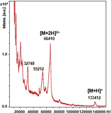
Until now, experimental molecular weight of HSulf-2 estimated to around 125 kDa relied on previously reported SDS-PAGE analysis, which showed the long chain at 75 kDa and the short chain hardly visible as a faint band at 50 kDa [26]. We confirmed this electrophoretic pattern (not shown), however, to provide an accurate mass value, HSulf-2 was submitted to MALDI-TOF MS analysis. Interestingly, the MALDI spectrum of the HSulf-2 showed in high m/z range a single low intensity peak at m/z 133,412, that we assigned to the mono-charged ion [M+H]+of the whole HSulf-2 molecule (Fig. 3). A major peak at m/z 66,410 dominated the spectrum, which could be attributed to the doubly charged ion [M+2H]2+of the entire HSulf-2 species. Based on these mono- and doubly-charged ions, an average experimental mass value of 133,115 ± 297 g mol−1was determined for thefirst time by MS for the whole HSulf-2 enzyme. This experimental mass value was higher that the molecular weight deduced from the amino acid com-position (98,170 g mol−1) suggesting a significant contribution of PTMs. An additional ion at m/z 55,212 was also detected, which could arise from possible variations in these modifications. The peak at m/z 66,410 exhibited a shoulder (at. m/z 65,420, and probably its doubly charged ion at m/z 32,748,Fig. 3) that remains unidentified. We cannot
rule out that the observed shoulder along the doubly charged ion of the entire HSulf-2 species could be an isolated chain, which remains linked to the enzyme.
A shift of the HSulf-2 bands on SDS-PAGE was previously reported after treatment by the N-glycosidase PNGase F [3,25], indicating the presence of N-glycans in agreement with the potential N-glycosylation sites along the HSulf-2 sequence (Scheme 1). We performed the trypsin in-gel digestion and nanoLC-ESI-MS/MS analysis of the resulting pep-tides on the most visible band attributed to the long chain, before and after treatment of HSulf-2 by PNGase F. The detected peptides belonged to the long chain (residues Phe 1 to Arg 514), ascertaining thus the identity of the main band on SDS-PAGE (Fig. S3). 46% of the long chain sequence was covered (i.e. 71% of the theoretical coverage expected by using trypsin), while the sequence coverage reached 66% (i.e. 100% of the theoretical coverage expected by using trypsin) after N-deglycosy-lation by PNGase F. The increased sequence coverage obtained after PNGase F treatment indicates that some cleavage sites in the long chain are protected from trypsin by N-glycan chains. Among the seven po-tential N-glycosylation sites found within the long chain (Asn 41, 88, 108, 125, 147, 174, 217),five on Asn 88, 108, 147, 174 and 217 were detected only in peptides obtained after N-deglycosylation. Deamida-tion introduced by PNGase F at Asn glycosylaDeamida-tion site was observed on MS/MS spectra of de-glycosylated peptides (Fig. S4,Table S4). Con-versely, Asn 125 is probably not a N-glycosylation site as it was de-tected in both glycosylated and N-deglycosylated forms of the long chain. The N-terminalfirst forty amino acids containing a potential N-glycosylation site were not covered. We assume that the various trypsin cleavage sites present within this N-terminal region may yield either too small peptides (≤6 residues, m/z above the chosen MS cutoff at 250) or the too acidic long peptide (PNIILVLTDDQDVELGSMQVMNK, with the potential site of glycosylation at Asn41) to be detected.
This study provides thefirst detailed structural data at the mole-cular level of the human endosulfatase HSulf-2 by mass spectrometry. The entire enzyme molecule was detected by MALDI-TOF allowing the determination of its average experimental mass at 133,115 Da, which indicated a significant contribution of post-translational modifications to the whole molecular mass. The protein sequence was covered by
Fig. 2. Sequence coverage of the whole HSulf-2 enzyme. Merged sequence map of HSulf-2 determined by combining the covered sequences obtained by the proteases trypsin, trypsin/lys-C Mix, Arg-C, chymotrypsin, Asp-N and Glu-C. C: formylglycine in the catalytic domain; N: potential N-glycosylation site; RS: furin cleavage site.
Fig. 3. MALDI-TOF mass spectrometry analysis of HSulf-2. Mass spectrum of HSulf-2 in positive ionization mode (100 kDa-filtrated Hsulf-2, mixed with si-napinic acid matrix, linear mode).
I. Seffouh et al. Biochemistry and Biophysics Reports 18 (2019) 100617
gested Asn 88 as potentially N-glycosylated, this residue was actually identified in several peptide sequences formed under in-solution di-gestion conditions, indicating that Asn 88 was either not N-glycosylated like Asn 125 or could be a heterogeneous glycosylation site. Further characterization of Hsulf-2 N-glycans and whole protein top-down analysis are currently under way. The weak MS ionization efficiency of the protein and the need for multiple proteases to achieve the full se-quence coverage in solution make HSulf-2 a tough protein to analyze and suggest tights folding and/or additional unusual modification of the protein backbone. This study provides the basis for further mass spectrometry investigations of the human endosulfatases.
Conflicts of interest
The authors declare that they have no conflicts of interest with the contents of this article.
Acknowledgements
We would like to thank V. Legros, D. Lebeau and W. Buchmann for help with running the Orbitrap and MALDI analysis. This work was supported in part by the CNRS and the GDR GAG (GDR 3739), the French Infrastructure for Integrated Structural Biology (FRISBI) ANR-10-INSB-05-01, the “Investissements d’avenir” program Glyco@Alps (ANR-15-IDEX-02), and by grants from the Agence Nationale de la Recherche (ANR-12-BSV8-0023 and ANR-17-CE11-0040) and Université Grenoble Alpes (UGA AGIR program).
Appendix A. Supplementary data
Supplementary data to this article can be found online athttps:// doi.org/10.1016/j.bbrep.2019.01.010.
Transparency document
Transparency document related to this article can be found online at https://doi.org/10.1016/j.bbrep.2019.01.010.
References
[1] K. Uchimura, The Sulfs: expression, purification, and substrate specificity, Methods Mol. Biol. 1229 (2015) 401–412,https://doi.org/10.1007/978-1-4939-1714-3_31. [2] G. Diez-Roux, A. Ballabio, Sulfatases and human disease, Annu. Rev. Genom. Hum.
Genet. 6 (2005) 355–379,https://doi.org/10.1146/annurev.genom.6.080604. 162334.
[3] M. Morimoto-Tomita, K. Uchimura, Z. Werb, S. Hemmerich, S.D. Rosen, Cloning and characterization of two extracellular heparin-degrading endosulfatases in mice and humans, J. Biol. Chem. 277 (2002) 49175–49185,https://doi.org/10.1074/ jbc.M205131200.
[4] G.K. Dhoot, M.K. Gustafsson, X. Ai, W. Sun, D.M. Standiford, C.P. Emerson, Regulation of Wnt signaling and embryo patterning by an extracellular sulfatase, Science 293 (2001) 1663–1666,https://doi.org/10.1126/science.293.5535.1663. [5] X. Ai, A.-T. Do, M. Kusche-Gullberg, U. Lindahl, K. Lu, C.P. Emerson, Substrate
specificity and domain functions of extracellular heparan sulfate 6-O-en-dosulfatases, QSulf1 and QSulf2, J. Biol. Chem. 281 (2006) 4969–4976,https://doi. org/10.1074/jbc.M511902200.
[6] C. Braquart-Varnier, C. Danesin, C. Clouscard-Martinato, E. Agius, N. Escalas, B. Benazeraf, X. Ai, C. Emerson, P. Cochard, C. Soula, A subtractive approach to characterize genes with regionalized expression in the gliogenic ventral
[10] B. Gorsi, S. Whelan, S.E. Stringer, Dynamic expression patterns of 6-O en-dosulfatases during zebrafish development suggest a subfunctionalisation event for sulf2, Dev. Dynam. 239 (2010) 3312–3323,https://doi.org/10.1002/dvdy.22456. [11] A. Wojcinski, H. Nakato, C. Soula, B. Glise, DSulfatase-1fine-tunes Hedgehog pat-terning activity through a novel regulatory feedback loop, Dev. Biol. 358 (2011) 168–180,https://doi.org/10.1016/j.ydbio.2011.07.027.
[12] T. Ohto, H. Uchida, H. Yamazaki, K. Keino-Masu, A. Matsui, M. Masu, Identification of a novel nonlysosomal sulphatase expressed in thefloor plate, choroid plexus and cartilage, Genes Cells 7 (2002),https://doi.org/10.1046/j.1356-9597.2001.00502. x173–85.
[13] S. Nagamine, S. Koike, K. Keino-Masu, M. Masu, Expression of a heparan sulfate remodeling enzyme, heparan sulfate 6-O-endosulfatase sulfatase FP2, in the rat nervous system, Brain Res. Dev. Brain Res. 159 (2005) 135–143,https://doi.org/ 10.1016/j.devbrainres.2005.07.006.
[14] S. Nagamine, K. Keino-Masu, K. Shiomi, M. Masu, Proteolytic cleavage of the rat heparan sulfate 6-O-endosulfatase SulfFP2 by furin-type proprotein convertases, Biochem. Biophys. Res. Commun. 391 (2010) 107–112,https://doi.org/10.1016/j. bbrc.2009.11.011.
[15] X. Ai, A.-T. Do, O. Lozynska, M. Kusche-Gullberg, U. Lindahl, C.P. Emerson, QSulf1 remodels the 6-O sulfation states of cell surface heparan sulfate proteoglycans to promote Wnt signaling, J. Cell Biol. 162 (2003) 341–351,https://doi.org/10.1083/ jcb.200212083.
[16] R.R. Vivès, A. Seffouh, H. Lortat-Jacob, Post-synthetic regulation of HS structure: the Yin and Yang of the sulfs in cancer, Front. Oncol. 3 (2014) 331,https://doi.org/ 10.3389/fonc.2013.00331.
[17] S.D. Rosen, H. Lemjabbar-Alaoui, Sulf-2: an extracellular modulator of cell signaling and a cancer target candidate, Expert Opin. Ther. Targets 14 (2010) 935–949,
https://doi.org/10.1517/14728222.2010.504718.
[18] A. Kleinschmit, T. Koyama, K. Dejima, Y. Hayashi, K. Kamimura, H. Nakato, Drosophila heparan sulfate 6-O endosulfatase regulates Wingless morphogen gra-dient formation, Dev. Biol. 345 (2010) 204–214,https://doi.org/10.1016/j.ydbio. 2010.07.006.
[19] I. Maltseva, M. Chan, I. Kalus, T. Dierks, S.D. Rosen, The SULFs, extracellular sul-fatases for heparan sulfate, promote the migration of corneal epithelial cells during wound repair, PLoS One 8 (2013) e69642, ,https://doi.org/10.1371/journal.pone. 0069642.
[20] Y.-H. Wang, C. Beck, Distinct patterns of endosulfatase gene expression during Xenopus laevis limb development and regeneration, Regen 2 (2015) 19–25,https:// doi.org/10.1002/reg2.27(Oxford, England).
[21] R. El Masri, A. Seffouh, H. Lortat-Jacob, R.R. Vivès, The “in and out” of glucosamine 6-O-sulfation: the 6th sense of heparan sulfate, Glycoconj. J. 34 (2017) 285–298,
https://doi.org/10.1007/s10719-016-9736-5.
[22] K. Uchimura, M. Morimoto-Tomita, A. Bistrup, J. Li, M. Lyon, J. Gallagher, Z. Werb, S.D. Rosen, HSulf-2, an extracellular endoglucosamine-6-sulfatase, selectively mo-bilizes heparin-bound growth factors and chemokines: effects on VEGF, FGF-1, and SDF-1, BMC Biochem. 7 (2006) 2,https://doi.org/10.1186/1471-2091-7-2. [23] X. Yue, J. Lu, L. Auduong, M.D. Sides, J.A. Lasky, Overexpression of Sulf2 in
idiopathic pulmonaryfibrosis, Glycobiology 23 (2013) 709–719,https://doi.org/ 10.1093/glycob/cwt010.
[24] A. Seffouh, F. Milz, C. Przybylski, C. Laguri, A. Oosterhof, S. Bourcier, R. Sadir, E. Dutkowski, R. Daniel, T.H. van Kuppevelt, T. Dierks, H. Lortat-Jacob, R.R. Vivès, HSulf sulfatases catalyze processive and oriented 6-O-desulfation of heparan sulfate that differentially regulates fibroblast growth factor activity, FASEB J. 27 (2013) 2431–2439,https://doi.org/10.1096/fj.12-226373.
[25] M.-A. Frese, F. Milz, M. Dick, W.C. Lamanna, T. Dierks, Characterization of the human sulfatase Sulf1 and its high affinity heparin/heparan sulfate interaction domain, J. Biol. Chem. 284 (2009) 28033–28044,https://doi.org/10.1074/jbc. M109.035808.
[26] R. Tang, S.D. Rosen, Functional consequences of the subdomain organization of the sulfs, J. Biol. Chem. 284 (2009) 21505–21514,https://doi.org/10.1074/jbc.M109. 028472.
[27] T. Dierks, B. Schmidt, L. V Borissenko, J. Peng, A. Preusser, M. Mariappan, K. von Figura, Multiple sulfatase deficiency is caused by mutations in the gene encoding the human C(alpha)-formylglycine generating enzyme, Cell 113 (2003) 435–444,
https://doi.org/10.1016/S0092-8674(03)00347-7.
[28] M.J. Appel, C.R. Bertozzi, Formylglycine, a post-translationally generated residue with unique catalytic capabilities and biotechnology applications, ACS Chem. Biol. 10 (2015) 72–84,https://doi.org/10.1021/cb500897w.
[29] T. Barbeyron, L. Brillet-Guéguen, W. Carré, C. Carrière, C. Caron, M. Czjzek, M. Hoebeke, G. Michel, Matching the diversity of sulfated biomolecules: creation of a classification database for sulfatases reflecting their substrate specificity, PLoS One 11 (2016) e0164846https://doi: 10.1371/journal.pone.0164846.