HAL Id: hal-02263774
https://hal.archives-ouvertes.fr/hal-02263774
Submitted on 5 Aug 2019HAL is a multi-disciplinary open access archive for the deposit and dissemination of sci-entific research documents, whether they are pub-lished or not. The documents may come from teaching and research institutions in France or abroad, or from public or private research centers.
L’archive ouverte pluridisciplinaire HAL, est destinée au dépôt et à la diffusion de documents scientifiques de niveau recherche, publiés ou non, émanant des établissements d’enseignement et de recherche français ou étrangers, des laboratoires publics ou privés.
Fathers over 40 and increased failure to conceive: the
lessons of in vitro fertilization in France
Elise de la Rochebrochard, Jacques de Mouzon, François Thépot, Patrick
Thonneau
To cite this version:
Elise de la Rochebrochard, Jacques de Mouzon, François Thépot, Patrick Thonneau. Fathers over 40 and increased failure to conceive: the lessons of in vitro fertilization in France. Fertility and Sterility, Elsevier, 2006, 85 (5), pp.1420-1424. �10.1016/j.fertnstert.2005.11.040�. �hal-02263774�
Publisher’s Version/PDF in open access
on editor web site:
http://www.fertstert.org/article/S0015-0282%2806%2900104-X/pdf
La Rochebrochard Elise (de), Mouzon Jacques (de), Thépot François, Thonneau Patrick and the FIVNAT Association, 2006, “Fathers over 40 and increase failure to conceive: the lessons of in vitro fertilization in France”, Fertility and Sterility, 85(5), p. 1420-1424. DOI: 10.1016/j.fertnstert.2005.11.040
Fathers over 40 and increased failure to conceive:
the lessons of in vitro fertilization in France
Elise de La Rochebrochard, PhD (a), Jacques de Mouzon, MD (a), François Thépot,
MD (b), Patrick Thonneau, MD (c) and the FIVNAT Group
(a) INED, INSERM, Unit 569, Bicêtre Hospital, 94275 Le Kremlin-Bicêtre, France (b) Laboratoire de Biologie de la Réproduction, CHU d’Amiens, 80000 Amiens, France (c) INSERM, Human Fertility Research Group, Paule de Viguier Hospital, 31059
FIVNAT is an association which was founded in 1986. Most French IVF centres belong to the FIVNAT association. The FIVNAT association is directed by a committee elected every two years. The current committee was elected in September 2004 and is composed of Philippe ARVIS, Jean-Philippe AYEL, Joëlle BELAISCH-ALLART, Jacques CHOUTEAU, Laurent JANNY, Rachel LEVY, François MOUCHEL, Jean-Luc POULY (chairman), Dominique ROYERE, Jean-Paul TAAR. The FIVNAT association is principally funded by Organon Pharmaceuticals Inc.
The authors declared no conflict of interests.
Correspondence to: Elise de La Rochebrochard, Inserm Unit 569, Bicêtre Hospital,
82 rue du Général Leclerc, 94275 Le Kremlin-Bicêtre, France. Tel. 33 (0) 1 45 21 23 33, fax 33 (0) 1 45 21 20 75, e-mail roche@ined.fr
Running title: Fathers over 40 and failure to conceive
Capsule. As an increasing number of couples choose to postpone childbearing, they
should be informed that paternal age over 40 years is an important risk factor for failure to conceive.
ABSTRACT
Objective: To investigate paternal age effect mediated by biological modifications
with use of data from assisted reproductive technologies.
Design: National IVF registry. Setting: France.
Patients: 1,938 men whose partners were totally sterile, with bilateral tubal
obstruction or absence of both tubes (in order to avoid bias sampling in analysis of paternal age) and treated by conventional IVF.
Intervention: None.
Main outcome measure(s): Risk of failure to conceive defined as absence of
intrauterine pregnancy.
Results: The odds ratio of failure to conceive for paternal age 40 years was 2.00
(95% CI: 1.10-3.61) when the woman was aged 35-37 years, 2.03 (95% CI: 1.12-3.68) for age 38-40 years, and 5.74 (95% CI: 2.16, 15.23) for age 41 years and over.
Conclusions: As an increasing number of couples choose to postpone childbearing,
they should be informed that paternal age over 40 years is an important risk factor for failure to conceive.
INTRODUCTION
In industrialized countries, demographers have observed a trend to delay childbearing, reflecting couples’ desire to have children at older ages. However, the risk of reproductive difficulties is clearly increased for couples who delay childbearing until after the age of 35. Maternal age over 35 years increases risks of infertility, miscarriage and ectopic pregnancy (1, 2). Moreover, a recent simulation model showed that assisted reproductive techniques (ART) “do not fully compensate for the years (and the chances of conceiving) lost" (3). This marked maternal age effect led to the conclusion that 35 years is the “amber light” in the reproductive life of women (4).
Paternal age was long almost ignored in studies of age effect on reproductive outcomes, but its potential role has recently been investigated. Some works have shown that increasing paternal age is accompanied by greater risk of delay in achieving pregnancy, of miscarriage and of late fetal death (5-8). In a recent review of the literature, we considered that 40 years could be the “amber light” in male reproductive life, as is 35 years for women’s reproductive life (9). The demonstrated effect of paternal age on risk of delay in achieving pregnancy could be the consequence of either biological modification of the male reproductive tract or of decrease in male sexual activity. When analyzing natural conception, it is very difficult to distinguish sexual and biological consequences of age. In order to analyze paternal age effect mediated by biological aging alone, data on medically assisted cycles provide a very interesting model.
Data on medically assisted reproduction have been used to confirm a biological effect of maternal age on the probability of conception (10). In order to avoid sampling bias in analysis of infertile couples, Schwartz et al. selected couples
requesting artificial insemination with donor semen (AID) because the men were totally sterile (azoospermic men only). Among these couples whose sterility was linked to male reproductive impairment, the authors hypothesized that the women’s fecundity was comparable to that of the general population, and so the maternal age effect in this population requiring AID could be extrapolated to the general population. This study confirmed that maternal age affected the probability of conception, mediated by biological aging of the women. It showed that this effect began as early as 30 years and became significant after age of 35. To confirm a biological effect of paternal age, the methodology of Schwartz et al. could be applied by selecting couples requiring medical assistance because the wife was totally sterile.
In order to confirm the hypothesis of a biological paternal age effect on the risk of failure to conceive, we studied ART data from the French national in vitro fertilization (IVF) registry by selecting couples requesting IVF because the woman was totally sterile, that is to say with bilateral tubal obstruction or absence of both tubes.
MATERIALS AND METHODS
Since 1986, the French National IVF Registry (FIVNAT) has collected information on aspiration cycles carried out in France (11). The FIVNAT registry received approval from the French Data Protection Authority (CNIL) on 17 December 1987 (declaration n° 174 168). IVF centers participate voluntarily in this registry. The 79 centers currently belonging to FIVNAT perform nearly 90% of the aspiration cycles in this country. We carried out data quality control on FIVNAT centers concerning fulfilling of key items and thus restricted our analysis to 59 centers (59/79=75%). In order to analyze paternal age, we investigated couples requesting conventional IVF in which the female partners were totally sterile, i.e. with bilateral tubal obstruction or absence of both tubes. To avoid bias due to changes in ART techniques (especially related to increasingly widespread use of intracytoplasmic sperm injection), we restricted our investigation to IVF performed since 2000. Finally, 1,938 couples treated by conventional IVF for bilateral tubal obstruction were included in this study.
We analyzed the risk of failure to conceive, defined as absence of intrauterine pregnancy confirmed by echography and an HCG level >1000 IU. Age effect was considered by using five-year age classes. As the age of 37 years has previously been demonstrated to be a cut-off point for the effect of maternal age on IVF success rate (12), we divided the group of women aged 35–40 into two sub-groups, 35-37 years and 38-40 years.
Age effects were analyzed based on odds ratios estimated by logistic regression using the SAS system (v8.02) package. Estimation of odds ratios relies on the method of maximum likelihood and confidence intervals for odds ratios were computed based on individual Wald tests. In a first logistic multivariate model, we
analyzed paternal age effect by controlling for maternal age effect. This model is based on the hypothesis that the paternal age effect is the same whether the woman is young, middle-aged or older. This hypothesis has been debated in some studies which indicated that the paternal age effect may be greater when the woman is aged 35 years and over than among younger women (7, 8). In order to take into account the possibility that paternal age effect may differ according to maternal age, we also used a second model which included an interaction factor between maternal age and paternal age.
RESULTS
As shown in table 1 and in table 2, the risk of failure to conceive clearly increased with maternal age and with paternal age in both models. In table 1, without male and female age interaction, a significant maternal age effect appeared in women aged 38-40 years and in women aged 41 years. The odds ratio (OR) for paternal age 40 years compared to <30 years was 1.70 (95% CI: 1.14-2.52).
In table 2, taking into account an interaction between male and female ages, the odds ratio of failure to conceive for paternal age 40 years was 2.00 (95% CI: 1.10-3.61) when the woman was aged 35-37 years, 2.03 (95% CI: 1.12-3.68) for age 38-40 years, and 5.74 (95% CI: 2.16, 15.23) for age 41 years and over.
DISCUSSION
Our results provide for the first time strong evidence for a paternal age effect on failure to conceive that is linked only to biological male aging (without confusion with sexual activity). We observed a clear tendency to increased risk of failure to conceive, especially when the fathers were aged over 40 years. Results in the first and last classes in table 2 (older woman with young man or young woman with older man) should be interpreted with caution because of the small number of couples in these classes. We thus analyzed table 2 by concentrating on classes with at least 30 couples. This revealed a clear increase in risk of failure to conceive with paternal age over 40 years when the woman was aged 35 years and over.
The paternal age effect was demonstrated here in a population of couples treated in IVF programmes and who were highly selected on the fertility characteristics of the woman (women who were totally sterile, that is to say with bilateral tubal obstruction or absence of both tubes). This finding can be extrapolated to the general population based on the hypothesis that these sterile women have no tendency to bond with men having any particular fertility characteristics. To the best of our knowledge, there is no biological or sociological evidence at the present time that could seriously question this hypothesis.
Our results on a paternal age effect after 40 years are in accordance with results recently published concerning the general population. In a European population-based study of couples attempting to conceive naturally, a significant odds ratio of 2.99 (95% CI: 2.77, 7.55) for risk of not having conceived after 12 months of attempting to achieve pregnancy was observed when the woman was aged 35-39 years and the man 40 years and over (7). A similar tendency was
daily probability of conception in couples composed of a woman aged 35-39 years and of a man in his late thirties or older (8).
It has been shown that couples having difficulty in conceiving also have an increased risk of miscarriage (19). Thus, the association between paternal age and failure to conceive raised the question of a possible association between paternal age and miscarriage. In the literature, an increased risk of miscarriage was observed in couples composed of a woman aged 35 years and over and of a man aged 40 years and over (OR = 6.73; 95% CI: 3.50, 12.95) (6). More recently, in a large Danish cohort, a two-fold increase of the risk of early fetal death was found when the father was aged 50 years and over compared with fathers aged 25-29 years, after controlling for various confounders and especially for maternal age (5). In the same cohort, the authors showed a paternal age effect as early as 45 years when considering late fetal deaths.
In a prospective American study of a cohort of more than 5,000 Californian women, the association between paternal age and risk of spontaneous abortion was analyzed by distinguishing between risk of fetal death during the first trimester of pregnancy and risk of fetal death during the beginning of the second trimester (up to 20 weeks of gestation) (20). The authors concluded that elevated paternal age ( 35 years) increased the risk of spontaneous abortion during the first trimester and at the beginning of the second trimester, with a suggestion that the association was stronger for deaths occurring during the first trimester.
Interestingly, there is a remarkable concordance in all these studies, stressing the fact that older fathers (≥ 40-45 years) have a key impact on both reproductive issues, failure to conceive and miscarriage. The mechanism for the paternal age effect remains to be explained. Previously, as for maternal age, the genetic
hypothesis had been emphasized (21, 22). After analysis of 11,535 pregnancies obtained by artificial insemination using donor spermatozoa, an increased risk of trisomy 21 for the fetus when the donor was aged ≥38 years has been suggested (23). The aneuploidy rate for both sex chromosomes and for autosomes 9 and 18 was also investigated by comparing 15 men aged 30 years and less and 8 men aged 60 years and older (24), but no significant differences between the two age groups were revealed in this recent study. So, the effect of paternal age on aneuploidy remains debatable and insight into this question may be gained in the future from analysis of aneuploidy mechanisms (25).
Cytogenetic analysis of semen specimens collected from donors has demonstrated an increased risk of frequency of numerical and structural aberrations in men aged 59-74 years compared with men aged 23-39 years (26). More recently, a review indicated that the paternal age effect may be mediated principally by structural chromosomal aberrations in sperm (27). Several authors have also suggested an increased risk of autosomal dominant diseases in children of fathers aged 40 years and older (28). Male genetic alterations could be mediated by age-related increases in germ cell mutations, impairment of DNA repair mechanisms and apoptotic processes (29-32).
On the other hand, morphological changes in the testis have been shown in aging men with decreased numbers of Leydig cells, arteriosclerotic lesions, thickening and hernia-like protrusions of the basal membrane of the seminiferous tubules, and fibrotic thickening of the tunica albuginea (33). These alterations in male reproductive tract function could induce a decrease in quality and quantity of spermatozoa production. A review comparing sperm parameters in men aged under
30 years and over 50 years demonstrated a clear decline in semen volume, sperm motility and sperm morphology with increasing age (34).
Once again, our results confirmed the well-established maternal age effect on the risk of failure to conceive (13). After controlling for paternal age, we found a clearly increased risk of failure to conceive in women after 37 years, in agreement with the literature (12). The cut-off at 37 years was also confirmed by the observed rate of oocyte atresia. Investigation of the number of follicles contained in ovaries obtained during surgery or in women who died suddenly had shown that the disappearance of ovarian follicles accelerated strongly after age of 37.5 years, at the time when the number of follicles fell below the critical figure of 25,000 (14).
The maternal age effect had been principally linked to genetic alteration of oocytes, especially abnormalities in the meiotic spindle of the oocyte, in women aged 37 years and older (15). More recently, the roles of cohesin and the premature separation of homologous chromatids have been put forward. Chromosome segregation during meiosis and mitosis is certainly one of the most important molecular and cellular processes that allow cells to transmit their genetic information across generations. Failure to maintain genetic stability during cell division leads to cell death or malignant transformation. Several authors have demonstrated a role of cohesin (a multi-subunit complex) in sister chromatid cohesion (16-18).
In industrialized countries, a tendency to postpone childbearing has been observed, leading to parents who are more advanced in age. Furthermore, the number of older couples requesting ART procedures has also increased. It had long been known that these couples must be informed that postponing childbearing beyond the age of 35 years for the woman significantly increases the risk of an adverse reproductive outcome (13). It now appears that this is only one aspect of the
age issue. In reproduction, age must no longer be considered as the affair of the woman, but as that of the couple. Just like maternal age over 35 years, paternal age over 40 years is a key risk factor in reproduction.
REFERENCES
1. Menken J, Trussell J, Larsen U. Age and infertility. Science 1986;233:1389-94. 2. Nybo Andersen AM, Wohlfahrt J, Christens P, Olsen J, Melbye M. Maternal age and fetal loss: population based register linkage study. BMJ 2000;320:1708-12.
3. Leridon H. Can assisted reproduction technology compensate for the natural decline in fertility with age? A model assessment. Hum Reprod 2004;19:1548-53.
4. Gosden R, Rutherford A. Delayed childbearing. BMJ 1995;311:1585-6.
5. Nybo Andersen AM, Hansen KD, Andersen PK, Davey Smith G. Advanced paternal age and risk of fetal death: a cohort study. Am J Epidemiol 2004;160:1214-22.
6. La Rochebrochard (de) E, Thonneau P. Paternal age and maternal age are risk factors for miscarriage; results of a multicentre European study. Hum Reprod 2002;17:1649-56.
7. La Rochebrochard (de) E, Thonneau P. Paternal age over 40 years: an important risk factor for infertility. Am J Obstet Gynecol 2003;189:901-5.
8. Dunson DB, Colombo B, Baird DD. Changes with age in the level and duration of fertility in the menstrual cycle. Hum Reprod 2002;17:1399-403.
9. La Rochebrochard (de) E, McElreavey K, Thonneau P. Paternal age over 40 years: the 'amber light' in the reproductive life of men? J Androl 2003;24:459-65.
10. Schwartz D, Mayaux MJ. Female fecundity as a function of age: results of artificial insemination in 2193 nulliparous women with azoospermic husbands. Federation CECOS. N Engl J Med 1982;306:404-6.
11. de Mouzon J, Bachelot A, Spira A. Establishing a national in vitro fertilization registry: methodological problems and analysis of success rates. Stat Med 1993;12:39-50.
12. Piette C, de Mouzon J, Bachelot A, Spira A, FiVNAT (French In Vitro National). In-vitro fertilization: influence of women's age on pregnancy rates. Hum Reprod 1990;5:56-9.
13. Practice Committee of the American Society for Reproductive Medicine (ASRM). Aging and infertility in women. Fertil Steril 2004;82:102-6.
14. Faddy MJ, Gosden RG, Gougeon A, Richardson SJ, Nelson JF. Accelerated disappearance of ovarian follicles in mid-life: implications for forecasting menopause. Hum Reprod 1992;7:1342-6.
15. Heffner LJ. Advanced maternal age--how old is too old? N Engl J Med 2004;351:1927-9.
16. Wang X, Dai W. Shugoshin, a guardian for sister chromatid segregation. Exp Cell Res 2005;310:1-9.
17. Michaelis C, Ciosk R, Nasmyth K. Cohesins: chromosomal proteins that prevent premature separation of sister chromatids. Cell 1997;91:35-45.
18. Haering CH, Nasmyth K. Building and breaking bridges between sister chromatids. Bioessays 2003;25:1178-91.
19. Gray RH, Wu LY. Subfertility and risk of spontaneous abortion. Am J Public Health 2000;90:1452-4.
20. Slama R, Bouyer J, Windham G, Fenster L, Werwatz A, Swan SH. Influence of paternal age on the risk of spontaneous abortion. Am J Epidemiol 2005;161:816-23.
21. American College of Obstetricians and Gynecologists (ACOG) Committee. ACOG committee opinion. Advanced paternal age: risks to the fetus. Number 189, October 1997. Committee on Genetics. Int J Gynaecol Obstet 1997;59:271-2.
22. Kuhnert B, Nieschlag E. Reproductive functions of the ageing male. Hum Reprod Update 2004;10:327-39.
23. Thepot F, Mayaux MJ, Czyglick F, Wack T, Selva J, Jalbert P. Incidence of birth defects after artificial insemination with frozen donor spermatozoa: a collaborative study of the French CECOS Federation on 11,535 pregnancies. Hum Reprod 1996;11:2319-23.
24. Luetjens CM, Rolf C, Gassner P, Werny JE, Nieschlag E. Sperm aneuploidy rates in younger and older men. Hum Reprod 2002;17:1826-32.
25. Martin RH. Mechanisms of nondisjunction in human spermatogenesis. Cytogenet Genome Res 2005;111:245-9.
26. Sartorelli EM, Mazzucatto LF, de Pina-Neto JM. Effect of paternal age on human sperm chromosomes. Fertil Steril 2001;76:1119-23.
27. Sloter E, Nath J, Eskenazi B, Wyrobek AJ. Effects of male age on the frequencies of germinal and heritable chromosomal abnormalities in humans and rodents. Fertil Steril 2004;81:925-43.
28. Friedman JM. Genetic disease in the offspring of older fathers. Obstet Gynecol 1981;57:745-9.
29. Morris ID, Ilott S, Dixon L, Brison DR. The spectrum of DNA damage in human sperm assessed by single cell gel electrophoresis (Comet assay) and its relationship to fertilization and embryo development. Hum Reprod 2002;17:990-8.
30. Singh NP, Muller CH, Berger RE. Effects of age on DNA double-strand breaks and apoptosis in human sperm. Fertil Steril 2003;80:1420-30.
31. Crow JF. The origins, patterns and implications of human spontaneous mutation. Nat Rev Genet 2000;1:40-7.
32. Rolf C, Nieschlag E. Reproductive functions, fertility and genetic risks of ageing men. Exp Clin Endocrinol Diabetes 2001;109:68-74.
33. Plas E, Berger P, Hermann M, Pfluger H. Effects of aging on male fertility ? Exp Gerontol 2000;35:543-51.
34. Kidd SA, Eskenazi B, Wyrobek AJ. Effects of male age on semen quality and fertility: a review of the literature. Fertil Steril 2001;75:237-48.
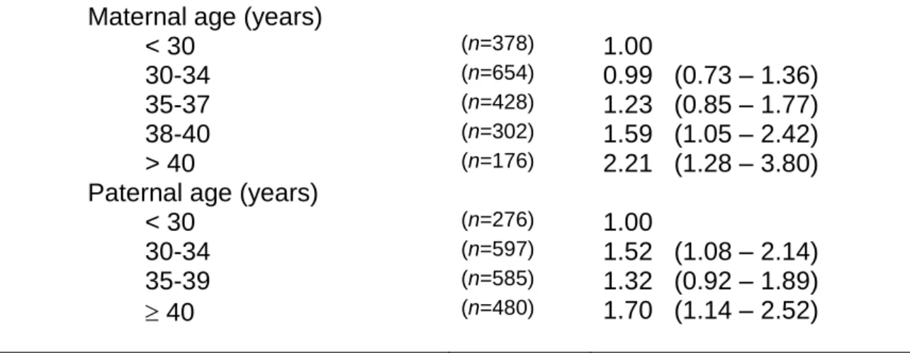
Table 1. Adjusted odds ratio (OR) and 95% confidence interval (95% CI) of
risk of failure to conceive after IVF attempts in a logistic regression model without maternal/paternal age interaction (n = 1,938)
Maternal age (years)
< 30 (n=378) 1.00
30-34 (n=654) 0.99 (0.73 – 1.36)
35-37 (n=428) 1.23 (0.85 – 1.77)
38-40 (n=302) 1.59 (1.05 – 2.42)
> 40 (n=176) 2.21 (1.28 – 3.80)
Paternal age (years)
< 30 (n=276) 1.00
30-34 (n=597) 1.52 (1.08 – 2.14)
35-39 (n=585) 1.32 (0.92 – 1.89)
Table 2. Adjusted odds ratio (OR) and 95% confidence interval (95% CI) of
risk of failure to conceive after IVF attempts in a logistic regression model with maternal/paternal age interaction (n = 1,938)
Paternal age Maternal age
(years) (years) < 30 30-34 35-37 38-40 > 40 < 30 1.00 (reference) (n=145) 0.79 (0.42, 1.51) (n=63) 1.62 (0.57, 4.57) (n=27) 1.29 (0.48, 3.43) (n=27) 0.49 (0.16, 1.50) (n=14) 30-34 1.44 (0.84, 2.46) (n=152) 1.34 (0.84, 2.13) (n=283) 1.49 (0.78, 2.85) (n=86) 1.47 (0.65, 3.33) (n=45) 5.34 (1.22, 23.42) (n=31) 35-39 0.78 (0.40, 1.50) (n=59) 1.24 (0.76, 2.02) (n=205) 1.33 (0.80, 2.22) (n=180) 3.05 (1.44, 6.48) (n=93) 2.16 (0.89, 5.20) (n=48) ≥ 40 1.25 (0.43, 3.62) (n=22) 1.36 (0.75, 2.46) (n=103) 2.00 (1.10, 3.61) (n=135) 2.03 (1.12, 3.68) (n=137) 5.74 (2.16, 15.23) (n=83)