Algerienne Democratic and Popular Republic
The Ministry of Higher Education and Research
University of MostaganemAbdelhamid Ibn Badis Scientifique
Faculty of Natural Sciences & Life Department of Biology
THESIS
For graduation
DOCTOR OF SCIENCE
Filiere: Biology Option: Biochemistry Presented ByMrs. Fatima LAICHE
Composition of the thesis jury
President: Mr Brahim .Lotmani Professor Univ MostaganemSupervisor: Mr Djebli Noureddine Professor Univ Mostaganem
Examiner: Mr Aoues AEKProfessor Univ Oran Es-Senia Oran
Examiner: Mr Taoufik Professor Univ Sahraoui Es-Senia Oran
Examiner: Mrs Hammadi Kheira MC-AUniv Mostaganem
University Year :2015 -2016
S
S
t
t
u
u
d
d
i
i
n
n
g
g
t
t
h
h
e
e
e
e
f
f
f
f
e
e
c
c
t
t
o
o
f
f
e
e
x
x
e
e
r
r
c
c
i
i
s
s
e
e
o
o
n
n
t
t
h
h
e
e
e
e
x
x
p
p
r
r
e
e
s
s
s
s
i
i
o
o
n
n
o
o
f
f
i
i
n
n
d
d
u
u
c
c
i
i
b
b
l
l
e
e
n
n
i
i
t
t
r
r
i
i
c
c
o
o
x
x
i
i
d
d
e
e
s
s
y
y
n
n
t
t
h
h
a
a
s
s
e
e
a
a
n
n
d
d
h
h
e
e
a
a
t
t
s
s
h
h
o
o
c
c
k
k
p
p
r
r
o
o
t
t
e
e
i
i
n
n
7
7
0
0
i
i
n
n
t
t
h
h
e
e
b
b
r
r
a
a
i
i
n
n
s
s
o
o
f
f
m
m
i
i
c
c
e
e
w
w
i
i
t
t
h
h
i
i
n
n
d
d
u
u
c
c
e
e
d
d
P
P
a
a
r
r
k
k
i
i
n
n
s
s
o
o
n
n
`
`
s
s
d
d
i
i
s
s
e
e
a
a
s
s
e
e
ii
Aeknowledgment
I WouId like to thank my supervisor professor Noureddine DJEBLI
For all kind, patiece and scientific supervision for all steps of this
thesis. I also would like to thank Dr. Ahed Alkhatib who offered his
Iaboratory and experience to produce this thesis. I also would like to
thank everyone helped me in this thesis.
iii
Dedication
I would like to dedicate this thesis to my husband Abdulla who has
supported me in all stages of my study, I also would like to dedicate
this work for my kids, the flowers of my life, to father and mother vestl
souls, to my brothers and sisters.
iv RESUME
La maladie de Parkinson est une maladie neurodégénérative commun. La carence en dopamine est considérée comme responsable du développement de la
maladie de Parkinson. L'entraînement physique a été associé à des améliorations chez les patients atteints de la maladie de Parkinson.L'objectif de la présente étude est d'explorer l'effet de l'entraînement sur l'expression de HSP70 et iNOS dans le cerveau des souris atteintes de la maladie de Parkinson induite.Quarante souris albinos ont été sélectionnés et ils sont attribuées en quatre groupes: contrôle sédentaire (CS, n = 10), exercice contrôle (EC, N = 10), et un groupe pour induire la maladie de Parkinson (GMP, N = 10) et un groupe de 10 souris albinos maladie de Parkinson qui suivé l'exercice (EPD, N = 10). Le Protocole de MPTP a été utilisé pour induire la maladie de Parkinson par des injections de 10 doses de MPTP (25 mg / kg) et du probénécide (250 mg / kg) pendant 5 semaines. Après la formation de protocoles et apres l'exercice sur tapis roulant est terminé.
Les échantillons de tissus cérébraux ont été évalués par immunohistochimie pour examiner l'expression de HSP70 dans les quatre groupes d'animaux.Les résultats de la présente étude montrent que l'expression de HSP70 a été réduite dans le cerveau des souris atteintes de la maladie de Parkinson de manière significative (P <0,05) en comparaison avec des groupes témoins. Les résultats ont également montré que l'entraînement physique a augmenté l'expression de HSP70 dans EC de façon significative (P <0,05) par rapport au groupe de contrôle, et non significative (P> 0,05) dans EPD par rapport à GMD.Bien que l'augmentation de l'expression de HSP70 dans la maladie de Parkinson nest été pas significatif donc il a un rôle potentiel dans l'amélioration de l'état des souris atteintes de la maladie de Parkinson et il peut avoir un rôle thérapeutique potentiel.Les données de la présente étude ont également montré une expression significative de iNOS dans les cerveaux de rats avec la maladie de Parkinson induite par rapport au groupe témoin (p 0,000) et par rapport au groupe de contrôle, et cette expression était significativement diminuée dans le groupe exercé (P 0,000).
v
ABSTRACT
Parkinson disease is a common neurodegenerative disease. Deficiency of dopamine is thought to be responsible for development of Parkinson Disease. Exercise training has been associated with improvements in patients with Parkinson Disease. The objective of the present study is to explore the effect of exercise training on the expression of HSP70 and iNOS in brains of mice with induced Parkinson Disease.
Forty albino mice were selected and assigned into four groups: Sedentary control (SC, N=10), exercised control (EC, N=10), Parkinson Disease (PD, N=10) and exercised Parkinson Disease (EPD, N=10). MPTP protocol was used to induce Parkinson Disease by injections of 10 doses of MPTP (25 mg/kg) and probenecid (250 mg/kg) over 5 weeks. After the protocols treadmill exercise training had been finished, samples from the brain tissues were assessed by immunohistochemistry to examine the expression of HSP70 in the four groups of animals.
The results of the present study showed that the expression of HSP70 was reduced in the brain of mice with Parkinson Disease significantly (P < 0.05) compared with control groups. The results also showed that exercise training increased the expression of HSP70 in EC significantly (P < 0.05) compared with control group, and insignificantly (P>0.05) in EPD compared with PD.
Although the increased expression of HSP70 in exercised Parkinson Disease was not significant, it has a potential role in improved the status of mice with Parkinson Disease and it may have a potential therapeutic role.
The data of the present study also showed significant expression of iNOS in brainsof rats with induced Parkinson Disease compared with control group (P 0.000) compared with control group, and this expression was significantly decreased in exercised group (P 0.000).
vi
صخلم
يبصعلا لكاتلا تاذ ةعئاشلا ةيبصعلا ضارملأا نموه نوسنكراب ضرم
.
صقن نأ دقتعيو
يدؤت يتلا ةيسيئرلا بابسلأا نم نيمابودلا
إ
ضرملا اذه روطت ىل
.
تاساردلا تنيب دقو
أ
نيرامتلا ن
نوسنكرابلا ىارمل يريرسلا ضاولا نيستت ي اًاماه اًارود بعمت ةياايرلا
.
ةساردلا هذه ت ده دقو
نييويتلا نيرشؤملا زيكرت ىمع ةياايرلا نيرامتلا ريثات فاشكتسا ىلا
(
HSP70
و
iNOS
)
ي
نوسنكرابلا ضرم ايي ثدتتسملا نارئللا ةممدا
.
ضبرأ ىلإ ايعيزوتو ارا نيعبرأ رايتخا مت دقو
تاعومجم
:
ةطباالا ةعومجملا
(
SC
،
N = 10
)
بيردتلا ةعومجم ،
(
ECN = 10,
)
ةعومجم ،
نوسنكراب ضرم
(
PD
،
N = 10
)
ةبردتملا نوسنكراب ضرم ةعومجمو
(
EPDN = 10,
)
.
مت دقلو
لوكوتورب مادختسا
MPTP
نقت قيرط نع نوسنكراب ضرم ثادتتسلا
10
تاعرج
(
25
غمم
/
ممك
)
ديسنيبوربلاو
(
250
غمم
/
غك
)
ةدمل
5
ضيباسأ
.
دعبو
أ
ةياايرلا تابيردتلا تيتنا ن
,
مت
أ
تانيع ذخ
نيرشؤملا زيكرت صتلل ةيئايميكلا ةيعانملا غابصلاا قيرط نع ايصت و غامدلا نم ةيجيسن
نييويتلا
(
HSP70
و
iNOS
)
ةساردلا تاعومجم ي
.
زيكرت نأ ةساردلا جئاتن تريظأو
HSP70
ي ضلخنا
أ
ضرمب ةباصملا نارئللا ةممد
لكشب نوسنكرابلا
إ
ماه يئاصت
(
P<0.05
)
ةرطيسلا ةعومجم ضم ةنراقم
.
اًاايأ جئاتنلا تريظأو
زيكرت تداز تانيرمتلا نأ
HSP70
بيردتلا ةعومجم ي
(
P<0.05
)
،ةرطيسلا ةعومجم ضم ةنراقم
(
P <0.05
)
نوسنكراب ةعومجم ضم ةنراقم ةبردتملا نوسنكراب ةعومجم ي
.
نم ةماه زيكارت دوجو ةساردلا هذه تانايب تريظأ امك
iNOS
ةباصلا نارئللا ةممدأ ي
ةرطيسلا ةعومجم ضم ةنراقم نوسنكرابلا ضرمب
(
P0.000
)
.
ظوتمم لكشب ضلخنا دق زيكرتلا اذهو
بيردتلا ةعومجم ي
(
P 0.000
)
.
تاجاتنتسلاا
:
ةساردلا تنيب
أ
يتصلا ضاولا نيستت ي دعاست ةياايرلا نيرامتلا ن
يويتلا رشؤملا زيكرت ةدايز للاخ نم كلذو نوسنكرابلا ىارمل
HSP70
أ
رشؤملا زيكرت ضلخ و
يويتلا
iNOS
ي
أ
نوسنكرابلا ضرمب ةباصملا نارئللا ةممد
.
دق ،كلذ ىمع ةولاعو
أ
جئاتن تراش
ةساردلا هذه
إ
ةيجلاع فادهاك تانيتوربلا هذه فاديتسا ةيناكما ىل
.
ةلادلا تاممكلا
:
،ةميقثلا نداعملا
شاعرلا ضرم ةصاصرلا
.
Recommendations
vii
1- Exerise traninig is recomemended regulary for wide spectrum of people either healthy or diseased.
2- Patients with PD are recommended to perform exercise training to improve their health status.
3- It is recommended to use HSP70 and iNOS inhibitors by pharmaceutical
companies as new therapeutic option fo PD.
viii
APV: Amino phosphonoValeric
AD: aldehyde dehydrogenase
ADHD: Attention defcit Hyperactivity disorder
AMD:acid mine drainage
ATSDR: Agency for Toxicsubstances and Disease Registry
BDNF: Brain- derived neurotrophic Factor
BG: Basal Ganglia
BLL: Blood Lead Level
CAMKII: Calcium\calmadulin Kinase II
CDC: Centers for Disease Control
COMT: catechol-O- methyltransferase
CREB:cAMP Response element binding protein
DA: Dopamine
eNOS: endothelial NOS
EPSCs: Excitatory postsynaptic Currents
GA: Geldanamycin
GABA: gamma-aminobutyric acid
GABA: glutamate amby-aminobutyric acid
HSP: heat shock protein iNOS: inducible NOS
IPSCs: Inhibitory postsynaptic Currents
IQ:Intelligence quotient
LBs: LewyBodies
L-DOPA: L-dihydroxyphenylalanine
MAKP: mitogen activated Protein Kinase
MAO: monoamine oxidase
MPTP: N-methyl-4-phenyl-1, 2, 3, 6-tetrahydropyridine
NINDS: National Institute of Neurological Disorders and Stroke
NMDAR: N-mehyle D-aspartate Receptor
nNOS: Neuronal NOS NO: Nitric oxide
ix
NR1: NMAD Receptor
Pb: Lead (Plumbum)
PD: Parkinson's disease ROS: reactive oxygen species
SH: sulphydrylgroupe
SPM: Suspended particulate matters
TH: Tyrosine hydroxylase
UCH-L1: Ubiquitin carboxyl-terminal hydrolase L1 UPDRS:The Unified Parkinson’s Disease Rating scale
VGCCs: Voltage – gated calcium channels
VPA: valproic acid
VTA: ventral tegmental area αSN :α-synuclein
x
List of figures
Figure 1: Neuropathology of Parkinson’s diseas…………
Figure 2: Dopamine synthesis from amino acid tyrosine by the action of tyrosine…………...
Figure 3:Basal ganglia (Striatum (Caudate and putamen), globus pallidus (GP) (externa (GPe) and interna (GPi)), substantia nigra (SN), and subthalamic nucleus (STN))……….
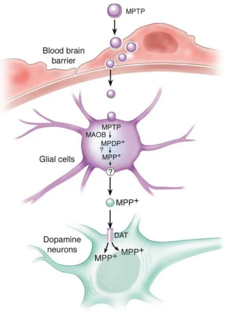
Figure 4: Direct pathway (CortextostriatumtoGpi and SNPrtoThalamustoCortex), Indirect pathway (cortextostriatumtoGPetoSTNtoGPi and SNPrtoThalamustoCortex)………. Figure 5:Schematic Representation of MPTP Metabolism……… Figure 6: Schematic Representation of MPP+ Intracellular Pathways Inside dopaminergic
neurons……….
Figure 7: Mechanisms of Neurodegeneration……… Figure 8: The modified human treadmill to train the rats according to our rehabilitation protocol... Figure 9: A histogram represents the average expression of nNOS in the striatum of
sedentary Parkinson's disease and sedentary control……….. Figure 10: Photograph of the striatum of the mouse brain without MPTP treatment……….. Figure 11:A histogram represents the average expression of nNOS in the striatum of
sedentary Parkinson's disease and exercised Parkinson's disease……….… Figure 12: Photograph of the striatum of the mouse brain after treatment with MPTP……… Figure 13:A histogram represents the average expression of nNOS in the striatum of
exercise control and exercised Parkinson's disease………. Figure 14: Photograph of the striatum of the mouse brain.……….………...
Figure 15: A histogram represents the average expression of nNOS in the striatum of
sedentary control and exercised control………... Figure 16: Photograph of the striatum of the mouse brain………... Figure 17: A histogram represents the average expression of iNOS in the striatum of
sedentary control and sedentary Parkinson………... Figure 18: Photograph of the striatum of the mouse brain…………..…………... Figure 19: A histogram represents the average expression of iNOS in the striatum of
sedentary control, exercised control, sedentary Parkinson and exercise Parkinson……… Figure 20: Photograph of the striatum of the mouse brain……….…………...
22
27
29
30
35
36
49
58
64
65
66
67
68
69
70
71
72
73
74
75
xi
Figure 21: A histogram represents the average expression of iNOS in the striatum
ofsedentary control and exercised
Parkinson………..
Figure 22: Photograph of the striatum of the mouse brain………. Figure 23: A histogram represents the average expression of iNOS in the striatum of
sedentary Parkinson and exercised Parkinson…….………
Figure 24: Photograph of the striatum of the mouse brain after treatment with MPTP………...
Figure 25: A histogram represents the average expression of HSP70 in the striatum of
sedentary control and sedentary Parkinson……….
Figure 26: The expression of HSP70 in EC group……….……...………... Figure 27: The expression of HSP70 in SPD group 40X………...…….………... Figure 28:The expression of HSP70 in EPD group, 40X………... Figure 29: The expression of HSP70 in SC group 40 X………….…..…….…………...
76
77
78
79
80
81
82
82
83
xii
SUMMARY
I- Chapter One: Heavy metals and neurotoxicity -
Introduction………..…..…....1
1. Heavy Metal Emission……….…..…...…2
1.2. Chemistry of Heavy Metal Pollution………...…..………3
1.3.Human Exposure through Food, Air and Water………...……..………3
1.4. Human Exposure through Industrial Products……….………..4
1.5. Occupational Exposure……….……..…4
1.6. Biochemistry of Toxicity……….………...5
1.7. Human Health and Heavy Metals Exposure……….…...5
2. Lead……….………...7
2.1.Neurotoxic efects of Pb2+: Results from Epidemiological Studies…...10
2.2. Neurotoxicity: Results from experimental animal studies………...….11
II- Chapter Two: Parkinson's Disease 1. Introduction……….. 19
1.1An Overview of Parkinson's Disease……….. 19
1.2 Epidemiology of PD……….. 20
1.3 Clinical picture………... 20
1.4 Pathology of PD………. 20
1.5 Etiology of PD………... 23
1.6 Management of PD……… 1.7 The relationship between physical and PD……… 1.8 Nitric oxide……… 1.9 Heat shock proteins……… 23 24 24 25 1.10 Significance of the study……….. 25
1.11 Study hypothesis……….. 25
xiii
Chapter Three: Review of Literature Dopamine
- Review of Literature……….. 2.1 Dopamine synthesis and release………
27 27
2.2 Basal Ganglia……… 28
2.2.1 Circuit connections of Basal Ganglia………. 29
2.3 Cardinal symptoms of Parkinson's disease……… 31
2.3.1 Tremor……… 2.3.2 Bradykinesia………... 2.3.3 Rigidity………... 2.3.4 Posturalinstability……….. 31 31 32 32 2.4 Stages of Parkinson's disease………. 32
2.5 Causes of Parkinson's disease……… 32
2.5.1 Genetic Causes of PD………... 33
2.5.2 Mitochondrial dysfunction……….. 36
2.5.3 Environmental factors………. 37
2.6 Nitric Oxide (NO)……….. 38
2.6.2 Nitric Oxide Synthase (NOS)………. 38
2.6.3 Function of NO………... 40
2.6.4 Nitric Oxide Synthase and Parkinson Disease……… 40
2.7 Parkinson Disease and Exercise……… 41
2.8 HSPs and PD Pathophysiology……….. 44
2.9 PD-Related Gene Mutations and Possible Association with HSPs... 45
2.10 Alpha-synuclein (αSN)……… 45
2.11 Parkin………... 46
2.12 Ubiquitin carboxyl-terminal hydrolase L1 (UCH-L1)………. 46
2.13 Protective Role of HSPs in PD……… 47
2.14 Hsp90………... 47
2.15 Hsp70………... 48
2.16 Small HSPs………... 50
2.17 Potential Target for the Treatment of PD……… 51
xiv
2.19 HSPs may promote the UPS in protein degradation……… 52 2.20 HSPs inducers and their potential application in PD……… 53
III- Material and methods
55 3.1 Chronic model of PD………. 56 3.2 Endurance exercise protocol……….. 57 3.3 Identification of striatal tissue……… 59 3.4 Brain section immunohistochemistry using nNOS and
iNOS antibodies 59
3.5 Analysis of tissues images 61
IV- Results 63
4.1 nNOS (N1) results 64 4.2 iNOS (N2) results 72 4.3 The Results for Heat Shock Protein (HSP70)
V- Discussion ofresults 84 VI- Conclusions VII- References VIII- Publication 91 93 126
Chapter One
Heavy metals and
1
INTRODUCTION
The term ―heavy metals‖ refers to any metallic element that has a relatively high density and is toxic or poisonous even at low concentration (Lenntech, 2004). ―Heavy metals‖ is a general collective term, which applies to the group of metals and metalloids with atomic density grea-ter than 4 g/cm3, or 5 times or more, greater than water (Huton and Symon,1986; Battarbee et al., 1988; Nriagu and Pacyna 1988; Nriagu, 1989; Garbarino et al., 1995, Hawkes, 1997). However, being a heavy metal has little to do with density but concerns chemical properties. Heavy metals include lead (Pb), cadmium (Cd), zinc (Zn), mercury (Hg), arsenic (As), silver (Ag) chromium (Cr), copper (Cu) iron (Fe), and the platinum group elements. Environment is defined as the totality of circumstances surrounding an organism or group of organisms especi-ally, the combination of external physical conditions that affect and influence the growth, development and survival of organisms (Farlex, 2005).
It consists of the flora, fauna and the abiotic, and includes the aquatic, terrestrial and atmospheric habitats. The environment is considered in terms of the most tangible aspects like air, water and food, and the less tangible, though no less important, the communities we live in (Gore, 1997).
A pollutant is any substance in theenvironment,which causes objection-able effects, impairing the welfare of the environment, reducing the quality of life and may eventually cause death. Such a substance has to be present in the enviro-nment beyond a set or tolerance limit, which could be either a desirableor acceptable limit. Hence, environ-mental pollution is the presence of a pollutant in the envi-ronment; air, water and soil, which may be poisonous or toxic and will cause harm to living things in the polluted (
Duru.ibe et al., 2007
).2
1. Heavy Metal Emission
Heavy metals can be emitted into the environment by both natural and anthropogenic causes. The major caus-es of emission are the anthropogenic sources specifically mining operations (Hutton and Symon, 1986; Battarbee et al., 1988; Nriagu, 1989). In some cases, even long aft-er mining activities have ceased, the emitted metals con-tinue to persist in the environment. Peplow (1999) repor-ted that hard rock mines operate from 5-15 years until the minerals are depleted, but metal contamination that occurs as aconsequence of hard rock mining persist for hundreds of years after the cessation of mining opera-tions. Apart from mining operations, mercury is introduc-ed into the environment through cosmetic products as well as manufacturing processes like making of sodium hydroxide. Heavy metals are emitted both in elemental and comp-ound (organic and inorganic) forms. Anthropogenic sour-esof emission are the various industrial point sources including former and present mining sites, foundries and smelters, combustion by-products and traffics (UNEP / GPA, 2004).
Cadmium is released as a by- product of zinc (and occasionally lead) refining; lead is emitted du-ring its mining and smelting activities, from automobile exhausts (by combustion of petroleum fuels treated with tetraethyl lead antiknock) and from old lead paints; mercury is emitted by the degassing of the earth’s crust. Generally, metals are emitted during their mining and processing activities (Lenntech, 2004). Environmental pollution by heavy metals is very promi-nent in areas of mining and old mine sites and pollution reduces with increasing distance away from mining sites (Peplow, 1999).
These metals are leached out and in sloppy areas, are carried by acid water downstream or run-off to the sea. Through mining activities, water bodies are most emphatically polluted (Garbarino et al., 1995; INECAR, 2000). The potential for contamination is incre-ased when mining exposes metal-bearing ores rather than natural exposure of ore bodies through
3
erosion (Garbarino et al., 1995), and when mined ores are dumped on the earth surfaces in manual dressing proce-sses. Through rivers and streams, the metals are trans-ported as either dissolved species in water or as an integral part of suspended sediments, (dissolved species in water have the greatest potential of causing the most deleterious effects).
They may then be stored in river bed sediments or seep into the underground water thereby contaminating water from underground sources, particu-larly wells; and the extent of contamination will depend on the nearness of the well to the mining site. Wells located near mining sites have been reported to contain heavy metals at levels that exceed drinking water criteria (Garbarino et al., 1995; Peplow, 1999).
1.2. Chemistryof Heavy Metal Pollution
Mining activities and other geochemical processes often result in the generation of acid mine drainage (AMD), a phenomenon commonly associated withmining activities. It is generated when pyrite (FeS2) and other sulphide minerals in the aquifer and present and former mining sites are exposed to air and water in the presence of oxidizing bacteria, such as Thiobacillus ferrooxidans, and oxidised to produce metal ions, sulphate and acidity (Ogwuegbu and Muhanga, 2005).
2FeS2 + 7O2 + 2H2O 2FeSO4 + 2H2SO4
2FeSO4 + 2H2SO4 Fe2(SO4)3 + SO2 + 2H2O Fe2(SO4)3 + 2FeAsS + 9/2O2 + 3H2O 2H3AsO4 + 4FeSO4 + S
1.3.Human Exposure Through Food, Air and Water
Heavy metal pollution of surface and underground water sources results in considerable soil pollution and pollution increases when mined ores aredumped on the ground surface for manual dressing (Garbarino et al.,1995;INECAR, 2000). Surface dumping exposes the metals to air and rain thereby generating
4
much AMD. When agricultural soils are polluted, these metals are taken upby plants and consequently accumulate in their tissues (Trueby, 2003).Animals that graze on such contaminated plants and drink from polluted waters, as well as marine lives that breed in heavy metal polluted waters also accumulate such metals in their tissues, and milk, if lactating (Habashi, 1992; Garbarino et al., 1995; Horsfall and Spiff, 1999; Peplow, 1999). Humans are in turn exposed to heavy metals by consuming contaminated plants and animals, and this has been known to result in various biochemical disorders. In summary, all living organisms within a given ecosystem are variously contaminated along their cycles of food chain.
1.4. Human Exposure through Industrial Products
Industrial products that are used in homes, and which have been produced with heavy metals are sources of human exposure to such heavy metals. Mercury expos-ure is through disinfectants (like mercurochrome), anti-fungal agents, toiletries, creams and organo-metallics (McCluggage, 1991); cadmium exposure is through nickel/cadmium batteries and artist paints; lead exposure is through wine bottle wraps, mirror coatings, batteries, old paints and tiles and linolein amongst others. Infants are more susceptible to the endangering effects of expo-sure to heavy metals.
1.5. Occupational Exposure
Heavy metal exposure occurs significantly by occupa-tional exposure. Workers of the mining and production of cadmium, chromium, lead, mercury, gold and silver have been reported to be thus exposed; also inhabitants around industrial sites of heavy metal mining and proces-sing, are exposed through air by suspended particulate matters (SPM) (Heyer, 1985; USDOL, 2004; Ogwuegbu and Muhanga, 2005).
5
1.6. Biochemistry of Toxicity
The poisoning effects of heavy metals are due to their interference with the normal body biochemistry in the normal metabolic processes. When ingested, in the acid medium of the stomach, they are converted to their stable oxidation states (Zn2+) and combine with the body’s biomolecules such as proteins and enzymes to form strong and stable chemical bonds. The equations below show their reactions during bond formation with the sulphydryl groups (-SH) of cysteine and sulphur atoms of methionine (-SCH3) (Ogwuegbu and Ijioma, 2003).
The hydrogen atoms or the metal groups in the above case are replaced by the poisoning metal and the enzyme is thus inhibited from functioning, whereas the protein–metal compound acts as a substrate and reacts with a metabolic enzyme.
1.7. Human Health and Heavy Metals Exposure
Metals, a major category of globally-distributed pollutants, are natural elements that have been extracted from the earth and harnessed for human industry and products for millenia. (An exception to metals being ―natural‖ is plutonium, the material at the heart of nuclear weapons,created by man through the processing of uranium.) (
Low et al., 2000
). Metals are notable for their wide environmental dispersion from such activity; their tendency to accumulate in select tissues of the human body; and their overall potential to be toxic even at relatively minor levels of exposure. Some metals, such as copper and iron, are essential to life and play irreplaceable roles in, for example, the functioning of critical enzyme systems (Kapaj et al., 2006
).Other metals are xenobiotics, i.e., they have no useful role in human physiology (and most other living organisms) and, even worse, as in the case of lead and mercury, may be toxic even at trace levels of exposure. Even those metals that are essential, however, have the potential to turn harmful at very high levels of exposure, a reflection of a very basic tenet of toxicology--―the dose
6
makes the poison.‖ One reflection of the importance of metals relative to other potential hazards is their ranking by the U.S. Agency for Toxic Substances and Disease Registry (ATSDR), which lists all hazards present in toxic waste sites according to their prevalence and the severity of their toxicity. The first, second, third, and sixth hazards on the list are heavy metals: lead, mercury, arsenic, and cadmium, respectively. Exposure to metals can occur through a variety of routes. Metals may be inhaled as dust or fume (tiny particulate matter, such as the lead oxide particles produced by the combustion of leaded gasoline) (
Peterson et al.,
2006
).Some metals can be vaporized (e.g., mercury vapor in the manufacture of fluorescent lamps) and inhaled. Metals may also be ingested involuntarily through food and drink. The amount that is actually absorbed from the digestive tract can vary widely, depending on the chemical form of the metal and the age and nutritional status of the individual. Once a metal is absorbed, it distributes in tissues and organs. Excretion typically occurs primarily through the kidneys and digestive tract, but metals tend to persist in some storage sites, like the liver, bones, and kidneys, for years or decades.The toxicity of metals most commonly involves the brain and the kidney, but othermanifestations occur, and some metals, such as arsenic, are clearly capable of causing cancer. An individual with metals toxicity, even if high dose and acute, typically has very general symptoms, such as weakness or headache. This makes the diagnosis of metals toxicity in a clinical setting very difficult unless a clinician has the knowledge and training to suspect the diagnosis and is able to order the correct diagnostic test. Chronic exposure to metals at a high enough level to cause chronic toxicity effects (such as hypertension in individuals exposed to lead and renal toxicity inindividuals exposed to cadmium) can also occur in individuals who have no symptoms (
ASTDR, 2005
).Much about metals toxicity, such as the genetic factors that may render some individuals especially vulnerable to metals toxicity, remains a subject of intense investigation. It is possible that low-level metals exposure contributes
7
much more towards the causation of chronic diseaseand impaired functioning than previously thought.This chapter focuses on exposure to the four ―heavy‖ metals on the ATSDR listmentioned above —lead, mercury, arsenic, and cadmium—as they are arguably the mostimportant metal toxins from a global, as well as U.S. perspective. Some additional remarks arealso made regarding a few other metals of concern. (Exposure to arsenic and lead in drinking wateris covered by John Balbus in Chapter 3, Water Quality and Water Resources)(
ASTDR, 2005
).2. Lead
Exposure For centuries, lead has been mined and used in industry and in household products. Modern industrialization, with the introduction of lead in mass-produced plumbing, solder used in food cans, paint, ceramic ware, and countless other products resulted in a marked rise in population exposures in the 20th century. The dominant source of worldwide dispersion of lead into the environment (and into people) for the past 50 years has clearly been the use of lead organic compounds as antiknock motor vehicle fuel additives. Since leaded gasoline was introduced in 1923, its combustion and resulting contamination of the atmosphere has increased background levels everywhere, including the ice cap covering Northern Greenland (Fig. 1), where there is no industry and few cars and people.
Although a worldwide phase-out of leaded gasoline is in progress ,it is still being used all over the world. The current annual worldwide production of lead is approximately 5.4 million tons and continues to rise. Sixty percent of lead is used for the manufacturing of batteries (automobile batteries, in particular), while the remainder is used in the production of pigments, glazes, solder,plastics, cable sheathing, ammunition, weights, gasoline additive, and a variety of other products.Such industries continue to pose a significant risk to workers, as well as surrounding communities.In response to these risks, many developed countries over the last 25 years have implemented regulatory action that has effectively decreased actual exposures to the generalpopulation. However, exposures remain
8
high or are increasing in many developing countriesthrough a rapid increase in vehicles combusting leaded gasoline and polluting industries (some ofwhich have been ―exported‖ by corporations in developed countries seeking relief fromregulations). Moreover, some segments of the population in developed countries (such as theU.S.) remain at high risk of exposure because of the persistence of lead paint, lead plumbing, andlead-contaminated soil and dust, particularly in areas of old urban housing. A number of factors can modify the impact of lead exposures. For example, water with alower pH (such as drinking water stemming from the collection of untreated ―acid rain‖) willleach more lead out of plumbing connected by lead solder than more alkaline water. Lead fromsoil tends to concentrate in root vegetables (e.g., onion)and leafy green vegetables (e.g., spinach).Individuals will absorb more lead in their food if their diets are deficient in calcium, iron, or zinc.Other more unusual sources of lead exposure also continue to be sporadically found, such asimproperly glazed ceramics, lead crystal, imported candies, certain herbal folk remedies, andvinyl plastic toys.ToxicityLead has been the intense focus of environmental health research for many decades.
Studies inhumans were greatly assisted by the development of methods (such as graphite furnace atomicabsorption spectroscopy) for the accurate and reliable measurement of lead in blood (measured inunits of micrograms per deciliter [mg/dL]), a technique that is now widely available and used forsurveillance and monitoring, as well as research. The general body of literature on lead toxicity indicates that, depending on the dose, leadexposure in children and adults can cause a wide spectrum of health problems, ranging fromconvulsions, coma, renal failure, and death at the high end to subtle effects on metabolism andintelligence at the low end of exposures. Children (and developing fetuses) appear to beparticularly vulnerable to the neurotoxic effects of lead.
A plethora of well-designed prospectiveepidemiologic studies has convincingly demonstrated that low-level lead exposure in children lessthan five
9
years of age (with blood lead levels in the 5-25 mg/dL range) results in deficits inintellectual development as manifested by lost intelligence quotient points.As a result, in theU.S., the Centers for Disease Control (CD) lowered the allowable amount of lead in a child’sblood from 25 to 10 mg/dL and recommended universal blood lead screening of all children between the ages of six months and five years.However, a number of issues still remain unresolved with respect to lead toxicity in children. Among the most important is the risk posed to the fetus posed by mobilization of long-ved skeletal stores of lead in pregnant women.Recent research has clearly demonstrated thataternal bone lead stores are mobilized at an accelerated rate during pregnancy and lactation ande associated with decrements in birth weight, growth rate, and mental development. Sinceone lead stores persist for decades,it is possible that lead can remain a threat to fetal healthany years after environmental exposure had actually been curtailed. In contrast to children, adults are generally allowed by regulations to be exposed to highermounts of lead. In the U.S., for example, the Occupational Safety and Health Administrationquires that the blood lead levels of exposed workers be maintained below 40 mg/dL as a way ofreventing toxic effects to nerves, the brain, kidney, reproductive organs, and heart. This standardprobably outdated, however. First, the standard does not protect the fetuses of women who become pregnant while on the job (or even if they leave the job for several years because of the isue of bone lead mobilization, as discussed above).
Second, recent epidemiologic studies have linked blood lead levels in the range of 7-40 mg/dL with evidence of toxicity in adults, such as neurobehavioral decrementsand renal impairments.Third, recent studies using a newlydeveloped technique, K-x-ray fluorescence, to directly measure bone lead levels (as opposed toblood lead levels) have provided evidence demonstrating that cumulative lead exposure inindividuals with blood lead levels well below 40 mg/dL is a major risk factor for the developmentof hypertension,cardiac conduction delays,and cognitive impairments.Finally, even as research progresses to delineate the full toxicologic implications of leadexposure, investigations at the interface of genetics and environmental health are beginning
10
touncover subgroups of individuals who may be particularly susceptible to the toxicity of lead.
2.1.Neurotoxic efects of Pb2+: Results from Epidemiological Studies
The neurological efects of Pb2+ in exposed children have been a driving factor in reducing the level of Pb2+ in the environment (
Gilbert and
Weiss, 2006
). In 1991 the United States Centers for Disease Control and Prevention (CDC) lowered the defnition of Pb2+ intoxication to 10 µg/dL BLL (the current regulatory level) motivated by the evidence from several studies that children with BLL of at least 10 µg/dL had impaired intellectual function (CDC, 1991). More recently, studies have shown that the dose-response of Pb2+ on IQ in children is non-linear, with lower exposures of Pb2+ resulting in a greater rate of IQ loss than at higher exposures (Canfield etal.,2003; Lanphear
et al., 2005;Hu et al., 2006;Jusko et al., 2008
). These data clearly demonstrate that the majority of the estimated IQ loss in Pb2+-exposed children occurs during the frst 10 µg/dL, and many studies have suggested a lack of a threshold for the efects of Pb2+ on IQ (Canfield etal.,2003; Lanphear et al.,
2005; Jusko et al., 2008
).A large, internationally-pooled analysis of Pb2+-exposed children estimated that children with BLLs of 10 µg/dL experience a defcit of about 6.2 IQ points relative to children with estimated BLLs of 1 µg/dL (
Lanphear et al.,
2005
).This is comparable to the defcit of 7.4 IQ points observed in children with BLLs of 10 µg/dL in another large study (
Jusko et al.,
2008
). On an individual level, a decrease in IQ of 6-7 points would be difcult to detect. However, the efect of a population decrease in IQ of this magnitude is quite signifcant. By shifing the normal distribution of IQ scores lower, the number of children with impaired intelligence would increase signifcantly while the number of exceptionally gifed children would decrease (Gilbert and Weiss,
2006
). Several researchers have studied this efect from an economical standpoint11
and suggest that the monetary cost of such an efect may total over 40 billion dollars for one age group alone. Over a 20-year period, one generation, this loss may amount to nearly 800 billion dollars (
Landrigan et al.,2002; Gilbert and
Weiss, 2006
). In addition to the cognitive defcits associated with Pb2+ exposure, children with elevated BLLs experience behavioral defcits. School children with elevated BLLs are more likely to act out in class, display antisocial behavior, and have trouble paying attention (Bellinger et al.,1994;
Needleman et al., 1996; Royet al., 2009
).Cumulative childhood Pb2+ exposure was associated with a higher incidence in behavioral problems in 8-year-old children (Leviton et al.,1993; Bellinger et al.,1994
). These behavioral efects appear to have a phenotype similar to attention-defcit hyperactivity disorder (ADHD).Furthermore, recent studies have identifed that childhood Pb2+ exposure is positivity associated with ADHD diagnosis (
Froehlich et al.,
2009; Roy et al., 2009
).The cognitive and behavioral defcits of Pb2+-exposed children persist even afer the cessation of Pb2+ exposure (White et
al., 2007
), and chelation therapy is unable to remediate the efect of Pb2+ on cognition (Chisolm et al., 1990; Rogan et al., 2001; Dietrich et al., 2004
). Prenatal and/or childhood Pb2+ exposure was associated with anti-social and delinquent behavior as adolescents (Dietrich et al., 2001
), an increased likelihood be an adjudicated delinquent (Needleman et al., 2002
), or to be arrested as an adult (Wright et al., 2008).Furthermore, childhood Pb2+ exposure may predict adult cognitive function (
Mazumdaret al., 2011
). Children who experience elevated Pb2+ levels are more likely to have decreased brain volume in adulthood in specifc brain regions (Cecil et al., 2008
). These changes could account for altered behavior and cognition in adults exposed to Pb2+ as children. Tus, developmental Pb2+ exposure in humans results in long-lasting efects on12
cognition and behavior even afer cessation of exposure. Possible Mechanism of Pb2+.
2.2. Neurotoxicity: Results from experimental animal studies
It is believed that Pb2+ targets the learning and memory processes of he brain by inhibiting the N-methyl-D-aspartate receptor (NMDAR), which is essential for hippocampus-mediated learning and memory (
Morris et al.,
1982, 1986
). The NMDAR is essential for learning spatial navigation tasks n animal models (Morris et al., 1986
),and animals which have been developmentally xposed to Pb2+ exhibit similar learning defcits as those with absent or mpaired NMDARs (Morris et al., 1982, 1986; Tsienet al., 1996
). The NMDAR is composed of an obligatory NR1 subunit and one or more accessory subunits from the NR2 and NR3 families. In the ippocampus, NR2A and NR2B are the most abundant NR2 family members. Pb2+ is a potent, non-competitive antagonist of the NMDAR (Alkondon et al., 1990;
Guilarte and Miceli, 1992; Guilarte, Miceli and Jett, 1994; Ruan et al.,
1998
). Evidence suggests that Pb2+ binds the Zn2+ regulatory site of the NMDAR in a voltage-independent manner (Guilarte et al., 1995; Yamada
et al.,1995; Gavazzo et al., 2008
).Since Zn2+ binds with high afnity at a regulatory site on the NR2A subunit (
Fayyazuddin et al., 2000
), but with lower afnity to the NR2B subunit (Rachline et al., 2005
), this suggests preferential sensitivity of NR2A-NMDARs for Pb2+ (Guilarte et al., 1995; Gavazzo et al., 2008
). In support of this hypothesis, electrophysiological studies on recombinant receptors demonstrate that Pb2+ more potently inhibits NR2A-NMDARs than NR2B-NMDARs (Yamada et al.,1995; Omelchenko et al., 1996
), or the tri-heteromeric form, NR1/NR2A/NR2B-NMDAR (Omelchenko et al., 1996
). In addition to acting as an NMDAR antagonist, Pb2+ exposure also disrupts normal NMDAR ontogeny. Chronic developmental Pb2+ exposure results in decreased NR2A content in the hippocampus (Guilarte and McGlothan, 1998;
13
Nihei MK, Guilarte, 1999; Nihei et al., 2000
), and altered expression of NR1 splice variants (Guilarte etal., 2000; Zhang et al., 2002; Guilarte and,
McGlothan, 2003
). In contrast, NR2B mRNA levels either remained unchanged or slightly increased in rats developmentally exposed to Pb2+ (Guilarte and McGlothan, 1998; Nihei MK, Guilarte, 1999; Nihei et al.,
2000; Zhang et al., 2002
). Together, these data suggest that Pb2+ delays the normal developmental witch of increased NR2A incorporation in NMDARs with synapse maturation (Toscano et al., 2002; Toscano and Guilarte., 2005
).Similar trends have also been observed in culturedneuron systems(
Xu
and Rajanna, 2006; Neal et al., 2011
) and suggest that Pb2+ exposure may cause lasting changes in NMDAR subunit composition and expression. In addition to hippocampal changes in NMDAR subunit expression and ontogeny, Pb2+ may alter the cellular distribution of NMDAR populations. We have shown that Pb2+ exposure during synaptic development in hippocampal cultures reduces the levels of synaptic NR2A-NMDARs with a concomitant increase in extrasynaptic NR2B-NMDARs (Neal et al., 2011). Tis is signifcant because the NR2 family members are linked to diferential MAPK signaling (Kim et al., 2005
), pro-death or pro-life signaling (Soriano et
al., 2008
), and diferential induction of nuclear gene expression (Hardingham et
al., 2002
). In particular, NR2A-NMDAR activation is linked to cell survival pathways and cyclic AMP response element binding protein (CREB) activation while NR2B-NMDAR activation is linked to cell death pathways and CREB shutof (Hardingham et al., 2002).
Thus, changes in synaptic localization of NMDARs by Pb2+ could alter downstream NMDAR-mediated signaling. Supporting this hypothesis, chronic developmental Pb2+ exposure results in altered MAPK signaling (Cordova et al., 2004
), calcium/calmodulin kinase II (CaMKII) activity (Toscano et al., 2005
), and altered CREB phosphorylation and binding afnity (Toscano et al., 2002;
2003
).CREB is a transcription factor for many immediate early genes (IEGs),14
which play an essential role in memory consolidation and are expressed as a result of NMDAR activity (
Bourtchuladze et al., 1994
). Altered IEG expression in animals exposed to Pb2+ has been observed (Kim et al.,2002
) indicating that altered CREB activity due to Pb2+-mediated disruption of NMDAR signaling may result in impaired learning and memory processes.Pb2+ exposure can cause defcits in neurotransmission.Rats chronically exposed to low levels of Pb2+ have reduced Ca2+-dependent glutamate and γ-aminobutyric acid (GABA) release in the hippocampus (
Lasley and Gilbert, 1996; 2002; Xiao et al., 2006
), which indicates presynaptic neuron dysfunction during Pb2+ exposure. In cultured hippocampal neurons (Braga, Pereira and Albuquerque, 1999
)and in brain slices (Xiao et al., 2006
), Pb2+ exposure impairs excitatory postsynaptic currents (EPSCs) and inhibitory postsynaptic currents (IPSCs).EPSCs and IPSCsare dependent upon neurotransmitter release from the presynaptic neuron, thus, reductions in EPSCs and IPSCs indicate a defcit in neurotransmission in both the glutamatergic and GABAergic systems as a result of Pb2+ exposure.A recent study from our laboratory has shown that Pb2+ exposure in cultured hippocampal neurons during synaptic development resulted in altered presynaptic protein expression and defcits in vesicular neurotransmitter release (
Neal et al., 2010
). Pb2+ exposure reduced the expression of key presynaptic proteins involved in vesicular release, such as synaptophysin (Syn) and synaptobrevin (Syb). Reductions of vesicular release proteins were associated with both glutamatergic and GABAergic synapses, consistent with electrophysiological observations regarding EPSC and IPSC generation during Pb2+ exposure (Braga, Pereira and
Albuquerque, 1999; Xiao et al., 2006
).Vesicular release in Pb2+-exposed neurons was signifcantly impaired relative to control conditions as determined by live-imaging studies using the synaptic vesicle dye FM 1-43 (
Neal et al., 2010
). Together, animal and15
cell culture studies indicate a role for Pb2+ in presynaptic dysfunction which results in reduced neurotransmission (
Neal and Guilarte, 2010
). One molecular mechanism by which Pb2+ may disrupt neurotransmission is by inhibiting neuronal voltage-gated calcium (Ca2+) channels (VGCCs) (Penget al., 2002
). Removal of extracellular Ca2+ from hippocampal slice cultures resulted in identical efects on IPSC frequency as Pb2+ exposure, suggesting that the Pb2+-induced inhibition of IPSC frequency occurred via reduction of Ca2+ infux through VGCCs (Xiao et al., 2006
). Inhibition of presynaptic VGCCs may prevent the necessary rise in internal Ca2+ required for fast, Ca2+-dependent vesicular release, thus interfering with neurotransmission. However, the efects of Pb2+ we observed on presynaptic protein expression were dependent on NMDAR activity, based on comparison studies with thespecifcNMDAR antagonistaminophosphonovaleric acid (APV,which does not inhibit VGCCs) which resulted in similar efects as Pb2+ exposure (Neal
et al., 2010
).Thus, while Pb2+ inhibits VGCCs, which may result in impaired neurotransmission, VGCC inhibition by Pb2+ is not exclusively responsible for the presynaptic efects of Pb2+ and long-term NMDAR inhibition plays an important role in these efects.An emerging theme in the mechanism of Pb2+ neurotoxicity is the disruption of intracellular Ca2+ dynamics. Inhibition of either VGCCs or NMDARs by Pb2+ would result in a signifcant reduction of Ca2+ entry into the cell. Tis is important because Ca2+ signaling is essential for synaptic development and plasticity (
Konur and Ghosh, 2005; Waites and
Garner, 2011
) and perturbation of these processes can lead to neurological disease states (Mirnics et al., 2001; Waites and Garner, 2011
). One key Ca2+-dependent pathway involved in synaptic development and neurotransmitter release is brain-derived neurotrophic factor (BDNF) signaling (Shieh et al., 1998; Shieh and Ghosh, 1999; Chen et al., 2003; Matsuda et
al., 2009
). BDNF is a trans-synaptic signaling molecule that is released from both axons and dendrites (Matsuda et al., 2009
). We have recently shown16
that BDNF levels are reduced in Pb2+-exposed cultures and that exogenous BDNF supplementation during Pb2+ exposure can fully mitigate the efects of Pb2+ on presynaptic function and protein expression (
Neal et al., 2010
). Furthermore, BDNF expression and release are dependent on Ca2+ signaling, and both NMDAR- and VGCC-dependent Ca2+ pathways have been implicated in BDNF neurotransmission (Jiang et al., 2005;Walz et al., 2006; Matsuda et
al., 2009
).Interestingly, NMDAR-dependent release of BDNF may play a greater role in dendritic BDNF release rather than axonic BDNF (
Matsuda et al., 2009
). This would support our hypothesis that NMDAR-dependent release of BDNF is disrupted during Pb2+ exposure (Neal et al., 2010; Neal and Guilarte, 2010
), since the majority of NMDARs are postsynaptically located (Wenthold et al.,
2003
).Regardless of whether Ca2+ disruption occurs via block of NMDAR or VGCC (or both), BDNF expression and release are impaired during Pb2+ exposure, which has efects on synaptic development (Neal et al., 2010
) and may cause long-term impairment of hippocampal function in vivo. Interestingly in an animal study investigating the efects of environmental enrichment on Pb2+ exposure, animals exposed to Pb2+ but living in an enriched environment did not exhibit the defcits in spatial learning tasks usually observed in rats chronically exposed to Pb2+ (Guilarte et al., 2003
). In fact, Pb2+-exposed rats living in an enriched environment performed equally as rats which were not exposed to Pb2+. Furthermore, the Pb2+-exposed rats living in enriched environments exhibited elevated mRNA levels of BDNF relative to Pb2+-exposed rats living in normal conditions.17
This indicates that BDNF may be implicated in vivo in the efects of Pb2+ on learning and memory.To summarize, Pb2+ remains a neurotoxiciant of concern due to its ubiquitious environmental presence and the absence of ―safe‖ levels of exposure. Pb2+ exposure can cause both behavioral and cognitive defcits in children at very low (<10 µg/dL blood lead) levels of exposure. Recent progress has been made in the understanding of the cellular mechanism of Pb2+ toxicity, but further work is needed to address intervention and/or remediation strategies.
18
Chapter two
Parkinson's Disease
19
Introduction
The purpose of the present study is to show how exercise benefits patients who have Parkinson's disease (PD). The present chapter introduces an overview of PD. It also covers introductory parts to both of stress proteins and iNOS and their roles in PD.
1.1
-AnOverviewofParkinson'sDisease
Parkinson’s disease (PD) is known as a chronic-progressive and disabling neurological disorder and the second most common neurodegenerative disease after Alzheimer’s disease (Tolosa, 2006).
From a pathologic point of view, PD can be defined by nigrostriatal loss of dopaminergic cells and Lewy bodies in the surviving cells on autopsy. Furthermore, PD may be manifested from a clinical point of view by a broad spectrum of motor and non-motor features. The four cardinal features of PD can be grouped under the acronym TRAP: tremor at rest, rigidity, akinesia (or bradykinesia) and postural instability. This syndrome is labeled ―parkinsonism‖ and may also occur in other medical conditions than idiopathic PD, such as dementia with Lewy bodies, cerebrovascular disease, the so called parkinsonian plus syndromes or as side effect after administration of neuroleptic medication. The presence of akinesia and one of the other symptoms are considered sufficient for the clinical diagnosis of parkinsonism. Diagnostic criteria have been developed by the UK Parkinson’s Disease Society Brain Bank and the National Institute of Neurological Disorders and Stroke (NINDS) (Tolosa, 2006). Other diagnostic criteria for clinical subgroups of the disease were suggested by Larsen et al (1994). According to a study conducted by Jankovic (2008), flexed posture and motor blocks (freezing) have been included among classic features of PD.
It has been shown that the diagnosis of PD is still based on the presence of a combination of cardinal motor features, associated and exclusionary symptoms, and response to levodopa (Rao, 2003).
20
The Unified Parkinson’s Disease Rating scale (UPDRS) is regarded as the
most established scale for assessing motor dysfunction, disability and impairment (Fahn, 1987 ).
1.2
-Epidemiology of PD
Studies indicated that the incidence and prevalence of PD increase with age, but the trend observed is that published numbers vary widely across studies and countries, which reflects differences in methodology and diagnostic criteria. Studies based on metaanalyses indicate that about 1.6% of persons
65 years of age or older are affected by the disease (Rijk, 1997). Other studies have indicated that the incidence studies give a rate of about 17 per 100 000 per year in the overall population and the highest incidence is generally between 70 and 79 years of age (Twelves, 2003).
According to a study published by Alves (2009), it has been found that the annual incidence rate to be 12.6 per 100 000 inhabitants, age-adjusted to the 1991European population structure.Other studies, as the study conducted by Taylor (2007), men are more likely to develop Parkinson’s disease than women.
1.3 -Clinical Picture
Disease is presented usually as unilateral and insidious. The course of disease is relentlessly progressive, with gradually increasing motor symptoms, and development of a range of non-motor symptoms, increasing functional impairment and disability, such as autonomic dysfunction, pain, skin problems, sleep disturbances and neuropsychiatric symptoms (Chaudhuri , 2006). Patients with PD usually suffer from significant functional impairment, a poor health-related quality of life (HRQOL), and increased mortality compared with the general population (Poewe, 1998).
1.4 -
Pathology of PD
Several brain regions and neurotransmitter systems have been identified to be involved in the pathogenesis of PD besides to the defining loss of nigrostriatal dopaminergic neurons. Braak (2004) proposed that there is a sequential rostral progression of the pathological involvement. Accordingly, in majority of cases, brain stem nuclei such as serotonergic raphe nuclei, the
21
adrenergic locus coeruleus, as well as dopaminergic nuclei such as ventral tegmental area are involved.
Other studies reported that the major cholinergic nuclei in the basal forebrain and limbic structures are also involved rather early in course, and in the final stages, neocortical involvement is common. Whereas the nigrostriatal pathology is the main cause of the motor symptoms, the widespread extra-striatal pathologies may contribute to the wide variety of non-motor symptoms in PD (Karagulle, 2008; Lerner, 2008; Frisina, 2009). It has been reported that the pathological hallmarks of PD are the loss of the nigrostriatal dopaminergic neurons and the presence of intraneuronal proteinacious cytoplasmic inclusions, termed ―Lewy Bodies‖ (LBs). The cell bodies of nigrostriatal neurons are in the SNpc, and they project, primarily to the putamen. The loss of these neurons, which normally contain conspicuous amounts of neuro melanin (Marsden, 1983), produces the classic gross neuropathological finding of SNpc depigmentation (Figure 1).
22
Figure1. Neuropathology of Parkinson’s Disease (A) Schematic representation
of the normal nigrostriatal pathway (in red). It is composed of dopaminergic neurons whose cell bodies are located in the substantia nigra pars compacta (SNpc; see arrows).
These neurons project (thick solid red lines) to the basal ganglia and synapse in the striatum (i.e., putamen and caudate nucleus). The photograph demonstrates the normal pigmentation of the SNpc, produced by neuromelanin within the dopaminergic neurons. (B) Schematic representation of the diseased nigrostriatal pathway (in red). In Parkinson’s disease, the nigrostriatal pathway
23
degenerates. There is a marked loss of dopaminergic neurons that project to the putamen (dashed line) and a much more modest loss of those that project to the caudate (thin red solid line). The photograph demonstrates depigmentation (i.e., loss of dark-brown pigment neuromelanin; arrows) of the SNpc due to the marked loss of dopaminergic neurons. (C) Immunohistochemical labeling of intraneuronal inclusions, termed Lewy bodies, in a SNpc dopaminergic neuron. Immunostaining with an antibody against -synuclein reveals a Lewy body (black arrow) with an intensely immunoreactive central zone surrounded by a faintly immunoreactive peripheral zone (left photograph). Conversely, immunostaining with an antibody against ubiquitin yeilds more diffuse immunoreactivity within the Lewy body (right photograph). Source: Dauer, Serge Przedborski (2003)
1.5-
Etiology of PD
Although the etiology of PD remains largely unknown, Etiology is largely unknown, but it is hypothesized that PD is caused by interplay of genetic and environmental causes. Recent findings regarding genetics of PD have enhanced the understanding of basic disease mechanisms, for example the exploring the central role of synuclein, the key element of the Lewy body. Mutations in the synuclein gene (SNCA) were found in autosomal dominant PD. Other findings revealed the identification of other mutations to contribute to familial cases of PD (Kurz, 2006).
1.6-
Management of PD
The current treatment of PD is mainly medical and its aim is to alleviate thesymptoms. A cure is not available. Treatment of Parkinson’s disease is complex because of the chronic-progressive course of the disease and the wide range of motor and non-motor symptoms demanding different strategies. Drugs include L-dopa, dopamine receptor agonists, anticholinergic drugs, and antiglutamatergic drugs (Rascol, 2002).
1.7-The relationship between physical activity and PD
Many studies have been carried out to investigate effectiveness of physical activity in collaboration with pharmacological treatment in PD patient. Although there is mounting evidence that physical activity can improve the patients’