HAL Id: hal-02481964
https://hal-amu.archives-ouvertes.fr/hal-02481964
Submitted on 26 Mar 2020
HAL is a multi-disciplinary open access
archive for the deposit and dissemination of
sci-entific research documents, whether they are
pub-lished or not. The documents may come from
teaching and research institutions in France or
abroad, or from public or private research centers.
L’archive ouverte pluridisciplinaire HAL, est
destinée au dépôt et à la diffusion de documents
scientifiques de niveau recherche, publiés ou non,
émanant des établissements d’enseignement et de
recherche français ou étrangers, des laboratoires
publics ou privés.
Meyer, The Isham Barcoding of Medical Fungi Working Group, Stephane
Ranque
To cite this version:
Minh Thuy Vi Hoang, Laszlo Irinyi, Sharon Chen, Tania Sorrell, Wieland Meyer, et al.. Dual DNA
Barcoding for the Molecular Identification of the Agents of Invasive Fungal Infections. Frontiers in
Microbiology, Frontiers Media, 2019, 10, �10.3389/fmicb.2019.01647�. �hal-02481964�
doi: 10.3389/fmicb.2019.01647
Edited by: Saad J. Taj-Aldeen, Hamad Medical Corporation, Qatar Reviewed by: Zheng Wang, Yale University, United States Huzefa A. Raja, The University of North Carolina at Greensboro, United States *Correspondence: Wieland Meyer wieland.meyer@sydney.edu.au Specialty section: This article was submitted to Fungi and Their Interactions, a section of the journal Frontiers in Microbiology Received: 10 May 2019 Accepted: 03 July 2019 Published: 18 July 2019 Citation: Hoang MTV, Irinyi L, Chen SCA, Sorrell TC, the ISHAM Barcoding of Medical Fungi Working Group and Meyer W (2019) Dual DNA Barcoding for the Molecular Identification of the Agents of Invasive Fungal Infections. Front. Microbiol. 10:1647. doi: 10.3389/fmicb.2019.01647
Dual DNA Barcoding for the
Molecular Identification of the
Agents of Invasive Fungal Infections
Minh Thuy Vi Hoang1,2, Laszlo Irinyi1,2,3, Sharon C. A. Chen1,3,4, Tania C. Sorrell1,2,3,
the ISHAM Barcoding of Medical Fungi Working Group and Wieland Meyer1,2,3,5*
1Molecular Mycology Research Laboratory, Centre for Infectious Diseases and Microbiology, Sydney Medical School,
Westmead Clinical School, Faculty of Medicine and Health, The University of Sydney, Sydney, NSW, Australia,2The
Westmead Institute for Medical Research, Westmead, NSW, Australia,3Marie Bashir Institute for Infectious Diseases
and Biosecurity, The University of Sydney, Sydney, NSW, Australia,4Centre for Infectious Diseases and Microbiology
Laboratory Services, Institute for Clinical Pathology and Medical Research, NSW Health Pathology, Westmead, NSW, Australia,5Research and Education Network, Westmead Hospital, Westmead, NSW, Australia
Invasive fungal infections, such as aspergillosis, candidiasis, and cryptococcosis, have significantly increased among immunocompromised people. To tackle these infections the first and most decisive step is the accurate identification of the causal pathogen. Routine identification of invasive fungal infections has progressed away from culture-dependent methods toward molecular techniques, including DNA barcoding, a highly efficient and widely used diagnostic technique. Fungal DNA barcoding previously relied on a single barcoding region, the internal transcribed spacer (ITS) region. However, this allowed only for 75% of all fungi to be correctly identified. As such, the translational elongation factor 1α (TEF1α) was recently introduced as the secondary barcode region to close the gap. Both loci together form the dual fungal DNA barcoding scheme. As a result, the ISHAM Barcoding Database has been expanded to include sequences for both barcoding regions to enable practical implementation of the dual barcoding scheme into clinical practice. The present study investigates the impact of the secondary barcode on the identification of clinically important fungal taxa, that have been demonstrated to cause severe invasive disease. Analysis of the barcoding regions was performed using barcoding gap analysis based on the genetic distances generated with the Kimura 2-parameter model. The secondary barcode demonstrated an improvement in identification for all taxa that were unidentifiable with the primary barcode, and when combined with the primary barcode ensured accurate identification for all taxa analyzed, making DNA barcoding an important, efficient and reliable addition to the diagnostic toolset of invasive fungal infections.
Keywords: identification, fungal DNA barcoding, dual barcoding system, internal transcribed spacer region, translational elongation factor 1α, ISHAM Barcoding Database, invasive fungal diseases
INTRODUCTION
While AIDS-associatedPneumocystis jirovecii pneumonia (Pjp) and cryptococcosis have declined
in developed countries due to widespread use of highly active antiretroviral treatment (Dromer
et al., 2004; Morris et al., 2004; Rajasingham et al., 2017), the overall burden of invasive fungal diseases (IFDs), especially candidemia and invasive aspergillosis has increased worldwide
(Patterson, 2005; Maschmeyer, 2006; Pfaller et al., 2006b;
Warnock, 2007;Pappas et al., 2010;Benedict et al., 2017). IFDs
alone cause about 1.6 million deaths/year (Brown et al., 2012).
The rise in incidence of IFDs is largely due to an increase in at-risk populations, especially immunocompromised individuals, such as recipients of solid organs or hematopoietic stem cell transplants, and patients with underlying chronic diseases (Kontoyiannis et al., 2010;Brown et al., 2012;Armstrong-James et al., 2014;Bitar et al., 2014;Schelenz et al., 2015). It is paramount that the management of invasive mycoses must be improved through advancements in prevention, diagnosis, treatment and
surveillance (Denning, 2016;Kneale et al., 2016;Cole et al., 2017).
The majority of the current fungal diagnostic techniques are inadequate for the identification of all pathogenic fungi, which is a pre-requisite for timely initiation of appropriate antifungal
therapy (Schelenz et al., 2015;Irinyi et al., 2016;Cole et al., 2017).
Culture-based identification techniques rely on morphological and phenotypic characteristics, are often inaccurate, and lack in most cases species-specific features. Phenotypic traits also fail to differentiate between closely related species or species complexes with near-identical morphological characteristics but distinguishable genetic traits, such as the casual agents of many mold infections, including aspergillosis and scedosporiosis (Tavanti et al., 2005). Further, not all pathogenic fungi grow under laboratory conditions. Morphology based diagnostics are also time-consuming (7–14 days), highly laborious, and heavily dependent on the level of mycological expertise of the microscopist, making them unsuitable for rapid and reliable
diagnosis (Irinyi et al., 2016).
Serological tests or fungal biomarkers, such as theAspergillus
antigen test or the Cryptococcus lateral flow assay (CrAg R
LFA, IMMY, Norman, OK, United States), are available, but they
are designed to identify specific pathogens (Chen et al., 2014;
Schelenz et al., 2015;Cadena et al., 2016).
To overcome the limitations of standard phenotypic diagnosis and identification methods, culture- and “expert-free” methods capable of identifying fungi directly from biological specimens are needed. Sequence-based identification has proven to be more accurate than conventional methods in diagnostic clinical
mycology (Ciardo et al., 2006;Balajee et al., 2007). Among the
applied molecular techniques, DNA barcoding is one of the most promising and efficient methods, as it enables rapid identification of species and recognition of cryptic species across all fungal genera. As such, DNA barcoding has recently been established as the gold standard identification technique for fungal species and has been proven to be more accurate than conventional
identification techniques (Ciardo et al., 2006; Balajee et al.,
2007). Barcodes are standardized, easily amplified, universal
short DNA sequences (500–800 bp), which are divergent at the species level enabling rapid identification by comparison with a validated reference sequence collection. To ensure consistency of identification, barcodes should be unique to a single species,
and stable within each species (Hebert et al., 2003). Additionally,
interspecies variation must exceed the intraspecies variation, generating a “break” in the distribution of distances, which is
referred to as the “barcoding gap” (Meyer and Paulay, 2005).
DNA barcoding and its associated references databases [BOLD (Hebert et al., 2003), UNITE (Kõljalg et al., 2013), RefSeq at
Genbank (Schoch et al., 2014), and the “ISHAM Barcoding
Database” (Meyer et al., 2018)], plays a central role in the
identification landscape, as it is the basis for all future methods, either culture dependent or culture independent (Figure 1).
After numerous candidate genetic loci were evaluated, with varying success rates, the internal transcribed spacer (ITS) region
was established as the primary fungal DNA barcode bySchoch
et al. (2012). The ITS region is composed of two non-coding and variable regions, ITS1 and ITS2, flanking the highly conserved 5.8S gene. They are located between the 18S [small subunit (SSU)]
and 28S [large subunit (LSU)] genes in the nrDNA repeat (White
et al., 1990). The advantage of the ITS region is, that it can be easily amplified from most fungal taxa, using universal primers, with the most commonly used ones being the ITS1, ITS2, ITS3,
ITS4, and ITS5 (White et al., 1990). Fungal-specific primers
were also designed to avoid cross reactivity with plant or animal
DNA, such as SR6R and LR1 (Vilgalys and Hester, 1990), V9D,
V9G, and LS266 (Gerrits van den Ende and de Hoog, 1999), IT2
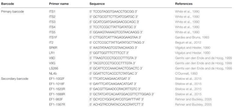
(Beguin et al., 2012), ITS1F (Gardes and Bruns, 1993) and NL4b (O’Donnell, 1993; Figure 2 and Table 1). To provide quality controlled reference primary fungal DNA barcode sequences the International Society for Human and Animal Mycology (ISHAM) “ITS DNA barcode database” was established in 2015 (Irinyi et al., 2015). The primary fungal DNA barcode region identifies up to 75% of the estimated ∼700 pathogenic fungal
species (de Hoog et al., 2014).
To address the shortcomings of the ITS region and hence close this identification gap, a secondary barcode was proposed
in 2015 (Stielow et al., 2015). Thetranslational elongation factor
1α (TEF1α) was selected due to its high species discrimination
across fungal taxa and the ability to design universal primers, such as EF1-1018F (Al33F)/EF1-1620R (Al33R), EF1-1002F
(Al34F)/EF1-1688R (Al34R) (Stielow et al., 2015), or
EF1-983F/EF1-1567R (Rehner and Buckley, 2005; Figure 2 and
Table 1). To accommodate the secondary fungal DNA barcode a dedicated database that would eventually include all medically relevant fungal species was established to complement the ISHAM-ITS database, which contains only quality-controlled
TEF1α sequences obtained from taxonomically verified fungal
cultures (Meyer et al., 2018). Both databases have been combined
within the “ISHAM Barcoding Database”,1which was launched
in November 2017 at the 7th International DNA barcoding conference at the Kruger National Park in South Africa. The combined dual barcode database provides the medical and veterinary community with quality-controlled primary and secondary fungal DNA barcodes reference sequences. This database is publicly available, and its quality-controlled sequences are shared with other major databases, including RefSeq within
GenBank (Benson et al., 2014) and UNITE (Kõljalg et al., 2013).
To date, the clinical utility and accuracy of the dual DNA barcoding system for identification of pathogenic fungi has not been assessed. This study aimed to increase the number of reference secondary barcode sequences for 1http://its.mycologylab.org/
FIGURE 1 | Current and potentially available techniques for fungal ID, showing the central role of the reference DNA barcode database and the pros and cons of all techniques. Crossed boxes indicate missing databases. DNA barcoded reference strains are fundamental to build up a spectral library.
FIGURE 2 | Schematic structure of the primary (ITS) and secondary (TEF1α) fungal DNA barcode regions indicating universal primers for their amplification.
pathogenic fungal species and to compare the accuracy and resolution of the primary and secondary fungal DNA barcodes separately or in combination, focusing on fungi causing invasive fungal infections.
MATERIALS AND METHODS
Cultures
To generate quality controlled TEF1α sequences 270 strains,
representing 90 human/animal pathogenic fungal species, were used (see Supplementary Table 1).
DNA Extraction
DNA was isolated and purified from cultures using either
previously described manual method (Ferrer et al., 2001) or
the Quick-DNA Fungal/Bacterial Kit (D6007, Zymo Research) according to the manufacturer’s instructions.
DNA Barcode Generation
The secondary fungal DNA barcoding region (TEF1α)
was amplified using the primers described by Stielow
et al. (2015), including: EF1-1018F (Al33F) (50
GAYTTCATCAAGAACATGAT 30
) and EF1-1620R (Al33R) (50
GACGTTGAADCCRACRTTGTC 30
TABLE 1 | Universal and fungal specific primers for the dual DNA barcoding scheme.
Barcode Primer name Sequence References Primary barcode ITS1 50
TCCGTAGGTGAACCTGCGG 30 White et al., 1990 ITS2 50 GCTGCGTTCTTCATCGATGC 30 White et al., 1990 ITS3 50 GCATCGATGAAGAACGCAGC 30 White et al., 1990 ITS4 50TCCTCCGCTTATTGATATGC 30 White et al., 1990
ITS5 50GGAAGTAAAAGTCGTAACAAGG 30 White et al., 1990
ITS1F 50CTTGGTCATTTAGAGGAAGTAA 30 Gardes and Bruns, 1993
IT2 50CCTCCGCTTATTGATATGCTTAGG 30 Beguin et al., 2012
SR6R 50AAGTATAAGTCGTAACAAGG 30 Vilgalys and Hester, 1990
LR1 50GGTTGGTTTCTTTCCT 30 Vilgalys and Hester, 1990
V9D 50TTAAGTCCCTGCCCTTTGTA 30 Gerrits van den Ende and de Hoog, 1999
V9G 50TACGTCCCTGCCCTTTGTA 30 Gerrits van den Ende and de Hoog, 1999
LS266 50GCATTCCCAAACAACTCGACTC 30 Gerrits van den Ende and de Hoog, 1999
NL4b 50GGATTCTCACCCTCTATGAC 30 O’Donnell, 1993
Secondary barcode EF1-1002F 50TTCATCAAGAACATGAT 30 Stielow et al., 2015
EF1-1018F 50GAYTTCATCAAGAACATGAT 30 Stielow et al., 2015
EF1-1620R 50GACGTTGAADCCRACRTTGTC 30 Stielow et al., 2015
EF1-1688R 50
GCTATCATCACAATGGACGTTCTTGGAG 30
Stielow et al., 2015 EF1-983F 50
GCYCCYGGHCAYCGTGAYTTYAT 30
Rehner and Buckley, 2005 EF1-1567R 50
ACHGTRCCRATACCACCRATCTT 30
Rehner and Buckley, 2005
EF1-1002F (Al34F) (50 TTC ATCAAGAACATGAT 30 ) and EF1-1688R (Al34R) (50 CTATCATCACAATGG ACGTTCTTGGAG 30
) being used together (Stielow et al., 2015). The primer set
Al33F-Al33R was first used to amplify the TEF1α region. If
amplification was unsuccessful then it was repeated using the second primer set Al34F-Al34R. Both primer pairs used the following PCR amplification protocol: 5 min initial denaturing
at 94◦
C, followed by 40 of 50 s at 94◦
C, 50 s annealing at
48◦
C, 50 s at 72◦
C and 7 min final extension at 72◦
C (Stielow et al., 2015). Amplification success was visualized through gel electrophoresis in a 1.5% agarose gel containing ethidium bromide (EtBr) with ultra-violet illumination. Successfully amplified PCR products were sent for commercial sequencing, e.g., Macrogen Inc., South Korea in both forward and reverse directions. Bidirectional sequenced were assembled and edited
using Sequencher R
ver. 5.3. (Gene Codes Corporation, Ann Arbor, MI, United States). Sequences were manually checked to resolve ambiguous bases on the forward and reverse trace files considering the PHRED scores received.
DNA Barcode Analysis
The sequences for each taxon were aligned with the program
CLUSTALW (Thompson et al., 1994) part of the software MEGA
ver. 7 (Larkin et al., 2007). Resulting alignments were checked
visually and edited when needed.
The intraspecies diversity was estimated by calculating the average nucleotide diversity (π) within species, where there were sequences available from more than three strains. The proportion of nucleotide differences in all haplotypes in the sample was derived using the software DnaSP ver. 5.10.01 (Librado and Rozas, 2009).
Individual analysis of the primary and secondary barcoding regions was performed through barcoding gap analysis.
Additionally, analysis was performed on the combined barcoding regions. Taxonomic groups were selected for analysis if represented by three or more species, and if those species were represented by three or more strains. Furthermore, taxonomic groups were required to include at least one species that has been demonstrated to cause invasive fungal infections. Only strains with both primary and secondary barcodes in the database where included in the analysis. When available, more specific taxonomic groups such as clades were selected over genera. Sequences for each genus were aligned and cut to equal
length using CLUSTALW as present in MEGA ver. 7 (Larkin
et al., 2007;Kumar et al., 2016). Genetic distances between each strain were calculated using the Kimura 2-parameter model (K2P) and the intraspecies and interspecies genetic distances
were compared (Kimura, 1980). Intra- and interspecies genetic
distances were graphed against frequency and analyzed for barcoding gaps. Barcoding gaps were defined by the presence of a distinct difference between the largest intraspecies genetic
distance and smallest interspecies genetic distance (Irinyi et al.,
2015), i.e., there was no common x value between the intra- and
interspecies groups.
The introduction of the secondary fungal DNA barcode was accessed for each genus on the basis that there would be an improvement in DNA barcoding if the secondary barcode generated a barcoding gap when the primary barcode did not, or if the overlap between the intra- and interspecies genetic distances was reduced.
RESULTS
The study produced 270 new quality controlled secondary fungal barcode sequences, covering 90 pathogenic fungal species
FIGURE 3 | Intraspecies variation for species, which are represented by more than three strains, in the internal transcribed spacer (ITS) (blue bars) primary fungal DNA barcode compared with the translation elongation factor 1α (TEF1α) (gray bars) secondary fungal DNA barcode.
(Supplementary Table 1). Overall, the PCR success rate was
high within the 270 TEF1 sequences. 220 secondary barcodes
were generated using the Al33F–Al33R primer pair and 50 were amplified with the Al34F–Al34R primer pair. There was no trend in amplification success in different fungal species. However, the Al34F–Al34R primer set was required to amplify all strains
of Aspergillus niger, Candida albicans, Candida dubliniensis,
Kluyveromyes marxianus, and Pichia kudriavzevii. There was unsuccessful amplification with both primer sets for some strains
ofCladosporium spp., Rhodotorula spp., and Trichosporon spp.
All sequences were submitted to the ISHAM Barcoding Database (see footnote 1). The length of the ITS and partial
TEF1α sequences in the database ranges 285–791 and 534–
1002 bp, respectively.
The analysis of the nucleotide diversity (π) of 43 fungal
species with more than three strains in the ISHAM barcoding
database showed that theTEF1α region is less diverse than the
ITS region in most species (Figure 3). The intraspecies variation
of TEF1α was for most species below 1.5%, confirming the
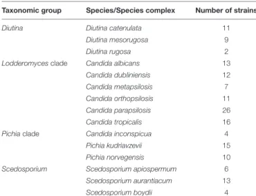
secondary barcode as a more discriminator marker. According to the selection criteria four different taxonomic groups were selected as proof of principle for the dual barcoding system.
These included the two genera,Diutina and Scedosporium, and
the two taxonomic clades,Lodderomyces and Pichia. The Diutina
genus was selected for analysis despite one of the species,Diutina
rugosa, being represented by only two strains as this genus is newly established that causes rare disease (Table 2). The species and number of strains included in these analyses are outlined in Table 2.
Diutina Species
Barcoding gap analysis of the genus Diutina demonstrated
no overlap between the intraspecies and interspecies genetic distances with either the primary or secondary fungal DNA barcodes (Figure 4). There was similarly no overlapping region
for the barcoding gap analysis of the combination of the primary and secondary barcodes (Figure 4). As such, both fungal barcodes and the combination of the barcodes generated appropriate barcoding gaps.
The Lodderomyces Clade
For the Lodderomyces clade, the intraspecies and interspecies
genetic distances, generated by the primary fungal barcode produced an overlapping region thereby indicating that there was no barcoding gap (Figure 4). The barcoding gap analysis for the
secondary DNA barcode (TEF1α) demonstrated no overlap in the
intraspecies and interspecies genetic distances forLodderomyces
thus showing a clear barcoding gap (Figure 4). TheLodderomyces
clade analysis using the combination of both the ITS andTEF1α
TABLE 2 | Number of fungal strains with primary and secondary barcodes present in the ISHAM Barcoding Database analyzed in this study.
Taxonomic group Species/Species complex Number of strains Diutina Diutina catenulata 11
Diutina mesorugosa 9 Diutina rugosa 2 Lodderomyces clade Candida albicans 13
Candida dubliniensis 12 Candida metapsilosis 7 Candida orthopsilosis 11 Candida parapsilosis 26 Candida tropicalis 16 Pichia clade Candida inconspicua 4
Pichia kudriavzevii 15 Pichia norvegensis 10 Scedosporium Scedosporium apiospermum 6
Scedosporium aurantiacum 13 Scedosporium boydii 4
FIGURE 4 | Barcoding gap analyses for fungal taxa including causative agents of invasive fungal disease with intraspecies (gray) and interspecies (blue) genetic distances calculated using the Kimura 2-parameter model. (A) Diutina genus (Diutina catenulata, Diutina mesorugosa, and Diutina rugosa). (B) Lodderomyces-clade (Candida albicans, Candida dublinsiensis, Candida metapsilosis, Candida orthopsilosis, Candida parapsilosis, and Candida tropicalis). (C) Pichia-clade (Candida inconspicua, Pichia kudriavzevii, and Pichia norvegensis).(D) Scedosporium genus (Scedosporium apiospermum, Scedosporium aurantiacum, and Scedosporium boydii).
regions resulted in the generation of a barcoding gap being generated (Figure 4).
The Pichia Clade
Barcoding gap analysis of the Pichia clade did not reveal any
overlapping regions in the genetic distances for either the ITS or
TEF1α regions (Figure 4). As there were no overlapping regions
this demonstrated that there are barcoding gaps for both the primary and secondary barcode. These results were similar when
both barcoding regions were combined as there was a barcoding gap produced (Figure 4).
Scedosporium Species
Barcoding gap analysis of the primary fungal DNA barcode showed an overlapping region (Figure 4). For the secondary barcode, the barcoding gap analysis revealed that there was no overlap between the intraspecies and interspecies genetic distances and so a barcoding gap was present
(Figure 4). The barcoding gap analysis of the combination of both barcodes, produced a barcoding gap as there was no overlap between the intraspecies and interspecies genetic distances (Figure 4).
DISCUSSION
ISHAM Barcoding Database Expansion
Accurate and rapid routine diagnostic tools are essential to reduce the burden of fungal disease. DNA barcoding requires reliable quality-controlled reference sequences for its implementation. As such, fungal DNA Barcoding databases need to have high taxonomic coverage and reliable sequences to ensure users can accurately match sample sequences to the correct reference sequences. Additionally, the more sequences from different strains for each species are represented, the higher is the accuracy of the identification, as this reflects more realistically the existing species variation.
In this study, we generated 270 secondary barcodes, which could be amplified reliably using either the primer set Al33F– Al33R or the primer set Al34f–A134R (Figure 2), with a few exceptions where no amplifications where obtained for some
strains ofCladosporium spp., Rhodotorula spp., or Trichosporon
spp. In those cases, modification of the amplification conditions, such as touchdown PCR, may be appropriate.
As a result of this study the ISHAM Barcoding Database contains, as of the May 10, 2019, 4091 primary (ITS) and
868 secondary barcode (TEF1α) sequences covering 640 and
129 pathogenic fungal species, respectively. From those 2681
ITS and 613 TEF1α sequences representing 252 species cause
invasive fungal diseases. There are an estimated 700 pathogenic fungal species, with the majority rarely causing infections. As such, the taxonomic coverage of ITS sequences within the “ISHAM Barcoding Database” is close to covering all
pathogenic fungal species, whilst the TEF1α sequences cover
less than one fifth. In addition, 59 species are currently only
represented by oneTEF1α sequence, which limits identification
due to underrepresented intraspecies variation. As the “ISHAM Barcoding Database” has only been recently expanded to
include TEF1α sequences, it is expected that as more TEF1α
sequences are submitted, these issues will be resolved. Currently the “ISHAM Barcoding Database” has enabled pragmatic implementation of the dual barcode scheme for the identification of pathogenic fungi.
Comparison of the Primary and
Secondary Barcodes
Since the establishment of the secondary fungal DNA barcode the improvement of the identification accuracy for pathogenic fungi has not been assessed. In this study, we compared for the first time the primary and secondary fungal DNA barcodes via barcoding gap analysis based on intraspecies and interspecies genetic distance values as calculated using the K2P model (Kimura, 1980). In addition, the combination of both barcodes was tested and compared to the individual barcodes.
Diutina Species (Previously Belonging to the Genus Candida)
The genus Diutina was established only recently. This group
of fungal species was previously part of the genus Candida
(Kurtzman et al., 2011; Khunnamwong et al., 2015). Diutina species are uncommon agents of disease, but are associated with
nosocomial infections, with fatality rates of up to 70% (Padovan
et al., 2013). Diutina catenulata and Diutina rugosa are well known causes of fungemia in immunocompromised patients
and the seriously ill (Radosavljevic et al., 1999; Minces et al.,
2009;Behera et al., 2010;Ha et al., 2018).D. rugosa is notable for its increasing resistance against multiple antifungal agents worldwide, thereby increasing the need for early detection and identification to enable timely initiation of effective treatment (Pfaller et al., 2006a).
An exception was made for the genusDiutina to be included
into this analysis, as D. rugosa is only represented by two
strains in the analysis (Table 2). The inclusion of the species
was important asDiutina mesorugosa and D. rugosa have been
proposed as synonyms with the variability of ITS sequences used
as supporting evidence (Ming et al., 2019).
Barcoding gap analysis indicates that these species can
be clearly identified as separate species. The Diutina species
are representative of the 75% of pathogenic fungi that
are accurately identified by the primary barcode (Irinyi
et al., 2016). The secondary barcode and the combination of both barcodes equally identified these species (Figure 4), demonstrating the same level of resolution as the primary
barcode (Stielow et al., 2015).
The Lodderomyces Clade
TheLodderomyces clade contains 21 Candida species, including
some of the major pathogenic fungal species (Kurtzman et al.,
2011). This clade contains three of the five most common
pathogenic Candida species: Candida albicans, Candida
dubliniensis, and Candida parapsilosis (Spampinato and
Leonardi, 2013). These species cause a wide spectrum of diseases, from cutaneous infections to fatal disseminated septicemia (Tavanti et al., 2005). Candida species are predominantly commensals in the body and as such these infections largely target immunocompromised patients and are commonly the
cause of nosocomial outbreaks (Brown et al., 2012).
Candida is a polyphyletic genus with a highly complex
taxonomic history (Kurtzman et al., 2011). In the latest edition
of The Yeasts A Taxonomic Study, 314 differentCandida species
were described excluding species with anamorph-teleomorph
linkages (Kurtzman et al., 2011). The genus Candida was
initially intended to include undifferentiated yeasts that cannot
be identified by phenotype (Berkhout, 1923). As such there
is little stability in the genus with some species being linked to others as anamorphs and the introduction of newly found species. With the advanced use of genomics in taxonomic studies
the composition of species and the Lodderomyces clade are
expected to change.
The barcoding gap analysis of the primary barcode was unable to generate a barcoding gap and so was unable to accurately identify all species of the clade (Figure 4).
Upon closer inspection, the lack of a barcoding gap was
due to the close relationships between Candida orthopsilosis
and Candida parapsilosis, which form part of the Candida
parapsilosis species complex. This species complex was
previously representing a single species, Candida parapsilosis,
with three subgroups that were then revised to be separate species based on various molecular techniques, including sequencing of the ITS region. The barcoding gap analysis of this study, however, did not reflect this differentiation and so ITS was unable to identify these different species. Barcoding gap analysis of the secondary barcode did produce
a barcoding gap and thereby demonstrated that TEF1α
could accurately identify all species of the Lodderomyces clade (Figure 4). As such, the introduction of the dual barcoding scheme resulted in an accurate identification of these pathogenic fungal species.
The Pichia Clade
ThePichia clade is composed of 20 species. In our study, Candida
inconspicua was also included in the analysis, as it is thought to
be the anamorph forPichia cactophila (Kurtzman and Robnett,
1998;Kurtzman et al., 2008, 2011).Pichia kudriavzevii and Pichia norvegensis are predominantly linked to nosocomial pathogens
and may cause outbreaks of infections (Nagarathnamma et al.,
2017). Candida inconspicua has also been found in clinical
samples whilst the other species of the Pichia clade have not
yet been found to be medically relevant (Kurtzman et al., 2011;
Guitard et al., 2013). These species also cause invasive infections in patients with a highly compromised immune systems, and are
often found under previous names in the literature (Kurtzman
et al., 2011;Guitard et al., 2013;Schuster et al., 2013;Sanclemente et al., 2015;Douglass et al., 2018).
Similarly, to the genusDiutina, barcoding gap analysis of the
Pichia clade revealed barcoding gaps for the primary barcode, secondary barcode and the combination of both barcodes (Figure 4), resulting in an accurate identification of all species
of thePichia clade.
Scedosporium Species
Fungi belonging to the genus Scedosporium cause a wide
variety of diseases from localized infections to invasive
diseases (Cortez et al., 2008). Scedosporium species mainly
cause infections in patients with a compromised immune systems largely due to underlying illnesses such as solid organ transplantation, cystic fibrosis, leukemia and bone
marrow transplantation (Tamm et al., 2001; Allen et al.,
2013; Kubisiak-Rzepczyk et al., 2013; Yu et al., 2013; Johnson et al., 2014). Scedosporium spp. can also cause infection in
immunocompetent patients (Ceccarelli et al., 2012;Agatha et al.,
2014;Cruysmans et al., 2015).
The taxonomy of the genus Scedosporium is complex and
has changed multiple times over the last decade. Previously,
the nomenclature of the genus ruled Scedosporium to be the
anamorph name whilst Pseudallescheria and Petriella were the
teleomorph names (Rainer and de Hoog, 2006; Cortez et al.,
2008; Lu et al., 2011). The two major clinical species were Scedosporium prolificans and Scedosporium apiospermum and its
teleomorph form, Pseudallescheria boydii (Cortez et al., 2008).
Multilocus sequencing, morphological analysis and physiological
testing later demonstrated thatS. apiospermum and P. boydii are
separate species and not anamorphs teleomorph pairs (Gilgado
et al., 2008). In 2011, the new nomenclature rules dictating
“one fugus = one name” led to the fact that Scedosporium
took precedence over Pseudallescheria as the genus name
(Hawksworth, 2011;Lackner et al., 2014). The composition of the
genus was also changed withS. prolificans being removed from
the genus and renamed to Lomentospora prolificans (Lackner
et al., 2014). The separation between S. apiospermum and S. boydii was reinforced, however, it was noted that there was no clinical difference between the species, as such all
species are referred to the S. apiospermum species complex
(Lackner et al., 2014).
In this study, there was no barcoding gap present for the
primary barcode indicating that Scedosporium species cannot
be accurately identified using this region alone (Figure 4). The predominant reason for the overlap in intraspecies and interspecies genetic distances was the high similarity between S. apiospermum and S. boydii. This was reflective of the nomenclature history as these species were previously thought to be an anamorph – teleomorph pair. Barcoding gap analysis of the secondary barcoding region introduced a barcoding gap and
hence allowed for the accurate identification of allScedosporium
species (Figure 4). As the separation of S. apiospermum and
S. boydii was established via multilocus sequence analysis, it is
reassuring that TEF1α can resolve all Scedosporium species to
the same degree (Gilgado et al., 2008). With the introduction of
the dual barcoding scheme, allScedosporium species can now be
accurately identified.
Assessment of the Dual Barcoding
Scheme
Meaningful implementation of the dual DNA barcoding scheme depends on demonstration of an improvement in the identification accuracy of fungal species causing infections when
adding the secondary barcode,TEF1α. To demonstrate this the
two fungal barcodes were analyzed separately and in combination from selected species, which had sequences from both barcodes being in the “ISHAM Barcoding Database,” using barcoding gap analysis through the generation of genetic distances by the K2P
model (Kimura, 1980).
Although K2P has been used in all DNA barcoding
studies since its introduction by Hebert et al. (2003), the
accuracy of this model has been questioned (Kimura, 1980;
Hebert et al., 2003). In the selection of ITS region as the primary fungal DNA barcode, the uncorrected p-distances were used for barcoding gap analysis whereas in the case of the secondary fungal DNA barcode (TEF1α) analysis
the K2P method was applied (Schoch et al., 2012; Stielow
et al., 2015). The K2P region was selected to generate barcoding gaps over uncorrected p-distances as K2P has been found to generate larger barcoding gaps for most
data sets (Srivathsan and Meier, 2012). Additionally, in
performance of K2P was found to be similar (Srivathsan and Meier, 2012). More complicated models have been applied in some studies in an attempt to improve the accuracy of the K2P model, however, the differences were minimal,
prompting us to use K2P in our study (Collins et al., 2012;
Barley and Thomson, 2016).
Comparison of the number of pathogenic fungal species and strains represented in the “ISHAM Barcoding Database”
highlighted the deficit in the number of TEF1α reference
sequences. Therefore, the value of this study is limited as only strains with both barcodes available were used for the analysis. More species represented by more sequences would give a more exact assessment about the value and resolution power of the dual barcoding concept.
DNA barcode gab analysis showed that two of the groups
(Diutina and Pichia) tested were accurately identified
using the primary barcode and the secondary barcode on its own, demonstrating that both barcodes had similar resolution. For the remaining two groups (Lodderomyces
and Scedosporium) the primary barcode was not able
to produce barcoding gaps whilst the secondary did, demonstrating that the secondary barcode had a higher resolution power. In addition, the combination of both barcodes increased the discriminatory power enabling a more accurate
identification. These results indicate, that the application of the dual barcoding system drastically improves species identification in cases where a single barcoding system is unable to do so.
Implementation of the Dual Barcoding
Scheme
We envisage that the proposed dual barcoding scheme can be applied in the routine diagnostic setting in a stepwise procedure. After a clinical specimen is obtained, the unknown fungal isolate is first assessed based on its morphologic and/or biochemical characteristics. Then those unknown fungal isolates which lack obvious morphological characteristics or result in unclear biochemical profiles should be subjected to DNA isolation and primary fungal DNA barcoding (ITS1/2 region). If the obtained sequence shows less than 98.5% identity to a given ITS reference sequence in the database, the secondary fungal DNA barcode
(TEF1α) should be obtained to achieve a final species
identification (Figure 5).
The dual fungal DNA barcoding scheme is an efficient diagnostic tool for the identification of agents of invasive mycoses and can be confidently implemented into routine
diagnostics. Its implementation along with the expansion
of the “ISHAM Barcoding Database” will lead to a
paradigm shift in fungal disease diagnostics, enabling
a highly accurate identification of the agents of fungal
disease. The timely initiation of appropriate therapy
will improve patient outcomes through the reduction in morbidity and mortality, prevent the use of unnecessary or inappropriate antifungal drugs, minimizing drug toxicity and resistance development and greatly reduce the associated health care cost.
CALL FOR DATA SUBMISSION
To achieve a 100% coverage of all pathogenic fungi and reflect the intraspecies variation for both the primary and secondary fungal DNA barcode we call for the submission
of quality-controlled reference sequences of the ITS
region and the TEF1α for inclusion into the “ISHAM
Barcode Database.” Please contact the curators of the database WM at wieland.meyer@sydney.edu.au and LI at laszlo.irinyi@sydney.edu.au.
MEMBERS OF THE ISHAM BARCODING
OF MEDICAL FUNGI WORKING GROUP
Michael Arabatzis, National and Kapodistrian University of Athens, Athens, Greece; Ian Arthur, PathWest Laboratory Medicine, Nedlands, WA, Australia; Jose F. Cano-Lira, Universitat Rovira i Virgili, Reus, Spain; Gianluigi Cardinali, Università degli Studi di Perugia, Perugia, Italy; Laura Rosio Castañón, Universidad Nacional Autónoma de México, Mexico City, Mexico; Sharon Chen, Westmead Hospital, University of Sydney, Sydney, NSW, Australia; Wen Chen, Agriculture and Agri-Food Canada, Ottawa, ON, Canada; Ariya Chindamporn, Chulalongkorn University, Bangkok, Thailand; Arnaldo L. Colombo, Universidade Federal de São Paulo, São Paulo, Brazil; Marie Desnos-Ollivier, Institut Pasteur, Paris, France; Wilhelm de Beer, FABI, Pretoria, South Africa; Sybren de Hoog, Westerdijk Fungal Biodiversity Institute, Utrecht, Netherlands; Françoise Dromer, Institut Pasteur, Paris, France; Dea Garcia-Hermoso, Institut Pasteur, Paris, France; Marieka Gryzenhout, University of the Free State, Bloemfontein, South Africa; Josep Guarro, Universitat Rovira i Virgili, Reus, Spain; Catriona Halliday, University of Sydney/Westmead Hospital, Sydney, NSW, Australia; Marijke Hendrickx, BCCM/IHEM, Scientific Institute of Public Health, Brussels, Belgium; Sabine Huhndorf, The Field Museum, Chicago, IL, United States; Laszlo Irinyi, Westmead Hospital, University of Sydney, Sydney, NSW, Australia; C. Andre Levesque, Agriculture and Agri-Food Canada, Ottawa, ON, Canada; Wieland Meyer, Westmead Hospital, University of Sydney, Sydney, NSW, Australia; Maria Luiza Moretti, University of Campinas, Campinas, Brazil; Mauro de Medeiros Muniz, Instituto de Pesquisa Clínica Evandro Chagas (IPEC), Fundação Oswaldo Cruz
(FIOCRUZ), Rio de Janeiro, Brazil; Analy Salles de Azevedo Melo, Universidade Federal de São Paulo, São Paulo, Brazil; Angela Satie Nishikaku, Universidade Federal de São Paulo, São Paulo, Brazil; Anne-Cécile Normand, Aix-Marseille University, Marseille, France; Célia Pais, University of Minho, Braga, Portugal; Renaud Piarroux, Aix-Marseille University, Marseille, France; Stéphane Ranque, Aix-Marseille University, Marseille, France; Barbara Robbertse, National Center for Biotechnology Information, Bethesda, MD, United States; Vincent Robert, Westerdijk Fungal Biodiversity Institute, Utrecht, Netherlands; Conrad L. Schoch, National Center for Biotechnology Information, Bethesda, MD, United States; Keith A. Seifert, Agriculture and Agri-Food Canada, Ottawa, ON, Canada; Célia Maria de Almeida Soares, Universidade Federal de Goiás, Goiânia, Brazil; Tania C. Sorrell, Westmead Hospital, University of Sydney, Sydney, NSW, Australia; John L. Spouge, National Center for Biotechnology Information, Bethesda, MD, United States; Dirk Stubbe, BCCM/IHEM, Scientific Institute of Public Health, Brussels, Belgium; Maria Lucia Taylor, Universidad Nacional Autónoma de México, Mexico City, Mexico; Conchita Toriello, Universidad Nacional Autónoma de México, Mexico City, Mexico; Aristea Velegraki, National and Kapodistrian University of Athens, Athens, Greece; Chompoonek Yurayart, Kasetsart University, Bangkok, Thailand; Rosely Maria Zancopé-Oliveira, Instituto de Pesquisa Clínica Evandro Chagas (IPEC), Fundação Oswaldo Cruz, FIOCRUZ, Rio de Janeiro, Brazil.
DATA AVAILABILITY
The primary and secondary fungal barcodes generated and analyzed for this study are available in the ISHAM Barcoding Database (http://its.mycologylab.org/).
AUTHOR CONTRIBUTIONS
WM, SC, and TS conceived the study. WM coordinated
the study and supervised the molecular and data
analysis. All members of the ISHAM working group for
“Barcoding of Medical Fungi” performed the sample
collections. MH and LI conducted the sample processing, all molecular studies, and data analysis. MH, LI, and WM performed the data interpretation. All authors wrote and corrected the manuscript.
FUNDING
This study was supported by the National Health and Medical Research Council of Australia (NH & MRC) grant (#APP1121936) to WM, SC, and TS, and Western Sydney Local Health District Research and Education Network Research Grant Scheme to WM, TS, SC, and LI. TS is a Sydney Medical School Foundation Fellow.
ACKNOWLEDGMENTS
The authors thank all members of the ISHAM working group for “Barcoding of Medical Fungi” for the contribution of strains and barcode sequences.
SUPPLEMENTARY MATERIAL
The Supplementary Material for this article can be found online at: https://www.frontiersin.org/articles/10.3389/fmicb. 2019.01647/full#supplementary-material
REFERENCES
Agatha, D., Krishnan, K. U., Dillirani, V. A., and Selvi, R. (2014). Invasive lung infection by Scedosporium apiospermum in an immunocompetent individual.Indian J. Pathol. Microbiol. 57, 635–637. doi: 10.4103/0377-4929. 142716
Allen, P., Koka, R., Kleinberg, M. E., and Baer, M. R. (2013). Scedosporium apiospermum soft tissue infection as the initial presentation of acute myeloid leukemia: a case report.J. Clin. Oncol. 31, e98–e100. doi: 10.1200/jco.2012.43. 9489
Armstrong-James, D., Meintjes, G., and Brown, G. D. (2014). A neglected epidemic: fungal infections in HIV/AIDS.Trends Microbiol. 22, 120–127. doi: 10.1016/j.tim.2014.01.001
Balajee, S. A., Sigler, L., and Brandt, M. E. (2007). DNA and the classical way: identification of medically important molds in the 21st century.Med. Mycol. 45, 475–490. doi: 10.1080/13693780701449425
Barley, A. J., and Thomson, R. C. (2016). Assessing the performance of DNA barcoding using posterior predictive simulations.Mol. Ecol. 25, 1944–1957. doi: 10.1111/mec.13590
Beguin, H., Pyck, N., Hendrickx, M., Planard, C., Stubbe, D., and Detandt, M. (2012). The taxonomic status ofTrichophyton quinckeanum and T. interdigitale revisited: a multigene phylogenetic approach. Med. Mycol. 50, 871–882. doi: 10.3109/13693786.2012.684153
Behera, B., Singh, R., IXess, I., Mathur, P., Hasan, F., and Misra, M. C. (2010). Candida rugosa: a possible emerging cause of candidaemia in trauma patients. Infection 38, 387–393. doi: 10.1007/s15010-010-0044-x
Benedict, K., Richardson, M., Vallabhaneni, S., Jackson, B. R., and Chiller, T. (2017). Emerging issues, challenges, and changing epidemiology of fungal disease outbreaks. Lancet Infect. Dis. 17, e403–e411. doi: 10.1016/S1473-3099(17) 30443-7
Benson, D. A., Clark, K., Karsch-Mizrachi, I., Lipman, D. J., Ostell, J., and Sayers, E. W. (2014). GenBank.Nucleic Acids Res. 42, D32–D37. doi: 10.1093/nar/ gkw1070
Berkhout, C. M. (1923). De Schimmelgeslachten Monilia, Oidium, Oospora en Torula. Utrecht: Rijksuniversiteit Utrecht.
Bitar, D., Lortholary, O., Le Strat, Y., Nicolau, J., Coignard, B., Tattevin, P., et al. (2014). Population-based analysis of invasive fungal infections, France, 2001–2010.Emerg. Infect. Dis. 20, 1149–1155. doi: 10.3201/eid2007.140087 Brown, G. D., Denning, D. W., Gow, N. A., Levitz, S. M., Netea, M. G., and
White, T. C. (2012). Hidden killers: human fungal infections.Sci. Transl. Med. 4:165rv113. doi: 10.1126/scitranslmed.3004404
Cadena, J., Thompson, G. R. III, and Patterson, T. F. (2016). Invasive Aspergillosis: current strategies for diagnosis and management.Infect. Dis. Clin. North Am. 30, 125–142. doi: 10.1016/j.idc.2015.10.015
Ceccarelli, L., Calisti, G., Delle Rose, D., Ricciardi, A., Maffongelli, G., Sordillo, P., et al. (2012). Dapsone hypersensitivity syndrome complicated by scedosporium apiospermum pneumonia in an immunocompetent patient.Infection 40, 459– 462. doi: 10.1007/s15010-011-0225-2
Chen, S. C., Meyer, W., and Sorrell, T. C. (2014).Cryptococcus gattii infections. Clin. Microbiol. Rev. 27, 980–1024. doi: 10.1128/CMR.00126-13
Ciardo, D. E., Schar, G., Bottger, E. C., Altwegg, M., and Bosshard, P. P. (2006). Internal transcribed spacer sequencing versus biochemical profiling for identification of medically important yeasts.J. Clin. Microbiol. 44, 77–84. doi: 10.1128/JCM.44.1.77-84.2006
Cole, D. C., Govender, N. P., Chakrabarti, A., Sacarlal, J., and Denning, D. W. (2017). Improvement of fungal disease identification and management: combined health systems and public health approaches.Lancet Infect. Dis. 17, e412–e419. doi: 10.1016/S1473-3099(17)30308-0
Collins, R. A., Boykin, L. M., Cruickshank, R. H., and Armstrong, K. F. (2012). Barcoding’s next top model: an evaluation of nucleotide substitution
models for specimen identification. Methods Ecol. Evol. 3, 457–465. doi: 10.1111/j.2041-210X.2011.00176.x
Cortez, K. J., Roilides, E., Quiroz-Telles, F., Meletiadis, J., Antachopoulos, C., Knudsen, T., et al. (2008). Infections caused by Scedosporium spp. Clin. Microbiol. Rev. 21, 157–197. doi: 10.1128/CMR.00039-07
Cruysmans, C., Rodriguez-Villalobos, H., Fomekong, E., Dumitriu, D., Nassogne, M. C., and Van der Linden, D. (2015). Epidural abscess caused byScedosporium apiospermum in an immunocompetent child. Pediatr. Infect. Dis. J. 34, 1277– 1278. doi: 10.1097/INF.0000000000000864
de Hoog, G. S., Guarro, J., Gené, J., and Figueras, A. A. (2014).Atlas of Clinical Fungi. Netherlands: Centraalbureau voor Schimmelcultures (CBS).
Denning, D. W. (2016). Minimizing fungal disease deaths will allow the UNAIDS target of reducing annual AIDS deaths below 500 000 by 2020 to be realized. Philos. Trans. R. Soc. Lond. B Biol. Sci. 371:0468. doi: 10.1098/rstb.2015.0468 Douglass, A. P., Offei, B., Braun-Galleani, S., Coughlan, A. Y., Martos, R.,
Ortiz-Merino, R. A., et al. (2018).Population genomics shows no distinction between pathogenicCandida krusei and environmental Pichia kudriavzevii: One species, four names.PLoS Pathog. 14:e1007138. doi: 10.1371/journal.ppat.1007138 Dromer, F., Mathoulin-Pelissier, S., Fontanet, A., Ronin, O., Dupont, B., and
Lortholary, O. (2004). Epidemiology of HIV-associated cryptococcosis in France (1985-2001): comparison of the pre- and post-HAART eras.Aids 18, 555–562. doi: 10.3201/eid2007.140087
Ferrer, C., Colom, F., Frases, S., Mulet, E., Abad, J., and Alio, J. (2001). Detection and identification of fungal pathogens by PCR and by ITS2 and 5.8S ribosomal DNA typing in ocular infections.J. Clin. Microbiol. 39, 2873–2879. doi: 10.1128/ JCM.39.8.2873-2879.2001
Gardes, M., and Bruns, T. D. (1993). ITS primers with enhanced specificity for basidiomycetes - application to the identification of mycorrhizae and rusts.Mol. Ecol. 2, 113–118. doi: 10.1111/j.1365-294X.1993.tb00005.x
Gerrits van den Ende, A. H. G., and de Hoog, G. S. (1999). Va∗
riability and molecular diagnostics of the neurotropic speciesCladophialophora bantiana. Stud. Mycol. 43, 151–162.
Gilgado, F., Cano, J., Gene, J., Sutton, D. A., and Guarro, J. (2008). Molecular and phenotypic data supporting distinct species statuses forScedosporium apiospermum and Pseudallescheria boydii and the proposed new species Scedosporium dehoogii. J. Clin. Microbiol. 46, 766–771. doi: 10.1128/JCM. 01122-07
Guitard, J., Angoulvant, A., Letscher-Bru, V., L’Ollivier, C., Cornet, M., Dalle, F., et al. (2013). Invasive infections due toCandida norvegensis and Candida inconspicua: report of 12 cases and review of the literature. Med. Mycol. 51, 795–799. doi: 10.3109/13693786.2013.807444
Ha, M. V., Choy, M. S., McCoy, D., Fernandez, N., and Suh, J. S. (2018). Candida catenulata candidaemia and possible endocarditis in a cirrhotic patient successfully de-escalated to oral fluconazole.J. Clin. Pharm. Ther. 43, 910–913. doi: 10.1111/jcpt.12728
Hawksworth, D. L. (2011). A new dawn for the naming of fungi: impacts of decisions made in Melbourne in July 2011 on the future publication and regulation of fungal names.IMA Fungus 2, 155–162. doi: 10.5598/imafungus. 2011.02.02.06
Hebert, P. D., Cywinska, A., Ball, S. L., and deWaard, J. R. (2003). Biological identifications through DNA barcodes.Philos. Trans. R. Soc. Lond. B Biol. Sci. 270, 313–321. doi: 10.1098/rspb.2002.2218
Irinyi, L., Lackner, M., de Hoog, G. S., and Meyer, W. (2016). DNA barcoding of fungi causing infections in humans and animals.Fungal Biol. 120, 125–136. doi: 10.1016/j.funbio.2015.04.007
Irinyi, L., Serena, C., Garcia-Hermoso, D., Arabatzis, M., Desnos-Ollivier, M., Vu, D., et al. (2015). international society of human and animal mycology (ISHAM)-ITS reference DNA barcoding database-the quality controlled standard tool for routine identification of human and animal pathogenic fungi.Med. Mycol. 53, 313–337. doi: 10.1093/mmy/myv008
Johnson, L. S., Shields, R. K., and Clancy, C. J. (2014). Epidemiology, clinical manifestations, and outcomes of Scedosporium infections among solid organ transplant recipients.Transpl Infect Dis. 16, 578–587. doi: 10.1111/tid.12244 Khunnamwong, P., Lertwattanasakul, N., Jindamorakot, S., Limtong, S., and
Lachance, M. A. (2015). Description ofDiutina gen. nov., Diutina siamensis, f.a. sp. nov., and reassignment ofCandida catenulata, Candida mesorugosa, Candida neorugosa, Candida pseudorugosa, Candida ranongensis, Candida rugosa and Candida scorzettiae to the genus Diutina. Int. J. Syst. Evol. Microbiol. 65, 4701–4709. doi: 10.1099/ijsem.0.000634
Kimura, M. (1980). A simple method for estimating evolutionary rates of base substitutions through comparative studies of nucleotide sequences.J. Mol. Evol. 16, 111–120. doi: 10.1007/BF01731581
Kneale, M., Bartholomew, J. S., Davies, E., and Denning, D. W. (2016). Global access to antifungal therapy and its variable cost.J. Antimicrob. Chemother. 71, 3599–3606. doi: 10.1093/jac/dkw325
Kõljalg, U., Nilsson, R. H., Abarenkov, K., Tedersoo, L., Taylor, A. F. S., Bahram, M., et al. (2013). Towards a unified paradigm for sequence-based identification of fungi.Mol. Ecol. 22, 5271–5277. doi: 10.1111/mec.12481
Kontoyiannis, D. P., Marr, K. A., Park, B. J., Alexander, B. D., Anaissie, E. J., Walsh, T. J., et al. (2010). Prospective surveillance for invasive fungal infections in hematopoietic stem cell transplant recipients, 2001-2006: overview of the transplant-associated infection surveillance network (TRANSNET) database. Clin. Infect. Dis. 50, 1091–1100. doi: 10.1086/651263
Kubisiak-Rzepczyk, H., Gil, L., Zawirska, A., Kubisiak-Michalska, A., Mol, A., Reich, A., et al. (2013).Scedosporium prolificans fungaemia in a patient with acute lymphoblastic leukaemia. J. Mycol. Med. 23, 261–264. doi: 10.1016/j. mycmed.2013.08.003
Kumar, S., Stecher, G., and Tamura, K. (2016). MEGA7: molecular evolutionary genetics analysis version 7.0 for bigger datasets.Mol. Biol. Evol. 33, 1870–1874. doi: 10.1093/molbev/msw054
Kurtzman, C., and Robnett, C. (1998). Identification and phylogeny of ascomycetous yeasts from analysis of nuclear large subunit (26S) ribosomal DNA partial sequences.Antonie Van Leeuwenhoek 73, 331–371. doi: 10.1023/A: 1001761008817
Kurtzman, C. P., Fell, J. W., and Boekhout, T. (2011).The Yeasts: A Taxonomic Study. Amsterdam: Elsevier.
Kurtzman, C. P., Robnett, C. J., and Basehoar-Powers, E. (2008). Phylogenetic relationships among species ofPichia, Issatchenkia and Williopsis determined from multigene sequence analysis, and the proposal of Barnettozyma gen. nov., Lindnera gen. nov. and Wickerhamomyces gen. nov. FEMS Yeast Res. 8, 939–954. doi: 10.1111/j.1567-1364.2008.00419.x
Lackner, M., Hagen, F., Meis, J. F. Gerrits van den Ende, A. H., Vu, D., Robert, V., et al. (2014). Susceptibility and diversity in the therapy-refractory genus scedosporium.Antimicrob. Agents Chemother. 58, 5877–5885. doi: 10.1128/ AAC.03211-14
Larkin, M. A., Blackshields, G., Brown, N. P., Chenna, R., McGettigan, P. A., McWilliam, H., et al. (2007). Clustal W and clustal X version 2.0.Bioinformatics 23, 2947–2948. doi: 10.1093/bioinformatics/btm404
Librado, P., and Rozas, J. (2009). DnaSP v5: a software for comprehensive analysis of DNA polymorphism data. Bioinformatics 25, 1451–1452. doi: 10.1093/ bioinformatics/btp187
Lu, Q., Gerrits van den Ende, A. H., Bakkers, J. M., Sun, J., Lackner, M., Najafzadeh, M. J., et al. (2011). Identification ofPseudallescheria and Scedosporium species by three molecular methods.J. Clin. Microbiol. 49, 960–967. doi: 10.1128/JCM. 01813-10
Maschmeyer, G. (2006). The changing epidemiology of invasive fungal infections: new threats. Int. J. Antimicrob. Agents 27(Suppl. 1), 3–6. doi: 10.1016/j. ijantimicag.2006.03.006
Meyer, C. P., and Paulay, G. (2005). DNA barcoding: error rates based on comprehensive sampling.PLoS Biol. 3:e422. doi: 10.1371/journal.pbio.0030422 Meyer, W., Irinyi, L., Hoang, M. T.<I. >V. </I. >, Robert, V., Garcia-Hermoso, D., Desnos-Ollivier, M., et al. (2018). Database establishment for the secondary fungal DNA barcode-translational elongation factor 1α (TEF1α). Genome 62, 160–169. doi: 10.1139/gen-2018-0083
Minces, L. R., Ho, K. S., Veldkamp, P. J., and Clancy, C. J. (2009). Candida rugosa: a distinctive emerging cause of candidaemia. A case report and review of the literature.Scand. J. Infect Dis. 41, 892–897. doi: 10.3109/00365540903161531
Ming, C., Huang, J., Wang, Y., Lv, Q., Zhou, B., Liu, T., et al. (2019). Revision of the medically relevant species of the yeast genus Diutina.Med. Mycol. 57, 226–233. doi: 10.1093/mmy/myy001
Morris, A., Lundgren, J. D., Masur, H., Walzer, P. D., Hanson, D. L., Frederick, T., et al. (2004). Current epidemiology of Pneumocystis pneumonia.Emerg. Infect. Dis. 10, 1713–1720. doi: 10.3201/eid1010.030985
Nagarathnamma, T., Chunchanur, S. K., Rudramurthy, S. M., Vineetha, K. R., Ramamurthy, K., Joseph, J., et al. (2017). Outbreak of Pichia kudriavzevii fungaemia in a neonatal intensive care unit.J. Med. Microbiol. 66, 1759–1764. doi: 10.1099/jmm.0.000645
O’Donnell, K. (1993). “Fusarium and its near relatives,” in The Fungal Holomorph: Mitotic, Meiotic and Pleomorphic Speciation in Fungal Systematics, eds D. R. Reynolds and J. W. Taylor (Wallingford: CAB International), 225–233. Padovan, A. C., Melo, A. S., and Colombo, A. L. (2013). Systematic review and
new insights into the molecular characterization of theCandida rugosa species complex.Fungal Genet. Biol. 61, 33–41. doi: 10.1016/j.fgb.2013.10.007 Pappas, P. G., Alexander, B. D., Andes, D. R., Hadley, S., Kauffman, C. A.,
Freifeld, A., et al. (2010). Invasive fungal infections among organ transplant recipients: results of the transplant-associated infection surveillance network (TRANSNET).Clin. Infect. Dis. 50, 1101–1111. doi: 10.1086/651262 Patterson, T. F. (2005). Advances and challenges in management of invasive
mycoses.Lancet 366, 1013–1025. doi: 10.1016/S0140-6736(05)67381-3 Pfaller, M. A., Diekema, D. J., Colombo, A. L., Kibbler, C., Ng, K. P., Gibbs, D. L.,
et al. (2006a).Candida rugosa, an emerging fungal pathogen with resistance to azoles: geographic and temporal trends from the ARTEMIS DISK antifungal surveillance program.J. Clin. Microbiol. 44, 3578–3582. doi: 10.1128/JCM. 00863-06
Pfaller, M. A., Pappas, P. G., and Wingard, J. R. (2006b). Invasive fungal pathogens: current epidemiological trends.Clin. Infect. Dis. 43(Suppl. 1), S3–S14. doi: 10.1086/504490
Radosavljevic, M., Koenig, H., Letscher-Bru, V., Waller, J., Maloisel, F., Lioure, B., et al. (1999).Candida catenulata fungemia in a cancer patient. J. Clin. Microbiol. 37, 475–477.
Rainer, J., and de Hoog, G. S. (2006). Molecular taxonomy and ecology of Pseudallescheria, Petriella and Scedosporium prolificans (Microascaceae) containing opportunistic agents on humans.Mycol. Res. 110(Pt 2), 151–160. doi: 10.1016/j.mycres.2005.08.003
Rajasingham, R., Smith, R. M., Park, B. J., Jarvis, J. N., Govender, N. P., Chiller, T. M., et al. (2017). Global burden of disease of HIV-associated cryptococcal meningitis: an updated analysis.Lancet Infect. Dis. 17, 873–881. doi: 10.1016/ S1473-3099(17)30243-8
Rehner, S. A., and Buckley, E. (2005). A Beauveria phylogeny inferred from nuclear ITS and EF1-alpha sequences: evidence for cryptic diversification and links to Cordyceps teleomorphs. Mycologia 97, 84–98. doi: 10.3852/mycologia.97.1.84 Sanclemente, G., Marco, F., Cervera, C., Hoyo, I., Colmenero, J., Pitart, C., et al.
(2015).Candida norvegensis fungemia in a liver transplant recipient. Rev. Iberoam. Micol. 32, 115–117. doi: 10.1016/j.riam.2013.11.005
Schelenz, S., Barnes, R. A., Barton, R. C., Cleverley, J. R., Lucas, S. B., and Kibbler, C. C. (2015). British society for medical mycology best practice recommendations for the diagnosis of serious fungal diseases.Lancet Infect. Dis. 15, 461–474. doi: 10.1016/S1473-3099(15)70006-X
Schoch, C., Seifert, K., Huhndorf, S., Robert, V., Spouge, J., Levesque, C., et al. (2012). Nuclear ribosomal internal transcribed spacer (ITS) region as a universal DNA barcode marker for Fungi.Proc. Natl. Acad. Sci. U.S.A. 109, 6241–6246. doi: 10.1073/pnas.1117018109
Schoch, C. L., Robbertse, B., Robert, V., Vu, D., Cardinali, G., Irinyi, L., et al. (2014). Finding needles in haystacks: linking scientific names, reference specimens and molecular data for Fungi.Database 2014:bau061. doi: 10.1093/database/bau061 Schuster, M. G., Meibohm, A., Lloyd, L., and Strom, B. (2013). Risk factors and
outcomes of Candida krusei bloodstream infection: a matched, case-control study.J. Infect. 66, 278–284. doi: 10.1016/j.jinf.2012.11.002
Spampinato, C., and Leonardi, D. (2013).Candida infections, causes, targets, and resistance mechanisms: traditional and alternative antifungal agents.BioMed. Res. Int. 2013:204237. doi: 10.1155/2013/204237
Srivathsan, A., and Meier, R. (2012). On the inappropriate use of Kimura-2-parameter (K2P) divergences in the DNA-barcoding literature.Cladistics 28, 190–194. doi: 10.1111/j.1096-0031.2011.00370.x
Stielow, J. B., Lévesque, C. A., Seifert, K. A., Meyer, W., Irinyi, L., Smits, D., et al. (2015). One fungus, which genes? Development and assessment of universal primers for potential secondary fungal DNA barcodes.Persoonia 35, 242–263. doi: 10.3767/003158515X689135
Tamm, M., Malouf, M., and Glanville, A. (2001). Pulmonary Scedosporium infection following lung transplantation.Transpl. Infect. Dis. 3, 189–194. doi: 10.1034/j.1399-3062.2001.30402.x
Tavanti, A., Davidson, A. D., Gow, N. A. R., Maiden, M. C. J., and Odds, F. C. (2005). Candida orthopsilosis andCandida metapsilosis spp. nov. to replace Candida parapsilosis groups II and III. J. Clin. Microbiol. 43, 284–292. doi: 10.1128/JCM.43.1.284-292.2005
Thompson, J. D., Higgins, D. G., and Gibson, T. J. (1994). CLUSTAL W: improving the sensitivity of progressive multiple sequence alignment through sequence weighting, position-specific gap penalties and weight matrix choice.Nucleic Acids Res. 22, 4673–4680. doi: 10.1093/nar/22.22.4673
Vilgalys, R., and Hester, M. (1990). Rapid genetic identification and mapping of enzymatically amplified ribosomal DNA from several Cryptococcus species. J. Bacteriol. 172, 4238–4246. doi: 10.1128/jb.172.8.4238-4246. 1990
Warnock, D. W. (2007). Trends in the epidemiology of invasive fungal infections. Nippon Ishinkin Gakkai Zasshi 48, 1–12. doi: 10.3314/jjmm.48.1
White, T. J., Bruns, T., Lee, S., and Taylor, J. W. (1990). “Amplification and direct sequencing of fungal ribosomal RNA genes for phylogenetics,” inPCR Protocols: a Guide to Methods and Applications, eds M. A. Innis, D. H. Gelfandm, J. J. Sninsky, and T. J. White (New York: Academic Press), 315–322. doi: 10.1016/ b978-0-12-372180-8.50042-1
Yu, Z., Hu, L., Jiang, M., Ning, H., Xu, C., Li, B., et al. (2013). Dermatic Scedosporium apiospermum infection after autologous bone marrow transplantation. Intern. Med. 52, 689–693. doi: 10.2169/internalmedicine. 52.8532
Conflict of Interest Statement: The authors declare that the research was
conducted in the absence of any commercial or financial relationships that could be construed as a potential conflict of interest.
Copyright © 2019 Hoang, Irinyi, Chen, Sorrell, the ISHAM Barcoding of Medical Fungi Working Group and Meyer. This is an open-access article distributed under the terms of the Creative Commons Attribution License (CC BY). The use, distribution or reproduction in other forums is permitted, provided the original author(s) and the copyright owner(s) are credited and that the original publication in this journal is cited, in accordance with accepted academic practice. No use, distribution or reproduction is permitted which does not comply with these terms.