HAL Id: hal-02646003
https://hal.inrae.fr/hal-02646003
Submitted on 29 May 2020
HAL is a multi-disciplinary open access archive for the deposit and dissemination of sci-entific research documents, whether they are pub-lished or not. The documents may come from teaching and research institutions in France or abroad, or from public or private research centers.
L’archive ouverte pluridisciplinaire HAL, est destinée au dépôt et à la diffusion de documents scientifiques de niveau recherche, publiés ou non, émanant des établissements d’enseignement et de recherche français ou étrangers, des laboratoires publics ou privés.
and leaves under cadmium stress
Latifa Boulila Zoghlami, Wahbi Djebali, Zouhaier Abbes, Hédia Hédiji,
Mickael Maucourt, Annick Moing, Renaud Brouquisse, Wided Chaibi
To cite this version:
Latifa Boulila Zoghlami, Wahbi Djebali, Zouhaier Abbes, Hédia Hédiji, Mickael Maucourt, et al.. Metabolite modifications in Solanum lycopersicum roots and leaves under cadmium stress. African Journal of Biotechnology, Academic Journals, 2011, 10 (4), pp.567-579. �hal-02646003�
DOI: 10.5897/AJB10.1275
ISSN 1684–5315 © 2011 Academic Journals
Full Length Research Paper
Metabolite modifications in Solanum lycopersicum
roots and leaves under cadmium stress
Latifa Boulila Zoghlami
1*, Wahbi Djebali
1, Zouhaier Abbes
1, Hedia Hediji
1, Mickaël Maucourt
2,
Annick Moing
2, Renaud Brouquisse
2,3and Wided Chaïbi
11
Unité de Recherche de Biologie & Physiologie Cellulaires Végétales. Département de Biologie, Faculté des Sciences de Tunis El Manar, Campus Universitaire, 1060 Tunis, Tunisia.
2
Unité de Recherche 619 Biologie du Fruit, INRA - Universités de Bordeaux 1 et Bordeaux 2, Centre INRA de Bordeaux, BP 81, F-33140 Villenave d’Ornon, France.
3
Unité de Recherche 1064 Interactions Biotiques & Santé Végétale, INRA, BP 167, F-06903 Sophia Antipolis, France.
Accepted 25 November, 2010
The effects of cadmium (Cd) were investigated on growth and metabolite profiling in roots and leaves of tomato (Solanum lycopersicum L., Var. Ibiza F1) plants exposed for 3 and 10 days to various CdCl2
concentrations (0 - 300 µM). The aim of this study was to describe metabolite modifications in response to Cd stress and to identify Cd stress markers in the roots and leaves of tomato plants. During the treatment, Cd accumulated significantly in the roots compared to stems and leaves. Plant growth (root, stem and leaf) decreased when Cd concentration increased. The analysis of 1H-NMR spectra of polar extracts showed clear differences between metabolites amounts (soluble sugars, organic and amino acids) in 30 and 300 µM Cd-treated plants versus control ones. Among soluble sugars and organic acids, glucose, fructose and citrate contents significantly increased, by a factor 2 to 5 in both leaves and roots of Cd treated plants during the first three days of the treatment and then only in roots. In addition, Cd induced qualitative and quantitative changes in amino acid contents in the roots. Asparagine, glutamine and branched chain amino acids (valine, isoleucine, phenylalanine and tryptophane) significantly accumulated after 10 days of Cd exposure. Asparagine content which increased by 26 fold in the roots of 300 µM Cd treated plants when compared with control ones, was found to be a good marker for Cd stress. In contrast, few modifications occurred in the leaves in response to Cd, except for tyrosine which content was highly increased (by 10 fold) after three days of treatment with 30 µM. Taken together, our results show that, the exposure of tomato plants to various Cd concentrations results in significant changes in primary metabolism compounds, especially in the accumulation of some amino and organic acids involved in cellular compartmentation and detoxification of Cd.
Key words: Cadmium, sugars, organic acids, amino acids, tomato (Solanum lycopersicum).
INTRODUCTION
Cadmium (Cd) is a widespread pollutant released into the environment by anthropogenic activities like industrial processes, phosphatic fertilizers and atmospheric deposi-
*Corresponding author. E-mail: Latifa_art@yahoo.fr. Tel: 0021697442229.
Abbreviations: DW, Dry weight; WC, water content; FW, fresh
weight; AA, amino acid; GS1, glutamine synthetase; H-NMR,
Proton-nuclear magnetic resonance spectroscopy.
tion (Wagner, 1993). In plants, Cd toxicity is associated with a large number of morphological, physiological and biochemical damages (Sanita di Toppi and Gabbrielli, 1999; Boulila et al., 2006; Djebali et al., 2005, 2008). Boulila et al. (2006) showed that, in roots, Cd inhibits competitively the uptake of the essential mineral ions such as Ca, Mg, K, Fe and Mn causing ion deprivation, growth inhibition and chlorosis (Sanita di Toppi and Gabbrielli, 1999; Boulila et al., 2006). In leaves, Cd accumulation leads to the alteration of photosynthetic metabolism either by interfering with different steps in the Calvin cycle (El Said Deef, 2008; Kieffer et al., 2009), by
acting on photosystem II or plastoquinone (Kieffer et al., 2009), or by interfering with ribulose-1,5-bisphosphate carboxylase oxygenase (RuBisCO) activation (Prasad, 1995). Cd ions have a good affinity for ligands such as phosphates, cysteinyl and histidyl side chains of proteins, purines, pteridines and porphyrins (Devi et al., 2007). Based on these properties, Cd can affect the activity of a large number of enzymes such as glucose-6-phosphate dehydrogenase, malic enzyme, isocitrate dehydro-genase, RuBisCO and carbonic anhydrase (Siedlecka et al., 1997; Sarry et al., 2006; Devi et al., 2007). It disturbs several metabolic and physiological processes in plant, like growth and photosynthesis (Sanita di Toppi and Gabbrielli, 1999). Cd also stimulates oxygen free radicals and reactive oxygen species causing thereby oxidative stress that is a common response in plants facing envi-ronmental stress, like drought, heat and cold (Fornazier et al., 2002). This oxidative stress modifies antioxidant enzyme activities, such as catalase, superoxide dismu-tase and peroxidase, and increases lipid peroxidation and proteolysis (Pena et al., 2006; Djebali et al., 2005, 2008; Polge et al., 2009). Aina et al. (2007) showed that, in rice cell cultures exposed to Cd treatment, an up-regulation of some proteins involved in the ubiquitin/proteasome pathway occurred. Similarly, in Arabidopsis thaliana cell cultures and leaves, several key enzymes involved either in the proteasome pathway or in the biosynthesis of amino acids such as glutamate, cysteine and glycine, have been shown to be up-regulated by Cd treatment (Sarry et al., 2006; Polge et al., 2009). These amino acids are important precursors required for phytochelatin synthesis involved in cellular compartmentation and detoxification of Cd (Sarry et al., 2006; Cobbett and Goldsbrough, 2002).
Generally, the cellular responses to chemical stress, such as the complexation of metal ions with specific ligands, might be in the same time (cause and effect) of metabolic changes, allowing a rapid repair of cell damages (Benavides et al., 2005). Thus, the modification in the cellular content metabolites (such as organic acids and/or amino acids), results from both degradation processes (proteolysis) triggered by metal toxicity and plant adaptative response to the metal stress (Benavides et al., 2005). Because changes in metabolic profile are ultimate result of external perturbations (Holmes et al., 2001; Bailey et al., 2003), qualitative and quantitative determination of some metabolites provides a global estimation of the biochemical status of the cell (Fiehn et al., 2000). The question raised in this study therefore, is how increasingly Cd concentrations affect metabolite profiles in tomato plants?
In the present study, we investigated the effects of Cd on growth and metabolite profiling of roots, stems and leaves of tomato (Solanum lycopersicum L.) plants. A proton nuclear magnetic resonance (1H-NMR) approach was used to investigate the metabolomic response of tomato plants to Cd. Changes in the main primary
metabolites (sugars, organic and amino acids) were analyzed and discussed.
MATERIALS AND METHODS
Growth conditions and Cd treatment
The plant material used in this work was tomato (S. lycopersicum L., var. Ibiza F1; Protagri Company, Tunis, Tunisia). About one hundred seeds were sterilized in 10% (v/v) hydrogen peroxide for 20 min and washed with abundant distilled water afterwards. Seeds were germinated for 7 days in vermiculite in controlled conditions. The photoperiod was 16 h, with a photosynthetic photon flux
density of 300 to 400 μmol photons m-2 s-1. The day/night
tempe-rature was 23/18°C and the relative hygrometry was maintained close to 75% of the saturation. For each condition, twelve uniform seedlings were transferred to hydroponic systems, in tanks containing 12 L of culture medium, continuously aerated with a mineral composition as described in Djebali et al. (2008). The pH of the medium was checked and adjusted daily to 5.3 and 5.6. When
seedlings were three weeks old, CdCl2 was added to the medium at
0, 0.3, 3, 30, or 300 µM. Cd containing solutions were renewed every 3 days. After 3 and 10 days of Cd exposure, roots, stems and mature leaves (leaves 2 and 3) from 6 plants were harvested. Roots were carefully and rapidly washed three times in distilled water. For metabolite analysis, roots and leaves were quickly frozen in liquid N2, ground into powder in liquid N2 and stored at -80°C for subsequent analysis. For dry weight (DW) and water content (WC) determination, roots, stems and leaves were rapidly weighted after harvest (fresh weight, FW), dried at 70°C for 3 days and then weighted for DW . WC was calculated as
WC = (FW – DW)/DW
Two independent experiments were carried out to analyze each parameter.
Cadmium (Cd) content analysis
50 mg of dried roots or leaves were ground into powder and
mineralized with an acid mixture (HNO3/HClO4, 3:1, v/v) as
described by Van Assche et al. (1988). Cd content was determined by atomic absorption spectrometry (Analyst 300, flame spectro-meter, Perkin-Elmer Corporation).
Metabolite extraction for proton nuclear magnetic resonance (1H-NMR) analyses
Metabolites from roots and leaves were extracted by the hot ethanol procedure according to Stitt and Ap-Rees (1978) with slight modifications. 50 mg of lyophilized powders were extracted succes-sively with 2 ml of 80 and 50% ethanol and pure water, at 80°C for 15 min. The supernatants of each extraction were mixed, centrifuged for 10 min at 30000 g and evaporated under vacuum overnight in a rotary evaporator. Dry extracts were solubilised in
500 µl 100 mM phosphate buffer in D2O, titrated to pH 6.0 with 1 M
KOD, purified on 200 mg of Chelex 100 resin (Biorad, Hercules, CA, USA) to chelate paramagnetic cations and improve spectrum resolution, especially in the citrate region. Samples were then lyophilized and stored in a dry atmosphere until analysis (Moing et al., 2004).
1
H-NMR spectroscopy
sodium salt of (trimethylsilyl) propionic -2, 2, 3, 3-d4 acid (TSP) at a final concentration of 0.01% for chemical shift calibration. Samples were then transferred into NMR tubes. NMR spectra were recorded on a Bruker Avance spectrometer (Wissembourg, France) at 500.162 MHz at 300 K, using a 5-mm DUAL 1H-13C probe. Sixty-four scans of 32 K data points were acquired with a spectral width of 6000 Hz with acquisition time of 2.73 s. Recycle delay was 20 s. The ERETIC method was used for quantification of absolute con-centrations of metabolites (Akoka et al., 1999), with calibration curves of C1H-(α + β) glucose. Preliminary data processing was conducted with XWINNMR software (Bruker Biospin, Karlsruhe, Germany). Free induction decays were fourier transformed (0.3 Hz line broadening), phased and baseline corrected. The resulting spectra were aligned by shifting the TSP signal to zero. Metabolite concentrations in the NMR tube were calculated in the metabolite mode of AMIX software (Bruker, Karlsruhe, Germany), which calcu-lates resonance areas. The resulting data matrix was exported into
Microsoft® excel and quantified using glucose calibration curve and
the proton amount corresponding to each resonance. The meta-bolite quantities in each sample were calculated from concen-trations in the NMR tube and sample dry weight. The concentration of each organic or amino acid was expressed as g of the acid form per weight unit.
Statistical analysis
For DW and WC, Cd content and metabolite analysis (organic acids, amino acids and sugars) data were obtained from two independent experiments. For each experiment, statistical analysis was carried out with 6 replicates. The results were similar for the two experiments. For metabolic profiling analysis (sugars, organic and amino acids), the data presented in this paper (expressed as the mean of the six replicates ± SD calculated at P < 0.05) are from the second experiment. The significance of the variations in all parameters studied was calculated through the Tukey’s (HSD) test (by SPSS for Windows version 13.0), with Cd concentration and treatment duration (3 and 10 days) as fixed factors.
RESULTS AND DISCUSSION
In this study, we investigated growth and metabolite pro-filing in roots, stems and leaves of tomato plants exposed to Cd. As estimated by Wagner (1993), non-polluted soils contain Cd concentrations ranging from 0.04 to 0.32 µM. One to three µM Cd in a soil solution may be considered as a moderate pollution and Cd concentrations higher than 3 µM are considered as a high pollution (Sanita di Toppi and Gabbrielli, 1999). Here, we analyzed plants exposed to low (0.3 µM), moderate (3 µM) and high (30 and 300 µM) Cd concentrations. For all the parameters investigated, the plants grown in the presence of 0.3 µM Cd did not show specific phenotype and were identical to the control (data not shown). Thus, for clarity and simplification, the data obtained for moderate and high Cd concentrations only (3, 30 and 300 µM) were reported in this study.
Effects of Cd on growth and water content
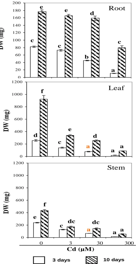
Root, stem and leaf biomass decreased with increasing Cd concentrations in the nutrient solution (Figure 1). A significant decline in dry weight (DW) was observed in
stems and leaves even at the lowest Cd concentrations (3 µM CdCl2) within the first 3 days (Figure 1). Root DW
was only affected in the presence of 30 and 300 µM Cd. When subjected to severe Cd stress (300 µM) for 10 days, leaf and root DW were reduced to 50 and 90% compared to their respective control (Figure 1). However, as previously shown by Djebali et al. (2008), at such a high Cd concentration, tomato plants are still able to recover when the stress is released. This means that the plants are able to cope with high Cd concentration for at least 10 days. As previously reported by Boulila et al. (2006) and Djebali et al. (2008) growth inhibition was accompanied by symptoms of chlorosis and necrotic areas (data not shown). In previous studies, Cd stress was found to modify the water balance (Sanita di Toppi and Gabbrielli, 1999). In tomato, moderate Cd concen-tration (3 µM) had no effect on water contents (WC) (Figure 2), whereas both root and shoot WC decreased in 30 and 300 µM Cd treated plants. Thus, at the most severe treatment (300 µM), the decrease of leaf and root WC was 70 and 80% when compared with their respec-tive control. These effects may be explained by the fact that, Cd is known to interfere with guard cell regulation and to affect water uptake capacity of roots (Perfus-Barbeoch et al., 2002).
Cadmium accumulation and distribution
During the treatment, Cd content increased in both leaves and roots as a function of external Cd concentrations (Figure 3). However, more than 80 to 90% of Cd was stored in the root system of the plant. After a 10 day period with the presence of 300 μM Cd, internal Cd concentrations reached 1.1 mg g-1 (DW) in leaves and 3.7 mg g-1 DW in roots, which accounted for 0.11 to 0.37% of the DW, respectively (Figure 3). The accu-mulation of Cd in the leaves indicates a significant transport of Cd from roots to leaves. However, only 20% of total Cd accumulated in plant was stored in leaf tissues. Commonly, in tomato plants, Cd is mainly retained in roots and only small amounts are transported to the shoots (Boulila et al., 2006; Abdel-Latif 2008, Djebali et al., 2008; Kieffer et al., 2009). Cadmium contents in tomato plants decrease in the following order: roots > stems > leaves > fruits > seeds (Blum, 1997). It is one of the strategies used by plants for avoiding Cd toxicity, particularly in photosynthetic tissues which are more sensitive to heavy metals than roots. Roots are considered as a barrier against Cd translocation to the aerial part of the plant by immobilization of toxic ions by means of the cell wall, extracellular carbohydrates (Sanita di Topi and Gabbrielli, 1999) or vacuolar seque-stration inside cortical cells (Djebali et al., 2002).
Effects of Cd on sugar contents
0 20 40 60 80 100 120 140 160 180 200 0 200 400 600 800 1000 1200 0 200 400 600 800 1000 1200
b
c
c
a
e
d
c
e
Root
Leaf
d
a
c
a
f
e
d
a
e
c
a
a
f
dc
dc
a
Stem
DW
(m
g)
DW
(m
g)
DW
(m
g)
0 3 30 300Cd (µM)
3 days 10 daysFigure 1. Growth of tomato roots, leaves and stems as a function of Cd treatment. Data are the mean of six samples ± SD. Values followed by the same letter are not significantly different (at p < 0.05) as described by Tukey’s (HSD) test.
0 2 4 6 8 10 12 14 16 18 0 2 4 6 8 10 12 14 16 18 0 2 4 6 8 10 12 14 16 18
WC
(ml
g
-1DW
)
WC
(ml
g
-1DW
)
WC
(ml
g
-1DW
)
0 3 30 300 Cd (µM)Stem
Root
Leaf
c b c a a e d e d c b c b a d c de e c ab c dc b a 3 days 10 daysFigure 2. Water content (WC) of tomato roots, leaves and stems as a function of Cd treatment. Data are the mean of six samples ± SD. Values followed by the same letter are not significantly different (at p < 0.05) as described by Tukey’s (HSD) test.
tose and sucrose) were analyzed by 1H-NMR spectro-scopy (Figure 4). In the roots of control plants, sucrose content was about 3 fold higher than that of glucose and
fructose (59, 19 and 19 µmol/g DW respectively; Figure 4). In plants treated for 10 days with 30 µM of Cd, glucose and fructose contents in roots were 2.6 and 4.8
3 days
10 days
µg
Cd
g
-1DW
µg
Cd
g
-1DW
a
a
b
a
c
d
a
Root
a a
b
a
b
c
a
Leaf
0 3 30 300
Cd (µM)
Figure 3. Cd accumulation in root and leaf tissues of tomato plants after 3 and 10 days of Cd treatments. Results are the mean of six samples ± SD. Histograms followed by the same letter are not significantly different (at p < 0.05) as described by Tukey’s (HSD) test.
fold higher, respectively, than in control, but sucrose content was not significantly modified (Figure 4). How-ever, in the roots of plants treated for 10 days with 300 µM CdCl2, glucose and fructose contents did not change
significantly compared to the control (12 and 10 µmol/g DW, respectively; Figure 4). Such an increase in glucose and fructose contents might result from an adaptation of carbon metabolism of the plant to the metal stress, as
already observed in rice where invertase activity was increased in response to Cd (Verma and Dubey, 2001). Thus, our data showed that, despite an inhibition of the growth of the roots (Figure 1), Cd stress was accom-panied with an accumulation of glucose, fructose and to a lesser extent, sucrose in the roots. In the leaves of control plants, glucose and fructose levels were two times higher than in roots (44 versus 19 µmol/g DW, and 35
Root
Leaf
Glucose
0 20 40 60 80Fructose
0 20 40 60 80Sucrose
0 20 40 60 80Glucose
0 20 40 60 80Fructose
0 20 40 60 80Sucrose
0 20 40 60 80b
a
b
a
c
d
b a
b
d
b
bc
cd
d
c
a
b
a
ab
d
a
c
c
b
cd
bc
a
de
ab
e
ab
e
bc
bc
cd
b
e
d
c
a
de
bc bc
a
de e
cd
b
A C E B D F 0 3 30 300Cd (µM)
0 3 30 300Cd (µM)
3 days
10 days
µ
m
ol
g
-1DW
µm
ol
g
-1DW
µ
m
ol
g
-1DW
Figure 4. Soluble sugar contents in tomato roots (A, C and E) and leaves (B, D and F) as a function of Cd treatment. Data are the means of six samples ± SD. Values followed by the same letter are not significantly different (at p < 0.05) as described by Tukey’s (HSD) test.
versus 16 µmol/g DW, respectively), whereas, that of sucrose was similar (56 versus 58 µmol/g DW) (Figure 4). The exposition to 30 µM Cd induced an increase in glucose, fructose and sucrose amounts after 3 days, but after 10 days their levels were similar to the control. As observed in the roots, 3 and 300 µM Cd treatment had only minor effect on sugar accumulation in leaf tissues (Figure 4).
In pea seedlings, Cd was also shown to induce an important reduction in shoot and root lengths together with an increase in sucrose content in the shoots (Devi et al., 2007). Similarly, in the shoots of rice plants submitted
to Cd, the accumulation of carbohydrates was combined with a decline of net photosynthetic rates (Moya et al., 1995). A recent study on poplar leaves showed that, the increase of various carbohydrate contents (glucose, fruc-tose, galacfruc-tose, sucrose and raffinose) and the modification of their metabolism could be one of the Cd effects leading to growth inhibition (Kieffer et al., 2009). Cell division and its regulation appear to be directly affected by the level of available carbohydrates and the form in which they are available (Francis and Halford, 2006). The regulation of primary carbohydrate metabo- lism and of the enzymes involved in carbon metabolism
plays an important role in determining carbohydrate composition and level and may have a large effect on growth (Koch, 2004). These observations suggest that, the use of carbohydrate is reduced in the presence of Cd (Bailey et al., 2003). In these conditions, photoassimilates are less needed for developmental processes and partially stored under sucrose or hexose forms, as well as more complex sugars, like raffinose (Kieffer et al., 2009).
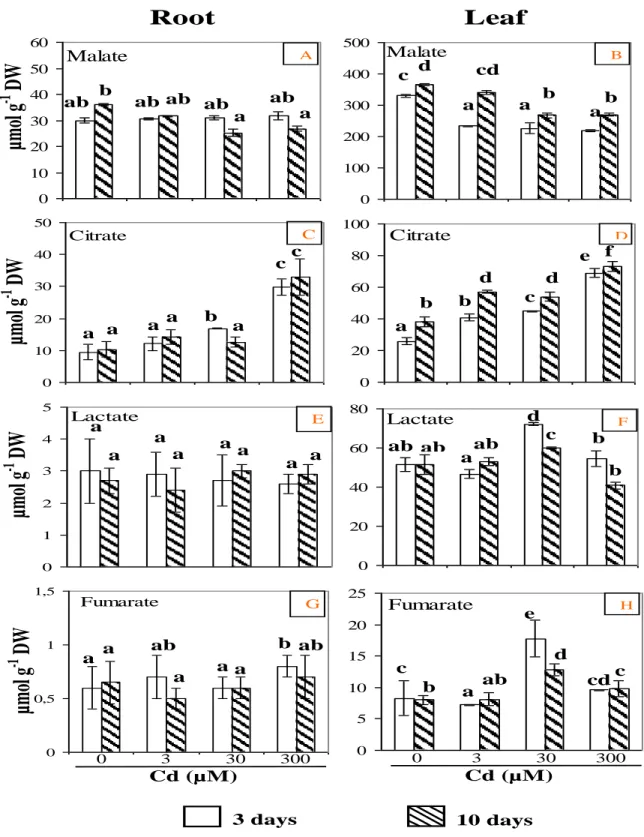
Effects of Cd on organic acid contents
The major organic acids present in root and leaf extracts (citrate, malate, lactate and fumarate) were identified and quantified by 1H- NMR spectroscopy. As reported in Figure 5, malate was the most abundant organic acid (68% of the total organic acid content) in the roots of control plants, followed by citrate (22%) and lactate (7%). Fumarate was present at low level (3%) and its concentration greatly changed depending on the extract. Similar organic acid contents have been reported in pea and buttercup tissues (Rivasseau et al., 2006). Organic acids, such as citrate, malate and fumarate, play a key role in the Krebs cycle which is the central energy yielding cycle of the cell. After a 10 day period of Cd treatment, malate contents only moderately decreased (10 to 25%), whereas lactate content was not significantly modified (Figure 5). Contrarily, Cd enhanced citrate content in root tissues. Thus, in roots treated with 300 µM Cd for 10 days, citrate quantity increased 3 fold (33 µmol.
g-1 DW) compared to control roots (Figure 5). In the leaves of control plants, organic acid contents were found to be higher than in roots (Figure 5). Thus, malate content in leaf tissues of control plants was 10 fold higher than in roots (330 versus 30 µmol.g-1 DW, respectively) (Figure 5), probably because of it’s involvement in respiration and photosynthesis processes. Upon Cd treatment, a significant decrease in malate level was observed, earlier in leaves than in roots. After 10 days, for the three Cd treatments, malate content decreased by 30% compared to the control. During the treatment, citrate accumulated in the leaves in a concentration dependent manner. At the most severe treatment (10 days with 300 µM), citrate quantity was 2.6 fold higher (73 µmol.g-1 DW) than in control leaves (Figure 5). Interestingly, in parallel to what was observed with soluble sugars (Figure 4), lactate and fumarate contents increased significantly by 1.4 and 2 fold, respectively, in the leaves of 30 µM Cd-treated plants as compared to the control (Figure 5).
The increase of major organic acid content was ob-served earlier in leaves than in roots (Figure 5). These observations suggest that, protective mechanisms against Cd ions are activated rapidly to limit metal delete-rious effects on leaves, especially on photosynthesis process which is important for growth. On the other hand, the accumulation of citrate in roots argues in favour of its
role in detoxification process by chelating metal ions as already reported (McCluskey et al., 2004). Fumarate is an intermediate organic acid in the tricarboxylic acid cycle and its variations could be related to changes in this cycle under Cd stress. In contrast, a decrease in malate content in both roots and leaves with increasing Cd concentrations could be explained by its cytosolic and vacuolar localisation in the cells (Gout et al., 1993). For this, malate is directly available for metabolic require-ments and its concentration may vary in response to metal stress because it is implied in different complex metabolic pathways (Rivasseau et al., 2006).
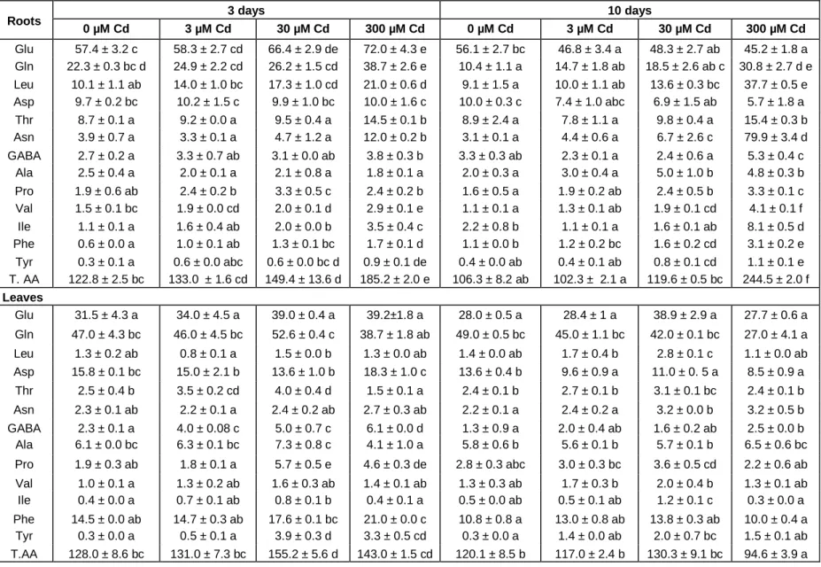
Effects of Cd on amino acid profile
The analysis of 1H-NMR spectra of tomato root and leaf extracts showed clear differences between amino acid (AA) profiling of control and Cd-treated plants. Thirteen different AA were identified and quantified (Table 1). In control roots, the major AA were glutamate (47% of total AA), glutamine (18%) followed by aspartate and leucine (8% of the total for each of them). Upon Cd treatment, total AA contents were increased in the roots of 30 and 300 µM Cd treated plants (Table 1). Thus, total AA content was 51 and 131% increased, respectively, after 3 and 10 days of 300 µM Cd treatment. For the lowest Cd concentration tested (3 µM), no significant change was observed after 3 and 10 days, which suggests that root amino acid metabolism was barely or not disturbed at that concentration. After 3 days of Cd exposure, aspa-ragine, glutamine, glutamate, leucine, valine, isoleucine, threonine, tyrosine, phenylalanine and proline quantity increased when compared with control. Aspartate and glutamate amount which are the synthesis precursors for asparagine and glutamine decreased significantly by 43 and 19%, respectively, in plants’ roots exposed for 10 days to 300 µM (Table 1). With the most severe treat-ment (300 µM Cd for 10 days), asparagine content in the roots was 26 fold higher than in control roots (79.9 versus 3.1 µmol.g-1 DW, respectively). Concomitantly, a 4 fold increase in leucine content and a 3 fold increase in glutamine, valine and isoleucine contents were observed (Table 1).
In control leaf tissues, the major AA were glutamine, glutamate, aspartate and phenylalanine, accounting for 37, 25, 12 and 11% of total AA, respectively (Table 1). Changes in AA contents in response to Cd stress were less important in leaves than in roots. After 3 days of treatment with 30 and 300 µM Cd, total AA content increased significantly by 21 and 10% when compared with control (Table 1). Under stress, glutamate, gluta-mine, aspartate and phenylalanine remained the major AA. Significant increases in proline, γ-aminobutyric acid (GABA, tyrosine, isoleucine and threonine were observed after 3 days of treatment under high Cd concentrations (30 and 300 µM, Table 1). However, under the same
1 Malate 0 10 20 30 40 50 60 Malate 0 100 200 300 400 500
µ
m
ol
g
-1DW
ab b ab ab ab a a ab c d a cd b a a b Citrate 0 10 20 30 40 50 Citrate 0 20 40 60 80 100 aµ
m
ol
g
-1DW
b c a a a a c = = = = = = = = = = a b b d c d e f Lactate 0 1 2 3 4 5 Lactate 0 20 40 60 80µ
m
ol
g
-1DW
a a a a a a a a ab a ab d c b ab b Fumarate 0 5 10 15 20 25 Fumarate 0 0,5 1 1,5 0 3 30 300 Cd (µM) 0 3 30 300 Cd (µM) c b a e d cd cµ
m
ol
g
-1DW
3 days 10 daysRoot
Leaf
ab a a a b ab ab a a A C E G B D F HFigure 5. Organic acid contents in tomato roots (A, C, E and G) and leaves (B, D, F and H) as a function of Cd treatment. Data are means of six samples ± SD. Values for the same compound sharing the same letters did not differ significantly at (p < 0.05) as described by Tukey’s (HSD) test.
Table 1. Individual and total free amino acid levels in tomato root and leaf tissues as a function of time and Cd treatment. (TAA, total amino acid). Values are expressed as mean of six samples ± SD. Values for the same compound sharing the same letters did not differ significantly at (p < 0.05) (Tukey’s test).
Roots 3 days 10 days
0 µM Cd 3 µM Cd 30 µM Cd 300 µM Cd 0 µM Cd 3 µM Cd 30 µM Cd 300 µM Cd Glu 57.4 ± 3.2 c 58.3 ± 2.7 cd 66.4 ± 2.9 de 72.0 ± 4.3 e 56.1 ± 2.7 bc 46.8 ± 3.4 a 48.3 ± 2.7 ab 45.2 ± 1.8 a Gln 22.3 ± 0.3 bc d 24.9 ± 2.2 cd 26.2 ± 1.5 cd 38.7 ± 2.6 e 10.4 ± 1.1 a 14.7 ± 1.8 ab 18.5 ± 2.6 ab c 30.8 ± 2.7 d e Leu 10.1 ± 1.1 ab 14.0 ± 1.0 bc 17.3 ± 1.0 cd 21.0 ± 0.6 d 9.1 ± 1.5 a 10.0 ± 1.1 ab 13.6 ± 0.3 bc 37.7 ± 0.5 e Asp 9.7 ± 0.2 bc 10.2 ± 1.5 c 9.9 ± 1.0 bc 10.0 ± 1.6 c 10.0 ± 0.3 c 7.4 ± 1.0 abc 6.9 ± 1.5 ab 5.7 ± 1.8 a Thr 8.7 ± 0.1 a 9.2 ± 0.0 a 9.5 ± 0.4 a 14.5 ± 0.1 b 8.9 ± 2.4 a 7.8 ± 1.1 a 9.8 ± 0.4 a 15.4 ± 0.3 b Asn 3.9 ± 0.7 a 3.3 ± 0.1 a 4.7 ± 1.2 a 12.0 ± 0.2 b 3.1 ± 0.1 a 4.4 ± 0.6 a 6.7 ± 2.6 c 79.9 ± 3.4 d GABA 2.7 ± 0.2 a 3.3 ± 0.7 ab 3.1 ± 0.0 ab 3.8 ± 0.3 b 3.3 ± 0.3 ab 2.3 ± 0.1 a 2.4 ± 0.6 a 5.3 ± 0.4 c Ala 2.5 ± 0.4 a 2.0 ± 0.1 a 2.1 ± 0.8 a 1.8 ± 0.1 a 2.0 ± 0.3 a 3.0 ± 0.4 a 5.0 ± 1.0 b 4.8 ± 0.3 b Pro 1.9 ± 0.6 ab 2.4 ± 0.2 b 3.3 ± 0.5 c 2.4 ± 0.2 b 1.6 ± 0.5 a 1.9 ± 0.2 ab 2.4 ± 0.5 b 3.3 ± 0.1 c Val 1.5 ± 0.1 bc 1.9 ± 0.0 cd 2.0 ± 0.1 d 2.9 ± 0.1 e 1.1 ± 0.1 a 1.3 ± 0.1 ab 1.9 ± 0.1 cd 4.1 ± 0.1 f Ile 1.1 ± 0.1 a 1.6 ± 0.4 ab 2.0 ± 0.0 b 3.5 ± 0.4 c 2.2 ± 0.8 b 1.1 ± 0.1 a 1.6 ± 0.1 ab 8.1 ± 0.5 d Phe 0.6 ± 0.0 a 1.0 ± 0.1 ab 1.3 ± 0.1 bc 1.7 ± 0.1 d 1.1 ± 0.0 b 1.2 ± 0.2 bc 1.6 ± 0.2 cd 3.1 ± 0.2 e Tyr 0.3 ± 0.1 a 0.6 ± 0.0 abc 0.6 ± 0.0 bc d 0.9 ± 0.1 de 0.4 ± 0.0 ab 0.4 ± 0.1 ab 0.8 ± 0.1 cd 1.1 ± 0.1 e T. AA 122.8 ± 2.5 bc 133.0 ± 1.6 cd 149.4 ± 13.6 d 185.2 ± 2.0 e 106.3 ± 8.2 ab 102.3 ± 2.1 a 119.6 ± 0.5 bc 244.5 ± 2.0 f Leaves Glu 31.5 ± 4.3 a 34.0 ± 4.5 a 39.0 ± 0.4 a 39.2±1.8 a 28.0 ± 0.5 a 28.4 ± 1 a 38.9 ± 2.9 a 27.7 ± 0.6 a Gln 47.0 ± 4.3 bc 46.0 ± 4.5 bc 52.6 ± 0.4 c 38.7 ± 1.8 ab 49.0 ± 0.5 bc 45.0 ± 1.1 bc 42.0 ± 0.1 bc 27.0 ± 4.1 a Leu 1.3 ± 0.2 ab 0.8 ± 0.1 a 1.5 ± 0.0 b 1.3 ± 0.0 ab 1.4 ± 0.0 ab 1.7 ± 0.4 b 2.8 ± 0.1 c 1.1 ± 0.0 ab Asp 15.8 ± 0.1 bc 15.0 ± 2.1 b 13.6 ± 1.0 b 18.3 ± 1.0 c 13.6 ± 0.4 b 9.6 ± 0.9 a 11.0 ± 0. 5 a 8.5 ± 0.9 a Thr 2.5 ± 0.4 b 3.5 ± 0.2 cd 4.0 ± 0.4 d 1.5 ± 0.1 a 2.4 ± 0.1 b 2.7 ± 0.1 b 3.1 ± 0.1 bc 2.4 ± 0.1 b Asn 2.3 ± 0.1 ab 2.2 ± 0.1 a 2.4 ± 0.2 ab 2.7 ± 0.3 ab 2.2 ± 0.1 a 2.4 ± 0.2 a 3.2 ± 0.0 b 3.2 ± 0.5 b GABA 2.3 ± 0.1 a 4.0 ± 0.08 c 5.0 ± 0.7 c 6.1 ± 0.0 d 1.3 ± 0.9 a 2.0 ± 0.4 ab 1.6 ± 0.2 ab 2.5 ± 0.0 b Ala 6.1 ± 0.0 bc 6.3 ± 0.1 bc 7.3 ± 0.8 c 4.1 ± 1.0 a 5.8 ± 0.6 b 5.6 ± 0.1 b 5.7 ± 0.1 b 6.5 ± 0.6 bc Pro 1.9 ± 0.3 ab 1.8 ± 0.1 a 5.7 ± 0.5 e 4.6 ± 0.3 de 2.8 ± 0.3 abc 3.0 ± 0.3 bc 3.6 ± 0.5 cd 2.2 ± 0.6 ab Val 1.0 ± 0.1 a 1.3 ± 0.2 ab 1.6 ± 0.3 ab 1.4 ± 0.1 ab 1.3 ± 0.3 ab 1.7 ± 0.3 b 2.0 ± 0.4 b 1.3 ± 0.1 ab Ile 0.4 ± 0.0 a 0.7 ± 0.1 ab 0.8 ± 0.1 b 0.4 ± 0.1 a 0.5 ± 0.0 ab 0.5 ± 0.1 ab 1.2 ± 0.1 c 0.3 ± 0.0 a Phe 14.5 ± 0.0 ab 14.7 ± 0.3 ab 17.6 ± 0.1 bc 21.0 ± 0.0 c 10.8 ± 0.8 a 13.0 ± 0.8 ab 13.8 ± 0.3 ab 10.0 ± 0.4 a Tyr 0.3 ± 0.0 a 0.5 ± 0.1 a 3.9 ± 0.3 d 3.3 ± 0.5 cd 0.3 ± 0.0 a 1.4 ± 0.0 ab 2.0 ± 0.7 bc 1.5 ± 0.1 ab T.AA 128.0 ± 8.6 bc 131.0 ± 7.3 bc 155.2 ± 5.6 d 143.0 ± 1.5 cd 120.1 ± 8.5 b 117.0 ± 2.4 b 130.3 ± 9.1 bc 94.6 ± 3.9 a
treatment asparagine, aspartate, alanine, leucine, valine, glutamine, glutamate and phenylalanine contents remain unchanged (Table 1). In the presence of 300 µM Cd, the prolongation of the stress period up to 10 days induced a 20% decrease in total AA contents in leaf tissues compared to control (94.6 versus 120 µmol.g-1 DW; Table 1). In contrast to roots, the majority of leaf AA (asparagine, glutamate, alanine, leucine, valine, threonine, phenylalanine and proline) was unaffected by 10 days of Cd treatment. However, significant reduction of glutamine and aspartate levels (45 and 37% from control, respectively) was observed in the presence of 300 µM CdCl2 (Table 1).
Globally, the increase in the content of several AA, especially in roots, could be explained by an increase in protein turnover and degradation (Smalle and Vierstra, 2004; Thompson and Vierstra, 2005). Proteolysis is involved in the degradation of proteins damaged by oxidative stress induced by Cd (Pena et al., 2006; Polge et al., 2009). The opposite effects induced by Cd in pro-tein content in the roots and the leaves, probably indicates that, protein turnover response to Cd is differently regulated in the two organs (Djebali et al., 2008). On the other hand, under Cd stress, some AA can be used for complexation of toxic ions inside the cell and play an important role in heavy metal homeostasis (Sanita di Toppi and Gabbrielli, 1999; Benavides et al., 2005). Proline accumulation observed in roots at the end of the experiment, confirmed the hypothesis that, one mechanism by which many plants and algae respond to and apparently detoxify heavy metals, is the production of proline (Chen et al., 2002; Abdel-Latif, 2008). This AA is an abiotic and biotic stress indicator in algae and in some plants (Verma, 1999). Many mechanisms were proposed to explain the role of proline in heavy metal stress conditions. Proline protective role in stressed plants is associated with reduced membrane and protein damages (Verma, 1999). Free proline has been proposed to act as an osmoprotectant (Taylor, 1996), a metal chelator (Farago and Mullen, 1979), an inhibitor of lipid peroxidation (Mehta and Gaur, 1999), a hydroxyl radical and singlet oxygen scavenger (Alia and Matysik, 2001) and an antioxidant especially in Cd stress cells (Siripornadulsil et al., 2002). In addition, the present work showed a spectacular increase in asparagine content in roots treated 10 days by 300 µM CdCl2 (26 fold, when
compared with control), but remain stable in leaf tissues (Table 1). Asparagine synthesis and accumulation appear to store the excess of nitrogen as an inert nitro-gen reserve under Cd stress and bacterial infection by
Pseudomonas syringae in tomato (Chaffei et al., 2004;
Olea et al., 2004). These authors also reported an up-regulation of asparagine synthetase and a strong aspa-ragine accumulation especially in tomato roots exposed to Cd (Chaffei et al., 2004). Simultaneously, they observed inducing of glutamine synthetase (GS1) activity, which suggested that, the existence of GS1/AS pathway,
representing a metabolic route for transferring ammonium released from protein catabolism into asparagine (Olea et al., 2004), an AA that may have a major role in nitrogen mobilization events such as natural senescence (Lea and Miflin, 1980), sugar starvation (Brouquisse et al., 1992), dark/light transitions (Sieciechowicz et al., 1988), or post-harvest conditions (King et al., 1990).
In roots, alanine, GABA and valine quantities increased after 10 days of Cd exposure. These three metabolites are implicated in coenzyme A formation and protein synthesis (Kupke et al., 2003), assuming metabolite accumulation could be interpreted as a metabolic im-balance and an alteration in coenzyme A biosynthesis (Broeckling et al., 2004). In addition, our data demonstrated a synchronic increase of valine, isoleucine (3 fold) and leucine (4 fold) in plants’ roots treated by 300 µM of Cd (Table 1). Valine, leucine and isoleucine are all produced by the same biosynthetic pathway (Broeckling et al., 2004), but the first enzymatic step uses different precursors for each branched chain amino acid (Broeckling et al., 2004). A linear and precise correlation was observed between leucine, isoleucine and valine amounts in response to perturbation with biotic and abiotic elicitors in Medicago truncatula cell culture (Broeckling et al., 2004). Probably, accumulation of bran-ched chain AA can be one consequence of perturbations in acetyl-coenzyme A biosynthesis and/or in glycolysis which are affected under stress conditions.
Conclusion
Summarising our results, it may be concluded that Cd stress triggered a concentration dependent reduction of tomato plant growth and a reduction of the WC of the tissues. In the roots and leaves, Cd triggered clear dis-turbance of the primary metabolism as shown by the modifications of several metabolites such as organic acids and amino acids. Among these metabolites, the strong accumulation of citrate in roots and leaves, tyrosine in leaves and asparagine in roots, was found to be dependent of the concentration of Cd. These three compounds appear to be good markers to assess the extent of Cd stress in tomato plants and could be potentially used to investigate Cd stress in other plants of interest.
ACKNOWLEDGEMENTS
The authors thank the Ministry of Higher Education, Scientific Research and Technology of Tunisia for finan-cial support. One part of this work was supported by the Comité Mixte de Coopération Universitaire Franco- Tunisien (grant n° 03G0911 for Latifa Boulila).The authors also thank the staff of UMR619 Biologie du Fruit (Bordeaux) for technical help and discussions, parti-
cularly Christelle Renaud and Dominique Rolin. The NMR analyses were performed on the Metabolome-Fluxome, Facility of Bordeaux Functional Genomics Centre.
REFERENCES
Abdel-Latif A (2008). Cadmium Induced Changes in Pigment Content, Ion Uptake, Proline Content and Phosphoenolpyruvate Carboxylase Activity in Triticum Aestivum Seedlings. Aust. J. Basic Appl. Sci. 2(1): 57-62.
Aina R, Labra M, Fumagalli P, Vannini C, Marsoni M, Cucchi U, Bracale M, Sgorbati S, Citterio S (2007). Thiol-peptide level and proteomic changes in response to cadmium toxicity in Oryza sativa L. roots. Environ. Exp. Bot. 5(3): 381-392.
Akoka S, Barantin L, Trierweiler M (1999). Concentration measurement by proton NMR using the ERETIC method. Anal. Chem. 71: 2554-2557. doi: 10.1021/AC981422I.
Alia MP, Matysik JL (2001). Effects of proline on the production of singlet oxygen. Amino acids, 21: 191-203.
Bailey NJC, Oven M, Holmes E, Nicholson JK, Zenk MH (2003). Metabolic analysis of the consequences of cadmium exposure in
Silene cucubalus cell cultures via 1H NMR spectroscopy and
chemometrics. Phytochemistry, 62: 851-858.
Benavides MP, Gallego SM, Tamaro ML (2005). Cadmium toxicity in plants. Brazilian J. Plant Physiol. 17 (1): 21-34.
Blum WH (1997). Cadmium uptake by higher plants. In: Proceedings of extended abstracts for the fourth international conferences on the Biogeochemistry of trace elements, Berkeley, USA. University of California.
Boulila Zoghlami L, Djebali W, Chaïbi W, Ghorbel MH (2006). Modifications physiologiques et structurales induitespar l’interaction cadmium-calcium chez la tomate (Lycopersicon esulentum). C. R. Biol. 329(9): 702-711.
Broeckling CD, Human DV, Farag AM, Smith JT, May GD, Mendes P, Dixon RA, Sumner WL (2004). Metabolic profiling of Medicago
truncatula cell cultures reveals the effects of biotic and abiotic
elicitors on metabolism. J. Exp. Bot. 56(410): 323-336.
Brouquisse R, James F, Pradet A, Raymond P (1992). Asparagine metabolism and nitrogen distribution during protein degradation in sugar starved maize root tips. Planta, 188: 384-395.
Chaffei C, Pageau K, Suzuki A, Gouia H, Ghorbel MH, Masclaux-Daubresse C (2004). Cadmium toxicity induced changes in nitrogen management in Lycopercicon esculentum leading to metabolic safeguard through an amino acid storage strategy. Plant Cell Physiol. 45(11): 1681-1693.
Chen GX, Wang N, Shao ZG, Shi GX (2002). Effects of phosphorus nutrition on physiological activity of Nymphaea tetragona Georgi and Trapa bispinosa Roxb.Leaves. J. NanJing normal university (Natural science) 25(1): 71-77.
Cobbett C, Goldsbrough P (2002). Phytochelatins and Metallothioeins: Roles in Heavy Metal Detoxification and Homeostasis. Ann. Rev. of Plant Biol. 53: 159-182.
Devi R, Munjral N, Gupta AK, Kaur N (2007). Cadmium induced changes in carbohydrate status and enzymes of carbohydrate metabolism, glycolysis and pentose phosphate pathway in pea. Environ. Exp. Bot. 61(2): 167-174.
Djebali W, Chaïbi W, Ghorbel MH (2002). Croissance, activité peroxydasique et modifications structurales et ultrastructurales induites par le cadmium dans la racine de tomate (Lycopersicon
esculentum). Can. J. Bot. 80: 942-953.
Djebali W, Zarrouk M, Brouquisse R, El Kahoui S, Limam F, Ghorbel MH, Chaïbi W (2005). Ultrastructure and lipid alterations induced by cadmium in tomato (Lycopersicon esculentum) chloroplast
membranes. Plant Biol. 7: 258-368.
Djebali W, Gallusci P, Polge C, Boulila L, Galtier N, Raymond P, Chaibi W, Brouquisse R (2008). Modifications in endopeptidase and 20S proteasome expression and activities in cadmium treated tomato (Solanum lycopersicum L.) plants. Planta, 227: 625-639.
El Said Deef H (2008). Effect of Cadmium and Zinc on Growth Parameters of Tomato Seedlings. Academic J. Plant Sci. 1(1): 05-11.
Farago ME, Mullen WA (1979). Plants which accumulate metals. Part IV.A possible copper-proline complexe from the roots of Armeria
maritima. Inorganica Chimica Acta, 32: 93-94.
Fiehn O, Kopka J, Dormann P, Altmann T, Tretheway RN, Willmitzer L (2000). Metabolite profiling for plant functional genomics. Nat. Biotechnol. 18: 1157-1161.
Fornazier C, Lobréaux S, Mari S, Briat JF, Lebrun M (2002). Metal resistance in yeast is mediated by the expression of a maize 20S proteasome _ subunit. Gene 293: 199-204.
Francis D, Halford N (2006). Nutrient Sensing in Plant Meristems. Plant Mol. Biol. 60: 981-993.
Gout E, Bligny R, Pascal N, Douce R (1993). 13C nuclear magnetic resonance studies of malate and citrate synthesis and compartmentation in higher plant cells. J. Bio. Chem. 268: 3986-3992.
Holmes E, Nicholson J, Tranter G (2001). Metabolic characterization of genetic variations in toxicological and metabolic responses using probabilistic neural networks. Chem. Res. Toxicol. 14: 182-191. Kieffer P, Dommes J, Hoffmann L, Hausman JF, Renaut J (2009).
Quantitative changes in protein expression of cadmium-exposed poplar plants. Proteomics, 8(12): 2514–2530.
King GA, Woollard DC, Irving DE, Borst WM (1990). Physiological changes in asparagus spear tips after harvest. Physiol. Plant. 80: 393-400.
Koch KE (2004). Sucrose metabolism: Regulatory mechanisms and pivotal roles in sugar sensing and plant development. Curr. Opin. Plant Biol. 7: 235-246.
Kupke T, Hernandez-Acosta P, Culianez-Macia FA (2003). 4’-Phosphopantetheine and coenzyme A biosynthesis in plants. J. Biol. Chem. 278: 38229-38237.
Lea PJ, Miflin BJ (1980). Transport and metabolism of asparagine and other nitrogen compounds within the plant. In: Stumpf PK & Conn EE, eds. Biochem. Plants, Academic Press, New York, pp. 569-607. McCluskey J, Herdman L, Skene KR (2004). Iron deficiency induces
changes in metabolism of citrate in lateral roots and cluster roots of
Lupinus albus. Physiol. Plant. 121(4): 286-294.
Mehta SK, Gaur JP (1999). Heavy-metal-induced proline accumulation and its role in ameliorating metal toxicity in Chlorella vulgaris. New Phytol. 143: 253-259.
Moing A, Maucourt M, Renaud C, Gaudillère M, Brouquisse R, Lebouteiller B, Gousset-Dupont A, Vidal J, Granot D, Denoyes-Rothan B, Lerceteau-Kïhler E, Rolin D (2004). Quantitative metabolic profiling by 1-dimentional 1H-NMR analyses: application to plant genetics and functional genomics. Functional Plant Biol. 31: 889-902. Moya JL, Ros R, Picazo I (1995). Heavy metal-hormone interactions in rice plants: Effects on growth, net photosynthesis, and carbohydrate distribution. J. Plant Growth Regul. 14(2): 61-67.
Olea F, Pérez-Garcia A, Canton FR, Rivera ME, Canas R, Avila C, Cazorala MF, Canovas MF, De-Vicente A (2004). Up-regulation and localization of asparagines synthetase in tomato leaves infectead by the bacterial pathogen Psedomonas synergae. Plant Cell. Physiol. 45(6): 770-780.
Pena LB, Pasquini LA, Tomaro ML, Gallego SM (2006). Proteolytic system in sunflower (Helianthus annuus L.) leaves under cadmium stress. Plant Sci. 171: 531-537.
Perfus-Barbeoch L, Leonhard N, Vavasseur A, Forestier C (2002). Heavy metal toxicity: cadmium permeates through calcium channels and disturbs the plant water status. Plant J. 32(4): 539-548. Polge C, Jaquinod M, Holzer F, Bourguignon J, Walling L, Brouquisse R
(2009). Evidence for the existence in Arabidopsis thaliana of the proteasome proteolytic pathway-Activation in response to cadmium. J. Biol. Chem. 284: 35412-35424.
Prasad MNV (1995). Cadmium toxicity and tolerance in vascular plants. Environ. Exp. Bot. 35(4): 525-545.
Rivasseau C, Boisson AM, Mongélard G, Couram G, Bastien O, Bligny R (2006). Rapid analysis of organic acids in plant extracts by capillary electrophoresis with indirect UV detection Directed metabolic analyses during metal stress. J. Chromatography A, 1129: 283-290. Sanita di Toppi L, Gabbrielli R (1999). Response to cadmium in higher
plants. Environ. Exp. Bot. 41: 105-130.
Sarry JE, Kuhn L, Ducruix C, Lafaye A, Junot C, Hugouvieux V, Jourdain A, Bastien O, Fievet JB, Vailhen D, Amekraz B, Moulin C,
Ezan E, Garin J, Bourguignon J (2006). The early responsesof
Arabidopsis thaliana cells to cadmium exposure explored by protein
and metabolite profiling analyses. Proteomics, 6(7): 2180-2198. Siedlecka A, Krupa Z, Samuelsson G, Oquist G, Gardstrom P (1997).
Primary carbon metabolism in Phaseolus vulgaris plants under Cd/Fe interaction. Plant Physiol. Biochem. 35: 951-957.
Siripornadulsil S, Traina S, Verman DPS, Sayre TR (2002). Molecular mechanisms of proline-mediated tolerance to toxic heavy metals in transgenic microalgae. Plant Cell, 14: 2837-2847.
Sieciechowicz KA, Joy KW, Ireland RJ (1988). The metabolism of asparagine in plants. Phytochemistry, 27: 663-671.
Smalle J, Vierstra RD (2004). The ubiquitin 26S proteasome proteolytic pathway. Annu. Rev. Plant Biol. 55: 555-590.
Stitt M, Ap-Rees T (1978). Pathways of carbohydrates oxidation in leaves of Pisum sativum and Triticum aestrivum. Photochemistry, 17: 1251-1256.
Taylor CB (1996). Proline and water deficit: Ups, downs, ins and outs. Plant Cell, 8: 1222-1224.
Thompson AR, Vierstra RD (2005). Autophagic recycling: lessons from yeast help to define the process in plants. Curr. Opin. Plant Biol. 8: 165-173.
Van Assche F, Cardinaels C, Clijsters H (1988). Induction of enzyme capacity in plants as a result of heavy metal toxicity: Dose-response relations in Phaseolus vulgaris L., treated with zinc and cadmium. Environ. Pollut. 52: 103-115.
Verma DPS (1999). Osmotic stress tolerance in plants: role of proline and sulfur metabolisms. In: molecular response to cold, drought, heat and salt stress in higher plants, Shinozaki K, Yamaguchi-Shinozaki K, eds. Austin, TX: R.G. Landers: 153-168.
Verma S, Dubey RS (2001). Effect of cadmium on soluble sugars and enzymes of their metabolism in rice. Biol. Plant. 44: 117-123. Wagner GJ (1993). Accumulation of cadmium in crop plants and its
consequences to human health. Adv. Agron. 51: 173-212.