HAL Id: inserm-00980901
https://www.hal.inserm.fr/inserm-00980901
Submitted on 19 Apr 2014
HAL is a multi-disciplinary open access
archive for the deposit and dissemination of
sci-entific research documents, whether they are
pub-lished or not. The documents may come from
teaching and research institutions in France or
abroad, or from public or private research centers.
L’archive ouverte pluridisciplinaire HAL, est
destinée au dépôt et à la diffusion de documents
scientifiques de niveau recherche, publiés ou non,
émanant des établissements d’enseignement et de
recherche français ou étrangers, des laboratoires
publics ou privés.
Sebastian Lülf, Julie Matz, Marie-Christine Rouyez, Annika Järviluoma, Kalle
Saksela, Serge Benichou, Matthias Geyer
To cite this version:
Sebastian Lülf, Julie Matz, Marie-Christine Rouyez, Annika Järviluoma, Kalle Saksela, et al..
Struc-tural basis for the inhibition of HIV-1 Nef by a high-affinity binding single-domain antibody.
Retro-virology, BioMed Central, 2014, 11 (1), pp.24. �10.1186/1742-4690-11-24�. �inserm-00980901�
R E S E A R C H
Open Access
Structural basis for the inhibition of HIV-1 Nef by
a high-affinity binding single-domain antibody
Sebastian Lülf
1,2, Julie Matz
3,4,5, Marie-Christine Rouyez
3,4,5, Annika Järviluoma
6, Kalle Saksela
6,
Serge Benichou
3,4,5and Matthias Geyer
1,2*Abstract
Background:The HIV-1 Nef protein is essential for AIDS pathogenesis by its interaction with host cell surface
receptors and signaling factors. Despite its critical role as a virulence factor Nef is not targeted by current antiviral strategies.
Results:We have determined the crystal structure of the complex formed by a camelid single-domain antibody
fragment, termed sdAb19, bound to HIV-1 Nef together with a stabilizing SH3 domain. sdAb19 forms a stoichiometric 1:1 complex with Nef and binds to a conformationally conserved surface at the C-terminus of Nef that overlaps with functionally important interaction sites involved in Nef-induced perturbations of signaling and trafficking pathways. The antibody fragment binds Nef with low nanomolar affinity, which could be attenuated to micromolar affinity range by site-directed mutagenesis of key interaction residues in sdAb19. Fusion of the SH3 domain to sdAb19, termed Neffin, leads to a significantly increased affinity for Nef and formation of a stoichiometric 2:2 Nef–Neffin complex. The 19 kDa Neffin protein inhibits all functions of Nef as CD4 and MHC-I downregulation, association with Pak2, and the increase in virus infectivity and replication.
Conclusions:Together, sdAb19 and Neffin thus represent efficient tools for the rational development of antiviral
strategies against HIV-1 Nef.
Keywords:HIV-1 Nef, Single-domain antibody, Crystal structure, Neffin
Background
The human immunodeficiency virus (HIV) is a persistent pathogen that caused an estimated 1.6 million people deaths in 2012 [1]. Of the fifteen proteins encoded by the HIV genome, the three viral enzymes, protease, integrase and reverse transcriptase are indispensable for the produc-tion of viral progeny. These enzymes are core targets of highly active anti-retroviral therapy (HAART) together with proteins mediating virus entry [2,3]. HAART allowed considerable success in reducing viral loads beyond detec-tion levels and elongating patient life expectancy, but the current therapy is unable to clear the virus due to the per-sistence of latent reservoirs [4]. Advances for a successful eradication strategy showed that HAART in combination
with targeted cytotoxic therapy was able to profoundly deplete productively infected cells of viral RNA [5]. In addition, many broad and potent donor-derived antibodies were uncovered in recent years, suggesting they could be valuable additions to anti-HIV-1 therapies [6]. Yet, the rapid emergence of drug resistant mutants and the in-creased worldwide spread of treatment resistant HIV-1 variants pose increasing problems to effective treatment of HIV-infected patients. One strategy to improve this situ-ation is the exploitsitu-ation of additional drug targets that could be added to the current regiment. Ideally, such tar-gets comprise viral factors, since interference with host cell factors may compromise physiological functions or even viability of host cells.
Besides the structural proteins, HIV-1 encodes four accessory proteins to facilitate immune evasion and opti-mize conditions for virus replication [7]. The accessory nef gene encodes a 24–35 kDa protein that is found in all primate lentiviruses and is critical for the full patho-genic potential of these viruses [8]. Nef affects membrane * Correspondence:matthias.geyer@caesar.de
1Center of Advanced European Studies and Research, Group Physical Biochemistry, Bonn, Germany
2Department of Physical Biochemistry, Max Planck Institute of Molecular Physiology, Dortmund, Germany
Full list of author information is available at the end of the article
© 2014 Lülf et al.; licensee BioMed Central Ltd. This is an Open Access article distributed under the terms of the Creative Commons Attribution License (http://creativecommons.org/licenses/by/2.0), which permits unrestricted use, distribution, and reproduction in any medium, provided the original work is properly credited. The Creative Commons Public Domain Dedication waiver (http://creativecommons.org/publicdomain/zero/1.0/) applies to the data made available in this article, unless otherwise stated.
trafficking in infected cells, e.g. by modulating the expres-sion of surface receptors such as CD4, CD8, CD28, MHC-I and MHC-MHC-IMHC-I, DC-SMHC-IGN and chemokine receptors in HIV-1 target cells [9]. In addition, Nef also affects signal transduction through interaction with cellular kinases like Pak2 and Hck to modulate signaling pathways in infected cells [9,10]. To achieve this multitude of activities, Nef has evolved as a versatile adaptor for protein interactions that lacks intrinsic enzymatic activity. The structure of HIV-1 Nef is characterized by its flexible loop regions that contain several sequence motifs as an N-terminal myristoylation site, a central poly-proline PxxP motif for SH3 domain binding and C-terminal motifs for interaction with clathrin-associated endosomal adaptor protein complexes [11].
Although compounds interfering with Nef's activity would be in multiple ways beneficial to the host, Nef is currently not a target of antiviral measures. The Nef protein is not essential for replication of HIV in the in-fected host, yet the protein promotes the progression to AIDS in humans by the different internalization profiles found in SIV or HIV infected cells for CD3 and CD4 T cell receptors [12]. Previously described Nef-interacting small molecular compounds bind Nef only with relatively low affinity, and display high cytotoxicity and/or interfere with only a subset of Nef interactions and functions [13,14]. The characterization of a camelid single-domain antibody fragment, termed sdAb19, which binds to HIV-1 Nef with high affinity, has provided an alternative ap-proach to inhibit the biological activities of Nef [15]. This 12.7 kDa antibody fragment interfered with the CD4 down-regulation activity of Nef, as well as with the as-sociation of Nef with Pak2 and the accompanying actin remodeling effects. In addition, sdAb19 was shown to counteract the Nef-dependent enhancement of virion infectivity and virus replication, and to be able to rescue Nef-mediated thymic CD4+T cell maturation defects in transgenic mice expressing Nef [15,16]. Here, we de-scribe the crystal structure of the sdAb19 single domain antibody in complex with HIV-1 NefSF2 and an engi-neered SH3 domain of Hck. We provide structural and functional evidence for the potent inhibition of Nef caused by occupation of a highly conserved surface epi-tope at the C-terminus of Nef. These data represent important findings for the rational development of new antiviral strategies targeting HIV-1 Nef.
Results
Architecture of the Nef–sdAb19–SH3B6complex
The Nef–antibody complex was formed by mixing a purified recombinant form of HIV-1 NefSF2(45–210) de-leted of the first 44 N-terminal residues with sdAb19, and adding the SH3 domain of human Hck, termed SH3B6 and engineered for high affinity binding to Nef, to this complex [17,18]. Analytical gel filtration showed
that addition of sdAb19 and SH3B6to NefSF2led to for-mation of a stoichiometric 1:1:1 complex whose elution volume at an apparent mass of 45 kDa corresponded well to the calculated mass of 41.6 kDa (Figure 1A). To characterize the tripartite SH3B6–Nef–sdAb19 complex formation, we determined the individual binding affin-ities between Nef and its two complex partners by iso-thermal titration calorimetry (ITC). The two interacting domains, SH3B6 and sdAb19, targeted Nef with similar individual affinities, showing dissociation constants of 19 nM and 39 nM, respectively, for binding to non-myristoylated NefSF2(45–210) (Figure 1B,C). To explore if myristoylation of Nef affects binding to sdAb19, we used the lipidated protein and performed ITC measure-ments (Table 1). Myristoylated Nef was prepared by coex-pression of full length NefSF2 with the N-myristoyl transferase and addition of myristic acid to the expression media [17]. The myristoylation reaction was confirmed by ESI mass spectrometry analysis (Additional file 1: Figure S1). However, Nef myristoylation showed no ef-fect on the binding affinity to sdAb19 (Additional file 1: Figure S2A and Table 1). A similar result was observed pre-viously for the binding of SH3B6to myrNef, which was not affected by the lipid modification of the viral protein [19].
The tripartite protein complex of HIV-1 NefSF2(45–210, ∆158-178, deleted of the C-terminal flexible loop encom-passing residues 158 to 178 of NefSF2), human Hck-SH3B6 (residues 79–138 of human Hck) and sdAb19 (residues 1–118) was purified by gel filtration and crystallized. The 2.1 Å structure was solved by molecular replacement using the Nef–SH3B6domain complex as a search model [19] (Materials and Methods, Additional file 1: Table S1). sdAb19 folds into a typical immunoglobulin domain closely resembling known llama single variable (VHH) structures [20-23]. The SH3B6–Nef–sdAb19 complex adopts an elon-gated shape and is formed between two subunits (chains A and B assigned to SH3B6and Nef, respectively) of one asymmetric unit cell with the antibody subunit from a symmetry mate unit cell (chain C' assigned to sdAb19) (Figure 1D). The Nef–sdAb19 interface covers an aver-age molecular surface area of 718 Å2, whereas Nef– SH3B6 covers an interface of 623 Å2, with no contacts formed between sdAb19 and SH3B6. This corresponds in total to 2,683 Å2 buried molecular surface area on the three proteins upon assembly into the tripartite complex. The buried interface area of sdAb19 upon binding to Nef corresponds to 12% of the total solvent accessible area of the antibody. The two cysteines C24 and C97 of sdAb19, located in close proximity on op-posing β-strands B and F, were found to be reduced and did not form an intramolecular disulfide bond in the crystal (Figure 1D and Additional file 1: Figure S3).
The camelid antibody was raised by immunization of the llama with recombinant Nef protein from the HIV-1
Lai allele, residues 57–205 [15]. To further characterize the sdAb19 binding specificity to different Nef alleles, we analyzed the commonly used NL4-3 and NA7 Nef proteins, which share a sequence identity of 85.7% and 89.0% with SF2 Nef, respectively. Whereas binding to
NefNL4–3was about 2-fold stronger compared to the SF2
allele, the dissociation constant of sdAb19 to NefNA7was determined to 118 nM (Additional file 1: Figure S2B,C and Table 1). The binding affinity to NA7 Nef was thus 3-fold weaker than the affinity determined for sdAb19– NefSF2 complex formation. Four homologous replace-ments occur between NL4-3 and SF2 Nef proteins in the binding interface to sdAb19, including the notable alter-ation from M198 to valine [24]. Likewise, four changes are found between NA7 and SF2 Nef, of which the non-homologous change from proline at position 154 in NefSF2 to alanine in NefNA7 is the most prominent. Overall, these changes appear to be moderate as the affinity varies only six-fold from the tightest binding allele, NL4-3, compared to the weakest binding allele, NA7. Off note, all Nef residues in the binding interface to sdAb19 are completely identical between Bru/Lai Nef and NL4-3 Nef alleles. As the average sequence identity
of Nef proteins from all HIV-1 subgroups is 84% [25], the diversity of the three analyzed Nef alleles SF2, NL4-3 and NA7 represents typical variations from the con-sensus sequence observed in the nature.
The C-terminal flexible loop of Nef is required for cellu-lar trafficking functions, as e.g. the internalization of CD4 molecules from the cell surface. Truncation of 21 residues within this C-terminal flexible loop of NefSF2reduced the binding affinity to 98 nM suggesting a minor contribution of the flexible loop to the Nef–sdAb19 binding interaction (Additional file 1: Figure S2D). The thermodynamic pa-rameters of the interactions and the binding stoichiome-tries are listed in Table 1.
Attenuation of sdAb19 binding by mutagenesis
sdAb19 targets Nef mainly by its three complementarity determining regions (CDRs) (Figure 2A). The buried surface area of Nef and sdAb19 involves 43 residues ac-cording to the PDBePISA survey (www.ebi.ac.uk/msd-srv/prot_int/) (Additional file 1: Figure S3). Of these, eleven residues in sdAb19 and eleven residues in Nef are contacting each other within a distance shell of 3.7 Å, indicating this surface patch as a conformational epitope
D
C N N C C N SH3B6 NefSF2 sdAb19 C97 C24 Δflexible loop 6 8 10 12 14 16 18 20 22 670 158 44 17 1.4 elution volume (ml) molecular mass standard (kDa) 0 0.1 0.2 0.3 0.4 absor pti o n28 0n m (au) Nef sdAb19 Nef+sdAb19 Nef+sdAb19+SH3B6A
B
time (min)C
0 10 20 30 0 0.5 1.0 1.5 2.0molar ratio [SH3B6] / [Nef]
0 -0.1 -0.2 -0.3 0 -5 -10 -15 -20 -25 µ c al / s ec kcal / m ol e o f inj ect a nt Kd= 18.7 nM time (min) 0 10 20 30 Kd= 38.6 nM 0 0.5 1.0 1.5 2.0
molar ratio [sdAb19] / [Nef]
0 -0.1 -0.2 -0.3 0 -2 -4 -6 -8 -10 µc a l / s e c kcal / m ol e o f inj ect ant -12 -0.4
Figure 1 Structure of the tripartite SH3B6–Nef–sdAb19 complex. (A) Size exclusion chromatography of Nef supplemented with sdAb19 and
SH3B6reveals the equimolar hetero-trimeric association of the three subunits. (B) ITC measurement of SH3B6binding to NefSF2. (C) Binding of the
camelid antibody sdAb19 to HIV-1 Nef showed a dissociation constant of 39 nM. (D) Crystal structure of HIV-1 NefSF2(beige) in complex with
camelid sdAb19 (green) and the SH3 domain of Hck (light blue). The two Nef interacting proteins bind to opposite surfaces of Nef. The position of the C-terminal flexible loop in Nef is indicated. Cysteines C24 and C97 in the canonical fold of sdAb19 are reduced in the crystal structure as shown in the final 2Fo–Fcelectron density map displayed at 1 σ (inset). The PDB accession number of the tripartite sdAb19 complex is 4ORZ.
for sdAb19. Core interacting residues form eight direct intermolecular hydrogen bonds and five salt bridges with only one water molecule buried in the binding interface (Figure 2B). Eight of the eleven directly interacting resi-dues in sdAb19 are located in the CDRs (Figure 2C). Only one residue, N35, of the canonical CDR1 (residues 29–35 according to the definition by Chothia et al. [26]) contributes to the interaction, whereas the majority of contacts are mediated by residues located in CDR2 (resi-dues 54–59). The non-canonical, hyper-variable CDR3 region (residues 100–107) instead appears rather short in sdAb19, and contributes only to a lesser extent to the interaction (Additional file 1: Figure S3).
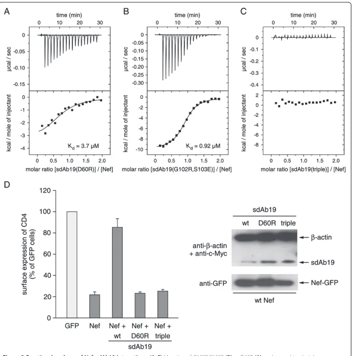
To probe the interaction with Nef, we selected three residues in sdAb19 for site directed mutagenesis. The ra-tional for choosing these sites was based on their loca-tion within a complementarity determining region, and the amino acid changes were designed to maximally affect the interaction without impairing the solubility of the antibody. A key residue of sdAb19 in the complex interface is D60, whose side chain carboxylic group forms ionic interactions with K148 of Nef and a hydrogen bond to Y139 (Figure 2B,C). To explore the contribution
of D60 in binding to Nef, we mutated this residue to ar-ginine, introducing thereby a charge reversal at this amino acid position while retaining the hydrophilic character of this surface residue. The dissociation constant of the sdAb19 D60R mutant for binding to NefSF2was increased to 3.7 μM as determined by ITC measurements, corre-sponding to a 100-fold weakening of the binding affinity (Figure 3A). In addition to this central residue, we chose two peripheral positions in sdAb19, G102 and S103, which were mutated to arginine and glutamic acid, respectively. This double mutation attenuated the binding affinity for Nef to 920 nM, corresponding to a 23-fold reduction com-pared to the native sdAb19 (Figure 3B). Only the combin-ation of all three mutcombin-ations, D60R/G102R/S103E, in sdAb19 finally led to a strong reduction in binding affinity, such that an interaction with Nef could no longer be de-tected by ITC (Figure 3C). These results showed that the binding capacity of sdAb19 to Nef can be experimentally scaled by introducing different substitutions into the key positions of this antibody fragment.
We tested the sdAb19 mutants in functional experi-ments for their effect on the Nef-mediated internaliza-tion of cell surface CD4. Whereas expression of Nef Table 1 Thermodynamic parameters of isothermal titration calorimetry measurements
Titration schemea Kd ΔG ΔH ΔS T ΔS n
(nM) (kcal/mol) (kcal/mol) (kcal/mol/deg) (kcal/mol) ([..]/[..]) a.) SH3B6–Nef–sdAb19 complex formation
SH3B6to NefSF2b 18.7 −10.55 −22.51 (±0.25) −0.040 −11.96 0.81 (±0.005)
sdAb19 to NefSF2 38.6 −10.11 −11.62 (±0.005) −0.005 −1.51 0.81 (±0.002)
b.) sdAb19 binding to Nef
sdAb19 to myrNefSF2 32.7 −10.20 −14.97 (±0.22) −0.016 −4.77 0.81 (±0.006)
sdAb19 to NefNL4–3 18.5 −10.37 −17.97 (±0.30) −0.025 −7.42 0.88 (±0.007)
sdAb19 to NefNA7 118 −9.45 −12.82 (±0.12) −0.011 −3.34 0.74 (±0.004)
sdAb19 to NefSF2(∆ flex. loop) 98 −9.55 −10.71 (±0.06) −0.004 −1.16 0.87 (±0.003)
c.) sdAb19 mutant binding
sdAb19 (G102R,S103E) to NefSF2 920 −8.25 −9.93 (±0.56) −0.006 −1.68 0.9 (±0.085)
sdAb19 (D60R) to NefSF2 3700 −7.40 −3.38 (±0.56) 0.014 4.02 0.74 (±0.009)
sdAb19 (triple) to NefSF2 – – – – – –
d.) NefSF2mutant binding
sdAb19 to NefSF2(K148E) 1700 −7.86 −8.77 (±0.26) −0.003 −0.89 0.74 (±0.015)
sdAb19 to NefSF2(M198K) – – – – – –
sdAb19 to NefSF2(L202K) – – – – – –
e.) Neffin binding to Nef
Neffin to NefSF2 1.6 −12.02 −38.26 (±0.17) −0.088 −26.23 0.66 (±0.001)
Neffin to NefNL4–3 3.9 −11.49 −33.19 (±0.20) −0.073 −21.71 0.65 (±0.002)
Neffin to NefNA7 14.4 −10.72 −34.69 (±0.12) −0.080 −23.97 0.67 (±0.001)
Neffin (triple) to NefSF2 23.3 −10.41 −18.13 (±0.12) −0.026 −7.72 1.08 (±0.004)
aall ITC measurements were performed at 25°C. bRecombinant Nef proteins refer to: Nef
alone in CD4 expressing cells potently stimulated the in-ternalization of CD4 leading to only a residual 20% CD4 expression remaining at the cell surface (Figure 3D), co-expression of sdAb19 blocked this effect and restored surface CD4 expression to levels observed in the control cells. In contrast, this capacity to counteract the effect of Nef on CD4 downregulation was lost by the D60R sdAb19 mutant and the triple-mutant D60R/G102R/ S103E, further establishing the critical role of these resi-dues for the functionality of sdAb19.
sdAb19 targets a C-terminal surface epitope on HIV-1 Nef The epitope on Nef that is recognized by sdAb19 en-compasses a surface patch toward the C-terminus of the viral protein. A ring of charged residues, E155, R188, K148, K192, H196, E201 and H203, surrounds hydrophobic residues I137, V150, M198 and L202 at its center as well
as the polar Y139 and the adjacent G134 (Figure 4A). The binding interface delineated on the surface representation of HIV-1 Nef is shown in Figure 4B. This surface patch is conserved based on the analysis of 1643 alleles of HIV-1 Nef proteins from subtype B [24]. For ten resi-dues of the sdAb19 binding interface the degree of se-quence conservation is between 96% and 99.9%, based on the analysis of homologous amino acid replacements (Figure 4B). Only three residues, M198 (89.2%), L202 (77.5%), and I137 (62.2%), share a smaller degree of se-quence conservation, with the isoleucine being mostly replaced by threonine. The high degree of sequence conservation of residues in the binding interface sug-gests that sdAb19 binds to almost all Nef alleles of the major HIV-1 subgroups in agreement with previous re-sults showing that sdAb19 was able to inhibit a broad panel of Nef proteins from different HIV-1 groups [15]. Figure 2 sdAb19 targets a C-terminal surface epitope on Nef. (A) Binding of sdAb19 to NefSF2. The three complementarity determining
regions are colored yellow (CDR1), red (CDR2), and blue (CDR3), respectively. (B) Residues of CDR2 are significantly involved in the interaction with Nef. Hydrogen bonds and salt bridges are displayed as dashed lines. (C) Interaction map of Nef and sdAb19 within a distance shell of 3.7 Å. Hydrophobic and polar interactions between main chain (dots) and side chain (bars) atoms are indicated by dashed lines colored grey and blue, respectively. For hydrogen bonds and salt bridges the inter-atomic distances are tabulated.
Three different residues in Nef, namely K148, M198 and L202, were mutated to probe their contribution to the recognition by the antibody fragment. As the corre-sponding residue to D60 in sdAb19, lysine 148 in Nef was mutated to glutamate in order to break the salt bridge formation by charge reversal (Figure 4C). This
highly conserved lysine is in the center of a basic patch on Nef that forms the binding interface to sdAb19 as shown in the electrostatic surface display (Figure 4D). The dissociation constant for K148E increased to 1.7 μM as determined by ITC experiments, corresponding to a 44-fold weaker binding affinity compared to the time (min) 0 10 20 30 0 -0.05 -0.10 -0.30 0 -2 -4 -6 -8 -10 µ ca l/ se c kca l/ m o le of in je ct a nt -0.15 -0.20 -0.25 0 0.5 1.0 1.5 2.0
molar ratio [sdAb19(G102R,S103E)] / [Nef] Kd= 0.92 µM 0 20 40 60 80 100 120 su rf a c e e x p re ssio n o f CD4 (%o f G F P c e lls) GFP Nef Nef + wt Nef + D60R Nef + triple sdAb19 time (min) 0 10 20 30 0 -0.05 -0.10 -0.30 0 -2 -4 -6 -8 -10 µ ca l/ se c kca l/ m o le of in je ct a nt -0.15 -0.20 -0.25 0 0.5 1.0 1.5 2.0
molar ratio [sdAb19(G102R,S103E)] / [Nef] Kd= 0.92 µM 0 20 40 60 80 100 120 su rf a c e e x p re ssio n o f CD4 (%o f G F P c e lls) GFP Nef Nef + wt Nef + D60R Nef + triple sdAb19 anti-β-actin + anti-c-Myc anti-GFP wt sdAb19 D60R triple β-actin sdAb19 Nef-GFP wt Nef
A
B
C
time (min) 0 10 20 30 0 0.5 1.0 1.5 2.0molar ratio [sdAb19(triple)] / [Nef]
0 -0.1 -0.2 0 -2 -4 -6 -8 2 µ c al / s ec kca l/ mol e of in je ct an t -0.3 -0.4 time (min) 0 10 20 30 0 0.5 1.0 1.5 2.0
molar ratio [sdAb19(D60R)] / [Nef]
0 -0.05 -0.10 0 -1 -2 -3 -4 µc a l / s e c kcal / m o le of in je ct ant -0.15 Kd= 3.7 µM
D
anti-β-actin + anti-c-Myc anti-GFP wt sdAb19 D60R triple β-actin sdAb19 Nef-GFP wt NefA
B
C
time (min) 0 10 20 30 0 0.5 1.0 1.5 2.0molar ratio [sdAb19(triple)] / [Nef]
0 -0.1 -0.2 0 -2 -4 -6 -8 2 µ c al / s ec kca l/ mol e of in je ct an t -0.3 -0.4 time (min) 0 10 20 30 0 0.5 1.0 1.5 2.0
molar ratio [sdAb19(D60R)] / [Nef]
0 -0.05 -0.10 0 -1 -2 -3 -4 µc a l / s e c kcal / m o le of in je ct ant -0.15 Kd= 3.7 µM
D
Figure 3 Functional analyses of Nef–sdAb19 interactions. (A-C) Mutation of G102R/S103E (B) or D60R (A) or the combined triple mutant D60R/G102R/S103E (C) in sdAb19 gradually attenuated binding to Nef as determined by ITC measurements. (D) Mutant sdAb19 proteins abrogate the inhibitory effect of sdAb19 on CD4 internalization. HeLa-CD4 cells were transfected with plasmids for expression of either Nef-GFP or GFP in combination with the plasmid for expression of wild-type or mutated sdAb19 (1:3 Nef:sdAb19 plasmid ratio). Transfected cells were stained with phycoerythrin-conjugated anti-CD4 at 4°C, and the surface expression of CD4 in Nef-GFP- or GFP-expressing cells was measured by flow cytometry (left panel). Results are expressed as the percentage of the mean fluorescence intensity determined in GFP-positive cells relative to that determined in GFP-negative cells. Values are the means from at least 3 independent experiments. Error bars represent 1 standard deviation from the means. Transfected cell lysates were analyzed by Western blot (right panel) using anti-c-Myc and anti-β-actin (top), or anti-GFP (bottom) antibodies.
wild type Nef protein (Figure 4E). Interestingly, the two-fold difference in binding affinity of the sdAb19 mutant D60R (3.7 μM) and the Nef mutant K148E (1.7 μM) could result from the different length of the two charged residues, which leads to either exposure (D60R) or re-traction (K148E) of the repulsive charge.
Mutation of either residue M198 or L202 in Nef to a large, basic lysine abrogated binding to sdAb19 in both cases (Additional file 1: Figure S4), underlining the im-portance of hydrophobic residues at these positions. These mutants however did not impair the ability of Nef to mediate CD4 internalization as shown when expressed
in CD4-positive cells (Additional file 1: Figure S5). This confirms on the one site the structural integrity of Nef upon these surface mutations. But it also suggests that residues M198 and L202 as part of the sdAb19 binding interface in Nef do not overlap with the CD4 binding surface of Nef. The inhibition of Nef-mediated down-regulation of CD4 by sdAb19 might instead occur at different sites of the large interaction surface be-tween Nef and sdAb19. Together, these results show that sdAb19 binds to a C-terminal surface epitope of Nef that overlaps with distinct functions of the viral protein. H203 L202 E201 M198 K192 R188 K148 Y139 E155 C NefSF2 Δflexible loop I137 V150 H196 G134
E
time (min) 0 10 20 30 0 -0.1 -0.2 -0.3 0 -2 -4 -6 -8 µc a l / sec kc al /m ol e of in ject a nt 0 0.5 1.0 1.5 2.0molar ratio [sdAb19] / [Nef(K148E)]
Kd= 1.7 µM
C
A
L202 M198 K148 D60 sdAb19 NefSF2 L52 H203(99.4%) L202(77.5%) E201 (98.8%) M198 (89.2%) K192 (96.2%) R188 (99.6%) K148 (99.9%) Y139(99.8%) E155 (96.7%) Δflexible loop I137 (62.2%) V150 (99.6%) NefSF2 H196 (99.7%) G134(99.6%)B
D
-4 kBT +4 kBT L202 M198 K148Figure 4 Delineation of the sdAb19 surface binding epitope on Nef. (A) Display of amino acids on the surface of NefSF2that interact with
sdAb19. Polar residues E155, R188, K148, K192, H196, E201 and H203 surround hydrophobic residues V150, M198, L202, I137 as well as G134 and the central Y139 to constitute the binding epitope on Nef. (B) Surface display of the interacting residues in NefSF2. Basic residues are colored blue,
acidic residue are colored red, and hydrophobic residues are colored yellow. The degree of sequence conservation for homologous residues in HIV-1 Nef alleles, subtype B, is shown in brackets. (C) Hydrophobic interactions are formed between M198 and L202 of Nef with L52 of sdAb19 in close proximity to the K148Nef–D60sdAb19salt bridge. (D) Electrostatic surface display of the binding interface for sdAb19 in HIV-1 NefSF2. The
elec-trostatic surface potential is colored from red (−4 kBT) to blue (+4 kBT). (E) Mutation K148E in Nef weakens the interaction by 44-fold compared to
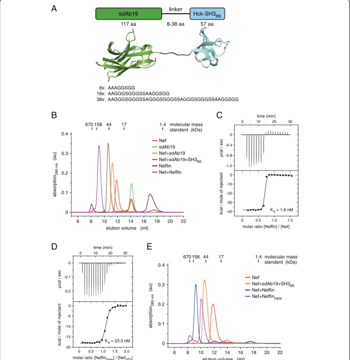
Neffin (sdAb19–SH3B6fusion) forms a 2:2 complex with Nef We previously showed that fusion of sdAb19 to SH3B6, termed Neffin, markedly potentiated the binding affinity to HIV-1 Nef, and increased the efficacy of inhibition against all Nef functions in infected cells [16,27]. As
both protein domains bind to different surface patches of Nef, the covalent linkage combined the two individual binding affinities. A scheme of the domain assembly using three different linker lengths between 8 and 38 residues is shown in Figure 5A. Unexpectedly, size
A
B
C
time (min)0 10 20 30
0 0.5 1.0 1.5
molar ratio [Neffin] / [Nef]
0 -0.4 -0.8 -1.2 0 -10 -20 -30 -40 µc a l / s e c kcal / m ol e o f inj ect a nt Kd= 1.6 nM 6 8 10 12 14 16 18 20 22 670 158 44 17 1.4 elution volume (ml) molecular mass standard (kDa) 0 0.1 0.2 0.3 0.4 abs o rp tio n280 n m (au) Nef sdAb19 Nef+sdAb19 Nef+sdAb19+SH3B6 Neffin Nef+Neffin sdAb19 Hck-SH3B6 117 aa linker 57 aa 8-38 aa 8x: AAAGGSGG 18x: AAGGGSGGGSSAAGGSGG 38x: AAGGGSGGGSSAGGGSGGGSSAGGGSGGGSSAAGGSGG 6 8 10 12 14 16 18 20 22 670 158 44 17 1.4 elution volume (ml) molecular mass standard (kDa) 0 0.1 0.2 0.3 0.4 absor p ti on 280 nm (a u ) Nef Nef+sdAb19+SH3B6 Nef+Neffin Nef+Neffintriple
D
E
0 0.5 1.0 1.5 2.0molar ratio [Neffintriple] / [NefSF2]
time (min) 0 10 20 30 0 -0.1 -0.2 µ c al / s ec kcal / m o le of inject ant Kd= 23.3 nM 0 -5 -10 -15 -20
Figure 5 Neffin binding to Nef results in a 2:2 complex formation. (A) Molecular architecture of the fusion of sdAb19 with SH3B6using a
flexible linker of 8, 18, or 38 amino acid length. (B) Analytical gel filtration of Nef with sdAb19 and SH3B6or Neffin (8 aa linker) reveals a significant size
increase for the Nef–Neffin complex. (C) Isothermal titration calorimetry confirms tight binding between Nef and Neffin exhibiting a Kdof 1.6 nM.
(D)The triple mutation D60R/G102R/S103E in the sdAb19 subunit increased the dissociation constant of Neffintriplebinding to Nef to the portion of the
SH3B6domain alone as determined by ITC measurements. (E) The elution volume of the Nef–Neffintriplecomplex corresponds to an equimolar 1:1
exclusion chromatography revealed a significantly earlier elution volume for the Nef–Neffin complex compared to the tripartite SH3B6–Nef–sdAb19 complex generated from individual protein assembly (Figure 5B). This ob-servation suggested the formation of a 2:2 Nef–Neffin complex. Variation of the linker length in Neffin up to 38 residues did not lead to formation of a 1:1 complex (Additional file 1: Figure S6), which might be explained by the opposite location of SH3 and sdAb19 binding surfaces on the structure of Nef. Of note, the very late retention of Neffin at an elution volume of 17 ml corre-sponds to the elution profile of the SH3 domain alone, confirming the building block construction strategy of this fusion protein. Using ITC experiments, the dissoci-ation constant between Nef and Neffin was determined to 1.6 nM (Figure 5C). While the increase in avidity based on the accumulated strength of both subunits might not be as high as observed in other cases, e.g. compared to the 4,000-fold increase seen for the multiva-lent combination of two VHHfragments [28], it should be noted that the affinity determined by ITC is at the lower resolution limit of this technique. Surface plasmon reson-ance of the NefNL4–3–Neffin interaction showed indeed binding in the picomolar affinity regime with very low dis-sociation rates [27].
To analyze the individual contributions of the two Neffin subunits to the complex formation with Nef, we introduced the sdAb19 triple mutation D60R/G102R/ S103E to Neffin, named Neffintriple. This mutation re-duced the binding affinity of Neffintriple to Nef to the contribution of the SH3B6domain alone (Figure 5D and Table 1), in line with the previous observation that the triple mutation in sdAb19 alone abrogated the interaction with Nef (Figure 3C). Size exclusion chromatography con-firmed indeed formation of a 1:1 Nef–Neffintriplecomplex taking the expected higher hydrodynamic volume of the complex by the unbound sdAb19 subunit into account (Figure 5E).
Neffintriple as well as the Neffin D60R mutant were tested in functional experiments to study their contribu-tion to the impairment of the Nef-induced CD4 internal-ization (Figure 6A). Whereas wild-type Neffin potently inhibited CD4 down-regulation by Nef, and fully re-stored CD4 surface expression levels for both NL4-3 and SF2 Nef alleles, the D60R mutant and the triple mutant failed to block CD4 down-regulation. This indicates that SH3 binding alone does not affect Nef's ability to inter-act with CD4 and connect it to the intracellular traffick-ing machinery, in line with previous observations [29]. Likewise, mutation of K148E, M198K or L202K in Nef, which strongly reduced binding to sdAb19, showed a gradually reduced susceptibility for the inhibition of CD4 internalization by wild-type Neffin (Figure 6B). These data indicate a reciprocal correlation between
sdAb19 binding and the ability of Nef to downregulate CD4. Nef(K148E), which was still able to bind sdAb19 but with a lower affinity (see Figure 4E), was functionally inhib-ited by Neffin only when high levels of Neffin were co-expressed at a 1:8 Nef:Neffin expression plasmid ratio. In contrast, the M198K and L202K mutants, which failed to display any affinity for sdAb19 (Additional file 1: Figure S4), were not or only poorly inhibited by Neffin even at the highest concentration (Figure 6B). Therefore, the for-mation of the 2:2 Nef–Neffin complex may lead to an additional coverage of Nef surfaces that would be other-wise accessible for interactions with host cell factors (Figure 6C). This effect might additionally contribute to the potency of Neffin for the inhibition of all HIV-1 Nef functions.
Discussion
Here we define the structural basis of HIV-1 Nef inhib-ition by the camelid-derived sdAb19 antibody fragment. sdAb19 binds to a C-terminal surface epitope on Nef that overlaps with multiple interaction sites of the viral protein. Nef was shown to target the Pak2 serine/threo-nine kinase by a C-terminal sequence motif that involves K192 and F195 residues [30]. We find that K192 and H196 in Nef are directly interacting with sdAb19. These overlapping surface interaction sites may thus well ex-plain how the interaction of Nef to Pak2 is impaired by the tight binding of sdAb19 [16], although other residues as the N-terminal VGF motif were shown to affect Pak2 binding as well [31].
The interaction with the cytoplasmic internalization motifs of CD4 and other T cell surface proteins has been mapped to a hydrophobic sorting motif recognition site between the two central helices in the core domain of Nef [32-34]. Although sdAb19 does not directly interact with residues of Nef that are supposed to mediate the inter-action with CD4, its close proximity to the C-terminal flexible loop is likely involved in inhibition of this function. The C-terminal flexible loop of Nef harbors a di-leucine based sorting motif as well as flanking acidic motifs whose presence is required for Nef trafficking functions via con-tacts with the clathrin-associated adaptor protein machin-ery [35,11]. Deletion of the central 21 residues in the flexible loop of Nef reduced the binding affinity to sdAb19 by three-fold. As sdAb19 directly interacts with E155 in the flexible loop of Nef, this could affect the interaction of Nef with the endocytic machinery, thus inhibiting the downstream effects of Nef on cell surface receptor in-ternalization. In line with this suggestion we previously showed that sdAb19 disrupted the direct interaction of Nef with endosomal adaptor protein complexes [16]. The antibody fragment however had no effect on the subcellu-lar localization of Nef as previously shown [15]. This ob-servation is in line with the finding that sdAb19 shows
similar binding affinities for an N-terminally truncated Nef 45–210 variant as well as the myristoylated full length protein. The data confirm that the myristate and the N-terminal polybasic patch that sustains membrane bind-ing [36-39] are free to interact with lipid compartments even in the Nef–sdAb19 complex. These observations might explain the inhibition of all internalization stimulat-ing functions of Nef by sdAb19 except for the downregu-lation of MHC-I, which supposedly does not occur at the plasma membrane [40]. This additional function is only abrogated through the coverage of the PxxP motif and flanking residues on the core domain of Nef by the SH3 domain moiety of Neffin [16].
The distinct 2:2 stoichiometry of the Nef–Neffin com-plex formation is surprising given that the two constitut-ing domains, sdAb19 and SH3B6, bind Nef with similar affinities. While the topology of the interaction as seen from the structure determination clearly shows how
both molecules bind to opposing sites of Nef separated by a long distance, it is perhaps unexpected that we did not observe strings of Nef–Neffin assemblies, where high aggregates would form through alternating domain interactions. Such aggregation strings would occur if the second Neffin molecule binding to a preformed Nef– (sdAb19-SH3B6)–Nef complex would recruit a third Nef subunit into this complex. Instead, the sharp elution profile at a 2:2 molecular mass suggests the specific and very tight quaternary Nef–Neffin complex formation. It seems reasonable to propose that such an assembly could additionally contribute to the inhibitory function of Neffin, exceeding the combined effects of sdAb19 and SH3 alone, as additional surfaces of Nef might be cov-ered through the 2:2 complex formation. A model re-garding how a 2:2 Nef–Neffin complex assembly could lead to additional coverage of Nef surfaces is illustrated in Figure 6C. Nef
+
sdAb19 SH3B6 Nef sd Ab19 SH3B6 Nef sdAb19 SH3 B6 0 20 40 60 80 100 120 sur fa c ee x p re s s io no f C D 4 (% o f GFP c e lls) GFP Nef Nef + wt Nef + D60R Nef + triple Neffin wt NefNL4-3 wt NefSF2A
C
anti-β-actin + anti-c-Myc anti-GFP wt Neffin D60R triple β-actin Neffin Nef-GFP wt NefNL4-3 0 20 40 60 80 100 120 s u rf a c ee xp re s sion o fC D 4 (% o f G F P c e lls )GFP wt Nef Nef(K148E) Nef(M198K) Nef(L202K) wt Neffin 1:0 1:4 1:8 Nef:Neffin plasmid ratio
B
Figure 6 Binding of sdAb19 in Neffin correlates with the inhibition of CD4 internalization. (A) HeLa-CD4 cells were transfected with plasmids for expression of either Nef-GFP or GFP in combination with the plasmid for expression of wild-type or mutated sdAb19 (1:3 Nef:sdAb19 plasmid ratio). Transfected cells were analyzed for CD4 cell surface expression and cell lysates were analyzed by Western blotting (inset panels) as in Figure 3D. (B) HeLa-CD4 cells were transfected with plasmids for expression of either Nef-GFP, mutant Nef-GFP or GFP alone (1:0 plasmid ratio) or in combination with increasing amounts of the plasmid for the expression of wild-type Neffin (1:4 or 1:8 plasmid ratio) and analyzed for CD4 cell surface expression. (C) Model of the complex formation between Nef and Neffin, consisting of the two individual Nef-binding domains sdAb19 and SH3B6. Formation of a stable 2:2 complex leads to coverage of additional surfaces in Nef that increases the inhibitory potential of Neffin against
Conclusions
The structural and functional characterization of sdAb19 and Neffin binding to Nef opens a broad avenue for their rational usage. sdAb19 and Neffin can be used (i) for basic research as biochemical tools for in vitro analysis at the molecular and cellular level, where they could impair intracellular signaling and trafficking pathways mediated by Nef in infected cells. (ii) They can also be used for in vivo evaluation of inhibition of the dysregulatory effects of Nef on the immune system in a relevant animal model such as HIV-1 infected “humanized” mice. These latter experiments should also give definitive answer regarding validation of Nef as a rational target for development of new antiretroviral strategies. (iii) sdAb19 can be used as a tool for small chemical compound identification or lead compound optimization in high throughput assays or drug design approaches based on the structure of the Nef– sdAb19 complex. Through binding to the molecular area recognized by sdAb19, these compounds could mimic the inhibitory activity of sdAb19 on Nef functions. (iv) sdAb19 or Neffin might ultimately be directly used in HIV treat-ment through expression in infected cells either by gene transfer or after development of nano- and micropar-ticle-based formulation strategies for efficient intracellular delivery. As both sdAb19 and Neffin proteins can be pro-duced in high amounts in E. coli, their easy and low cost production and the possibility to tune their binding affi-nity to Nef from the sub-nanomolar to the high micro-molar affinity range might facilitate a broad application in both academic and pharmaceutical research.
Methods
Plasmid cloning and protein production
Bacterial expression plasmids for HIV-1 NefSF2, the SH3 domain of human Hck, and llama sdAb19 were described previously [19,27,41]. Expression and purification of full length myristoylated NefSF2 protein (myr2-210, C59S, C210A), an N-terminal deletion construct of NefSF2 (45–210, C59S, C210A), and a NefSF2 variant contain-ing a deletion of the C-terminal flexible loop (45–210, ∆158-178, C59S, C210A) was performed similarly as described [17,41]. sdAb19 (1–118) was expressed as GST-fusion protein in E.coli and purified by affinity chromatography as described [27].
NefSF2–sdAb19–SH3B6crystallization
Initial screening for crystallization conditions of the NefSF2–sdAb19–SH3B6 complex was carried out using a Mosquito robot (TTP Labtech) with the sitting-drop method at 293 K and a concentration of 5–15 mg/ml. About 0.1 μl of protein solution was mixed with 0.1 μl of reservoir solution from a 70 μl reservoir in 96-well Hamp-ton 3553 crystallization plates. Initial crystals of NefSF2– sdAb19–SH3B6 could be obtained in 0.2 M potassium
formate and 20% polyethylene glycol (PEG) 3350. Crys-tal conditions were optimized to 0.2 M potassium for-mate, 17.5% polyethylene glycol (PEG) 3350 and 0.35 M ammonium chloride grown by hanging-drop vapor dif-fusion in Linbro crystallization plates. Crystals grew under these conditions within 12 days to a size of 250 × 40 × 40 μm. For cryo-protection, crystals were trans-ferred to a solution that contained the reservoir buffer with additional 25% ethylene glycol. After 5–10 sec-onds, crystals were flash-cooled in liquid nitrogen. Structure determination
The native diffraction data were measured from crystals cooled to cryogenic temperature of 100 K and were re-corded on beamline X10SA (PXII) of the Swiss Light Source (SLS) equipped with an MAR 225 CCD detector (oscillation width per frame, 1°; 400 frames collected) to 2.1 Å resolution. The NefSF2–sdAb19–SH3B6 crystals were of space group P41and had unit cell parameters of a = 73.07, b = 73.07 and c = 71.25 Å. Assuming the pres-ence of one tripartite complex in the asymmetric unit, the solvent content of the crystals is 52.02%. The XDS package [42] was used to process, integrate, and scale the collected data. The structure was solved with molecu-lar replacement using the NefSF2–SH3B6(PDB entry 3RBB [19]) as a search model in PHASER [43]. The model was refined by alternating cycles of manual rebuilding in COOT [44] and minimization in REFMAC5 [45]. Data collection statistics and refinement parameters are given in Additional file 1: Table S1. Protein interfaces and ac-cessible surface areas were calculated with the program PISA (http://www.ebi.ac.uk/pdbe/). Molecular diagrams were drawn using PyMOL (http://www.pymol.org/). Size exclusion chromatography
Analytical gel filtrations of recombinant NefSF2, SH3B6, sdAb19, Neffin, and all mutants thereof were performed using a multicomponent Waters 626 LC system (Waters, MA) equipped with a Superdex S75 (10/300 GL) column (GE Healthcare). Typically, 100 μl of a 1.5 mg/ml protein solution was loaded onto the column that was equilibrated in 10 mM Tris/HCl (pH 9.0), 100 mM NaCl buffer prior to injection of the protein samples. Gel filtrations were run at a flow rate of 0.5 ml per minute in 10 mM Tris/ HCl (pH 9.0), 100 mM NaCl onto the S75 column at 4°C or 20°C. The optical density was monitored at a wave-length of 280 nm over the time course of the experiment. Gel filtration experiments were performed repeated times. Isothermal titration calorimetry
Interaction of HIV-1 NefSF2with SH3B6,sdAb19 or Neffin was performed by isothermal titration calorimetry using a MicroCal iTC200 microcalorimeter (GE Healthcare). Measurements were carried out in 20 mM Tris/HCl buffer
(pH 9.0), 100 mM NaCl at 25°C. SH3B6,sdAb19 or Neffin at a concentration of 200 μM were stepwise injected from the syringe to 20 μM NefSF2 placed in the measurement cell. The change in heating power was observed over the reaction time until equilibrium was reached. Data were analyzed using the software provided by the manufacturer. Cell culture, transfection, flow cytometry, and
immunoblot analysis
Plasmids for expression in mammalian cells of wild-type Nef fused to the green fluorescent protein (Nef-GFP), as well as wild-type Myc-tagged forms of sdAb19 and Neffin, have been described previously [15,16]. Plasmids for ex-pression of Nef, sdAb19 and Neffin mutants were con-structed by PCR-mediated site-directed mutagenesis using specific primers and the wild-type expression vectors as templates. Cell culture experiments were performed in HeLa cells stably expressing CD4 similarly as described [15,16]. CD4 cell surface staining and flow cytometry ana-lysis were performed as described previously [15]. Protein expression was analyzed on transfected cell lysates by Western blot using anti–c-Myc (9E10; Roche) or anti-GFP (sc-8334; Santa Cruz Biotechnology Inc.) antibodies [16]. Accession numbers
The atomic coordinates and structure factors of the HIV-1
NefSF2–HckSH3-B6–sdAb19 complex have been deposited
in the Protein Data Bank under accession code 4ORZ.
Additional file
Additional file 1: Table S1. Data collection and refinement statistics of the SH3B6–Nef–sdAb19 crystal structure. Figure S1. Analytical
characterization of HIV-1 Nef myristoylation. Figure S2. Isothermal titration calorimetry measurements of sdAb19 titrated to HIV-1 Nef proteins of different virus strains. Figure S3. Sequence and secondary structure display of the SH3B6–NefSF2–sdAb19 complex structure. Figure S4. ITC measurements of sdAb19 titrated to Nef mutants. Figure S5. Cell surface down-regulation of CD4 is not changed by Nef mutations that affect sdAb19 binding. Figure S6.Size exclusion chromatography of Neffin constructs and complex formation with Nef. Additional references.
Abbreviations
CDR:Complementarity determining region; GFP: Green fluorescent protein; HAART: Highly active anti-retroviral therapy; HIV: Human immunodeficiency virus; MFI: Mean fluorescence intensity; Nef: Negative factor; Neffin: Nef inhibitor; sdAb19: Single domain antibody 19; SIV: Simian immunodeficiency virus; TCR: T-cell receptor.
Competing interests
The authors declare that they have no competing interests. Authors’ contributions
SL purified proteins, performed binding studies, crystallized the protein complex and determined the crystal structure. JM and MCR performed cell surface expression experiments under the supervision of SB. AJ and KS contributed reagents and expertise. KS, SB and MG analyzed data. MG designed of the study and wrote the manuscript. All authors discussed the results and commented on the manuscript. All authors read and approved the final manuscript.
Acknowledgements
We thank Karin Vogel-Bachmayr and Emilie Trinh for expert technical assistance and Dr. Ingrid Vetter for help with diffraction data analysis. This work was supported by grants from Inserm-Transfert and SATTidfInnov to S.B. Author details
1Center of Advanced European Studies and Research, Group Physical Biochemistry, Bonn, Germany.2Department of Physical Biochemistry, Max Planck Institute of Molecular Physiology, Dortmund, Germany.3Inserm U1016, Institut Cochin, Paris, France.4CNRS, UMR8104, Paris, France. 5Université Paris-Descartes, Sorbonne Paris-Cité, Paris, France.6Department of Virology, Haartman Institute, University of Helsinki, Helsinki, Finland. Received: 15 December 2013 Accepted: 4 March 2014 Published: 13 March 2014
References
1. UNAIDS Report on the Global AIDS Epidemic.; 2013. http://www.unaids.org. 2. Gupta RK, Hill A, Sawyer AW, Cozzi-Lepri A, Von Wyl V, Yerly S, Lima VD,
Günthard HF, Gilks C, Pillay D: Virological monitoring and resistance to first-line highly active antiretroviral therapy in adults infected with HIV-1 treated under WHO guidelines: a systematic review and meta-analysis. Lancet Infect Dis2009, 9:409–417.
3. Flexner C: HIV drug development: the next 25 years. Nat Rev Drug Discov 2007, 6:959–966.
4. Sigal A, Baltimore D: As good as it gets? The problem of HIV persistence despite antiretroviral drugs. Cell Host Microbe2012, 12:132–138. 5. Denton PW, Long JM, Wietgrefe SW, Sykes C, Spagnuolo RA, Snyder OD,
Perkey K, Archin NM, Choudhary SK, Yang K, Hudgens MG, Pastan I, Haase AT, Kashuba AD, Berger EA, Margolis DM, Garcia JV: Targeted cytotoxic therapy kills persisting HIV infected cells during ART. PLoS Pathog2014, 10:e1003872.
6. West AP Jr, Scharf L, Scheid JF, Klein F, Bjorkman PJ, Nussenzweig MC: Structural Insights on the Role of Antibodies in HIV-1 Vaccine and Therapy. Cell2014, 156:633–648.
7. Malim MH, Emerman M: HIV-1 accessory proteins–ensuring viral survival in a hostile environment. Cell Host Microbe2008, 3:388–398.
8. Kirchhoff F: Immune evasion and counteraction of restriction factors by HIV-1 and other primate lentiviruses. Cell Host Microbe2010, 8:55–67. 9. Kirchhoff F, Schindler M, Specht A, Arhel N, Münch J: Role of Nef in primate
lentiviral immunopathogenesis. Cell Mol Life Sci2008, 65:2621–2636. 10. Foster JL, Garcia JV: HIV-1 Nef: at the crossroads. Retrovirology 2008, 5:84. 11. Geyer M, Fackler OT, Peterlin BM: Structure–function relationships in HIV-1
Nef. EMBO Rep2001, 2:580–585.
12. Schindler M, Münch J, Kutsch O, Li H, Santiago ML, Bibollet-Ruche F, Müller-Trutwin MC, Novembre FJ, Peeters M, Courgnaud V, Bailes E, Roques P, Sodora DL, Silvestri G, Sharp PM, Hahn BH, Kirchhoff F: Nef-mediated suppression of T cell activation was lost in a lentiviral lineage that gave rise to HIV-1. Cell2006, 125:1055–1067.
13. Witkowski W, Verhasselt B: Contributions of HIV-1 Nef to immune dysregulation in HIV-infected patients: a therapeutic target? Expert Opin Ther Targets2013, 17:1345–1356.
14. Lülf S, Horenkamp FA, Breuer S, Geyer M: Nef surfaces: where to interfere with function. Curr HIV Res2011, 9:543–551.
15. Bouchet J, Basmaciogullari SE, Chrobak P, Stolp B, Bouchard N, Fackler OT, Chames P, Jolicoeur P, Benichou S, Baty D: Inhibition of the Nef regulatory protein of HIV-1 by a single-domain antibody. Blood2011, 117:3559–3568. 16. Bouchet J, Hérate C, Guenzel CA, Vérollet C, Järviluoma A, Mazzolini J, Rafie S,
Chames P, Baty D, Saksela K, Niedergang F, Maridonneau-Parini I, Benichou S: Single-Domain Antibody-SH3 Fusions for Efficient Neutralization of HIV-1 Nef Functions. J Virol2012, 86:4856–4867.
17. Breuer S, Gerlach H, Kolaric B, Urbanke C, Opitz N, Geyer M: Biochemical indication for myristoylation-dependent conformational changes in HIV-1 Nef. Biochemistry2006, 45:2339–2349.
18. Hiipakka M, Poikonen K, Saksela K: SH3 domains with high affinity and engineered ligand specificities targeted to HIV-1 Nef. J Mol Biol1999, 293:1097–1106.
19. Horenkamp FA, Breuer S, Schulte A, Lülf S, Weyand M, Saksela K, Geyer M: Conformation of the Dileucine-Based Sorting Motif in HIV-1 Nef Revealed by Intermolecular Domain Assembly. Traffic2011, 12:867–877.
20. Spinelli S, Frenken L, Bourgeois D, De Ron L, Bos W, Verrips T, Anguille C, Cambillau C, Tegoni M: The crystal structure of a llama heavy chain variable domain. Nat Struct Biol1996, 3:752–757.
21. Hinz A, Lutje Hulsik D, Forsman A, Koh WW, Belrhali H, Gorlani A, de Haard H, Weiss RA, Verrips T, Weissenhorn W: Crystal structure of the neutralizing Llama V(HH) D7 and its mode of HIV-1 gp120 interaction. PLoS One2010, 5:e10482.
22. Lutje Hulsik D, Liu YY, Strokappe NM, Battella S, El Khattabi M, McCoy LE, Sabin C, Hinz A, Hock M, Macheboeuf P, Bonvin AM, Langedijk JP, Davis D, Forsman Quigley A, Aasa-Chapman MM, Seaman MS, Ramos A, Poignard P, Favier A, Simorre JP, Weiss RA, Verrips CT, Weissenhorn W, Rutten L: A gp41 MPER-specific llama VHH requires a hydrophobic CDR3 for neutralization but not for antigen recognition. PLoS Pathog2013, 9:e1003202. 23. Muyldermans S: Nanobodies: natural single-domain antibodies. Annu Rev
Biochem2013, 82:775–797.
24. O'Neill E, Kuo LS, Krisko JF, Tomchick DR, Garcia JV, Foster JL: Dynamic evolution of the human immunodeficiency virus type 1 pathogenic factor, Nef. J Virol2006, 80:1311–1320.
25. Geyer M, Peterlin BM: Domain assembly, surface accessibility and sequence conservation in full length HIV-1 Nef. FEBS Lett2001, 496:91–95. 26. Chothia C, Lesk AM, Gherardi E, Tomlinson IM, Walter G, Marks JD, Llewelyn
MB, Winter G: Structural repertoire of the human VH segments. J Mol Biol 1992, 227:799–817.
27. Järviluoma A, Strandin T, Lülf S, Bouchet J, Mäkelä AR, Geyer M, Benichou S, Saksela K: High-affinity target binding engineered via fusion of a single-domain antibody fragment with a ligand-tailored SH3 single-domain. PLoS One 2012, 7:e40331.
28. Hultberg A, Temperton NJ, Rosseels V, Koenders M, Gonzalez-Pajuelo M, Schepens B, Ibañez LI, Vanlandschoot P, Schillemans J, Saunders M, Weiss RA, Saelens X, Melero JA, Verrips CT, Van Gucht S, De Haard HJ: Llama-de-rived single domain antibodies to build multivalent, superpotent and broadened neutralizing anti-viral molecules. PLoS One2011, 6:e17665. 29. Fackler OT, Moris A, Tibroni N, Giese SI, Glass B, Schwartz O, Kräusslich HG:
Functional characterization of HIV-1 Nef mutants in the context of viral infection. Virology2006, 351:322–339.
30. Agopian K, Wei BL, Garcia JV, Gabuzda D: A hydrophobic binding surface on the human immunodeficiency virus type 1 Nef core is critical for association with p21-activated kinase 2. J Virol2006, 80:3050–3061. 31. Meuwissen PJ, Stolp B, Iannucci V, Vermeire J, Naessens E, Saksela K, Geyer
M, Vanham G, Arien KK, Fackler OT, Verhasselt B: Identification of a highly conserved valine-glycine-phenylalanine amino acid triplet required for HIV-1 Nef function. Retrovirology2012, 9:34.
32. Grzesiek S, Stahl SJ, Wingfield PT, Bax A: The CD4 determinant for downregulation by HIV-1 Nef directly binds to Nef. Mapping of the Nef binding surface by NMR. Biochemistry1996, 35:10256–10261.
33. Khalid M, Yu H, Sauter D, Usmani SM, Schmokel J, Feldman J, Gruters RA, van der Ende ME, Geyer M, Rowland-Jones S, Osterhaus AD, Kirchhoff F: Efficient Nef-mediated downmodulation of TCR-CD3 and CD28 is associated with high CD4+ T cell counts in viremic HIV-2 infection. J Virol 2012, 86:4906–4920.
34. Schmökel J, Li H, Shabir A, Yu H, Geyer M, Silvestri G, Sodora DL, Hahn BH, Kirchhoff F: Link between primate lentiviral coreceptor usage and Nef function. Cell Rep2013, 5:997–1009.
35. Craig HM, Pandori MW, Guatelli JC: Interaction of HIV-1 Nef with the cellular dileucine-based sorting pathway is required for CD4 down-regulation and optimal viral infectivity. Proc Natl Acad Sci USA1998, 95:11229–11234. 36. Gerlach H, Laumann V, Martens S, Becker CF, Goody RS, Geyer M: HIV-1 Nef
membrane association depends on charge, curvature, composition and sequence. Nat Chem Biol2010, 6:46–53.
37. Welker R, Harris M, Cardel B, Kräusslich HG: Virion incorporation of human immunodeficiency virus type 1 Nef is mediated by a bipartite membrane-targeting signal: analysis of its role in enhancement of viral infectivity. J Virol1998, 72:8833–8840.
38. Giese SI, Woerz I, Homann S, Tibroni N, Geyer M, Fackler OT: Specific and distinct determinants mediate membrane binding and lipid raft incorporation of HIV-1(SF2) Nef. Virology2006, 355:175–191. 39. Akgun B, Satija S, Nanda H, Pirrone GF, Shi X, Engen JR, Kent MS:
Conformational transition of membrane-associated terminally acylated HIV-1 Nef. Structure2013, 21:1822–1833.
40. Roeth JF, Williams M, Kasper MR, Filzen TM, Collins KL: HIV-1 Nef disrupts MHC-I trafficking by recruiting AP-1 to the MHC-I cytoplasmic tail. J Cell Biol2004, 167:903–913.
41. Breuer S, Schievink SI, Schulte A, Blankenfeldt W, Fackler OT, Geyer M: Molecular design, functional characterization and structural basis of a protein inhibitor against the HIV-1 pathogenicity factor Nef. PLoS One 2011, 6:e20033.
42. Kabsch W: Xds. Acta Crystallogr D Biol Crystallogr 2010, 66:125–132. 43. McCoy AJ, Grosse-Kunstleve RW, Adams PD, Winn MD, Storoni LC, Read RJ:
Phaser crystallographic software. J Appl Crystallogr2007, 40:658–674. 44. Emsley P, Lohkamp B, Scott WG, Cowtan K: Features and development of
Coot. Acta Crystallogr D Biol Crystallogr2010, 66:486–501.
45. Murshudov GN, Vagin AA, Dodson EJ: Refinement of macromolecular structures by the maximum-likelihood method. Acta Crystallogr D Biol Crystallogr1997, 53:240–255.
doi:10.1186/1742-4690-11-24
Cite this article as:Lülf et al.: Structural basis for the inhibition of HIV-1 Nef by a high-affinity binding single-domain antibody. Retrovirology 2014 11:24.
Submit your next manuscript to BioMed Central and take full advantage of:
• Convenient online submission
• Thorough peer review
• No space constraints or color figure charges
• Immediate publication on acceptance
• Inclusion in PubMed, CAS, Scopus and Google Scholar
• Research which is freely available for redistribution
Submit your manuscript at www.biomedcentral.com/submit