HAL Id: inserm-00130900
https://www.hal.inserm.fr/inserm-00130900
Submitted on 6 Mar 2007
HAL is a multi-disciplinary open access archive for the deposit and dissemination of sci-entific research documents, whether they are pub-lished or not. The documents may come from teaching and research institutions in France or abroad, or from public or private research centers.
L’archive ouverte pluridisciplinaire HAL, est destinée au dépôt et à la diffusion de documents scientifiques de niveau recherche, publiés ou non, émanant des établissements d’enseignement et de recherche français ou étrangers, des laboratoires publics ou privés.
Survival and proliferation factors of normal and
malignant plasma cells.
Bernard Klein, Karin Tarte, Michel Jourdan, Karene Mathouk, Jérôme
Moreaux, Eric Jourdan, Eric Legouffe, John de Vos, Jean François Rossi
To cite this version:
Bernard Klein, Karin Tarte, Michel Jourdan, Karene Mathouk, Jérôme Moreaux, et al.. Survival and proliferation factors of normal and malignant plasma cells.. International Journal of Hematology, Springer Verlag, 2003, 78 (2), pp.106-13. �inserm-00130900�
SURVIVAL AND PROLIFERATION FACTORS OF NORMAL AND MALIGNANT
PLASMA CELLS
Bernard Klein1, Karin Tarte1, Michel Jourdan1, Karene Mathouk1, Jerome Moreaux1, Eric Jourdan2, Eric Legouffe3, John De Vos1, Jean François Rossi3
1. INSERM U475 and Unit for Cellular and Gene therapy, CHU Montpellier, 34285 Montpellier. France
2. Service de Médecine Interne B, CHU de Nîmes, 30900, Nîmes, France.
3. Service d'Hématologie et Oncologie Médicale, CHU Montpellier, Hôpital Lapeyronie, 34295 Montpellier, France
Acknowledgements: this work was supported by a grant from Equipe labellisée, la Ligue contre le cancer, Paris, France
Corresponding author. Pr Bernard Klein INSERM U475 99Rue Puech Villa
34197 Montpellier, France Email: klein@montp.inserm.fr
Running title: Myeloma growth factors
Key words: myeloma, IL-6, IGF-1, HGF, EGF, BAFF
HAL author manuscript inserm-00130900, version 1
HAL author manuscript
Abstract
Since the first identifications of interleukin(IL)-6 as a myeloma cell growth factor by Dr Kawano’s and Dr Klein’s groups 14 years ago, numerous studies have emphasized its major role in the emergence of malignant plasma cells in vivo and in the generation of normal plasma cells.
Four transcription factors control B cell differentiation into plasma cells. The B cell transcription factor pax-5 is mainly responsible for a B cell phenotype and bcl-6 represses the plasma cell transcription factor blimp-1 and plasma cell differentiation. Bcl-6 expression is triggered by CD40 and IL-4 activation. A lack of CD40 and IL-4 activation yields a down regulation of bcl-6 expression and IL-6 stimulation an upregulation of blimp-1, mainly through STAT3 activation. Blimp-1 will further downregulate bcl-6 and pax-5 expression and makes it possible plasma cell differentiation. IL-6 as well as IL-10 upregulate XBP-1. XBP-1 is another transcription factor involved in plasma cell differentiation whose gene expression is shut down by pax-5.
These plasma cell transcription factors blimp-1 and XBP-1 are upregulated and the B cell transcription factors bcl-6 and pax-5 downregulated in malignant cells compared to B cells. Apart for this recent identification of these four transcription factors, the factors involved in normal plasma cell generation are mostly unknown.
Regarding malignant plasma cells, three categories of growth factors have been identified. 1) the IL-6 family cytokines, IL-10 and IFNα that activate the JAK/STAT and MAPK pathways. 2) growth factors activating the PI-3 kinase/AKT and MAPkinase pathways, unlike the JAK/STAT pathway (insulin like growth factor 1, hepatocyte growth factor and members of the epidermal growth factor family able to bind syndecan-1 proteoglycan). 3) BAFF or APRIL that activate the NF-kappaB and PI-3 kinase/AKT pathways. BAFF and APRIL bind to BAFF receptor and TACI and are major B cell survival factors. Recent data indicate that
these various growth factors may cooperate together to provide optimum signalling, eventually because ther are colocalized together and with cytoplasmic transduction elements in caveolin-linked membrane caveolae.
The identification of these myeloma cell growth factors and of the associated transduction pathways should provide novel therapeutic targets in multiple myeloma.
Since 15 years, numerous studies have been devoted to the study of myeloma growth factors. These myeloma growth factors may be specific to the myeloma clone or involved in the generation of normal plasma cells. In a first part, we will briefly review the recent knowledge of the biology of normal plasma cells and then discuss the major growth factors involved in multiple myeloma (MM).
1. Biology of normal plasma cells
Mature plasma cells are mostly located in the bone marrow where they represent 0.25% bone marrow mononuclear cells and in the lamina propria of mucosae. Due to their rarity, the process of generation of normal plasma cells and their biology are poorly known. Plasma cells are generated in the lymph node where B cells with a high affinity antigen receptor (Ig) are selected by the antigen through mutations of the Ig variable genes. Selected B cells are then induced to become either a memory B cell or a plasmablastic cell. The plasmablastic cells are supposed to migrate rapidly to the bone marrow where they can find additional survival and differentiation factors making it possible their long-term survival and differentiation into mature plasma cells. A hallmark of mature plasma cells is their large Ig secretion, a high expression of the syndecan-1 proteoglycan that is not expressed on B cells and a lack of most B cell markers except CD19. These PC also largely express CD38.
The intercellular communication signals that are critical to induce B cell differentiation into plasmablastic cell are poorly known. Plasmablastic cells can be greatly expanded in vivo in some patients with acute inflammation. These plasmablastic cells are highly proliferating and
short living. They comprise syndecan-1- immature plasmablastic cells that can yield
syndecan-1+ plasmablastic cells. 1 We recently developed an in-vitro model of generation of polyclonal plasmablastic cells (PPC) starting from healthy donor’s or MM patient's peripheral
blood B cells. 2 This model consists of culturing memory B cells with a CD40 ligand
transfectant, interleukin(IL)-4, IL-2, IL-10 and IL-12 for 4 days before induction of plasma cell differentiation is induced by removal of CD40 stimulation and change in cytokine combination: removal of IL-4 and addition of IL-2, IL-6 and IL-10 and IL-12. These plasmablastic cells are highly proliferating but apoptose at days 7-8 of culture. This model should be critical to better understand the mechanisms controlling the survival of plasmablastic cells and mature plasma cells in the bone marrow 3. In particular, we recently compared gene expression profiles of PPC and bone marrow plasma cells and identified several autocrine and paracrine loops, associated with an induction of anti-apoptotic proteins, that can be involved in the prolonged survival of mature plasma cells 4.
As illustrated up in Figure 1, memory B cells express Bcl-6 and Pax-5 whereas these transcription factors are donwregulated in plasmablastic cells. On the contrary, plasmablastic cells expressed the plasma cell transcription factors Blimp-1 and XBP-1. Actually, according
Figure 1: Transcription factors involved in plasma cell differentiation
to the data of the literature, 3 one can hypothesize that, in germinal center B cells, IL-4
upregulates bcl-6 transcription through STAT6 phosphorylation and CD40 stimulation blocks Bcl-6 degradation. Bcl-6 in turn blocks Blimp-1 gene expression.
Bcl-6
XBP-1 Blimp-1Pax-5
Bcl-6XBP-1
Blimp-1
Pax-5IL-4
STAT6CD40L
IL-10
IL-6
STAT3 CD40 CD27CD27L
Memory B cells
Plasmablastic cells
A1, c-myc, Spi-B, Id3, CIITA B cell genes
Removal of IL-4 and CD40 signals make it possible a downregulation of bcl-6 and an expression of blimp-1 that is triggered by IL-6 and IL-10 activating the JAK/STAT pathway, mainly STAT3. Blimp-1 is a transcriptional repressor that downregulates bcl-6 and Pax5 as well as numerous other B cell genes. Pax-5 is critical for B cell maintenance and its overexpression may block plasma cell phenotype in plasma cell lines. Pax-5 directly represses
XBP-1 gene that encodes for a second major plasma cell transcription factor whose gene
targets are poorly unidentified. In our model of PPC generation, we found that activated B cells coexpress CD70 and CD27 suggesting that an activation of CD27 together with IL-10
takes part in the process of plasmablastic cell generation. 2 Indeed, CD27 is expressed on
memory B cells and highly on plasma cells 5 and triggering CD27 with CD70, the CD27
ligand, together with interleukin (IL)-10 induces plasma cell differentiation in vitro. 6 IL-6 also plays a major role, in part by inducing STAT3 phosphorylation that will trigger Blimp-1
expression and probably through induction of XBP-1 transcription7. Recently, XBP-1 was
described as an inducer of IL-6 production 8 suggesting the existence of an amplification loop between IL-6 and XBP-1.. Jego et al. using plasmablastic cells from patients with reactive plasmacytosis showed a major role of IL-6 in plasma cell differentiation. 1 In this model, the differentiation of syndecan-1- plasmablastic cells into syndecan-1+ early plasma cells was blocked with antibodies to IL-6. This property of IL-6 is not surprising since IL-6 gene was
initially cloned in 1988 as a B cell differentiation factor. 9 In addition, transgenic mice
expressing an IL-6 gene driven by an Eµ promoter develop massive polyclonal plasmacytosis 10whereas IL-6 knock out mice have a defect in the production of high affinity antibodies. 1112
As pointed above, the polyclonal plasmablastic cells generated in our in vitro model rapidly apoptose in vitro, on days 7-8 after starting the cultures of B cells, 3-4 days after removal of CD40 stimulation, despite the addition of various cytokines: IL-6, sIL-6R, IL-10, IL-2, IL-12.
This apoptosis is associated with a rapid downregulation of several genes coding for anti-apoptotic proteins, the A1 protein of the bcl-2 family member and the c-IAP2 inhibitor of caspase activity. Conversely, we found an upregulation of the Bik, caspase 3 and caspase 10 genes, coding for pro-apoptotic proteins. The down regulation of A1 is likely a direct consequence of Blimp-1 expression that blocks A1 transcription.
Thus in conclusion, only two intercellular communication pathways have been described for normal plasma cells: IL-6 and activation of CD27 and IL-10. It is not presently known whether the factors known to induce the growth of malignant plasma cells - IGF-1, EGF family, HGF, BAFF/APRIL - are also involved in the biology of normal plasma cells. Nor are known the transduction pathways that are activated in normal plasma cells resulting in their cell survival and proliferation. We can expect that at least a part of the growth factors recently identified for malignant plasma cells are also involved in normal plasma cell biology.
2. Myeloma cell survival and proliferation factors
Numerous studies have been devoted to the identification of myeloma cell growth factors and to the signalling pathways leading to survival and/or proliferation of myeloma cells. A first category of factors activates the JAK/STAT and MAP kinase pathways (mainly IL-6). Another category involves the PI-3 kinase/AKT as well as MAPkinase and NF-kappa B pathways.
2.1. Factors activating the JAK/STAT and MAP kinase pathways: 6, cytokines of
IL-6 family, interferon alpha
IL-6 binds to a specific receptor (IL-6R) and the complex IL-6/IL-6R binds and induces the
homodimerization of the gp130 IL-6 transducer. 13 A remarkable feature of IL-6R is that its
soluble form (sIL-6R) is an agonist molecule. It binds IL-6 with the same affinity as
membrane IL-6R and the complex IL-6/sIL-6R binds and activates gp130. 13 The evidences of a major role of IL-6 in the survival and proliferation of malignant plasma cells accumulated since the initial reports by others and us 15 years ago. 14,15 These evidences are the following:
1) Antibodies to IL-6 block the myeloma cell proliferation and reduce by 50% the number of myeloma cells in culture of patients’ bone marrow cells in vitro 14-16
2) Injection of anti-IL-6 monoclonal antibody inhibited myeloma cell proliferation in
patients with terminal disease 17,18 if the antibody was injected at a sufficient
concentration to block the large IL-6 production in vivo. 19
3) Serum levels of IL-6 and soluble IL-6R are increased in patients with MM in association with a poor prognosis. 20,21
4) The bone marrow environment of patients with MM, mainly by monocytes, myeloid cells and stromal cells, overproduces IL-6. 15,22 This production of IL-6 by the tumor environment is mostly mediated by IL-1 that is produced by monocytes and myeloma cells. 22,23 IL-1 induces PGE2 synthesis that further triggers IL-6 production. 23 Thus inhibitors of IL-1 as the IL-1 receptor antagonists or of PGE2 synthesis might be interesting to block IL-6 production in patients with MM. A similar mechanism was shown in the model of murine plasmacytoma in BALB/C mice. The generation of plasmacytomas was blocked by chronic administration of indomethacin that inhibited
PGE2 synthesis and the large IL-6 production by the inflammatory environment. 24
Myeloma cells can also directly trigger IL-6 production by direct contact with the bone marrow stromal cells by unidentified mechanisms. 25,26
5) Cell lines whose survival is dependent on addition of exogenous IL-6 can be obtained from patients with extramedullary proliferation. 27
6) Mice transgenic with an IL-6 gene driven by the Eµ promoter develop massive
polyclonal plasmacytosis. 28 When crossed with murine BALB/c mice that
spontaneously develop plasmacytomas, these crossed mice develop malignant plasma cell prolferation. 29 In addition, knock out of IL-6 gene abrogated the generation of
malignant plasmacytomas in BALB/C mice primed with mineral oil. 30
Other cytokines of the IL-6 family are also myeloma cell growth factors due to the expression of specific receptors: OSM, CNTF, IL-11, LIF. 31 But these factors are likely not involved in the emergence of the disease in vivo as they are weakly produced by the tumor or its environment. 32 In our hands, interferon-alpha (IFNα) is also a myeloma cell survival factor
that is independent of IL-6. 33,34 IFNα activated the JAK/STAT and MAP kinase pathways as
IL-6, in particular STAT3 phosphorylation. 34 Other groups found that IFNα could block
myeloma cell proliferation. This discrepancy might be explained by the ability of IFNα to induce P19 inhibitor in some cell lines yielding to apoptosis. 35 Finally, we found that IL-10, a potent plasma cell differentiation factor, is also a myeloma cell growth factor. 36 IL-10 works through induction of autocrine loops of IL-6 family cytokines. 37
The myeloma cell survival activity of these cytokines is partly mediated by the phosphorylation of STAT3 by JAK kinases activated by the gp130 IL-6 transducer or IFN receptor. Blockade of JAK/STAT pathway by AG490 inhibits STAT3 phosphorylation and
induces myeloma cell apoptosis. 38 STAT3 binding elements are found in the promoters of
several anti-apoptotic proteins: Mcl-1, Bcl-2, Bcl-xL. Among ten anti-apoptotic and
pro-apoptotic proteins, we found that only Mcl-1 was regulated by IL-6 or IFNα. 39 Other groups
suggested that bcl-xL was the main anti-apoptotic protein controlled by IL-6 in myeloma cells 40,41 but a recent study emphasizes that only a blockade of Mcl-1, unlike bcl-2 or bcl-xL could
inhibit myeloma cell survival. 42 In addition, we found that induction of the constitutive
production of Mcl-1 by retroviral vector is sufficient to promote myeloma cell proliferation
independently of IL-6 43. IL-6 was reported to activate AKT kinase in myeloma cells that is
able to trigger various signalling pathways. 44 AKT activation can be mediated by STAT3 phosphorylation and ras pathway that can trigger PI-3 kinase activation. 45 In our experience, we found a weak AKT phosphorylation in only some IL-6-dependent cell lines (results not shown). Actually, the IL-6-induced AKT phosphorylation in myeloma cells is weak and
transient as compared to that induced by IGF-1. 46 PI-3 kinase mediated AKT phosphorylation
appears critical in promoting proliferation of myeloma cell lines since PI-3 kinase inhibitors abrogate it unlike MAP kinase inhibitors. 47,48
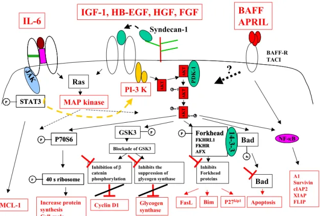
These transduction pathways activated by IL-6 and other myeloma cell growth factors are schematized in the figure 2.
Figure 2: Growth factors and transduction pathways involved in myeloma cell survival and proliferation
2.2. Factors activating the PI-3 kinase/AKT pathway: Insulin like growth factor 1,
heparin binding growth factors
2.2.1. Insulin like growth factor 1 (IGF-1). IGF-1 is a survival and proliferation factor for
most myeloma cell lines. 49-51 Its effect is independent of an activation of the JAK/STAT
AK T AK T AK T AK T AKT T P AKTAKT T P P AKT T P P S AKTAKT T P P P S PP S Forkhead FKHRL1 FKHR AFX P Forkhead FKHRL1 FKHR AFX P P Bad PPP 14 -3 -3 14 -3 -3 Inhibits Forkhead proteins
FasL Bim P27kip1
Bad Apoptosis GSK3 PPP P70S6 P P70S6 P P Inhibits the suppression of glycogen synthase Inhibition of β catenin phosphorylation Glycogen synthase Cyclin D1 Blockade of GSK3 40 s ribosome P 40 s ribosome P P Increase protein synthesis Cell cycle PD K-1 PD K-1 PI-3 K IGF-1, HB-EGF, HGF, FGF IL-6 MAP kinase STAT3 P STAT3 P P MCL-1 JA K JA K Ras Syndecan-1 A1 Survivin cIAP2 XIAP FLIP NF-κB NF-κB BAFF-R TACI BAFF APRIL
?
pathway. 51,52 IGF-1 induced the PI-3 kinase pathway and in particular the phosphorylation of
AKT protein. 44,52 IGF-1 also induces MAP kinase phosphorylation. 51,52 The myeloma
growth factor activity of IGF-1 is blocked by an inhibitor of PI-3 kinase pathway unlike a
MAP kinase inhibitor. 47,48 One mechanism of action of AKT is the phosphorylation of the
pro-apoptotic protein Bad that induces its sequestration by the 14-3-3 protein and prevents its
migration to mitochondrial membrane. 52,53 Other proteins are phosphorylated by the PI-3
kinase/AKT pathway in myeloma cells: the P70S6-kinase, forkhead proteins and the glycogen
synthase kinase-3 beta (GSK3β). 47,48,53 Phosphorylation of these proteins contributes to
blockade of apoptosis and activation of cell cycle in various models. In particular, IGF-1
induces cyclin D1 and Skp2 expression and downregulation of P27kip1 in myeloma cells. 48
In addition, it was shown in one myeloma cell line that the PI-3K/AKT pathway may activate the NF-kappa B pathway and expression of several targets of NF-kappa B involved in cell survival: A1/Bfl1, cIAP2, XIAP, survivin, FLIP. 46
Transfection of myeloma cells with an activated AKT enhances tumor growth and protects from DEX-induced apoptosis and expression of a AKT dominant negative results in inhibition
of IL-6 induced proliferation of myeloma cells. 54 The importance of the PI-3 kinase/AKT
pathway for the survival and proliferation of myeloma cells is emphasized by
deletion/mutation of PTEN gene in some myeloma cells. 55 PTEN is a phosphatase inhibiting
the PI-3 kinase/AKT pathway and its deletion results in a high activation of PI-3K/AKT pathway.
IGF-1 plays likely a major role in myeloma in vivo. Indeed, IGF-1 serum levels are predictive
of a poor survival in patients with MM although they are not increased. 56 IGF-1 is mostly
produced by the liver but also by osteoblasts in the bone matrix where myeloma cells survive and proliferate in vivo. The biology of IGF-1 is complex since four IGF binding proteins,
mostly IGF-BP3, circulate at high concentrations and neutralize IGF-1. 57 Cells may also
express IGF-binding protein that contribute to the biological activity of IGF-1 and disrupts the circulating IGF/IGF-BP complexes. Using Atlas macroarrays, we found that myeloma cells variably express IGF-BP3 or IGF-BP4 genes. 58
2.2.2. Heparin binding factors.
A hallmark of plasma cell differentiation is the expression of the proteoglycan syndecan-1. 59,60 This heparan-sulfate protein has many biological activities and in particular, is able to
bind heparin-binding growth factors and to present them to their specific receptors. 61 Thus, it is not surprising that several recently-reported myeloma cell growth factors are heparin-binding growth factors.
2.2.2.1. Heparin-binding epidermal growth factor like growth factor (HB-EGF). Using Atlas macroarrays, we found that myeloma cell lines overexpressed HB-EGF gene compared to EBV-transformed B cell lines or normal plasmablastic cells and that inhibitors of HB-EGF can block the IL-6-dependent survival of these myeloma cell lines. 58 Actually, we found that HB-EGF cooperates with IL-6 to trigger an optimal survival of myeloma cells, likely through
an interaction between the transducer chains, gp130 and EGF receptors. 62 HB-EGF can
activate two of the four members of the EGF receptor family, ErbB1 and ErbB4, which are variably express by myeloma cells. HB-EGF triggers the PI-3K/AKT pathway in myeloma cells, unlike STAT3 phosphorylation (unpublished data). Several coreceptors can enhance the binding of HB-EGF and increase its biological activity: syndecan-1, the tetraspanin CD9 and
the integrin α3β1. 63 Using Affymetrix microarrays and FACS analysis, we found that
myeloma cells compared to B cells or plasmablastic cells overexpress these coreceptors. 2 In addition, since there are 11 members of the EGF family able to activate the ErbB receptors, 64 it is likely that other EGF members may be involved in myeloma cell biology. Several inhibitors of EGF activity (humanized monoclonal antibodies, inhibitors of ErbB kinase activity) are actually investigated clinically in patients with epithelial cancers. 65 Our recent
data indicate that ErbB inhibitors can potentiate dexamethasone-induced apoptosis of myeloma cell lines and of primary myeloma cells of most patients and suggest that they might improve treatment of patients with MM.
2.2.2.2. Hepatocyte growth factor (HGF). A recent study has shown that HGF is also a growth factor for myeloma cell lines. 66 HGF activity is blocked by removal of heparan sulfate chains of syndecan-1 with heparitinase. This result indicates that syndecan-1 is critical to capture heparin-binding HGF and to present it to its receptor, cMet. Whether HGF cooperates with IL-6 to trigger myeloma cell survival was not investigated. Noteworthy, the XG-1 cell line used in this study was initially obtained in our laboratory and produces a low amount of autocrine IL-6 33 that is sufficient to induce the HB-EGF activity62. HGF is likely involved in the biology of myeloma. Indeed, serum level of HGF is increased and is a prognostic factor in
patients with MM. 67 As HGF increases bone resorption, it may also be involved in the
abnormal osteoclast resorption in patients with MM. 68
2.2.2.3. Fibroblast growth factor (FGF). A role of FGF in myeloma is suggested by the finding of a t(4;14) translocation affecting the FGF receptor type 3 (FGR3) in 15% of patients
with MM. 69 Whether FGR3 translocations have prognostic value is controversial. 69,70 In
addition, mutations of FGR3 making it possible a ligand independent FGR3 activation are found in some myeloma cell lines. 71 These mutations are involved in thanatophoric dysplasia.
Although serum levels of FGF2 are increased in myeloma, 72 no direct evidence of a role of
FGF or FGFR3 expression on the survival or proliferation of human myeloma cells has been published yet. FGFs likely play an important role in the myeloma biology because they bind syndecan-1 as HB-EGF or HGF and since activation of FGR3 may induces the PI-3K/AKT pathway 73 that is critical for myeloma cell survival and proliferation. Indirect evidences using the murine B9 hybridoma or 3T3 cells suggest that a constitutive FGR3 expression may increase resistance to dexamethasone or tumorogenicity. 74,75
2.3. Factors activating NF-kappa B: BAFF family.
BAFF and APRIL belong to the TNF family and activate at least three receptors of the TNF receptor family: BAFF-R, BCMA and TACI. BAFF proteins are critical for the survival of normal B cells, tumor B cells of chronic lymphoid leukaemia and may be involved in
Systematic Lupus Erythematosus 76. Activation of BAFF receptor family results in triggering
the NF-kappa B pathway and likely other unidentified pathways. 77Using DNA microarray,
others and we found an over expression of two BAFF receptors, BCMA and TACI. 2,78 This
observation prompts us to look for a role of the BAFF family in the survival/proliferation of myeloma cells. We found that BAFF or APRIL is a potent survival and proliferation factor of myeloma cells, depending on expression of BAFF-R or TACI on myeloma cells. In addition,
BAFF can protect myeloma cells from dexamethasone-induced apoptosis. 79 These data
suggest that BAFF inhibitor could be useful in the treatment of patients with MM in association with dexamethasone.
2.4. Cross-activation of growth factor receptors and potential clinical applications
The contribution of these various myeloma growth factors to myeloma disease can be simplified by considering the transduction pathways that are critical to promote myeloma survival and cell cycle. As indicated above, at least four transduction cascades are activated by these various factors, the JAK/STAT pathway induced by IL-6 cytokines and IFNα, the PI-3 kinase/AKT pathway strongly activated by IGF-1 and heparin binding factors and weakly induced by IL-6, the NF-kappa B pathway activated by IGF-1 and BAFF proteins and the MAP kinase pathway induced by all factors. We recently pointed out cooperation between
IL-6 and HB-EGF to trigger myeloma cell survival and proliferation. 62 The effect of HB-EGF
is dependent on weak gp130 activation by IL-6. This cooperation likely reflects a cross talk of the transduction elements and activation of various anti-apoptotic proteins. 62
This was recently shown for gp130 IL-6 transducer and IFN receptor or IGF receptor. Indeed, Jelinek’s group showed that IFN-α could activate the phosphorylation of endogenous gp130. This cross-activation is not reciprocal as IL-6 cannot cross-phosphorylate endogenous IFN
receptor. 80 The same group also showed that IFNα induced a cross-phosphorylation of
endogenous ErbB3 receptor in a myeloma cell line. 81 This ability of IFNα to induce these
cross-phosphorylations of other transducer chains unlike IL-6 or IGF-1, is linked to its ability
to trigger a large and long-lasting activation of JAK1 and Tyk2 kinases compared to IL-6. 80
More recently, it was shown that the gp130 and IGF-1R are both colocalized in caveolin-associated membrane caveolae in human myeloma cells together with PI-3 kinase and src kinase and that abrogation of caveolae by cholesterol inhibitors blocks IL-6 or IGF-1 induced
transduction, in particular PI-3K/AKT pathway. 82 Of major interest, the caveolin 1 gene is
overexpressed on malignant plasma cells compared to normal B cells or plasma cells. Taken together these data suggest that gp130 IL-6 transducers, IGF-1 receptors and EGF receptors and eventually coreceptors such as syndecan-1 and CD9 are colocalized within
caveolin-associated caveolae. In particular, myeloma cells overexpress the CD9 gene 2 coding for a
tetraspanin involved in the formation of membrane multimolecular complexes. 83
3. References
1. Jego G, Robillard N, Puthier D, Amiot M, Accard F, Pineau D, Harousseau JL, Bataille R, Pellat-Deceunynck C. Reactive plasmacytoses are expansions of plasmablasts retaining the capacity to differentiate into plasma cells. Blood. 1999;94:701-712.
2. Tarte K, De Vos J, Thykjaer T, Zhan F, Fiol G, Costes V, Reme T, Legouffe E, Rossi JF, Shaughnessy J, Jr., Orntoft TF, Klein B. Generation of polyclonal plasmablasts from peripheral blood B cells: a normal counterpart of malignant plasmablasts. Blood. 2002;100:1113-1122.
3. Calame KL, Lin KI, Tunyaplin C. Regulatory mechanisms that determine the development and function of plasma cells. Annu Rev Immunol. 2003;21:205-230.
4. Tarte K, Zhan F, De Vos J, Klein B, Shaughnessy J, Jr. Gene expression profiling of plasma cells and plasmablasts: toward a better understanding of the late stages of B-cell differentiation. Blood. 2003;102:592-600.
5. Jung J, Choe J, Li L, Choi YS. Regulation of CD27 expression in the course of germinal center B cell differentiation: the pivotal role of IL-10. Eur J Immunol. 2000;30:2437-2443.
6. Agematsu K, Nagumo H, Oguchi Y, Nakazawa T, Fukushima K, Yasui K, Ito S, Kobata T, Morimoto C, Komiyama A. Generation of plasma cells from peripheral blood memory B cells: synergistic effect of interleukin-10 and CD27/CD70 interaction. Blood. 1998;91:173-180
7. Wen XY, Stewart AK, Sooknanan RR, Henderson G, Hawley TS, Reimold AM, Glimcher LH, Baumann H, Malek LT, Hawley RG. Identification of c-myc promoter-binding protein and X-box binding protein 1 as interleukin-6 target genes in human multiple myeloma cells. Int J Oncol. 1999;15:173-178.
8. Iwakoshi NN, Lee AH, Vallabhajosyula P, Otipoby KL, Rajewsky K, Glimcher LH. Plasma cell differentiation and the unfolded protein response intersect at the transcription factor XBP-1. Nat Immunol. 2003;4:321-329.
9. Yamasaki K, Taga T, Hirata Y, Yawata H, Kawanishi Y, Seed B, Taniguchi T, Hirano T, Kishimoto T. Cloning and expression of the human interleukin-6 (BSF-2/IFN beta 2) receptor. Science. 1988;241:825-828.
10. Suematsu S, Matsuda T, Aozasa K, Akira S, Nakano N, Ohno S, Miyazaki J, Yamamura K, Hirano T, Kishimoto T. IgG1 plasmacytosis in interleukin 6 transgenic mice. Proc Natl Acad Sci U S A. 1989;86:7547-7551.
11. Kopf M, Baumann H, Freer G, Freudenberg M, Lamers M, Kishimoto T, Zinkernagel R, Bluethmann H, Köhler G. Impaired immune and acute-phase responses in interleukin-6-deficient mice. Nature. 1994;368:339-342
12. Reimold AM, Iwakoshi NN, Manis J, Vallabhajosyula P, Szomolanyi-Tsuda E, Gravallese EM, Friend D, Grusby MJ, Alt F, Glimcher LH. Plasma cell differentiation requires the transcription factor XBP-1. Nature. 2001;412:300-307.
13. Hirano T. Interleukin 6 and its receptor: ten years later. Int Rev Immunol. 1998;16:249-284.
14. Kawano M, Hirano T, Matsuda T, Taga T, Horii Y, Iwato K, Asaoka H, Tang B, Tanabe O, Tanaka H, Kuramoto A, Kishimoto T. Autocrine generation and essential requirement of BSF-2/IL-6 for human multiple myeloma. Nature. 1988;332:83-85
15. Klein B, Zhang XG, Jourdan M, Content J, Houssiau F, Aarden L, Piechaczyk M, Bataille R. Paracrine rather than autocrine regulation of myeloma-cell growth and differentiation by interleukin-6. Blood. 1989;73:517-526
16. Zhang XG, Bataille R, Widjenes J, Klein B. Interleukin-6 dependence of advanced malignant plasma cell dyscrasias. Cancer. 1992;69:1373-1376
17. Klein B, Wijdenes J, Zhang XG, Jourdan M, Boiron JM, Liautard J, Merlin M, Clement C, Morel-Fournier B, Lu ZY, Mannoni P, Sany J, Bataille R. Murine anti-interleukin-6 monoclonal antibody therapy for a patient with plasma cell leukemia. Blood. 1991;78:1198-1204
18. Bataille R, Barlogie B, Lu ZY, Rossi JF, Lavabre-Bertrand T, Beck T, Wijdenes J, Brochier J, Klein B. Biologic effects of anti-interleukin-6 (IL-6) murine monoclonal antibody in advanced multiple myeloma. Blood. 1995;86:685-691
19. Lu ZY, Brailly H, Wijdenes J, Bataille R, Rossi JF, Klein B. Measurement of whole body interleukin-6 (IL-6) production: prediction of the efficacy of anti-IL-6 treatments. Blood. 1995;86:3123-3131
20. Bataille R, Jourdan M, Zhang XG, Klein B. Serum levels of interleukin-6, a potent myeloma cell growth factor, as a reflect of disease severity in plasma cell dyscrasias. J.Clin.Invest. 1989;84:2008-2011
21. Gaillard JP, Bataille R, Brailly H, Zuber C, Yasukawa K, Attal M, Maruo N, Taga T, Kishimoto T, Klein B. Increased and highly stable levels of functional soluble interleukin-6 receptor in sera of patients with monoclonal gammopathy. Eur.J.Immunol. 1993;23:820-824
22. Portier M, Rajzbaum G, Zhang XG, Attal M, Rusalen C, Wijdenes J, Mannoni P, Maraninchi D, Piechaczyk M, Bataille R, Klein B. In vivo interleukin-6 gene expression in the tumoral environment in multiple myeloma. Eur.J.Immunol. 1991;21:1759-1762
23. Costes V, Portier M, Lu ZY, Rossi JF, Bataille R, Klein B. Interleukin-1 in multiple myeloma: producer cells and their role in the control of IL-6 production. Br.J Haematol. 1998;103:1152-1160
24. Hinson RM, Williams JA, Shacter E. Elevated interleukin 6 is induced by prostaglandin E2 in a murine model of inflammation: possible role of cyclooxygenase-2. Proc.Natl.Acad.Sci.U.S.A. 1996;93:4885-4890
25. Uchiyama H, Barut BA, Mohrbacher AF, Chauhan D, Anderson KC. Adhesion of Human Myeloma-Derived Cell Lines to Bone Marrow Stromal Cells Stimulates Interleukin-6 Secretion. Blood. 1993;82:3712-3720
26. Lokhorst HM, Lamme T, de Smet M, Klein S, de Weger RA, van Oers R, Bloem AC. Primary tumor cells of myeloma patients induce interleukin-6 secretion in long-term bone marrow cultures. Blood. 1994;84:2269-2277
27. Zhang XG, Gaillard JP, Robillard N, Lu ZY, Gu ZJ, Jourdan M, Boiron JM, Bataille R, Klein B. Reproducible obtaining of human myeloma cell lines as a model for tumor stem cell study in human multiple myeloma. Blood. 1994;83:3654-3663
28. Suematsu S, Matsuda T, Aozasa K, Akira S, Nakano N, Ohno S, Miyasaki JI, Yamamura KI, Hirano T, Kishimoto T. IgG1 plasmacytosis in interleukin-6 transgenic mice. Proc.Natl.Acad.Sci.USA. 1989;86:7547-7551
29. Suematsu S, Matsusaka T, Matsuda T, Ohno S, Miyazaki J, Yamamura K, Hirano T, Kishimoto T. Generation of plasmacytomas with the chromosomal translocation t(12;15) in interleukin 6 transgenic mice. Proc.Natl.Acad.Sci.U.S.A. 1992;89:232-235
30. Lattanzio G, Libert C, Aquilina M, Cappelletti M, Ciliberto G, Musiani P, Poli V. Defective development of pristane-oil-induced plasmacytomas in interleukin-6-deficient BALB/c mice. Am.J Pathol. 1997;151:689-696
31. Zhang XG, Gu ZJ, Lu ZY, Yasukawa K, Yancopoulos GD, Turner K, Shoyab M, Taga T, Kishimoto T, Bataille R, Klein B. Ciliary neurotropic factor, interleukin 11, leukemia inbitory factor, and oncostatin M are growth factors for human myeloma cell lines using the interleukin 6 signal transducer gp130. J.Exp.Med. 1994;179:1337-1342
32. Klein B. Growth factors in the pathogenesis of multiple myeloma. In: Garthon L, Durie BGM, eds. Multiple Myeloma. London: Edward Arnold Publishers; 1996:73-82
33. Jourdan M, Zhang XG, Portier M, Boiron JM, Bataille R, Klein B. IFN-alpha induced autocrine production of IL-6 in myeloma cell lines. J.Immunol. 1991;147:4402-4407
34. Ferlin-Bezombes M, Jourdan M, Liautard J, Brochier J, Rossi JF, Klein B. IFN-alpha is a survival factor for human myeloma cells and reduces dexamethasone-induced apoptosis. J Immunol. 1998;161:2692-2699
35. Arora T, Jelinek DF. Differential myeloma cell responsiveness to interferon-alpha correlates with differential induction of p19(INK4d) and cyclin D2 expression. J.Biol.Chem. 1998;273:11799-11805
36. Lu ZY, Zhang XG, Wijdenes J, Morel-Fournier B, Harousseau JL, Bataille R, Rossi JF, Klein B. Interleukin-10 is a growth factor for human myeloma cells. Blood. 1995;85:2521-2527
37. Gu ZJ, Costes V, Lu ZY, Zhang XG, Pitard V, Moreau JF, Bataille R, Wijdenes J, Rossi JF, Klein B. Interleukin-10 is a growth factor for human myeloma cells by induction of an oncostatin M autocrine loop. Blood. 1996;88:3972-3986
38. De Vos J, Jourdan M, Tarte K, Jasmin C, Klein B. JAK2 tyrosine kinase inhibitor tyrphostin AG490 downregulates the mitogen-activated protein kinase (MAPK) and signal transducer and activator of transcription (STAT) pathways and induces apoptosis in myeloma cells. Br.J.Haematol. 2000;109:823-828
39. Jourdan M, Vos JD, Mechti N, Klein B. Regulation of Bcl-2-family proteins in myeloma cells by three myeloma survival factors: interleukin-6, interferon-alpha and insulin-like growth factor 1. Cell Death Differ. 2000;7:1244-1252.
40. Catlett-Falcone R, Landowski TH, Oshiro MM, Turkson J, Levitzki A, Savino R, Ciliberto G, Moscinski L, Fernandez-Luna JL, Nunez G, Dalton WS, Jove R. Constitutive activation of Stat3 signaling confers resistance to apoptosis in human U266 myeloma cells. Immunity. 1999;10:105-115
41. Puthier D, Derenne S, Barille S, Moreau P, Harousseau JL, Bataille R, Amiot M. Mcl-1 and bcl-xL are co-regulated by IL-6 in human myeloma cells. Br.J Haematol. 1999;107:392-395
42. Derenne S, Monia B, Dean NM, Taylor JK, Rapp MJ, Harousseau JL, Bataille R, Amiot M. Antisense strategy shows that Mcl-1 rather than Bcl-2 or Bcl-x(L) is an essential survival protein of human myeloma cells. Blood. 2002;100:194-199.
43. Jourdan M, Veyrune JL, Vos JD, Redal N, Couderc G, Klein B. A major role for Mcl-1 antiapoptotic protein in the IL-6-induced survival of human myeloma cells. Oncogene. 2003;22:2950-2959.
44. Tu Y, Gardner A, Lichtenstein A. The phosphatidylinositol 3-kinase/AKT kinase pathway in multiple myeloma plasma cells: roles in cytokine-dependent survival and proliferative responses. Cancer Res. 2000;60:6763-6770.
45. Pfeffer LM, Mullersman JE, Pfeffer SR, Murti A, Shi W, Yang CH. STAT3 as an adapter to couple phosphatidylinositol 3-kinase to the IFNAR1 chain of the type I interferon receptor. Science. 1997;276:1418-1420
46. Mitsiades CS, Mitsiades N, Poulaki V, Schlossman R, Akiyama M, Chauhan D, Hideshima T, Treon SP, Munshi NC, Richardson PG, Anderson KC. Activation of NF-kappaB and upregulation of intracellular anti-apoptotic proteins via the IGF-1/Akt signaling in human multiple myeloma cells: therapeutic implications. Oncogene. 2002;21:5673-5683.
47. Qiang YW, Kopantzev E, Rudikoff S. Insulinlike growth factor-I signaling in multiple myeloma: downstream elements, functional correlates, and pathway cross-talk. Blood. 2002;99:4138-4146.
48. Pene F, Claessens YE, Muller O, Viguie F, Mayeux P, Dreyfus F, Lacombe C, Bouscary D. Role of the phosphatidylinositol 3-kinase/Akt and mTOR/P70S6-kinase pathways in the proliferation and apoptosis in multiple myeloma. Oncogene. 2002;21:6587-6597.
49. Georgii-Hemming P, Wiklund HJ, Ljunggren O, Nilsson K. Insulin-like growth factor I is a growth and survival factor in human multiple myeloma cell lines. Blood. 1996;88:2250-2258
50. Jelinek DF, Witzig TE, Arendt BK. A role for insulin-like growth factor in the regulation of IL-6- responsive human myeloma cell line growth. J.Immunol. 1997;159:487-496
51. Ferlin M, Noraz N, Hertogh C, Brochier J, Taylor N, Klein B. Insulin-like growth factor induces the survival and proliferation of myeloma cells through an interleukin-6-independent transduction pathway. Br J Haematol. 2000;111:626-634.
52. Ge NL, Rudikoff S. Insulin-like growth factor I is a dual effector of multiple myeloma cell growth. Blood. 2000;96:2856-2861.
53. Hideshima T, Nakamura N, Chauhan D, Anderson KC. Biologic sequelae of interleukin-6 induced PI3-K/Akt signaling in multiple myeloma. Oncogene. 2001;20:5991-6000.
54. Hsu JH, Shi Y, Hu L, Fisher M, Franke TF, Lichtenstein A. Role of the AKT kinase in expansion of multiple myeloma clones: effects on cytokine-dependent proliferative and survival responses. Oncogene. 2002;21:1391-1400.
55. Ge NL, Rudikoff S. Expression of PTEN in PTEN-deficient multiple myeloma cells abolishes tumor growth in vivo. Oncogene. 2000;19:4091-4095.
56. Standal T, Borset M, Lenhoff S, Wisloff F, Stordal B, Sundan A, Waage A, Seidel C. Serum insulinlike growth factor is not elevated in patients with multiple myeloma but is still a prognostic factor. Blood. 2002;100:3925-3929.
57. Duan C. Specifying the cellular responses to IGF signals: roles of IGF-binding proteins. J Endocrinol. 2002;175:41-54.
58. De Vos J, Couderc G, Tarte K, Jourdan M, Requirand G, Delteil MC, Rossi JF, Mechti N, Klein B. Identifying intercellular signaling genes expressed in malignant plasma cells by using complementary DNA arrays. Blood. 2001;98:771-780.
59. Wijdenes J, Vooijs WC, Clement C, Post J, Morard F, VIta N, Laurent P, Sun RX, Klein B, Dore JM. A plasmocyte selective monoclonal antibody (B-B4) recognizes syndecan-1. Br.J.Haematol. 1996;94:318-323
60. Costes V, Magen V, Legouffe E, Durand L, Baldet P, Rossi JF, Klein B, Brochier J. The Mi15 monoclonal antibody (anti-syndecan-1) is a reliable marker for quantifying plasma cells in paraffin-embedded bone marrow biopsy specimens. Hum Pathol. 1999;30:1405-1411.
61. Zimmermann P, David G. The syndecans, tuners of transmembrane signaling. Faseb J. 1999;13:S91-S100.
62. Wang YD, De Vos J, Jourdan M, Couderc G, Lu ZY, Rossi JF, Klein B. Cooperation between heparin-binding EGF-like growth factor and interleukin-6 in promoting the growth of human myeloma cells. Oncogene. 2002;21:2584-2592.
63. Davis-Fleischer KM, Besner GE. Structure and function of heparin-binding EGF-like growth factor (HB-EGF). Front Biosci. 1998;3:d288-299.
64. Normanno N, Bianco C, De Luca A, Salomon DS. The role of EGF-related peptides in tumor growth. Front Biosci. 2001;6:D685-707.
65. Mendelsohn J, Baselga J. The EGF receptor family as targets for cancer therapy. Oncogene. 2000;19:6550-6565.
66. Derksen PW, Keehnen RM, Evers LM, van Oers MH, Spaargaren M, Pals ST. Cell surface proteoglycan syndecan-1 mediates hepatocyte growth factor binding and promotes Met signaling in multiple myeloma. Blood. 2002;99:1405-1410.
67. Seidel C, Borset M, Turesson I, Abildgaard N, Sundan A, Waage A. Elevated serum concentrations of hepatocyte growth factor in patients with multiple myeloma. The Nordic Myeloma Study Group. Blood. 1998;91:806-812
68. Hjertner O, Torgersen ML, Seidel C, Hjorth-Hansen H, Waage A, Borset M, Sundan A. Hepatocyte growth factor (HGF) induces interleukin-11 secretion from osteoblasts: a possible role for HGF in myeloma-associated osteolytic bone disease. Blood. 1999;94:3883-3888.
69. Avet-Loiseau H, Li JY, Facon T, Brigaudeau C, Morineau N, Maloisel F, Rapp MJ, Talmant P, Trimoreau F, Jaccard A, Harousseau JL, Bataille R. High incidence of translocations t(11;14)(q13;q32) and t(4;14)(p16;q32) in patients with plasma cell malignancies. Cancer Res. 1998;58:5640-5645.
70. Rasmussen T, Hudlebusch HR, Knudsen LM, Johnsen HE. FGFR3 dysregulation in multiple myeloma: frequency and prognostic relevance. Br J Haematol. 2002;117:626-628.
71. Chesi M, Nardini E, Brents LA, Schrock E, Ried T, Kuehl WM, Bergsagel PL. Frequent translocation t(4;14)(p16.3;q32.3) in multiple myeloma is associated with increased expression and activating mutations of fibroblast growth factor receptor 3. Nat.Genet. 1997;16:260-264
72. Sato N, Hattori Y, Wenlin D, Yamada T, Kamata T, Kakimoto T, Okamoto S, Kawamura C, Kizaki M, Shimada N, Ote Y, Hata J, Ikeda Y. Elevated level of plasma basic fibroblast growth factor in multiple myeloma correlates with increased disease activity. Jpn J Cancer Res. 2002;93:459-466.
73. Hart KC, Robertson SC, Donoghue DJ. Identification of tyrosine residues in constitutively activated fibroblast growth factor receptor 3 involved in mitogenesis, Stat activation, and phosphatidylinositol 3-kinase activation. Mol Biol Cell. 2001;12:931-942.
74. Chesi M, Brents LA, Ely SA, Bais C, Robbiani DF, Mesri EA, Kuehl WM, Bergsagel PL. Activated fibroblast growth factor receptor 3 is an oncogene that contributes to tumor progression in multiple myeloma. Blood. 2001;97:729-736.
75. Pollett JB, Trudel S, Stern D, Li ZH, Stewart AK. Overexpression of the myeloma-associated oncogene fibroblast growth factor receptor 3 confers dexamethasone resistance. Blood. 2002;100:3819-3821.
76. Mackay F, Kalled SL. TNF ligands and receptors in autoimmunity: an update. Curr Opin Immunol. 2002;14:783-790.
77. Mackay F, Browning JL. BAFF: a fundamental survival factor for B cells. Nat Rev Immunol. 2002;2:465-475.
78. Claudio JO, Masih-Khan E, Tang H, Goncalves J, Voralia M, Li ZH, Nadeem V, Cukerman E, Francisco-Pabalan O, Liew CC, Woodgett JR, Stewart AK. A molecular compendium of genes expressed in multiple myeloma. Blood. 2002;100:2175-2186.
79. Tarte K, Moreaux J, Legouffe E, Rossi JF, Klein B. BAFF is a Survival Factor for Multiple Myeloma Cells. Blood. 2002
80. French JD, Walters DK, Jelinek DF. Transactivation of gp130 in Myeloma Cells. J Immunol. 2003;170:3717-3723.
81. Walters DK, French JD, Arendt BK, Jelinek DF. Atypical expression of ErbB3 in myeloma cells: cross-talk between ErbB3 and the interferon-alpha signaling complex. Oncogene. 2003;22:3598-3607.
82. Podar K, Tai YT, Cole CE, Hideshima T, Sattler M, Hamblin A, Mitsiades N, Schlossman RL, Davies FE, Morgan GJ, Munshi NC, Chauhan D, Anderson KC. Essential role of caveolae in interleukin-6- and insulin-like growth factor I-triggered Akt-1-mediated survival of multiple myeloma cells. J Biol Chem. 2003;278:5794-5801.
83. Charrin S, Le Naour F, Oualid M, Billard M, Faure G, Hanash SM, Boucheix C, Rubinstein E. The major CD9 and CD81 molecular partner. Identification and characterization of the complexes. J Biol Chem. 2001;276:14329-14337.