Development and Application of the Metalloprotease
Activity Multiplexed Bead-Based Immunoassay (MAMBI)
The MIT Faculty has made this article openly available.
Please share
how this access benefits you. Your story matters.
Citation
Ahrens, Caroline C. et al. "Development and Application of the
Metalloprotease Activity Multiplexed Bead-Based Immunoassay
(MAMBI)." Biochemistry 58, 38 (September 2019): 3938–3942 ©
2019 American Chemical Society
As Published
http://dx.doi.org/10.1021/acs.biochem.9b00584
Publisher
American Chemical Society (ACS)
Version
Author's final manuscript
Citable link
https://hdl.handle.net/1721.1/125912
Terms of Use
Creative Commons Attribution-Noncommercial-Share Alike
Development and Application of the Metalloprotease Activity Multiplexed Bead-based Immunoassay (MAMBI)
Caroline C. Ahrens1,¶#, Evan L. Chiswick1#, Ravindra C. Kodihalli1,‡, Miles A. Miller1,+, Julie Y.
Ramseier1,€ Keith B. Isaacson1,2, Douglas A. Lauffenburger1, Linda G. Griffith1, *
1Department of Biological Engineering and Center for Gynepathology Research, Massachusetts Institute
of Technology, Cambridge, Massachusetts 02139, United States
2Newton Wellesley Hospital, Minimally Invasive Gynecology Surgery Center, Wellesley, Massachusetts
02462 United States
# These authors contributed equally.
Abstract
Metalloproteinases (MMPs) are zinc-dependent endopeptidases that cleave various proteins to regulate normal and diseased cellular functions, and as such they play significant roles in human tissue development, homeostasis, and the pathogenesis of many diseases including cancers, endometriosis, and arthritis among others. Most MMPs are produced as zymogenic latent enzymes that must be cleaved to activate their catalytic regions, and localized endogenous protein inhibitors further regulate activity. Accordingly, they operate within recursive networks to degrade extracellular matrix (ECM) proteins and regulate cell signaling by cleaving growth factors and receptors at the cell surface and in the local pericellular environment. Thus, high-resolution information about the concentrations of specific active MMPs, revealing their intricate regulatory networks, may improve disease diagnosis and treatment. Here, we introduce a new and readily mastered method to measure MMP activities in multiplex fashion. We integrate aspects of activity-based enzyme labeling with commercial high-throughput, multiplexed protein quantification to yield the Metalloproteinase Activity Multiplexed Bead-based Immunoassay (MAMBI). Assays of recombinant active MMP-1, 2, 3, 7, 8, 9, 12 and 13 establish the sensitivity and selectivity of MAMBI detection. Levels of active native MMPs are similarly characterized in conditioned cell culture medium, menstrual effluent, and uterine tissue. In a single MAMBI assay (5 µl), we achieve sensitivities equal to leading single-plex MMP activity detection strategies (e.g., 10-15 M for MMP-1). We also
demonstrate high throughput inhibitor screening via MAMBI in complex, patient-derived samples.
Although MMPs are tantalizing therapeutic targets because of their widespread involvement in pathology 1-5, over 50 clinical inhibitor trials have failed 1, likely because of the complex networks they
function within 6. MMP activity levels, rather than transcript or protein levels, are recognized as disease
diagnostic and prognostic markers6, but current technologies – zymography, fluorescence resonance
energy transfer (FRET)-based polypeptide cleavage, and activity-based probes (ABPs) – are limited by tradeoffs among throughput, cost, sensitivity, and compatibility with in situ measurement 7-14.
We introduce Metalloprotease Activity Multiplexed Bead-based Immunoassay (MAMBI) as an effective approach for detecting MMP activity levels. MAMBI integrates ABP profiling (ABPP) of MMP activity 4, 9, 12-13, 15 with multiplex bead array assays (MBAA) developed for total protein
quantification. MAMBI measures the amounts of active MMPs as in ABPP techniques, while maintaining ability to monitor multiple specific proteins from low-abundance samples as in MBAA assays.
To show utility of the MAMBI approach, we characterize MMP activities in uterine tissues, where MMP activities mediate menstruation and are associated with pathologies2-3. We quantify relative
levels of specific MMP activities in conditioned cell culture media, peritoneal fluid aspirates, menstrual effluent, and uterine biopsies, and explore extension of MAMBI to screen inhibitor potencies and selectivities.
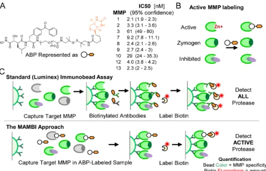
The MAMBI approach begins by incubating sample with an ABP (Figure 1). Here we use a trifunctional biotin-tagged hydroxamate benzophenone ABP 4 containing an active-site targeting warhead
based on potent MMP inhibitors, marimastat and GM6001, shown previously to target active MMPs promiscuously9 (Figure 1). The inhibition profile of the ABP against an MMP panel closely follows that
reported for inhibitors having similar warheads (Figure 1a, S1)9. While we focus on this tight-binding
ABP, a range of probes can be employed (Figure S2).
Figure 1: Overview of MAMBI. a) Chemical structure of the ABP and IC50 values measured by substrate cleavage inhibition
and schematics of b) the MAMBI approach to active MMP ABP labeling, and c) comparison of traditional bead-based total MMP and MAMBI active-only MMP detection workflows; multiplex assays employ panels of beads targeting different MMPs, but only one bead of the panel is shown.
After samples are ABP-labeled, a modified MBAA technique follows to quantify the levels of specific active MMPs. Commercial MBAA beads capture both active and inactive forms of target MMPs (Figure 1c, upper). The capture beads are next incubated with biotin-labeled secondary antibodies and then fluorescent streptavidin. Multi-color flow cytometry measures two fluorescent signals for each bead: a fiduciary signal from the bead to indicate the target protein identity; and the fluorescent streptavidin
signal to indicate the bound target amount. In MAMBI, instead of the secondary antibody, the ABP
structurally contains a biotin group (Figure 1a), which binds to fluorescently-labelled streptavidin to produce fluorescent signal proportional to the amount of active target MMP (Figure 1c, lower). When cocktails of MAMBI-processed beads are assayed in this way, the bead-localized streptavidin fluorescent intensity correlates to the level of the active form of a given MMP in the original solution.
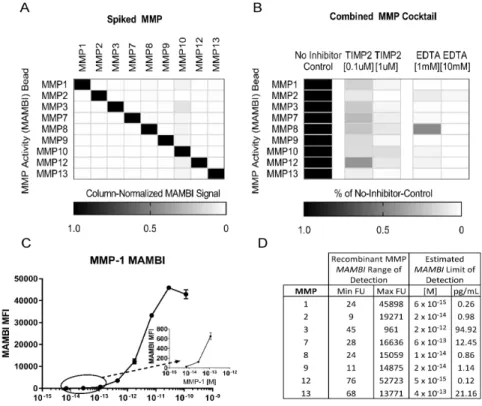
For implementation, we identified a panel of commercially-available MBAA beads for multiplexed capture of MMPs labeled with ABPs. Single active recombinant MMPs (5 nM, 5 µL total volume) were incubated with the ABP (30 min at 37 °C, 500 nM final ABP concentration). Each solution was then incubated with the cocktail of capture MBAA beads (60 minutes, RT, 25 µL total vol) and the bead fluorescence was quantified. MBAA beads targeting MMP-1, 2, 3, 7, 8, 9, 12 and 13 showed limited cross-reactivity. MMP-10 beads showed low signal to noise for rMMP-10 (Figure 2a), hence the
appearance of cross-reactivity between MMP-10 and other MMP beads (Figure S3). An inhibitor challenge further characterized the multiplexed MAMBI technique as responsive only to active MMPs. Addition of ethylenediaminetetraacetic acid (EDTA), a small molecule Zn2+ chelator known to inhibit
MMPs prevented subsequent ABP binding and corresponding streptavidin fluorescence for all bead types (Figures 2b, S4).
Figure 2: MAMBI process validation: a) An optimized cocktail of MAMBI beads shows high selectivity for detecting single recombinant MMPs in solution, b) A cocktail of recombinant MMPs +/- EDTA or TIMP-2 prior to incubation with ABP c) Dose-response curve to estimate limit of detection for MMP-1. Inset shows magnification of circumscribed data points, d) Observed MAMBI absolute limits of detection from dilutions of APMA-activated MMPs in reference solution.
UV-mediated crosslinking of the ABP benzophenone group to the MMP active sites is standard for proteomic-based active MMP assays, protecting the probe from dissociation during prolonged, multi-step sample preparation required for mass spectrometry detection4, 9, 12. In contrast, MAMBI sample
preparation (mild buffers, <4 hours) is less likely to result in probe dissociation (Figure S4). Accordingly, UV treatment did not increase the absolute intensity of MMP-associated streptavidin signal (Figure S5), indicating that UV treatment did not substantially impact the measurement of ABP binding to active MMPs during our protocol. The tight binding of ABP to the active MMPs in this screen (Figure 1a), combined with the gentle processing, allow robust detection without UV crosslinking.
To determine the sensitivity and dynamic range of the bead cocktail for active MMP
characterization, we used dilutions of an APMA-activated (Figure S6) recombinant MMP cocktail. Each MMP-specific capture bead, when assayed with MAMBI, showed consistently low background
fluorescence both in off-target and MMP-free negative controls. Detection limits for each MMP were approximated by the concentration of the standard diluent having signal at least two standard error above the signal from enzyme-free negative controls (Figure 2c, d). Values in Figure 2 are conservative upper limits of assay sensitivity based on full enzyme activity, but recombinant MMPs are well-known to exhibit only a fraction of maximal activity 16 (Figure S7-8). Standard curves generated from recombinant
cocktail solutions (5 µl) suggest that MAMBI can detect MMP-1, 2, 7, 8, 9, 12, and 13 at picomolar concentrations (Figure 2d). MAMBI also finds levels of active MMP-7, and collagenases (MMP-1 and MMP-8), which are difficult to detect by gelatin zymography. Moreover, zymography usually artificially activates MMPs via disruption of TIMP binding; MAMBI preserves TIMP binding, thus measuring activities more representative of in situ behavior. MAMBI thus may illuminate processes relatively inaccessible by current methods.
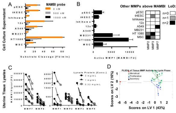
The MAMBI protocol developed with recombinant proteins was next adapted for measuring active native MMPs in biological samples, modifying certain steps to account for phenomena that might influence stability of the ABP during sample preparation. Biological replicates of conditioned medium were assayed from endometrial cell types [primary endometrial stromal cells (pESC); immortalized endometrial stromal cells (HESC), Ishikawa endometrial epithelial cell line and the 12Z endometriotic epithelial line], cancer cell lines [A549, MD-MBA-321 and HT1080] and an hTERT-immortalized mesenchymal stem cell (MSC) line. Protein-normalized samples (1000 µg protein/ml) showed robust aggregate MMP activity, measured as proteolytic activation of a commercially-available fluorogenic substrate (Figure 3a)7 (Figure S9,10). Apparent substrate activation by MMPs was suppressed by addition
of the ABP (500 nM), and in some cases was further decreased with additional ABP (Figure 3a), demonstrating potent inhibition via MMP active site binding.
MAMBI assays of the same samples ascertain comparative contributions of a subset of these MMPs. MAMBI detected active MMP-1 in all samples, along with minimal levels of active MMP-2 in medium from MSC cultures, MMP-3 in both biological replicates from HT1080-derived samples, and strong MAMBI MMP-7 signal in medium from A549 cells (Figure 3b). MMPs detected in only one biological replicate had signal closely above background levels. MAMBI did not detect MMP-9 and 12 in supernatant samples. Notably, no sample had signal above background levels when tested with the commercial active MMP-1 detection kit (Fluorkine E, R&D Systems) even for increased sample volume (20 µl compared with 5 µl used for MAMBI). MAMBI also detected MMP-1 and -7 in clarified aspirates of peritoneal fluid from endometriosis patients (Figure S11), addressing a key technical gap for these low-abundance samples6.
Figure 3: Detection of Endogenous MMP Activity with MAMBI: a) Fluorogenic substrate cleavage of conditioned cell culture medium +/- ABP inhibition (error is SD of n=2 biological replicates), b) Catalogue of active MMPs in conditioned cell culture medium detected by MAMBI where error in absolute MAMBI signal of MMP-1 is SD of n=2 biological replicates, c) Active MMPs detected by MAMBI in dilutions of total protein-normalized uterine tissue lysates from 8 distinct donors, d) PLSDA of 17 uterine tissue lysates using cycle phase as the categorical y-block.
M M P 7 M M P 8 0 1 0 0 0 0 2 0 0 0 0 3 0 0 0 0 4 0 0 0 0 5 0 0 0 0 M A M B I S ig n a l [F U ] M M P 1 M M P 2 M M P 3 M M P 9 0 5 0 0 1 0 0 0 2 0 0 0 4 0 0 0 6 0 0 0 3 m g / m L 1 m g / m L 0 . 3 3 m g / m L L y s a t e P r o t e i n [ C o n c . ] A B C D
Other MMPs above MAMBI LoD:
M M P 2 M M P 3 M M P 7 pESC tHESC Ishikawa 12z A549 MB231 HT 1080 MSC 0 0.2 0.4 0.6 0.8 1.0 n=0 n=1 n=2 C e ll C ul tur e S up e rna ta nt MAMBI probe U te ri ne Ti ss ue Ly sa te s -6 -5 -4 -3 -2 -1 0 1 2 Scores on LV 1 (43%) Sc o re s o n L V 2 (1 6% ) 0 1 -2 -3 0 2 0 4 0 6 0 8 0 M S C H T 1 0 8 0 M B 2 3 1 A 5 4 9 1 2 z I s h i k a w a t H E S C p E S C S u b s t r a t e C l e a v a g e [ F U / m i n ] 0 n M 5 0 0 n M 1 5 0 0 n M 0 5 0 0 0 1 0 0 0 0 M S C H T 1 0 8 0 M B 2 3 1 A 5 4 9 1 2 z I s h i k a w a t H E S C p E S C A c t i v e M M P 1 [ M A M B I F U ]
We next characterized MMP activity in uterine tissue, where MMP expression levels vary during the menstrual cycle 3. Others have noted that endogenous TIMPs interfere with assays of MMP activity
from tissue lysates 5. To mitigate TIMP inactivation of MMPs during processing, high molar excess of
ABP was added to tissue samples prior to and throughout homogenization. MAMBI identified robust activity in all eight donor samples for MMP-1, 2, 3, 7, 8, and 9, with notably high activity observed for MMP-7 and 8 (Figure 3c). Detection of the active protease forms was dose-dependent (Figure 3c), and could be blocked by treatment with either EDTA or the broad spectrum MMP inhibitor GM6001 (Figure S12). Despite protein normalization of lysates (e.g. 3mg/mL) substantial difference in MMP activities were observed, likely reflecting variations among donors and their respective menstrual cycle phases (Figure S13). Indeed, multivariate analysis of additional samples using partial least-squares discriminant analysis (PLSDA) shows that samples cluster according to cycle phase (Figure 3d). Our findings
characterizing active native MMPs in uterine samples, especially multiplexed detection of endogenous MMP activity in tissue samples, represent a significant advancement in active MMP monitoring readily adaptable to other complex tissue and disease models.
The MAMBI approach is also valuable for assessing potential MMP inhibitors. Recent efforts to develop MMP inhibitor screening platforms have extended established techniques of monitoring
fluorescent substrate cleavage 17 to competitive-ABPP assays, wherein inhibitor decreases ABP binding.
Competitive-ABPP approaches hold potential to simultaneously monitor relative inhibition of multiple enzymes in physiologically-relevant settings, but MMP inhibitor competitive-ABPP screens have only been demonstrated in well-defined single-enzyme samples 13 or in lower-throughput formats relying on
mass spec-based 9, 12 or gel-based 15-16 analysis. Hence, in ‘Competitive-MAMBI’ assays, we extend
competitive-ABPP assays to allow high-throughput characterization of MMP inhibitors in complex MMP mixtures.
Competitive-MAMBI assays were used to estimate relative potencies of commercially-available inhibitors against mixtures of recombinant and native MMPs. Inhibition of recombinant catalytic domain MMPs (Figure S7) by ten small molecule MMP inhibitors (Figure S14) was first characterized (Figure 4a), then further studied for a subset in diluted menstrual effluent (Figure 4c) -- a complex donor-derived sample possessing numerous active native MMPs 3. MAMBI assays of diluted effluent identified MMP-1,
2, 3 and 7 as having sufficiently high native active MMP levels to include in competitive-MAMBI assays, as well as non-saturating levels of MMPs indicated by serial dilutions (Figure 4b). For all samples, we incubated active MMPs with ABP and varied concentrations of inhibitor, then used MAMBI to determine ABP binding to the assayed MMP. Each inhibitor was titrated at 0x, 0.33x, 1x, 3x or 15x to the ABP (constant at 0.5 µM) or at 0, 0.17, 0.5, 1.5 and 7.5 µM, respectively, and incubated with the MMP
solution and ABP probe. Relative potencies are quantified by the loss of ABP labeling to target MMP due to inhibitor addition. Inhibitor binding for each condition is reported as the complement of fractional MAMBI signal observed for that condition with and without competitive inhibition (Figure 4a, c, S15).
Figure 4: Selectivity of MMP inhibitors against a panel of MMPs evaluated by competitive-MAMBI: a) Inhibition profile for recombinant MMPs, b) MAMBI activity across dilutions of clarified menstrual effluent, and c) Inhibition profile for MMPs in menstrual effluent.
Close correlation between inhibition profiles of the commercial MMP inhibitors detected by competitive-MAMBI versus established relative selectivities supports its utility in screening (Figure 4a, c, S15). In competitive-MAMBI, Marimastat and Batimastat showed expected profiles as promiscuous inhibitors exhibiting inhibitory activity toward all proteases tested.
In both recombinant and effluent-derived samples Batimastat showed stronger inhibitory activity against MMP-3 compared with Marimastat, consistent with their known comparative potencies toward MMP-3 (Batimastat IC50 = 20 nM; Marimastat IC50 = 200 nM) 1. ARP 101 showed increased potency
towards MMP-2 and MMP-3 compared with the closely-related but less lipophilic ARP 100, consistent with its described range of activity18. Ro 32-3555 showed expected acute potency towards MMP-1 and
MMP-7 in both recombinant and effluent-derived MMP samples, whereas MMP Inhibitor 3 VII and UK 370106 exhibited expected inhibition of MMP-3 (Figure S15-16). Gelatinase inhibitors SB-3CT and MMP Inhibitor 2 did not prevent binding of ABP to tested MMPs, which we attribute to their low overall potency compared with the ABP (Figure S16).
The strong overall concordance between published and observed inhibition profiles supports interpretation of the MAMBI assay as quantifying relative levels of active proteases. Compared to established approaches for inhibitor screening, competitive-MAMBI offers improved throughput and multiplexing, minimal sample requirements, and ready adoption to more complex and physiologically relevant protease samples.
In conclusion, we have integrated components of activity-based protein profiling and bead-based immunoassays to achieve multiplexed active MMP detection in complex samples. Our MAMBI approach detects relative levels of active recombinant MMP-1, 2, 3, 7, 8, 9, 12, and 13, and native forms of those same MMPs, in small volumes of cell culture medium, menstrual fluid effluent, and uterine tissue.
Marimastat Batimastat ARP 100 ARP 101 PF 356231 UK 370106 SB 3CT Ro 32-3555 MMP Inhib. 2 Recombinant MMPs
A
InhibitorB
Binding (Calculated Fraction)C
Marimastat Batimastat ARP 101 MMP Inhib. 3 VII UK 370106 Ro 32-3555 Menstrual Effluent MMPs MMP1 MMP2 MMP3 MMP7 0 500 1000 1500 2000 2500 M A M B I S ig n al [ F U ] MMP1 MMP2 MMP3 MMP7 0 50 100 150 200 4% 2% 1% MMP1 MMP2 MMP3 MMP7 0 50 100 150 200 4% 2% 1% MMP Inhib. 3 VII Ratio Inhibitor to MAMBI probe MMP1 MMP2 MMP3 MMP7 0 50 100 150 200 4% 2% 1% Ratio Inhibitor toMAMBI probe Inhibitor Binding
(Calculated Fraction) M M P 1 M M P 2 M M P 3 M M P 7 1 5 x 3 x 1 x 0 . 3 x 1 5 x 3 x 1 x 0 . 3 x 1 5 x 3 x 1 x 0 . 3 x 1 5 x 3 x 1 x 0 . 3 x 1 5 x 3 x 1 x 0 . 3 x 1 5 x 3 x 1 x 0 . 3 x 0 . 2 5 0 . 5 0 0 . 7 5 1 . 0 0 M M P 1 M M P 2 M M P 3 M M P 7 M M P 9 M M P 1 2 1 5 x 3 x 1 x 0 . 3 x 1 5 x 3 x 1 x 0 . 3 x 1 5 x 3 x 1 x 0 . 3 x 1 5 x 3 x 1 x 0 . 3 x 1 5 x 3 x 1 x 0 . 3 x 1 5 x 3 x 1 x 0 . 3 x 1 5 x 3 x 1 x 0 . 3 x 1 5 x 3 x 1 x 0 . 3 x 1 5 x 3 x 1 x 0 . 3 x 1 5 x 3 x 1 x 0 . 3 x 0 0 . 2 5 0 . 5 0 0 . 7 5 M M P 1 M M P 2 M M P 3 M M P 7 1 5 x 3 x 1 x 0 . 3 x 1 5 x 3 x 1 x 0 . 3 x 1 5 x 3 x 1 x 0 . 3 x 1 5 x 3 x 1 x 0 . 3 x 1 5 x 3 x 1 x 0 . 3 x 1 5 x 3 x 1 x 0 . 3 x 0 . 2 5 0 . 5 0 0 . 7 5 1 . 0 0
Moreover, competitive-MAMBI extends the MAMBI approach to evaluate the relative potency and selectivity of a panel of inhibitors across multiple MMPs.
Methods
See Supporting Information.
ASSOCIATED CONTENT
*S Supporting Information Additional methods and supporting figures and tables. available free of charge at http:// pubs.acs.org.
■
AUTHOR INFORMATION
Corresponding Author *(L.G.) E-mail: [email protected] ■
The authors declare no competing financial interest. ■
ACKNOWLEDGMENTS
We thank Marcia Moss and Fred Rasmussen (Biozyme, Inc., Apex, NC) for reagents and advice, Anthony Guidi and Luis Velasquez (Newton Wellesley Hospital) for assistance with dissection of uteri and Linda Stockdale for menstrual fluid collection. NIH R01 EB010246, the Manton Foundation, and the Begg Fund provided funding support.
References
1. Vandenbroucke, R. E.; Libert, C., Is there new hope for therapeutic matrix metalloproteinase inhibition? Nat Rev Drug Discov 2014, 13 (12), 904-27.
2. Becker, C. M.; Louis, G.; Exarhopoulos, A.; Mechsner, S.; Ebert, A. D.; Zurakowski, D.; Moses, M. A., Matrix metalloproteinases are elevated in the urine of patients with endometriosis. Fertil Steril 2010,
94 (6), 2343-6.
3. Osteen, K. G.; Yeaman, G. R.; Bruner-Tran, K. L., Matrix metalloproteinases and endometriosis.
Seminars in reproductive medicine 2003, 21 (2), 155-64.
4. Ravindra, K. C.; Ahrens, C. C.; Wang, Y.; Ramseier, J. Y.; Wishnok, J. S.; Griffith, L. G.; Grodzinsky, A. J.; Tannenbaum, S. R., Chemoproteomics of matrix metalloproteases in a model of cartilage
degeneration suggests functional biomarkers associated with posttraumatic osteoarthritis. J Biol Chem 2018, 293 (29), 11459-11469.
5. Crawford, B. D.; Pilgrim, D. B., Ontogeny and regulation of matrix metalloproteinase activity in the zebrafish embryo by in vitro and in vivo zymography. Dev Biol 2005, 286 (2), 405-14.
6. Miller, M. A.; Meyer, A. S.; Beste, M. T.; Lasisi, Z.; Reddy, S.; Jeng, K. W.; Chen, C.-H.; Han, J.; Isaacson, K.; Griffith, L. G.; Lauffenburger, D. A., ADAM-10 and -17 regulate endometriotic cell migration via concerted ligand and receptor shedding feedback on kinase signaling. Proceedings of the National
7. Miller, M. A.; Barkal, L.; Jeng, K.; Herrlich, A.; Moss, M.; Griffith, L. G.; Lauffenburger, D. A., Proteolytic Activity Matrix Analysis (PrAMA) for simultaneous determination of multiple protease activities. Integr Biol (Camb) 2011, 3 (4), 422-438.
8. Lombard, C.; Saulnier, J.; Wallach, J., Assays of matrix metalloproteinases (MMPs) activities: a review. Biochimie 2005, 87 (3-4), 265-72.
9. Saghatelian, A.; Jessani, N.; Joseph, A.; Humphrey, M.; Cravatt, B. F., Activity-based probes for the proteomic profiling of metalloproteases. Proc Natl Acad Sci U S A 2004, 101 (27), 10000-5.
10. Hu, H. Y.; Gehrig, S.; Reither, G.; Subramanian, D.; Mall, M. A.; Plettenburg, O.; Schultz, C., FRET-based and other fluorescent proteinase probes. Biotechnol J 2014, 9 (2), 266-81.
11. Klein, T.; Geurink, P.; Overkleeft, H.; Kauffman, H.; Bischoff, R., Functional proteomics on zinc-dependent metalloproteinases using inhibitor probes. ChemMedChem 2009, 4 (2), 164-70.
12. Sieber, S. A.; Niessen, S.; Hoover, H. S.; Cravatt, B. F., Proteomic profiling of metalloprotease activities with cocktails of active-site probes. Nat Chem Biol 2006, 2 (5), 274-81.
13. Antczak, C.; Radu, C.; Djaballah, H., A profiling platform for the identification of selective metalloprotease inhibitors. J Biomol Screen 2008, 13 (4), 285-94.
14. Sanman, L. E.; Bogyo, M., Activity-based profiling of proteases. Annu Rev Biochem 2014, 83, 249-73.
15. Chan, E. W. S.; Chattopadhaya, S.; Panicker, R. C.; Huang, X.; Yao, S. Q., Developing Photoactive Affinity Probes for Proteomic Profiling: Hydroxamate-based Probes for Metalloproteases. Journal of the
American Chemical Society 2004, 126 (44), 14435-14446.
16. Nakai, R.; Salisbury, C. M.; Rosen, H.; Cravatt, B. F., Ranking the selectivity of PubChem screening hits by activity-based protein profiling: MMP13 as a case study. Bioorg Med Chem 2009, 17 (3), 1101-8. 17. Lauer-Fields, J. L.; Minond, D.; Chase, P. S.; Baillargeon, P. E.; Saldanha, S. A.; Stawikowska, R.; Hodder, P.; Fields, G. B., High throughput screening of potentially selective MMP-13 exosite inhibitors utilizing a triple-helical FRET substrate. Bioorganic & Medicinal Chemistry 2009, 17 (3), 990-1005. 18. Nuti, E.; Casalini, F.; Avramova, S. I.; Santamaria, S.; Cercignani, G.; Marinelli, L.; La Pietra, V.; Novellino, E.; Orlandini, E.; Nencetti, S.; Tuccinardi, T.; Martinelli, A.; Lim, N. H.; Visse, R.; Nagase, H.; Rossello, A., N-O-isopropyl sulfonamido-based hydroxamates: design, synthesis and biological evaluation of selective matrix metalloproteinase-13 inhibitors as potential therapeutic agents for osteoarthritis. J
Med Chem 2009, 52 (15), 4757-73.