Abstract
Metallo-
β-lactamases (MBLs) hydrolyze almost all classes of β-lactam antibiotic, including
carbapenems
—currently first choice drugs for opportunistic infections by Gram-negative
bacterial pathogens. MBL inhibitor development is complicated by the diversity within this
group of enzymes, and by the appearance of new enzymes that continue to be identified
both as chromosomal genes and on mobile genetic elements. One such newly discovered
MBL is DIM-1, a mobile enzyme originally discovered in the opportunist pathogen
Pseudo-monas stutzeri but subsequently identified in other species and locations. DIM-1 is a
sub-class B1 MBL more closely related to the TMB-1, GIM-1 and IMP enzymes than to other
clinically encountered MBLs such as VIM and NDM; and possesses Arg, rather than the
more usual Lys, at position 224 in the putative substrate binding site. Here we report the
crystallization and structure determination of DIM-1. DIM-1 possesses a binuclear metal
center with a 5 (rather than the more usual 4) co-ordinate tri-histidine (Zn1) site and both
4-and 5-co-ordinate Cys-His-Asp- (Zn2) sites observed in the two molecules of the
crystallo-graphic asymmetric unit. These data indicate a degree of variability in metal co-ordination
geometry in the DIM-1 active site, as well as facilitating inclusion of DIM-1 in
structure-based MBL inhibitor discovery programmes.
Introduction
Acquired metallo-β-lactamases (MBLs) present a global public health challenge [
1
]. Their high
hydrolytic activity against carbapenems, (broad-spectrum
β-lactams of ultimate choice for
Gram-negative infections), and their non-susceptibility to serine
β-lactamase inhibitors, make
their dissemination significant in the rise of antibiotic resistant pathogens. Sequence data define
three MBL subclasses (B1
–B3 [
2
]) with B1 including almost all enzymes whose genes are found
on mobile genetic elements, and thus capable of rapid dissemination across geographic and
spe-cies boundaries. B1 MBLs from the IMP, VIM and NDM groups have already been found in
OPEN ACCESS
Citation: Booth MPS, Kosmopoulou M, Poirel L, Nordmann P, Spencer J (2015) Crystal Structure of DIM-1, an Acquired Subclass B1 Metallo- β-Lactamase from Pseudomonas stutzeri. PLoS ONE 10(10): e0140059. doi:10.1371/journal.pone.0140059 Editor: Anna Roujeinikova, Monash University, AUSTRALIA
Received: April 22, 2015 Accepted: September 20, 2015 Published: October 9, 2015
Copyright: © 2015 Booth et al. This is an open access article distributed under the terms of the
Creative Commons Attribution License, which permits unrestricted use, distribution, and reproduction in any medium, provided the original author and source are credited.
Data Availability Statement: Crystal structures (co-ordinates and structure factors) have been deposited in the Protein Data Bank (www.rcsb.org/pdb) with accession codes 4WD6 and 4ZEJ.
Funding: This study was supported by the British Society for Antimicrobial Chemotherapy (www.bsac. org; grant no. GA820 to J.S.), the U.K. Medical Research Council (www.mrc.ac.uk; grant no. G1100135 to J.S.), and the U.S. National Institutes of Allergy and Infectious Disease (http://www.niaid.nih. gov/; grant no. 1R01AI100560-01 to J.S.). The funders had no role in study design, data collection
multiple hosts, both Enterobacteriaceae [
3
] and non-fermenters (Pseudomonas aeruginosa,
Aci-netobacter baumannii [
4
–
6
]), while additional mobile enzymes [
7
–
9
] continue to be identified.
DIM-1 (Dutch IMipenemase) was identified in a carbapenem non-susceptible isolate of the
occasional opportunist pathogen Pseudomonas stutzeri, recovered from a patient undergoing
surgery for tibial osteomyelitis [
10
]. The carbapenemase gene, bla
DIM-1(GenBank:
GU323019.1), resided on a class 1 integron on a 70 kb plasmid, suggesting a capability for
dis-semination, and has subsequently been identified in isolates of multiple species (Escherichia
coli, Enterobacter cloacae, Comomonas testosteroni) of African origin and in a single strain of P.
stutzeri from India [
11
,
12
]. Independent acquisition events from a currently unknown
envi-ronmental reservoir have been suggested to account for these apparently unconnected
identifi-cations in widely separated loidentifi-cations [
11
]. The bla
DIM-1gene encodes a subclass B1 MBL
resembling TMB-1 (67% identity [
7
]) GIM-1 (54% identity [
13
]) and IMP-1 (48% identity
[
14
]; percentage identities are for the mature, processed polypeptides), but more distantly
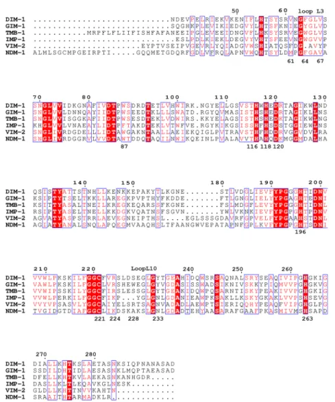
related to VIM-1 and NDM-1. An alignment of selected B1 MBLs is presented in
Fig 1
.
Like other B1 MBLs, DIM-1 is a broad-spectrum enzyme, hydrolyzing penicillins,
cephalo-sporins and carbapenems, although compared to some enzymes turnover of some penicillins is
reduced and activity is weaker against cephalosporins, e.g. ceftazidime or cefepime, with bulky,
charged, 3’ substituents [
10
]. In common with GIM-1 and TMB-1, DIM-1 is distinguished
from other B1 enzymes by possession of arginine at position 224, rather than the lysine
(Lys224) found in enzymes such as the IMP and NDM groups. Recent crystal structures [
16
,
17
] show NDM-1 Lys224 to make electrostatic interactions with the C3 carboxylate of
hydro-lyzed carbapenems, suggesting that this substitution might affect substrate binding. Here we
report the crystal structure of DIM-1, facilitating its inclusion in structure-based inhibitor
dis-covery programmes.
Materials and Methods
Protein Expression
DIM-1 was purified from E. coli TOP10 containing the plasmid pXD-1 [
10
] by ion-exchange
and size exclusion chromatography in a modification of previously published procedures.
Briefly, 2 L of Luria-Bertani broth were inoculated with 10 mL / L of an overnight culture and
grown for 24 h at 37°C with shaking at 160 rpm. Cells were harvested by centrifugation (4 000
g, 20 min, 4°C), washed in phosphate-buffered saline (PBS) and frozen until required. Thawed
pellets were resuspended in 25 mL / L Buffer A (50 mM Tris-Cl pH 7.5) containing protease
inhibitors (Complete EDTA-free, Roche Life Science, Burgess Hill, U.K.) and lysed by
sonica-tion. The lysate was clarified by centrifugation (15,000 g, 30 min, 4°C) and dialyzed overnight
(4°C) against 1 L buffer A. The dialysate was loaded onto a 50 mL Q-sepharose column (Sigma,
Poole, U.K.) which was washed in buffer A until absorbance returned to baseline. Bound
pro-tein was eluted in buffer A using a 300 mL 0–0.5 M NaCl gradient; DIM-1 eluted at
approxi-mately 0.1 M NaCl as identified by hydrolysis of the chromogenic cephalosporin nitrocefin
[
18
]. DIM-1 containing fractions were concentrated by centrifugal ultrafiltration, loaded onto
a 300 mL Superdex-75 column pre-equilibrated in Buffer B (50 mM Tris-Cl 200 mM NaCl pH
7.5) and eluted in the same buffer. The purity of the final preparation was greater than 95% as
adjudged by SDS—PAGE [
19
]. The purified protein was concentrated for crystallization trials
by centrifugal ultrafiltration.
Crystallization and Structure Solution
Sparse matrix crystallization screening with the Morpheus suite [
20
] used a Pheonix robot
(Art-Robbins) to set sitting drops (0.5
μL each of DIM-1 (15 mg / mL) and reservoir solution)
and analysis, decision to publish, or preparation of the manuscript.
Competing Interests: The authors have declared that no competing interests exist.
in 96-well plates with 100
μL well volume. Crystals appeared after 5–10 days’ incubation
(20°C) in 20% polyethylene glycol (PEG) 550 monomethyl ether (MME) / 10% PEG 20 000; 50
mM HEPES / 50 mM MOPS pH 7.5; were mounted in rayon loops, snap-frozen and stored in
liquid nitrogen until required. For soaking experiments, ceftazidime (Sigma, Poole, U.K.)
dis-solved to 250 mM in dimethyl sulfoxide (DMSO) was added to the drop to a final
concentra-tion of 85 mM and 33% DMSO and crystals frozen after 1 hour incubaconcentra-tion at 20°C. Diffracconcentra-tion
data were collected on an ADSC Quantum 4 detector mounted on beamline I03 of Diamond
Light Source (Didcot, U.K.) and reflections integrated, scaled and merged using HKL2000 [
21
].
Phases were obtained by molecular replacement using PHASER [
22
] with IMP-1 (PDB
acces-sion 1DDK; [
23
]) as a search model. The model was built using Coot [
24
] and refined using
Refmac5 [
25
] as implemented in the CCP4 software suite [
26
] with TLS
(Translation/Libra-tion/Screw) groups identified by the TLSMD server [
27
,
28
]. The quality of the final model was
assessed using PROCHECK [
29
] and Molprobity [
30
]. Data collection and refinement statistics
Fig 1. Alignment of DIM-1 with Selected Subclass B1 Metallo-β-Lactamases. Invariant residues are highlighted on a red background; conservative substitutions are colored red. Residue numbers according to the Class Bβ-Lactamase (BBL) standard numbering scheme are positioned above the sequences; individual residues discussed in the text are numbered below the sequences. This Figure was prepared using ESPript [15]. doi:10.1371/journal.pone.0140059.g001
are given in
Table 1
. Co-ordinates and structure factors have been deposited with the Protein
Data Bak (
www.rcsb.org
) with accession codes 4WD6 (native structure) and 4ZEJ
(ceftazi-dime-exposed structure).
Results and Discussion
DIM-1 crystallized in space group P 2
12
12
1with crystals containing two molecules in the
asymmetric unit (
Table 1
). The structure was solved by molecular replacement and could be
built as two polypeptide chains. Two complete data sets were collected, a native data set from a
crystal diffracting to 2.2 Å resolution (dataset 1) and a second, higher resolution data set
(data-set 2) arising from one of a number of crystals that had been soaked with the cephalosporin
cef-tazidime in DMSO, but for which no electron density corresponding to a bound
β-lactam
could be resolved. As this crystal diffracted to higher resolution (1.8 Å) and yielded higher
quality data (
Table 1
) this structure was also solved and refined. Non-crystallographic
symme-try was not employed at any stage in the refinement process; therefore our data provide
struc-tures for four independently refined DIM-1 monomers. The final models contain 219 (residues
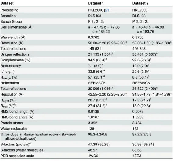
Table 1. Data Collection and Refinement Statistics.
Dataset Dataset 1 Dataset 2
Processing HKL2000 [21] HKL2000
Beamline DLS I03 DLS I03
Space Group P 212121 P 212121 Cell Dimensions (Å) a = 47.72 b = 47.86 c = 185.22 a = 46.40 b = 46.98 c = 183.76 Wavelength (Å) 0.9763 0.9763 Resolution (Å) 50.00–2.20 (2.28–2.20)a 50.00–1.80 (1.86–1.80)a Total reflections 149 531 496 348 Unique reflections 21 133 (1 504)a 38 481 (3 667)a Completeness (%) 94.5 (68.4)a 99.6 (96.6)a Redundancy 7.1 (5.9)a 12.9 (7.0)a I / (sig. I) 32.5 (6.6)a 29.6 (2.5)a Rmerge(%) 5.1 (25.1)a 8.8 (50.1)a
Refinement REFMAC5 REFMAC5
Total reflections 20 006 (1 016)a 36 522 (2 499)a Resolution (Å) 42.55–2.20 (2.26–2.20)a 91.88–1.79 (1.84–1.79)a Rcryst(%) 20.7 (23.9)a 17.2 (21.7)a Rfree(%)b 27.4 (34.2)a 18.9 (22.8)a RMS bond length (Å) 0.0138 0.0078 RMS bond angle (Å) 1.6167 1.2289 Protein atoms 3 392 3 434 Water molecules 126 192
% residues in Ramachandran regions (favored/ allowed/disallowed)
95.3/4.2/0.5 97.2/2.3/0.5
B-factors (protein)c 47.38 (55.26) 30.98 (39.81)
B-factors (water molecules) 48.57 38.68
PDB accession code 4WD6 4ZEJ
aData for the highest resolution shell are in parentheses. bR
freewas calculated with 5% of the reflections omitted. cFigures in parentheses are for chain B.
39
–294 according to the standard class B β-lactamase (BBL) numbering scheme [
2
]) amino
acids per chain, apart from chain B of the lower resolution structure (dataset 1) for which weak
electron density meant that residues 25
–28 (60–64; BBL residue numberings are given in
parentheses throughout this paper) inclusive were not modelled. Overall Cα rms deviations
between the four chains were a maximum 0.36 Å. For the four chains (A and B of dataset 1 and
A and B of dataset 2, respectively) 95/ 94/ 97/ 97% of residues lie within the most favored
regions of the Ramachandran plot and a further 4/ 6/ 2/ 2% within the additionally allowed
regions [
30
]. In three of the four chains Asp-48 (84) lies in the disfavored Ramachandran
region due to its involvement in a buried hydrogen bond network, involving Ser-33 (69),
Asn-34 (70), Thr-76 (115), Arg-82 (121) and Gly-199 (262) that is common to most members of
the B1 MBL subfamily and connects the two halves of the protein beneath the active site [
31
].
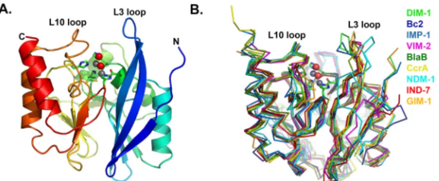
DIM-1 adopts the
αβ/βα sandwich fold of the MBL hydrolase superfamily, with the active
site situated in the groove between the two domains and containing two zinc ions (
Fig 2A
).
Superposition (using SSMsuperpose [
32
]) with other available B1 MBL structures (
Fig 2B
)
indicates that DIM-1 most closely resembles GIM-1 (C
α rmsd 0.87 Å) and IMP-1 (Cα rmsd
0.91 Å). Among B1 enzymes the MBL fold is well conserved, with significant differences
appar-ent at the N-terminus, in the length of the C-terminal
α-helix, and in the flexible loop L3
(resi-dues 25–32 (60–68)) that connects the β2 and β3 strands. Crystallographic [
23
,
33
–
35
],
mutagenic and kinetic [
36
,
37
], NMR [
38
,
39
] and molecular dynamics [
40
] evidence indicates
that this loop adjusts its conformation on
β-lactam or inhibitor binding and contributes to
sub-strate affinity and specificity. In the lower resolution (dataset 1) DIM-1 structure the L3 loop
could not be modelled for chain B, but in chain A it extends over the active site groove in a
“closed” conformation more typical of MBL complexes [
17
,
34
,
35
,
41
] than of other
uncom-plexed B1 MBL structures. These differences may arise from differences in crystal
contacts-while the apex of loop L3 of chain A lies close both to chain B and symmetry related molecules,
fewer contacts are made by the equivalent subunit of chain B. In the higher resolution structure
obtained from dataset 2, the complete loop L3 could be modeled for both chains, but chain B
featured elevated B-factors consistent with a reduced involvement in crystal contacts.
Estab-lishing whether the observed conformation of the DIM-1 L3 loop arises primarily from crystal
contacts, or indeed reflects that adopted on substrate and/or inhibitor binding, must await
determination of structures for the relevant complexes.
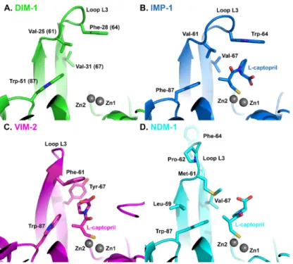
Fig 2. Crystal Structure of DIM-1. A. Overall fold of the enzyme. Protein backbone is color-ramped from blue (N-) to red (C-terminus). Active site residues are rendered as sticks (carbon atoms in green, other atom colors as standard). Zinc ions (gray) and water molecules (red) are shown as spheres. B. Superposition of B1 MBL structures: DIM-1 (green); BcII (pdb accession 1BC2 ([43], blue); IMP-1 (1DDK [23], teal); VIM-2 (1KO3 [44], magenta); BlaB (1M2X [45], dark green); CcrA (1ZNB [46], yellow); NDM-1 (3Q6X [41], cyan); IND-7 (3L6N [47], red) and GIM-1 (2YNT [48], orange). This Figure was generated using Pymol (www.pymol.org). doi:10.1371/journal.pone.0140059.g002
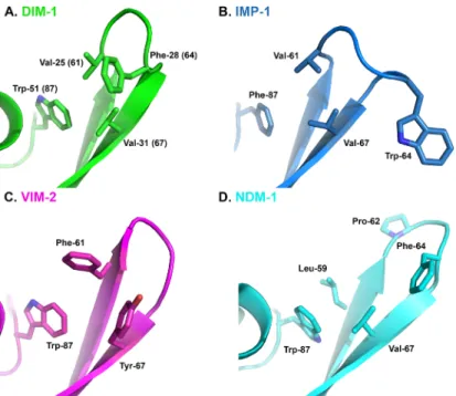
Our structure also confirms that DIM-1 features hydrophobic aliphatic amino acids (valine)
at positions 25 (61) and 31 (67) at the base of the L3 loop, and an aromatic residue
(phenylala-nine) at position 28 (64) at its apex. In this DIM-1 resembles IMP-1 (and similar enzymes such
as GIM-1 [
13
] or KHM-1 [
9
]) rather than the VIM enzymes (where positions 25 (61) and 31
(67) are occupied by aromatic residues) or NDM-1 (where the L3 loop contains proline at
posi-tion 26 (62) and adopts a different conformaposi-tion;
Fig 3
). As discussed above, inhibitor binding
to enzymes such as CcrA [
33
] or BcII [
39
] can involve conformational changes in the L3 loop,
and be affected by mutations within it [
36
]. Moreover, comparisons of inhibition of multiple
B1 enzymes that differ in their L3 loops (including NDM, IMP and VIM, but not DIM,
enzymes) by a series of hydroxythiazole compounds identified differences between enzymes of
up to two orders of magnitude in IC
50values [
42
]. Unfortunately, limited structural data are
available that permit direct comparison of inhibitor binding to different MBL targets. However,
structures for complexes of the thiol L-captopril with IMP-1 (pdb id 4C1F), NDM-1 (pdb
4EXS [
17
]) and VIM-2 (pdb 4C1D) show that this adopts different conformations that are in
part imposed by the proximity of different residues on the L3 loop (residues Met-61 of
NDM-1; Val-61 of IMP-1 and Phe-62 and Tyr-67 of VIM-2) to the captopril pyrrolidine ring.
Com-parison of these structures with that of DIM-1 (
Fig 4
) suggests that positioning of Phe-28(64)
on loop L3 over the active site, together with the presence of Val-25 (61), Val-31 (67) and
Trp-51(87) would support an orientation for the binding of the captopril pyrrolidine to DIM-1 that
is similar to that adopted upon binding to IMP-1, and that differs from those observed with
VIM-2 or NDM-1. It will be of interest to establish the extent to which this similarity between
the L3 loops of IMP-1 and DIM-1 results in similar inhibition profiles, and similar modes of
inhibitor binding.
Loop L10 (residues 160–176 (223–239)) on the opposite side of the active site groove also
shows features that distinguish DIM-1 from related enzymes. The DIM-1 L10 loop lacks
Trp-Fig 3. L3 Loop Conformations in Subclass B1 Metallo-β-Lactamases. A. DIM-1 (green); B. IMP-1 (1DDK [23], teal); C. VIM-2 (1KO3 [44], magenta); D. NDM-1 (3Q6X [41], cyan). This Figure was generated using Pymol.
165 (228), which in GIM-1 restricts this part of the active site cavity [
48
] and instead has serine
at this position. Like GIM-1, DIM-1 possesses tyrosine, rather than the more usual asparagine,
at position 170 (233), which further constrains the active site groove and provides a
hydropho-bic surface for substrate interactions, but abolishes the possible electrostatic contacts between
substrate and the Asn-170 (233) side chain that have been proposed for some other B1
enzymes ([
17
,
35
,
49
]. The DIM-1 active site also features a close electrostatic contact between
the side chains of Glu-78 (119) and Lys-111 (165), forming a bridge across the active site
groove in the vicinity of the probable binding site for substituent groups at the R1 position of
the
β-lactam core (i.e. the C6/C7 substituents of penicillins or cephalosporins, respectively).
Taken together, these factors create an active site in DIM-1 that is narrower and more defined
than the relatively wide open groove of many other B1 MBLs (
Fig 5
).
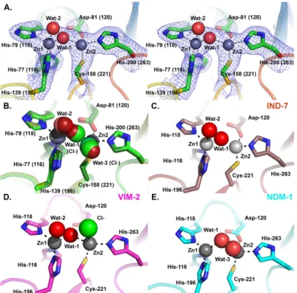
Electron density maps (
Fig 6A
) unequivocally identify two metal ions in the DIM-1 active
site, consistent with the binuclear species being the physiological form of B1 MBLs [
51
,
52
]. As
no particular precautions were taken to avoid oxidation of the ligand Cys-158 (221), which is
associated with loss of the metal ion in the Zn2 site in certain B1 MBLs, we surmise that this
occurs less readily in DIM-1 than in some other enzymes such as BcII, SPM-1 or VIM-2 [
31
,
45
]. All active site metal ions were refined as zinc, with occupancies of 1.0. This procedure
yielded B-factors for the individual zinc ions of 42.58 and 48.04 Å
2(chain A Zn1 and Zn2
respectively, dataset 1); 50.73 and 57.25 Å
2(chain B Zn1 and Zn2 respectively, dataset 1); 23.43
and 26.41 Å
2(chain A Zn1 and Zn2 respectively, dataset 2); and 27.13 and 36.58 Å
2(chain B
Zn1 and Zn2 respectively, dataset 2). This compares with B-factors for the complete individual
protein chains of 47.38 and 55.26 Å
2(chains A and B; dataset 1) and 30.98 and 39.81 Å
2(chains
A and B; dataset 2). The consistency between the B-values obtained for the modelled zinc ions
and the respective protein chains suggests that refining the bound metal ions as zinc is
reason-able. However, while we consider zinc to be the most likely metal ion to be present in the
DIM-Fig 4. Comparison of DIM-1 Active Site with L-Captopril-bound Complexes of Subclass B1 Metallo- β-Lactamases. A. DIM-1 (green); B. IMP-1 (4C1F, teal); C. VIM-2 (4C1D, magenta); D. NDM-1 (4EXS [17], cyan). This Figure was generated using Pymol.
1 active site, the available data cannot preclude the possibility that some fraction of other
metallated species may be present.
In the four independently refined DIM-1 chains the two metal ions lie approximately 4.00
Å–4.40 Å apart (distances in
Table 2
) and are liganded by the conserved residues His-77 (116),
His-79 (118) and His-139 (196) (tri-histidine or Zn1 site); and Asp-81 (120), Cys-158 (221)
and His-200 (263); Cys-His-Asp or Zn2 site). A
“bridging” water molecule (or hydroxide ion)
that is generally proposed to be the nucleophile in the hydrolytic reaction [
53
], is positioned
between the two metal ions (
Fig 6A
, Wat1), lying closer to Zn1 that Zn2. (Zinc:ligand distances
are given in
Table 2
). (In chain A of the higher resolution structure (dataset 2) this
“bridging”
ligand was modelled as a chloride ion (chloride was present in the buffer used for size exclusion
chromatography)). These features are largely typical of available crystal structures for binuclear
B1 MBLs.
B1 MBLs of known structure show a variety of co-ordination geometries at both the Zn1
and Zn2 sites. In the majority of structures the Zn1 site is tetrahedral [
46
,
48
,
54
]. However, in
three of the four refined active sites DIM-1 contains a pentaco-ordinate Zn1 center with a
sec-ond water molecule (
Fig 6A
, Wat2) acting as an additional ligand but at a distance (~2.4–2.5 Å
from Zn1) and with elevated B-factors (where both Wat1 and Wat2 are present (dataset 1)
Wat2 B-factors 44.31 Å
2and 46.13 Å
2compared to Wat1 B-factors 34.26 Å
2and 39.77 Å
2in
chains A and B, respectively) indicative of weaker binding. In the remaining molecule (chain B
of dataset 2) this water molecule is absent and the Zn1 site is clearly tetrahedral. The Zn2 site
also differs between the four molecules. In the lower resolution structure (dataset 1) this is
clearly tetrahedral (four co-ordinate) in molecule A, while in molecule B a second,
“apical”
water molecule (
Fig 6A
, Wat3) is present, as observed in other MBL structures [
43
,
46
,
55
], to
create a pentaco-ordinate center. In the higher resolution structure solved from dataset 2 the
position of Wat3 is occupied by chloride ions in both chains, and in both cases Zn2 is five
co-ordinated (
Fig 6B
).
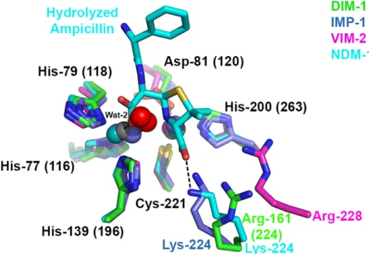
Fig 5. Active Site Grooves of B1 Metallo-β-Lactamases. Figure shows surfaces of A. DIM-1; B. GIM-1 (pdb accession 2YNT [48]) C. IMP-1 (1DDK [23]) and D. NDM-1 (3Q6X [41]) colored according to
electrostatic potential from red (- 0.5 V) to blue (+ 0.5 V). Hydrolyzed ampicillin bound to NDM-1 is shown as sticks (carbon atoms green, other atom colors as standard). This Figure was generated using CCP4MG [50]. doi:10.1371/journal.pone.0140059.g005
Five co-ordinate Zn1 sites have been observed in some other B1 MBL crystal structures,
notably those of IND-7 (
Fig 6C
, [
47
]) and reduced VIM-2 (
Fig 6D
, [
44
]). Notably, Yamaguchi
et al [
47
] used comparisons with structures of biomimetic copper complexes [
56
] to describe
the co-ordination geometry about IND-7 Zn1. Calculation of the structural parameter
τ ((β −
α)/60), where angle α refers to His79 (118) ND1—Zn1—His139 (196) NE2 and β to O (Wat2)
—Zn1—His77 (116) NE2, can be used to represent the distortion from square to trigonal
pyra-midal co-ordination geometry. (The value of
τ is considered to be 0 for an ideal square pyramid
and 1 for an ideal trigonal bipyramid). On this basis the IND-7, reduced VIM-2 and DIM-1
Zn1 sites can all be described as distorted trigonal bipyramidal, with values of
τ all above 0.85.
In all three structures Wat2 is relatively distant from Zn1 compared to other ligands, consistent
with proposals [
47
] that this defines the position occupied by the carbonyl oxygen of incoming
β-lactams and hence may be readily displaced when substrates bind. Superposition with
struc-tures of NDM-1 complexes with hydrolysed
β-lactams (e.g. 3Q6X; [
41
]
Fig 7
) indeed locates
DIM-1 Wat-2 in the approximate position of the C7 carboxylate formed on
β-lactam
hydroly-sis and thus supports this assertion.
The structural parameter
τ can also be used to describe the geometry of the Zn2 site. As
described above, defining angles
α as (Wat1 (Cl
-))—Zn2—His200 (263) NE2 and β as O (Wat3
Fig 6. DIM-1 Active Site and Comparison with Selected B1 Metallo-β-Lactamases. A. Stereogram of DIM-1 active site (dataset 1 structure). Carbon atoms are colored green, zinc ions gray, water molecules red, other atom colors as standard. Electron density map shown is 2|Fo|-|Fc|.ϕcalc, contoured at 1.0σ. B. Overlay
of DIM-1 active sites for the four independently refined molecules. Structures from dataset 1 are in darker, and dataset 2 in lighter, shades. Dashed lines show metal:ligand interactions in chain A of dataset 1. Note presence of additional water molecule (Wat-3; dataset 1 chain B) or chloride ions (dataset 2 structures) bound to Zn2 ion, and bridging chloride ion in dataset 2 chain A structure. C. Active site of IND-7 (3L6N [47]). Note five- and four-fold co-ordination of Zn1 and Zn2, respectively. D. Active site of VIM-2 (1KO3 [44]). Not five-fold co-ordination of both Zn2+ions. E. Active site of NDM-1 (3PSU [54]) Note four- and five-fold co-ordination of Zn1 and Zn2, respectively. This Figure was generated using Pymol.
(Cl
-))
—Zn2—Asp81 (120) OD2 gives values of τ ranging from 0.18 to 0.37 for the three chains
where Zn2 is five-co-ordinate. This indicates that the DIM-1 Zn2 site can be described as a
dis-torted square pyramid rather than the trigonal bipyramid observed in e.g. VIM-2 (
Fig 6D
pdb
1KO3 [
44
]). Under these criteria the Zn2 sites of NDM-1 (
Fig 6E
, pdb 3SPU [
54
]) and CcrA
Table 2. Metal:ligand distances (Å).
Structure Dataset 1 Dataset 2
Zn1:His77(116) 2.35a(2.19)b 2.16a(2.18)b Zn1:His79(118) 2.05 (2.34) 2.05 (2.16) Zn1:His139(196) 2.12 (2.32) 2.09 (2.07) Zn1:Wat1 1.81 (1.89) 2.26c(1.99) Zn1:Wat2 2.53 (2.40) 2.52 Zn2:Asp81(120) 2.33 (2.60) 2.03 (2.09) Zn2:Cys168(221) 2.16 (2.11) 2.19 (2.13) Zn2:His200(263) 2.24 (2.12) 2.15 (2.20) Zn2:Wat1 2.44 (2.37) 2.27c(2.75) Zn2:Wat3 (2.43) 2.47c(2.87)c Zn1:Zn2 3.97 (4.02) 4.22 (4.39) Wat-1:Asp81(120) 2.94 (3.24) 2.98 (3.04) aChain A.
bFigures in parentheses are for chain B. cRefined as a chloride ion
doi:10.1371/journal.pone.0140059.t002
Fig 7. Overlay of B1 Metallo-β-Lactamase Active Sites Showing Hydrolyzed β-Lactam Binding. Figure shows DIM-1, IMP-1 (1DDK [23], carbon, zinc and waters teal); VIM-2 (1KO3 [44], carbon, zinc and waters magenta) and NDM-1:ampicillin complex (3Q6X [41], carbon, zinc and waters cyan). Interactions of hydrolyzed ampicillin with NDM-1 are shown as dashed lines. Note differing positions of Arg-161 (224) (DIM-1), Lys-224 (IMP-1, NDM-1) and Arg-228 (VIM-2). This Figure was generated using Pymol.
2YNT and 2YNW [
48
]) and see recent near-atomic resolution structures of BcII (3.50 Å, 1.20
Å resolution, pdb id 4C09) and VIM-2 (3.51 Å, 1.29 Å resolution, pdb id 4BZ3). Increased
sep-arations have been observed for structures determined under more acidic conditions [
16
,
57
],
and the metal
—metal distance is also observed to increase in some complexes with hydrolyzed
β-lactams (e.g. NDM-1:ampicillin pdb 3Q6X 4.58 Å [
41
] compared to unliganded NDM-1
3.56
–3.97 Å pdb 3SPU), indicating that the MBL active site architecture is able to adapt to
accommodate bound ligands. It is notable that other structures in which, as is the case for
data-set 2, chloride ions are observed in the MBL active site (e.g. 1KO3 [
44
] or 2BG2 [
57
]) also
fea-ture relatively large separations between the two zinc ions (4.20 Å and 4.09 Å respectively).
Our observation of a variety of metal co-ordination arrangements in DIM-1, and the range of
architectures observed in the zinc sites of other enzymes, together highlight the flexibility of
co-ordination geometry about both metal ions of the B1 MBL active site (for a review see [
58
]).
This is also consistent with proposals that metal co-ordination geometry can change during
β-lactam binding and hydrolysis [
59
–
61
].
In common with some other mobile B1 MBLs (e.g. GIM-1 and TMB-1; [
7
,
13
]) DIM-1
fea-tures an arginine at residue 161 (224), rather than the lysine found at the equivalent position in
most other enzymes (e.g. NDM-1, IMP-1, SPM-1, IND-7). Structures of inhibitor and
β-lactam
complexes of the IMP-1 and NDM-1 enzymes [
17
,
23
,
41
], as well as evidence from directed
mutagenesis studies [
62
,
63
], implicate this lysine in substrate interactions via the invariant
car-boxylate at the
β-lactam C3 (penicillin) or C4 (cephalosporin) position. Structural comparisons
(
Fig 7
) suggest that DIM-1 Arg161 (224) NH1 might make similar contacts, although we note
that, relative to Zn2 (also implicated in binding substrate carboxylate) DIM-1 Arg161 (224)
NH1 is not identically located to Lys224 NZ of e.g. IMP-1 or NDM-1. Steady-state kinetic data
[
7
,
10
,
13
] show enzymes with Arg224 to be less efficient than other B1 MBLs against some
substrates, in particular penicillins (for which k
catvalues are notably reduced [
7
]). It is possible
that interactions involving Arg224, rather than Lys224, result in binding of some substrates in
orientations that are less optimal for hydrolysis.
In summary, the DIM-1 crystal structure presented here displays similarities and differences
when compared to those for other B1 MBLs. While the overall fold and binuclear zinc center
are common to all B1 MBLs, the DIM-1 structure highlights the variability evident in the active
site architecture around the L3 and L10 loops and in the identity and location of residues
(DIM-1 Arg161 (224)) likely to be involved in binding the
β-lactam C2/C3 carboxylate.
More-over, our data provide further evidence of variability in the co-ordination of both of the
cata-lytic zinc ions in B1 MBLs. Possession of a relatively plastic active site which differs
significantly between individual enzymes is one reason why MBLs remain challenging targets
for inhibitor design. The availability of structural information for enzymes, such as DIM-1,
whose active sites contain such distinguishing features and that, by virtue of their mobilization
and presence in pathogens, are a present challenge to clinical
β-lactam use, is thus a stepping
stone to development of broad spectrum inhibitors effective across the full range of MBLs.
Acknowledgments
We thank Diamond Light Source for access to beamline I03 (proposal number MX313) that
contributed to the results presented here, and Philip Hinchliffe, Ramya Salimraj and Martin
Challand for their careful reading of the manuscript.
Author Contributions
Conceived and designed the experiments: MPSB JS. Performed the experiments: MPSB.
Ana-lyzed the data: MPSB MK JS. Contributed reagents/materials/analysis tools: LP PN. Wrote the
paper: JS.
References
1. Cornaglia G, Giamarellou H, Rossolini GM. Metallo-beta-lactamases: a last frontier for beta-lactams? The Lancet infectious diseases. 2011; 11(5):381–93. doi:10.1016/S1473-3099(11)70056-1PMID:
21530894.
2. Galleni M, Lamotte-Brasseur J, Rossolini GM, Spencer J, Dideberg O, Frere JM. Standard numbering scheme for class B beta-lactamases. Antimicrob Agents Chemother. 2001; 45(3):660–3. PMID:
11181339.
3. Nordmann P, Naas T, Poirel L. Global spread of Carbapenemase-producing Enterobacteriaceae. Emerg Infect Dis. 2011; 17(10):1791–8. doi:10.3201/eid1710.110655PMID:22000347; PubMed Cen-tral PMCID: PMC3310682.
4. Castanheira M, Deshpande LM, Costello A, Davies TA, Jones RN. Epidemiology and carbapenem resistance mechanisms of carbapenem-non-susceptible Pseudomonas aeruginosa collected during 2009–11 in 14 European and Mediterranean countries. J Antimicrob Chemother. 2014. doi:10.1093/ jac/dku048PMID:24603963.
5. Edelstein MV, Skleenova EN, Shevchenko OV, D'Souza J W, Tapalski DV, Azizov IS, et al. Spread of extensively resistant VIM-2-positive ST235 Pseudomonas aeruginosa in Belarus, Kazakhstan, and Russia: a longitudinal epidemiological and clinical study. The Lancet infectious diseases. 2013; 13 (10):867–76. Epub 2013/07/13. doi:10.1016/s1473-3099(13)70168-3PMID:23845533.
6. Bonnin RA, Poirel L, Nordmann P. New Delhi metallo-beta-lactamase-producing Acinetobacter bau-mannii: a novel paradigm for spreading antibiotic resistance genes. Future Microbiol. 2014; 9(1):33–41. doi:10.2217/fmb.13.69PMID:24328379.
7. El Salabi A, Borra PS, Toleman MA, Samuelsen O, Walsh TR. Genetic and biochemical characteriza-tion of a novel metallo-beta-lactamase, TMB-1, from an Achromobacter xylosoxidans strain isolated in Tripoli, Libya. Antimicrob Agents Chemother. 2012; 56(5):2241–5. doi:10.1128/AAC.05640-11PMID:
22290947; PubMed Central PMCID: PMC3346670.
8. Pollini S, Maradei S, Pecile P, Olivo G, Luzzaro F, Docquier JD, et al. FIM-1, a new acquired metallo-beta-lactamase from a Pseudomonas aeruginosa clinical isolate from Italy. Antimicrob Agents Che-mother. 2013; 57(1):410–6. doi:10.1128/AAC.01953-12PMID:23114762; PubMed Central PMCID: PMC3535987.
9. Sekiguchi J, Morita K, Kitao T, Watanabe N, Okazaki M, Miyoshi-Akiyama T, et al. KHM-1, a novel plas-mid-mediated metallo-beta-lactamase from a Citrobacter freundii clinical isolate. Antimicrob Agents Chemother. 2008; 52(11):4194–7. doi:10.1128/AAC.01337-07PMID:18765691; PubMed Central PMCID: PMC2573105.
10. Poirel L, Rodriguez-Martinez JM, Al Naiemi N, Debets-Ossenkopp YJ, Nordmann P. Characterization of DIM-1, an integron-encoded metallo-beta-lactamase from a Pseudomonas stutzeri clinical isolate in the Netherlands. Antimicrob Agents Chemother. 2010; 54(6):2420–4. doi:10.1128/AAC.01456-09
PMID:20308383; PubMed Central PMCID: PMC2876379.
11. Deshpande LM, Jones RN, Woosley LN, Castanheira M. Retrospective molecular analysis of DIM-1 metallo-beta-lactamase discovered in Pseudomonas stutzeri from India in 2000. Antimicrob Agents Chemother. 2014; 58(1):596–8. doi:10.1128/AAC.01541-13PMID:24145536; PubMed Central PMCID: PMC3910751.
12. Leski TA, Bangura U, Jimmy DH, Ansumana R, Lizewski SE, Li RW, et al. Identification of blaOXA-(5) (1)-like, blaOXA-(5)(8), blaDIM-(1), and blaVIM carbapenemase genes in hospital Enterobacteriaceae isolates from Sierra Leone. J Clin Microbiol. 2013; 51(7):2435–8. doi:10.1128/JCM.00832-13PMID:
1096/fj.12-224014PMID:23363572; PubMed Central PMCID: PMC3633820.
17. King DT, Worrall LJ, Gruninger R, Strynadka NC. New Delhi metallo-beta-lactamase: structural insights into beta-lactam recognition and inhibition. J Am Chem Soc. 2012; 134(28):11362–5. doi:10.1021/ ja303579dPMID:22713171.
18. O'Callaghan CH, Morris A, Kirby SM, Shingler AH. Novel method for detection of beta-lactamases by using a chromogenic cephalosporin substrate. Antimicrob Agents Chemother. 1972; 1(4):283–8. PMID:4208895.
19. Laemmli UK. Cleavage of structural proteins during the assembly of the head of bacteriophage T4. Nature. 1970; 227(5259):680–5. PMID:5432063
20. Gorrec F. The MORPHEUS protein crystallization screen. J Appl Crystallogr. 2009; 42(Pt 6):1035–42. doi:10.1107/S0021889809042022PMID:22477774; PubMed Central PMCID: PMC3246824. 21. Otwinowski Z, Minor W. Processing of X-ray Diffraction Data Collected in Oscillation Mode. Method
Enzymol. 1997; 276:307–26.
22. McCoy AJ, Grosse-Kunstleve RW, Adams PD, Winn MD, Storoni LC, Read RJ. Phaser crystallographic software. J Appl Cryst. 2007; 40(4):658–74. doi:10.1107/S0021889807021206
23. Concha NO, Janson CA, Rowling P, Pearson S, Cheever CA, Clarke BP, et al. Crystal structure of the IMP-1 metallo beta-lactamase from Pseudomonas aeruginosa and its complex with a mercaptocarbox-ylate inhibitor: binding determinants of a potent, broad-spectrum inhibitor. Biochemistry. 2000; 39 (15):4288–98. PMID:10757977.
24. Emsley P, Lohkamp B, Scott WG, Cowtan K. Features and development of Coot. Acta Crystallogr D Biol Crystallogr. 2010; 66(4):486–501.
25. Murshudov GN, Vagin AA, Dodson EJ. Refinement of Macromolecular Structures by the Maximum-Likelihood Method. Acta Crystallogr D Biol Crystallogr. 1997; 53(3):240–55.
26. Winn MD, Ballard CC, Cowtan KD, Dodson EJ, Emsley P, Evans PR, et al. Overview of the CCP4 suite and current developments. Acta Crystallogr D Biol Crystallogr. 2011; 67(Pt 4):235–42. doi:10.1107/ S0907444910045749PMID:21460441; PubMed Central PMCID: PMC3069738.
27. Painter J, Merritt EA. Optimal description of a protein structure in terms of multiple groups undergoing TLS motion. Acta Crystallogr D Biol Crystallogr. 2006; 62(Pt 4):439–50. doi:10.1107/
S0907444906005270PMID:16552146.
28. Painter J, Merritt EA. TLSMD web server for the generation of multi-group TLS models. Journal of Applied Crystallography. 2006; 39:109–11. doi:10.1107/S0021889805038987
WOS:000234679400018.
29. Laskowski RA, MacArthur MW, Moss DS, Thornton JM. PROCHECK: a program to check the stereo-chemical quality of protein structures. J Appl Cryst. 1993; 26(2):283–91.
30. Chen VB, Arendall WB 3rd, Headd JJ, Keedy DA, Immormino RM, Kapral GJ, et al. MolProbity: all-atom structure validation for macromolecular crystallography. Acta Crystallogr D Biol Crystallogr. 2010; 66(Pt 1):12–21. Epub 2010/01/09. S0907444909042073 [pii] doi:10.1107/S0907444909042073
PMID:20057044; PubMed Central PMCID: PMC2803126.
31. Murphy TA, Catto LE, Halford SE, Hadfield AT, Minor W, Walsh TR, et al. Crystal structure of Pseudo-monas aeruginosa SPM-1 provides insights into variable zinc affinity of metallo-beta-lactamases. J Mol Biol. 2006; 357(3):890–903. PMID:16460758.
32. Krissinel E, Henrick K. Secondary-structure matching (SSM), a new tool for fast protein structure align-ment in three dimensions. Acta Crystallogr D Biol Crystallogr. 2004; 60(Pt 12 Pt 1):2256–68. PMID:
15572779.
33. Toney JH, Fitzgerald PM, Grover-Sharma N, Olson SH, May WJ, Sundelof JG, et al. Antibiotic sensiti-zation using biphenyl tetrazoles as potent inhibitors of Bacteroides fragilis metallo-beta-lactamase. Chem Biol. 1998; 5(4):185–96. PMID:9545432.
34. Toney JH, Hammond GG, Fitzgerald PM, Sharma N, Balkovec JM, Rouen GP, et al. Succinic acids as potent inhibitors of plasmid-borne IMP-1 metallo-beta-lactamase. J Biol Chem. 2001; 276(34):31913– 8. PMID:11390410.
35. Yamaguchi Y, Jin W, Matsunaga K, Ikemizu S, Yamagata Y, Wachino J, et al. Crystallographic investi-gation of the inhibition mode of a VIM-2 metallo-beta-lactamase from Pseudomonas aeruginosa by a mercaptocarboxylate inhibitor. J Med Chem. 2007; 50(26):6647–53. doi:10.1021/jm701031nPMID:
18052313.
36. Moali C, Anne C, Lamotte-Brasseur J, Groslambert S, Devreese B, Van Beeumen J, et al. Analysis of the Importance of the Metallo-beta-Lactamase Active Site Loop in Substrate Binding and Catalysis. Chem Biol. 2003; 10(4):319–29. PMID:12725860.
37. Yang Y, Keeney D, Tang X, Canfield N, Rasmussen BA. Kinetic properties and metal content of the metallo-beta-lactamase CcrA harboring selective amino acid substitutions. J Biol Chem. 1999; 274 (22):15706–11. PMID:10336469.
38. Scrofani SD, Chung J, Huntley JJ, Benkovic SJ, Wright PE, Dyson HJ. NMR characterization of the metallo-beta-lactamase from Bacteroides fragilis and its interaction with a tight-binding inhibitor: role of an active-site loop. Biochemistry. 1999; 38(44):14507–14. PMID:10545172.
39. Karsisiotis AI, Damblon CF, Roberts GC. Solution structures of the Bacillus cereus metallo-beta-lacta-mase BcII and its complex with the broad spectrum inhibitor R-thiomandelic acid. Biochem J. 2013; 456 (3):397–407. doi:10.1042/BJ20131003PMID:24059435; PubMed Central PMCID: PMC3898119. 40. Salsbury FR Jr., Crowley MF, Brooks CL 3rd. Modeling of the metallo-beta-lactamase from B. fragilis:
structural and dynamic effects of inhibitor binding. Proteins. 2001; 44(4):448–59. PMID:11484222. 41. Zhang H, Hao Q. Crystal structure of NDM-1 reveals a common {beta}-lactam hydrolysis mechanism.
FASEB J. 2011. doi:10.1096/fj.11-184036
42. Makena A, van Berkel SS, Lejeune C, Owens RJ, Verma A, Salimraj R, et al. Chromophore-linked sub-strate (CLS405): probing metallo-beta-lactamase activity and inhibition. ChemMedChem. 2013; 8 (12):1923–9. doi:10.1002/cmdc.201300350PMID:24166830.
43. Fabiane SM, Sohi MK, Wan T, Payne DJ, Bateson JH, Mitchell T, et al. Crystal structure of the zinc-dependent beta-lactamase from Bacillus cereus at 1.9 A resolution: binuclear active site with features of a mononuclear enzyme. Biochemistry. 1998; 37(36):12404–11. PMID:9730812.
44. Garcia-Saez I, Docquier J-D, Rossolini GM, Dideberg O. The three-dimensional structure of VIM-2, a Zn-beta-lactamase from Pseudomonas aeruginosa in its reduced and oxidised form. J Mol Biol. 2008; 375(3):604–11. PMID:18061205
45. Garcia-Saez I, Hopkins J, Papamicael C, Franceschini N, Amicosante G, Rossolini GM, et al. The 1.5-A structure of Chryseobacterium meningosepticum zinc beta-lactamase in complex with the inhibitor, D-captopril. J Biol Chem. 2003; 278(26):23868–73. PMID:12684522.
46. Concha NO, Rasmussen BA, Bush K, Herzberg O. Crystal structure of the wide-spectrum binuclear zinc beta-lactamase from Bacteroides fragilis. Structure. 1996; 4(7):823–36. PMID:8805566. 47. Yamaguchi Y, Takashio N, Wachino J, Yamagata Y, Arakawa Y, Matsuda K, et al. Structure of
metallo-beta-lactamase IND-7 from a Chryseobacterium indologenes clinical isolate at 1.65-A resolution. J Bio-chem. 2010; 147(6):905–15. doi:10.1093/jb/mvq029PMID:20305272
48. Borra PS, Samuelsen O, Spencer J, Walsh TR, Lorentzen MS, Leiros HK. Crystal structures of Pseudo-monas aeruginosa GIM-1: active-site plasticity in metallo-beta-lactamases. Antimicrob Agents Che-mother. 2013; 57(2):848–54. Epub 2012/12/05. doi:10.1128/aac.02227-12PMID:23208706; PubMed Central PMCID: PMCPMC3553699.
49. Brown NG, Horton LB, Huang W, Vongpunsawad S, Palzkill T. Analysis of the functional contributions of Asn233 in metallo-beta-lactamase IMP-1. Antimicrob Agents Chemother. 2011; 55(12):5696–702. doi:10.1128/AAC.00340-11PMID:21896903; PubMed Central PMCID: PMC3232802.
50. McNicholas S, Potterton E, Wilson KS, Noble ME. Presenting your structures: the CCP4mg molecular-graphics software. Acta Crystallogr D Biol Crystallogr. 2011; 67(Pt 4):386–94. doi:10.1107/
S0907444911007281PMID:21460457; PubMed Central PMCID: PMC3069754.
51. Badarau A, Page MI. Loss of enzyme activity during turnover of the Bacillus cereus beta-lactamase cat-alysed hydrolysis of beta-lactams due to loss of zinc ion. Journal of biological inorganic chemistry: JBIC: a publication of the Society of Biological Inorganic Chemistry. 2008; 13(6):919–28. doi:10.1007/ s00775-008-0379-2PMID:18449576.
52. Gonzalez JM, Meini MR, Tomatis PE, Medrano Martin FJ, Cricco JA, Vila AJ. Metallo-beta-lactamases withstand low Zn(II) conditions by tuning metal-ligand interactions. Nat Chem Biol. 2012; 8(8):698–700. doi:10.1038/nchembio.1005PMID:22729148; PubMed Central PMCID: PMC3470787.
53. Wang Z, Fast W, Benkovic SJ. On the mechanism of the metallo-beta-lactamase from Bacteroides fra-gilis. Biochemistry. 1999; 38(31):10013–23. PMID:10433708.
57. Davies AM, Rasia RM, Vila AJ, Sutton BJ, Fabiane SM. Effect of pH on the active site of an Arg121Cys mutant of the metallo-beta-lactamase from Bacillus cereus: implications for the enzyme mechanism. Biochemistry. 2005; 44(12):4841–9. PMID:15779910.
58. Karsisiotis AI, Damblon CF, Roberts GC. A variety of roles for versatile zinc in metallo-beta-lactamases. Metallomics: integrated biometal science. 2014; 6(7):1181–97. doi:10.1039/c4mt00066hPMID:
24696003.
59. Bicknell R, Schaffer A, Waley SG, Auld DS. Changes in the coordination geometry of the active-site metal during catalysis of benzylpenicillin hydrolysis by Bacillus cereus beta-lactamase II. Biochemistry. 1986; 25(22):7208–15. PMID:3099831.
60. Garrity JD, Bennett B, Crowder MW. Direct evidence that the reaction intermediate of metallo-beta-lac-tamase L1 is metal bound. Biochemistry. 2005; 44(3):1078–87. PMID:15654764.
61. Yang H, Aitha M, Marts AR, Hetrick AM, Bennett B, Crowder MW, et al. Spectroscopic and Mechanistic Studies of Heterodimetallic Forms of Metallo-ss-Lactamase NDM-1. J Am Chem Soc. 2014. doi:10. 1021/ja410376sPMID:24754678.
62. Materon IC, Palzkill T. Identification of residues critical for metallo-beta-lactamase function by codon randomization and selection. Protein Sci. 2001; 10(12):2556–65. PMID:11714924.
63. Yanchak MP, Taylor RA, Crowder MW. Mutational analysis of metallo-beta-lactamase CcrA from Bac-teroides fragilis. Biochemistry. 2000; 39(37):11330–9. PMID:10985778.