HAL Id: hal-01864127
https://hal.archives-ouvertes.fr/hal-01864127
Submitted on 29 Aug 2018
HAL is a multi-disciplinary open access archive for the deposit and dissemination of sci-entific research documents, whether they are pub-lished or not. The documents may come from teaching and research institutions in France or abroad, or from public or private research centers.
L’archive ouverte pluridisciplinaire HAL, est destinée au dépôt et à la diffusion de documents scientifiques de niveau recherche, publiés ou non, émanant des établissements d’enseignement et de recherche français ou étrangers, des laboratoires publics ou privés.
Physical forces determining the persistency and centring
precision of microtubule asters
Hirokazu Tanimoto, Jeremy Sallé, Louise Dodin, Nicolas Minc
To cite this version:
Hirokazu Tanimoto, Jeremy Sallé, Louise Dodin, Nicolas Minc. Physical forces determining the persis-tency and centring precision of microtubule asters. Nature Physics, Nature Publishing Group, 2018, 14 (8), pp.848 - 854. �10.1038/s41567-018-0154-4�. �hal-01864127�
Physical Forces Determining the Persistency and Centering Precision of
Microtubule Asters
Authors:
Hirokazu Tanimoto1,§, Jeremy Sallé1, Louise Dodin1 and Nicolas Minc1,*
Affiliations:
1 Institut Jacques Monod, CNRS UMR7592 and Université Paris Diderot, 15 rue Hélène Brion,
75205 Paris Cedex 13, France
§ Current affiliation: Department of Materials Science, Yokohama City University *Correspondence to: Nicolas Minc, nicolas.minc@ijm.fr
Abstract:
How large cellular structures move purposefully in a crowded and fluctuating cytoplasm remains unclear. In early embryos, microtubule asters can reach hundreds of micrometers and move at high speeds to find the geometrical center of large egg and blastomere cells, a process essential for the fidelity of cell division and development. Aster centration in these cells therefore represents a prime and ubiquitous example of directed cytoskeletal motion with acute
physiological constraints on precision. We found that positional fluctuations of migrating sea urchin sperm asters were small, anisotropic, and associated with the stochasticity of dynein-dependent forces moving the aster. Using in vivo magnetic tweezers to directly measure aster forces inside cells, we derived a linear aster force-velocity relationship and evidenced a spring-like active mechanism stabilizing aster transverse position. The large frictional coefficient and spring constant quantitatively accounted for the amplitude and growth characteristics of athermal positional fluctuations, demonstrating that aster mechanics ensure noise-suppression to promote persistent and precise centration. These findings define generic biophysical regimes of active cytoskeletal mechanics underlying the accuracy of cell division and early embryonic
Microtubule (MT) asters are star-shaped cytoskeletal structures composed of MT polymers radiating from an organizing center called the centrosome. They contribute to the spatial organization of crucial functions in eukaryotic cells, ranging from cell migration to nuclear centration and mitotic spindle orientation 1-3. One highly conserved property of MT asters is their ability to probe the geometrical boundaries of the cell to move and position themselves in the exact cell center. This was best highlighted in seminal in vitro work reconstituting aster growth and centration in microfabricated wells of a few microns in size. In those studies, pushing forces resulting from astral MT polymerization against the chamber wall 4,5 or pulling forces provided by MT minus-end directed dynein motors attached to the wall surface 6 allowed asters to target the chamber center.
A stereotypical and ubiquitous in vivo counterpart for aster centration occurs soon after
fertilization in most animal embryos7. In this context, the fertilizing sperm brings the male
pro-nucleus and its associated centrosomal material into the side of the egg, which results in the nucleation of a “sperm aster” that continuously grows and moves to the egg center. This
centration motion is critical to position the nucleus and subsequent spindle and division plane in the exact cell center. Contrary to in vitro situations, studies in systems including worm, frog, fish, and echinoderm embryos have suggested that aster centration in those cells may not
primarily involve MT polymerization or cortical dynein forces8-12. Rather, a prominent model is that most of the forces are provided by dynein motors working along astral MTs in bulk
cytoplasm 11,13-15. Dynein motors generate plus-end directed traction forces, probably as complexes with endomembrane components such as lysosome vesicles, yolk granules 13, via frictional interactions with the viscous cytoplasm. As longer MTs may associate with more dyneins, they may exert larger pulling forces on the centrosome. This length-dependent system, coupled to MT length asymmetries caused by cellular boundaries, provides a self-organization design for asters to target the cell center 11,13,14.
One outstanding physical problem implicated in aster centration in early embryos arises from the unusually large size of egg cells and early blastomeres 3,15. These cells are typically 10-100 times larger than somatic cells 16 or in vitro microchambers 6 , yet achieve aster centration on a
time-scale of only a few tens of minutes. Because of the physiological importance of aster centration in early embryos, these parameters set extreme constraints on motion persistency, speed, and centering precision. Given cytoplasmic crowding, extrinsic cellular noise, and intrinsic stochasticity of molecular elements involved 17,18, how moving asters may satisfy those constraints inside cells remains mysterious overall.
Here we exploited the centration of sea urchin sperm asters as a quantitative model system to derive the biophysical principles ensuring robust aster centration. By combining high-resolution tracking and direct intracellular aster force measurement, we find that aster motion is associated with large forces and small active positional fluctuations. This work demonstrates how aster mechanics may ensure noise-suppression to promote persistent and precise centration.
We first employed high-resolution microscopy (spatial resolution ~20 nm, temporal resolution ~50 ms, see supplementary information) (Fig. S1 and movie S1) to track the motion of male pro-nuclei attached to sperm MT asters in fertilized sea urchin eggs. We confirmed that aster speed was, on average, constant along the longitudinal centering direction (X-axis) and zero along the transverse axis (Y-axis) 11. Aster trajectory appeared smooth overall, but did exhibit some minor excursions away from the centering axis, which rapidly resorbed (Fig. 1, a and b).
To quantitatively examine the stochastic fluctuations around the mean motion, we detrended aster trajectory by subtracting its local velocity and computed the residual displacements, X and Y, as a function of lag time, t (eq. S2) 19. The probability distribution functions (PDFs) of X
and Y were nearly similar for t < 30 sec, and were well described by Gaussian functions (Fig. 1c). For t > 30 sec, while the PDF of Y kept a near-constant shape, the PDF of X appeared to deviate from a Gaussian and had a non-zero mean, which could reflect more complex behaviors such as higher order slow changes in the aster mean speed.
We characterized the statistical properties of aster fluctuations by plotting the second order moment of X and Y (mean-squared residual displacement, MSrD) as a function of t (Fig. 1d). Both MSrDs were flat at t < 1 sec, due to measurement noise. They then grew linearly above the measurement noise with a slope close to 1 for 1 < t < 30 sec, suggesting diffusive dynamics with similar diffusion coefficients along both axes: 𝐷𝑥 = 1.7*10-3 m2/sec and 𝐷𝑦 = 1.8*10-3 m2/sec. These results indicate the existence of uniform random forces which cause asters to
fluctuate in a diffusive manner.
The two MSrDs had different behaviors at t > 30 sec. While the longitudinal fluctuations kept growing, the transverse fluctuations saturated, likely reflecting a positional feedback that stabilizes aster trajectory transversely (Fig. 1d). Accordingly, these transverse fluctuations were well described by a random walk under spring-like restoration forces (Fig. 1d, inset) 20, so that
MSrD𝑦(𝛿𝑡) = 2𝜏𝐷𝑦(1 − 𝑒−𝛿𝑡/𝜏), (Eq. 1)
with a saturation amplitude 2𝜏𝐷𝑦 = 0.17m2 andsaturation timescale 𝜏 = 46 sec. This
corresponded to the typical size and restoration time of excursion events away from the centering axis. The saturation amplitude allowed for estimating a mean deviation distance of the aster from the centering axis of √2𝜏𝐷𝑦 = 0.41 µm, which was typically less than 1% of the cell radius, demonstrating a remarkable centration precision. Thus, aster fluctuations are small and anisotropic with characteristics determined by a balance between random forces and viscous dampening, and additional spring-like feedback in the transverse axis.
To discern if the observed fluctuations reflected thermal noise or active processes, we
manipulated cytoskeletal components using specific chemical inhibitors (Fig. 1, d and e and Fig. S3-4). To separately characterize the diffusive fluctuations from the effects of positional
exhibit purely diffusive behavior. Strikingly, addition of 100 M of Ciliobrevin D, which inhibits dynein activity and halts aster motion 11, decreased positional fluctuation amplitude by almost an order of magnitude. Thus dynein force-generation events which drive aster centration may also add active noise to this motion because of their stochastic nature. Actin
depolymerization with 20 µM Latrunculin B affected cell cortex and cell shape 11, but did not affect aster motion or fluctuation amplitude. This suggests that dynein drives aster fluctuations within the bulk cytoplasm not from the cortex, and by associating with cytoplasmic elements independent of actin 20,21. Finally, treatment with 20 µM Nocodazole to depolymerize MTs also halted aster motion, but caused the sperm nucleus to fluctuate more than in controls, suggesting that MTs may contribute to a large fraction of aster viscous drag. Importantly, in Nocodazole and Ciliobrevin D treatments, longitudinal and transverse MSrDs both grew diffusively for the entire timescale without saturation (Fig. 1d and fig. S4). This indicates that MTs and dynein contribute to the transverse spring-like feedback.
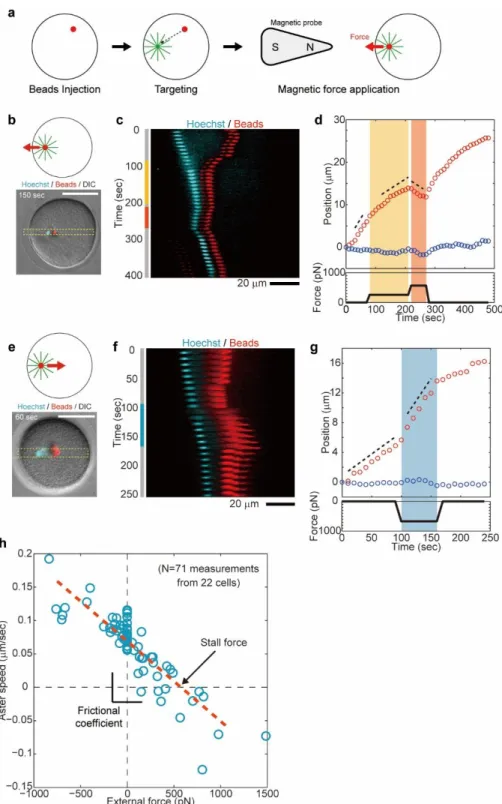
To understand how those kinetic properties may emerge from the mechanical properties of moving asters, we set out to directly measure the physical forces of asters inside cells. We modified a magnetic tweezer strategy recently used to measure forces for mitotic spindle maintenance in C. elegans 22 to be able to apply larger forces of several hundreds of pN to moving asters in arbitrary directions. These modifications rested on the injection of magnetic beads with highly persistent MT minus-end targeting activity, which rapidly aggregated and bound to the aster center upon fertilization in a MT- and dynein-dependent manner 23. Application of large calibrated forces was achieved by bringing a sharpened steel piece
connected to a magnet to a controlled distance from the internalized beads (Fig. 2a and figs. S5 to S9) (see supplementary information).
Application of external longitudinal forces against aster-centering motion caused aster speed to decrease in a dose-dependent manner (Fig. 2, b-d, Fig. S10 and movie S2). In these experiments, we focused on a short timescale response by computing aster speed within typically ~ 30 sec after force application, to minimize long-term aster adaptive responses. In Fig. 2b-d, we first applied a 260 pN force, which dropped aster longitudinal speed Vx by almost a factor 2 without
altering Vy. This force was subsequently increased to 570 pN which further decreased Vx to a
negative value, thus reverting aster motion. After the force was released, the aster restored a centering velocity close to its original value, suggesting that external forces did not grossly perturb aster organization. Conversely, applying external forces along the centering direction caused asters to accelerate (Fig. 2, e-g, Fig. S11 and movie S3). In Fig. 2e-g, we applied a 670 pN force in the positive X direction which increased aster speed by nearly 2-fold.
Systematic repetition of these measurements allowed derivation of an aster force-velocity relationship for a wide range of external forces, from +1500 pN to –700 pN (a positive force corresponds to a rear pull). Consequent changes in longitudinal aster speed Vx varied from -0.13
to 0.2 m/sec and collapsed into a single linear curve (Fig. 2h and fig. S12). These results indicate that aster motion is governed by a simple linear friction law, so that
𝑉𝑥 =1𝛾(𝐹𝑎𝑠𝑡𝑒𝑟− 𝐹𝑒𝑥𝑡𝑒𝑟𝑛𝑎𝑙). (Eq. 2)
Importantly, this linear relationship holds for external forces applied along and against aster centering motion, suggesting that contributions from compressive MT forces at the aster rear, close to the cortex, may be negligible in these experiments. Using those results, we determined an aster stall force which is equal to the aster endogenous force of 𝐹𝑎𝑠𝑡𝑒𝑟 = 580 +/- 21 pN, and a
frictional coefficient 𝛾 of 8400 +/- 280 pN*sec/m (+/- indicates the standard error in fitting parameters unless specified). Detached bead aggregates with a similar size to the male pro-nucleus moved much faster than asters under the same forces, indicating that most of this friction may be associated with MTs in the aster (fig. S6). These results demonstrate that the centering motion of sperm asters obeys a simple linear friction law involving large self-propelling forces and drag.
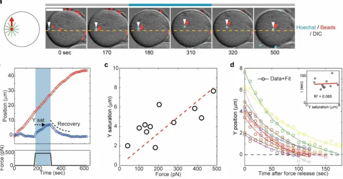
Fluctuation analyses in the transverse axis supported the existence of a spring-like feedback mechanism stabilizing aster position around the centering axis. To characterize this feedback, we applied magnetic forces perpendicular to the motion direction (Fig. 3,a and b, fig. S13 and movie S4). In Fig. 3a-b, we applied a 470 pN force in the Y positive direction for 140 sec. The external force did not affect aster motion along the X-axis, but caused a continuous drift in Y which eventually saturated ~7 µm away from the X-axis. Remarkably, after force cessation the Y position restored to its original value within tens of seconds (Fig. 3b). Computing the maximum Y-displacement at saturation as a function of various applied forces yielded a linear force-displacement curve (Fig. 3c). These results directly demonstrate the existence of a linear spring stabilizing aster position around the centration axis, with a spring constant of = 59 +/- 2.8 pN/m. The stiffness of this spring is ~4 times higher than in C. elegans 22, plausibly revealing different force-generation mechanisms. Accordingly, the transverse speed Vy following force
application was comparable to the changes in Vx in the longitudinal force experiments, ruling out
a major contribution of MT compressive forces to this transverse feedback. In addition, it has been shown that MT laser severing along the transverse axis in this system yields aster motion away from the site of ablation 11. These data support that this centering spring is mostly associated with MT pulling forces.
These transverse force experiments are consistent with a Kelvin–Voigt model, in which an elastic spring and viscous dashpot are connected in parallel 22 (Fig. 4). This model predicts that
the mean-squared displacement driven by internal random forces should saturate in an exponential manner, as observed in our fluctuation analysis, with a timescale equal to the relaxation timescale upon displacement by an external load. Accordingly, quantification of the recovery dynamics after force cessation revealed a restoring kinetic well described by an exponential relaxation with a single characteristic timescale, = 72 +/- 18 sec (+/- indicates standard deviation), independent of the initial Y-offset (Fig. 3d). This timescale was close to the saturation timescale observed in the transverse fluctuations ( = 46 sec), supporting the
consistency between our passive and active characterizations of the transverse positional feedback. Given that fluctuation saturation depended on MTs and dynein, these results suggest that the positional feedback maintaining the aster around the centering axis relies on dynein pulling forces on MTs.
These findings may be consistent with the length-dependent mechanism proposed to drive aster centration in sea urchins and other embryos 8-14,24-26. This system has the properties of an ‘active
spring’ with respect to cell geometry: a displacement away from the cell center yields a length imbalance on the two sides of the aster and creates a dynein-dependent restoration force proportional to the displacement 8. During aster centration, this spring is expected to function
only along the transverse axis, because MTs reach the cortex along this axis, while front MTs do not reach to the opposite side until the very end of centration (Fig. 4) 11. Using the simplest linear length-dependency for MT forces, 𝐹𝑀𝑇 = 𝛼𝐿𝑀𝑇, we can relate the spring constant 𝜅 to the
length-dependency factor, so that𝛼 = 𝜅 2⁄ . This analysis indicates that ~30 pN forces are generated per 1 m-depth region of the aster surface (corresponding to a volume of 5*103 m3 for an aster radius of 20 m). This suggests a lower bound of 100-200 dynein motors involved in moving and positioning these asters, much higher than in previous indirect estimates 13,27.
Aster mechanical properties along the two different axes appeared to be largely consistent. For asters to move to the cell center, front MTs should be longer than those at the back, because their growth is not restricted by the cortex. We recently estimated a difference in length between front and rear MTs of about 𝛿𝐿 ~ 10 µm 11. This would correspond to a net force of 𝐹
𝑎𝑠𝑡𝑒𝑟 = 𝛼𝛿𝐿 ~
300 pN; smaller yet close to the direct longitudinal force measurement calculated above. Furthermore, the relaxation timescale in the transverse force experiments allows for defining a frictional coefficient given by γ = κ ∙ τ = 4200 pN*sec/m. This value is comparable to but twice smaller than that obtained from direct measurements in the longitudinal axis. Because the time-window used to compute the frictional coefficient in the transverse axis (>100 sec) is larger than in the longitudinal axis (~30 sec), we envision that the differences in γ could reflect some shape changes of the aster, which could modify the effective drag over this longer timescale. Together, these findings indicate that the viscous dampening in the transverse position is essentially the same as the drag associated with aster longitudinal centering motion, and that length-dependent dynein-MT forces may account for both transverse feedback and net directed force.
The frictional coefficient of MT asters is remarkably high, 60 times larger than the reported value for static mitotic spindles in C. elegans 22. The linear relationship between aster force and
velocity suggests a viscous interaction between the aster-dynein complex and the cytoplasm. Based on the measured cytoplasmic viscosity of fertilized sea urchin eggs 28, this friction would amount to that of an object with a hydrodynamic radius of ~440 m, typically ~20 times larger than the aster physical radius. Such large friction may not be readily explained by internal structures of the aster. Given the high density of astral MTs, their association with
endomembranes such as the endoplasmic reticulum 15, an aster may be viewed as a sphere with low permeability 29,30. The frictional coefficient of asters is thus expected be close to that of a non-permeable counterpart, and much smaller than the sum of the drag of individual MTs. One possible source for such a large frictional coefficient is the confinement set by the cell boundary: the hydrodynamic interaction between the aster and cell boundaries could significantly reduce the hydrodynamic mobility of the aster. In support of this, recent quantitative hydrodynamic simulations suggest that even a moderate cell-confinement (aster/cell size ~ 0.5) can lead to a
10-30 times increase of aster frictional coefficient 30. Our experimental results may thus highlight the overlooked physical effect of cell-confinement on the mobility of intracellular structures.
Finally, we propose a simple model which explains how measured mechanical properties may account for aster positional fluctuations. Aster motion kinetics can be represented by one length-scale defined by dividing the diffusion coefficient by the mean speed, 𝐷𝑥/𝑉𝑥 = 20 nm. This small length-scale reflects the high persistency of aster motion. To illustrate how this characteristic length-scale may emerge from aster mechanics, we introduced a simple model for aster
longitudinal motion (Fig. 4 and Supporting text). In this model, a single force-generation event created by a moving dynein on a MT causes a fixed aster displacement, 𝛿𝑑, towards or away from the cell center. We assume that force-generation obeys a first-order reaction, (i.e. it is limited by either the binding or the activation of dynein), with a reaction rate 𝐾𝑓 (𝐾𝑟)
proportional to the front (rear) aster radius 𝐿𝑓 (𝐿𝑟). Using Poisson statistics 31, we can express 𝛿𝑑
as a function of 𝐷𝑥/𝑉𝑥 as (see supporting information):
𝛿𝑑 = 2𝐷𝑥
𝑉𝑥
𝐾𝑓−𝐾𝑟
𝐾𝑓+𝐾𝑟. (Eq. 3).
Assuming force balance between the aster and a single dynein-cargo complex, and using typical cargo vesicle parameters (radius r ~ 0.5 m and run-length l ~ 5 m 11) and aster shape
asymmetry 𝐿𝑟⁄ ~ 0.8 𝐿𝑓 11, this model predicts an aster hydrodynamic radius as R = (𝑙 ∙ 𝑟) 𝛿 𝑑
⁄ ~620 m, which is comparable to our direct measurement of ~440 m. This result demonstrates how the large aster frictional coefficient may suppress the motion error caused by dynein active fluctuations.
In conclusion, based on the comprehensive characterization of aster kinetics and forces, we find that aster centration in large embryos is extremely precise, significantly more so than in well-controlled in vitro aster centration assays4-6, and that this precision is achieved by unusually large endogenous forces and friction. Based on the measured frictional coefficient, we estimate that the cell dedicates an energy of at least γ𝑉2 ~ 1000 ATP molecules/sec for centering MT asters.
This large frictional coefficient enables asters to take an ensemble average of stochastic dynein force generation events and ensures motion persistency. The transverse feedback further suppresses fluctuations and maintains aster trajectory along a precise axis related to cell geometry. As demonstrated by external force applications, this active feedback can bring back the aster along its centering trajectory even after accidental deviations larger than those caused by intrinsic dynein fluctuations. Most fertilizing embryos are associated with large rotational flows, shape changes and other cytoplasmic re-organization7. Our results suggest that sperm MT asters may be equipped with near-optimal physical designs to stabilize their motion in such an unfavorable environment to rapidly and precisely target the geometrical center of the large egg cell.
References:
1 Bornens, M. The centrosome in cells and organisms. Science 335, 422-426, doi:10.1126/science.1209037 335/6067/422 [pii] (2012).
2 Tang, N. & Marshall, W. F. Centrosome positioning in vertebrate development. J Cell Sci 125, 4951-4961, doi:10.1242/jcs.038083
125/21/4951 [pii] (2012).
3 Mitchison, T. et al. Growth, interaction, and positioning of microtubule asters in extremely large vertebrate embryo cells. Cytoskeleton (Hoboken), doi:10.1002/cm.21050 (2012).
4 Holy, T. E., Dogterom, M., Yurke, B. & Leibler, S. Assembly and positioning of microtubule asters in microfabricated chambers. Proc Natl Acad Sci U S A 94, 6228-6231 (1997).
5 Faivre-Moskalenko, C. & Dogterom, M. Dynamics of microtubule asters in microfabricated chambers: the role of catastrophes. Proc Natl Acad Sci U S A 99, 16788-16793, doi:10.1073/pnas.252407099
252407099 [pii] (2002).
6 Laan, L. et al. Cortical Dynein Controls Microtubule Dynamics to Generate Pulling Forces that Position Microtubule Asters. Cell 148, 502-514, doi:10.1016/j.cell.2012.01.007 (2012).
7 Gilbert, S. Developmental Biology. 9th edition., Vol. 9th Ed (Sinauer Associates, 2010).
8 Hamaguchi, M. S. & Hiramoto, Y. Analysis of the Role of Astral Rays in Pronuclear Migration in Sand Dollar Eggs by the Colcemid‐UV Method. Development, growth & differentiation 28, 143-156 (1986). 9 Kimura, A. & Onami, S. Computer simulations and image processing reveal length-dependent pulling force
as the primary mechanism for C. elegans male pronuclear migration. Dev Cell 8, 765-775, doi:S1534-5807(05)00095-X [pii]
10.1016/j.devcel.2005.03.007 (2005).
10 Longoria, R. A. & Shubeita, G. T. Cargo transport by cytoplasmic dynein can center embryonic centrosomes. PLoS ONE 8, e67710 (2013).
11 Tanimoto, H., Kimura, A. & Minc, N. Shape-motion relationships of centering microtubule asters. J Cell
Biol 212, 777-787, doi:10.1083/jcb.201510064
jcb.201510064 [pii] (2016).
12 Wuhr, M., Tan, E. S., Parker, S. K., Detrich, H. W., 3rd & Mitchison, T. J. A model for cleavage plane determination in early amphibian and fish embryos. Curr Biol 20, 2040-2045,
doi:10.1016/j.cub.2010.10.024 S0960-9822(10)01288-1 [pii] (2010).
13 Minc, N., Burgess, D. & Chang, F. Influence of cell geometry on division-plane positioning. Cell 144, 414-426, doi:S0092-8674(11)00017-1 [pii]
10.1016/j.cell.2011.01.016 (2011).
14 Pierre, A., Salle, J., Wuhr, M. & Minc, N. Generic Theoretical Models to Predict Division Patterns of Cleaving Embryos. Dev Cell 39, 667-682, doi:S1534-5807(16)30830-9 [pii]
10.1016/j.devcel.2016.11.018 (2016).
15 Wuhr, M., Dumont, S., Groen, A. C., Needleman, D. J. & Mitchison, T. J. How does a millimeter-sized cell find its center? Cell Cycle 8, 1115-1121, doi:8150 [pii] (2009).
16 Zhu, J., Burakov, A., Rodionov, V. & Mogilner, A. Finding the cell center by a balance of dynein and myosin pulling and microtubule pushing: a computational study. Mol Biol Cell 21, 4418-4427, doi:10.1091/mbc.E10-07-0627
E10-07-0627 [pii] (2010).
17 Fulton, A. B. How crowded is the cytoplasm? Cell 30, 345-347, doi:0092-8674(82)90231-8 [pii] (1982). 18 Brangwynne, C. P., Koenderink, G. H., MacKintosh, F. C. & Weitz, D. A. Cytoplasmic diffusion:
molecular motors mix it up. J Cell Biol 183, 583-587, doi:10.1083/jcb.200806149 jcb.200806149 [pii] (2008).
19 Winkler, F. et al. Fluctuation Analysis of Centrosomes Reveals a Cortical Function of Kinesin-1. Biophys J
109, 856-868, doi:10.1016/j.bpj.2015.07.044
20 Pecreaux, J. et al. The Mitotic Spindle in the One-Cell C. elegans Embryo Is Positioned with High Precision and Stability. Biophys J 111, 1773-1784, doi:S0006-3495(16)30800-1 [pii]
10.1016/j.bpj.2016.09.007 (2016).
21 Almonacid, M. et al. Active diffusion positions the nucleus in mouse oocytes. Nat Cell Biol 17, 470-479, doi:10.1038/ncb3131
ncb3131 [pii] (2015).
22 Garzon-Coral, C., Fantana, H. A. & Howard, J. A force-generating machinery maintains the spindle at the cell center during mitosis. Science 352, 1124-1127, doi:10.1126/science.aad9745
352/6289/1124 [pii] (2016).
23 Hamaguchi, M. S., Hamaguchi, Y. & Hiramoto, Y. Microinjected polystyrene beads move along astral rays in sand dollar eggs. Development, growth & differentiation 28, 461-470 (1986).
24 Kimura, K. & Kimura, A. Intracellular organelles mediate cytoplasmic pulling force for centrosome centration in the Caenorhabditis elegans early embryo. Proc Natl Acad Sci U S A 108, 137-142, doi:1013275108 [pii]
10.1073/pnas.1013275108 (2011).
25 Shinar, T., Mana, M., Piano, F. & Shelley, M. J. A model of cytoplasmically driven microtubule-based motion in the single-celled Caenorhabditis elegans embryo. Proc Natl Acad Sci U S A 108, 10508-10513, doi:10.1073/pnas.1017369108
1017369108 [pii] (2011).
26 Barbosa, D. J. et al. Dynactin binding to tyrosinated microtubules promotes centrosome centration in C. elegans by enhancing dynein-mediated organelle transport. PLoS Genet 13, e1006941,
doi:10.1371/journal.pgen.1006941 PGENETICS-D-17-00891 [pii] (2017).
27 Grill, S. W., Howard, J., Schaffer, E., Stelzer, E. H. & Hyman, A. A. The distribution of active force generators controls mitotic spindle position. Science 301, 518-521, doi:10.1126/science.1086560 301/5632/518 [pii] (2003).
28 Hiramoto, Y. Mechanical properties of the protoplasm of the sea urchin egg. II. Fertilized egg. Exp Cell
Res 56, 209-218 (1969).
29 Nazockdast, E., Rahimian, A., Needleman, D. & Shelley, M. Cytoplasmic flows as signatures for the mechanics of mitotic positioning. Mol Biol Cell 28, 3261-3270, doi:10.1091/mbc.E16-02-0108 mbc.E16-02-0108 [pii] (2017).
30 Nazockdast, E., Rahimian, A., Zorin, D. & Shelley, M. A fast platform for simulating semi-flexible fiber suspensions applied to cell mechanics. J Comput Phys 329, 173-209 (2017).
31 Svoboda, K., Mitra, P. P. & Block, S. M. Fluctuation analysis of motor protein movement and single enzyme kinetics. Proc Natl Acad Sci U S A 91, 11782-11786 (1994).
Supplementary Materials:
Materials and Methods Supporting text
Supporting figures and captions S1 to S13 Captions for Movies S1 to S4
Movies S1 to S4
Additional information:
Supplementary information is available in the online version of the paper. The experimental protocol was approved by Isabelle Le Parco, head of the animal facility at Institut Jacques Monod. Correspondence and requests for raw data, materials, and codes should be addressed to N. M.
Acknowledgements:
The authors acknowledge M. Coppey and J. Azimzadeh for technical support, and S. Dmitrieff, T. Strick, M. Piel, M. Thery and K. Laband for careful reading of the manuscript. This research was supported by the CNRS and grants from the “Mairie de Paris emergence” program, the FRM “amorçage” grant AJE20130426890 and the European Research Council (CoG Forcaster N° 647073).
Author contributions:
H.T., L.D. and J.S. performed experiments. H.T. analyzed the data and developed the model. H.T. and N.M. designed the research and wrote the manuscript.
Fig. 1. Fluctuation analysis of centering MT asters. (a and b) High-resolution tracking of sperm
MT asters during the constant speed centration phase. The representative 3 min trajectory corresponds to 3600 time points. Aster XY position was defined using X as the centering axis. (c) PDFs of residual displacements along X and Y for different t. Bold lines are best-fit Gaussian distributions. (d) X and Y MSrDs for control and Ciliobrevin D-treated samples plotted as a function of t in log scale. The broken line indicates a slope of 1. Inset: Transverse MSrD (blue curve) of controls superimposed with a fit of the model from Eq. 1 (broken line), plotted on a linear scale. The absolute magnitude of the residual error between the fit and the data is also depicted (green curve). (e) Contributions of cytoskeletal components to aster positional fluctuations. The fluctuation amplitude was characterized by computing the MSrD at t=5 sec.
Fig. 2. Force-velocity relationships of MT asters. (a) Aggregates of injected magnetic beads
were targeted to the aster center to directly apply magnetic forces to centering asters. (b-g) External magnetic forces were applied to asters either against (b-d) or along (e-g) the centering direction. The 1D kymographs (c and f) and time-evolution of aster XY positions (d and g) show how applied forces consistently change aster longitudinal speed. (h) Aster longitudinal speed Vx plotted as a
function of external force amplitude. N=71 measurements from 22 cells. The red broken line indicates a linear fit.
Fig. 3. Direct demonstration of a transverse feedback stabilizing asters along their centering direction. (a and b) External forces were applied orthogonally to the centering direction along the
Y-axis for 140 sec. The applied force causes a drift towards the magnet tip. (c) Aster Y saturation (shown in b) plotted as a function of external force amplitude. N=10 measurements from 10 cells. The spring constant κ=59 pN/m was determined from the slope of the linear fit (red broken line).
(d) Recovery dynamics of aster Y position. Aster Y position after force cessation was plotted as a
function of time. N=10 measurements from 10 cells. Solid lines indicate best fits with exponential function (eq. S6), yielding a mean recovery timescale τ = 72 +/- 18 sec. Inset: Recovery timescale plotted as a function of Y saturation. The correlation analysis indicates that there is no correlation between the two variables.
Fig. 4. Aster mechanics ensure persistent and precise aster centration. The large frictional
coefficient of asters suppresses active fluctuation along the longitudinal axis. This process was analyzed using a simple Poisson model, in which a single dynein-force generation event causes an aster step motion (see main and supporting text). Along the transverse axis, fluctuations are further suppressed by a dynein-dependent feedback mechanism, which stabilizes the centering direction with respect to cell geometry.