HAL Id: hal-03165653
https://hal.archives-ouvertes.fr/hal-03165653
Submitted on 23 Mar 2021
HAL is a multi-disciplinary open access
archive for the deposit and dissemination of sci-entific research documents, whether they are pub-lished or not. The documents may come from teaching and research institutions in France or abroad, or from public or private research centers.
L’archive ouverte pluridisciplinaire HAL, est destinée au dépôt et à la diffusion de documents scientifiques de niveau recherche, publiés ou non, émanant des établissements d’enseignement et de recherche français ou étrangers, des laboratoires publics ou privés.
Co-ordination of reaching movements in patients with a
cervical spinal cord injury.
Gilles Hoffmann, Isabelle Laffont, Agnès Roby-Brami
To cite this version:
Gilles Hoffmann, Isabelle Laffont, Agnès Roby-Brami. Co-ordination of reaching movements in pa-tients with a cervical spinal cord injury.. Cahiers de Psychologie Cognitive - Current Psychology of Cognition, Marseille : ADRSC, 2002, 21 (2-3), pp.305-340. �hal-03165653�
1
Coordination of reaching movements
in patients with a cervical spinal cord injury
Published in:
Cahiers de Psychologie Cognitive / Current Psychology of Cognition
2002, 21 (2-3), 305-340
Gilles Hoffmann,1 Isabelle Laffont, 1,2 and Agnes Roby-Brami 11 1. CNRS & Université René Descartes, Paris, France 2. Hôpital Raymond Poincaré, 92380 Garches, France
Abstract
The aim of this work is to study the mechanisms of motor adaptation to a perturbation of the mechanical conditions of the execution of an action. For this purpose, we analysed the joint kinematics of prehension movements performed by tetraplegic patients. In these patients, movement perturbation resulted from the extensive paralysis of the upper limb due to a lesion of the cervical spinal cord, as well as from the effect of a musculotendinous transfer that some patients underwent in order to have the function of paralysed muscles restored. The prehension movements were recorded by means of electromagnetic field captors fixed to the subjects’ upper limb. Tetraplegic patients were able to efficiently produce prehension movements, which shows that they have adapted to the new dynamic conditions of their upper limb. They are led to develop new motor strategies in order to compensate for the loss of the function of the paralysed muscles. The most manifest of these strategies is the grasping of objects by tenodesis. This strategy imposes that the action of prehension, which is planned according to the properties of the subject’s body and to the constraints of the task, is performed in a sequential way in patients, rather than in parallel as it has been shown in healthy subjects. Moreover, the smooth hand velocity profile observed in tetraplegic patients suggests that their central nervous system (CNS) has considered the dynamic constraints of the upper limb in order to produce an efficient prehension movement. The analysis of joint coordination suggests that for the prehension movement, the CNS controls joint rotations, rather than hand displacement or joint torques. Finally, the relative preservation of the shoulder movements in all the patients, as well as the similarity of elbow movements between operated patients and non-operated patients suggests that the shoulder is the motor of elbow extension during the approach of the object, and that the function of the triceps muscle may be to stabilise the elbow in order to facilitate its control.
2
INTRODUCTION
A traumatic injury at the cervical level of the human spinal cord results in a sensorimotor impairment of the four limbs, called tetraplegia. A major consequence of a traumatic spinal cord lesion is the interruption of the descending motor pathways that convey motor commands from the brain to the motor neurons of the effector muscles. As a consequence, the muscles that are innervated by motor neurons located in the metameres1 that are situated below the level of the injury are paralysed.
The degree of paralysis of the upper limb muscles depends on the precise location of the spinal cord injury, the higher the location of the lesion the more important the paralysis. For example, a complete lesion at the C6 level induces a paralysis of the intrinsic hand muscles (C8), of the triceps brachii, the main extensor of the elbow, and of the wrist flexors (C7), whereas the wrist extensors (C6), the elbow flexors (C5) and the shoulder muscles are preserved. However, despite this extensive impairment of muscle function, the tetraplegic patients are able to use their upper limb in a relatively efficient way in daily life, owing to an active and prolonged rehabilitation (Welch, Lobley, O‘Sullivan, & Freed, 1986). In particular, they are able to reach for and to grasp light objects. In order to limit the functional sequelae of the spinal cord injury, a surgical procedure of musculotendinous transfer is now commonly proposed to these patients (review in Revol, Briand, Cormerais, Pedelucq, Busner, & Servant, 1997). This procedure is based on the large muscular redundancy of the upper limb. It consists in a modification of the anatomical insertion of the distal tendon of a healthy muscle, in order to compensate the paralysis of another muscle. After a C6 level spinal injury, the first step of the procedure is to “reanimate" the triceps brachii by means of a transfer of either the posterior deltoid (a shoulder muscle) or the biceps brachii (an elbow flexor muscle). This operation is followed by an intensive three months period of rehabilitation. Two more surgeries are then needed to enhance hand function. Clinical evaluations have shown that the patients are able to contract their muscle in the new anatomical position and function. After surgery, the ability of the upper limb is improved and the patients are in general satisfied with the functional outcome (Revol et al., 1997).
The upper limb function in tetraplegic patients is a pertinent experimental paradigm for investigating the mechanisms of sensorimotor adaptation of upper limb movements to a change in the limb dynamics. Firstly, after injury, paralysed muscles can no longer be voluntarily contracted, so that the dynamic conditions under which movements of the upper limb occur are profoundly perturbed. Secondly, the musculotendinous transfer also results in a profound dynamic perturbation of the conditions of movement, as the dynamic effect of the force produced by the transferred muscle no longer is its original one. As noted above, despite these important dynamic perturbations, tetraplegic patients are able to successfully produce some upper limb movements of everyday life. However, there are little fundamental studies of motor function in tetraplegic patients. Most of them mainly analysed the electr0myographic (EMG) activity of the transferred muscle and concluded that the transferred muscle could adopt very variable patterns of activity, corresponding to the original or to the new anatomical position (Illert, Trauner, Weller, & Wiedemami, 1986).
We therefore decided to study prehension movements made by tetraplegic patients in the most "natural" possible context, in order to analyse the mechanisms of this adaptation and to better
1 The human spinal cord is segmented into twenty-nine metameres: eight cervical Cl-C8, twelve thoracic
Tl-T12, five lumbar L1-L5, four sacral Sl- S4. The muscles of the upper limb are innervated by the cervical segments. The motor neurons corresponding to a particular muscle are gathered in a localised nucleus, which is located within one or several adjacent metameres.
3
understand how the central nervous system (CNS) controls movements of the upper limb. For this purpose, we performed a kinematic analysis of prehension during non-constrained reaching to grasp movements in tetraplegic patients before and after musculotendinous transfer. We also aimed at developing quantitative methods to assess the effects of a musculotendinous transfer that could be usable in clinical practice.
METHODS Participants
Experiments were carried out at the Service de Médecine Physique et de Réadaptation at the Hopital Raymond Poincare in Garches. Eleven right-handed tetraplegic patients participated in the present study. They were nine men and two women, were aged between 24 and 34 years, and had sustained a traumatic cervical spinal cord injury between 7 and 120 months ago. Table l summarises the clinical data. The condition of a muscle can be assessed by an analytical examination, which is called "testing" in clinical practice. In short, subjects are asked to voluntarily contract the muscle while the experimenter evaluates the outcome of this contraction by a score on a five-point scale2 (Tobis &
Hong, 1990).
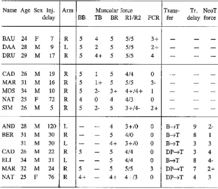
Table 1: Personal and clinical data of tetraplegic patients.
Thick horizontal lines separate the three groups of patients: top: group A patients, middle: group B patients without musculotendinous transfer, bottom: group B patients with transfer. Delay with regard to spinal cord injury and to transfer is
indicated in months. Principle of muscle force evaluation is explained in the text. The nature of the musculotendinous transfer (X—>T) indicates which muscle X has been transferred to the paralysed triceps. Inj. delay: injury delay; DP: Deltoideus posterior; BB: Biceps brachii; TB: Triceps brachii; BR: Brachio radialis; R1: Extensor carpi radialis longus; R2:
Extensor carpi radialis brevis; FCR: Flexor carpi radialis; NeoTriceps (neoT) refers to muscle X after musculotendinous transfer.
2 O: no contraction; 1: contraction, but no movement; 2: movement, but only if the effect of gravity is
suppressed by the posture of the limb; 3: movement possible against gravity; 4: movement possible against a moderate resistance; 5: normal movement.
4
Eight patients (AND, BER, CAD, ELI, MAR, MOS, NAT, SIM) had a C6 level lesion with an initially almost complete paralysis of the triceps brachii (score of 1~2). Five of these eight patients (CAD, MAR, MOS, NAT, SIM) were studied before surgery. They were unable to voluntarily extend their elbow, and voluntary contraction of the triceps brachii merely resulted in a palpable contraction of the muscle. Triceps paralysis is an indication for a surgical musculotendinous transfer, which was proposed to these patients. Six of the eight patients (AND, BER, CAD, ELI, MAR, NAT) were studied after surgery, three of them having been studied before surgery as well (CAD, MAR, NAT). The transfer was performed either on the right or on the left upper limb, or on both of them in one patient (BER). In four patients, the biceps brachii was the transferred muscle and in three patients it was the posterior deltoid. After surgery, patients improved their ability to voluntarily extend their elbow, The manual muscle test showed that all of them, except one (AND) were able to at least partly extend their elbow against gravity (score of 2+ or more). Three other patients (BAU, DAA, DRU) had a slightly lower level lesion (C6-C7). Their triceps brachii was relatively well preserved, so as to allow them to produce a voluntary extension of their elbow (score of 2-4). There was no indication for a musculotendinous transfer onto the triceps muscle for these C6-C7 patients. The studies of patients after musculotendinous transfer at the elbow were carried out while the surgeries aimed at improving their hand function had not yet been performed. Five healthy right-handed subjects participated in the study. They were aged between 22 and 52. None of them had neurological or orthopaedic disorders. All of them had their right arm studied.
Movement recording apparatus
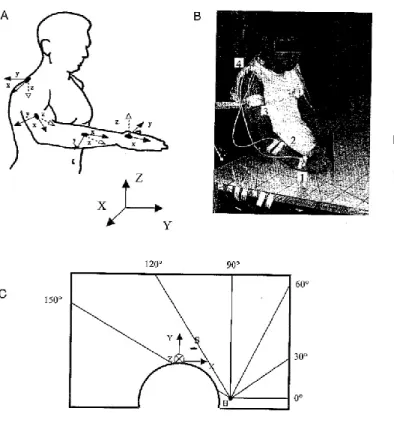
Movements were analysed by means of a three-dimensional kinematic analysis method: the Spatial Tracking System (STS). This method is based upon the utilisation of electromagnetic field receivers (Fastrack by Polhemus) and allows measuring both the position and the orientation of four receivers fixed to the subject’s arm, with regard to a steady reference, with a 30 Hz sample frequency (Figure 1A). Position is measured in Cartesian coordinates (x, y, z). Orientation is measured in nautical Euler’s angles (azimuth, elevation, roll). Azimuth is rotation about the vertical axis of the global reference frame, elevation is rotation in the vertical plane, which has been previously rotated by azimuth, and roll is rotation about the main axis within the limb segment. The four receivers were positioned in the following way: one on the dorsal side of the hand (receiver l), one on the dorsal side of the forearm (receiver 2), one on the lateral side of the upper arm (receiver 3), one on the acromion (receiver 4) (Figures lA and lB).
Experimental set-up
Subjects were seated in front of a horizontal experimental table, of a fixed height and with a circular cut-out part to allow the subject’s wheelchair to approach the table (Figure 1B,C). The steady STS reference was attached under the table, in front of the subject. Six lines were drawn on the surface of the table, converging at the point that corresponded to the vertical projection on the table of the subject’s centre of the humerus head of the studied arm (Figure 1C). The humerus is the bone of the upper arm, and its head is at the centre of the main shoulder joint which connects the scapula and the humerus. The six lines on the table were oriented in directions 0°, 30°, 60°, 90°, 120°, 150°, respectively, with regard to the X-axis of the reference frame, when the right arm was studied. When the left arm was studied, the set-up of the table was symmetrical. The place of the object to be grasped was normalised according to the subject's morphology: it corresponded to the point of vertical projection on the table of the head of the metacarpal bones (that is, of the centre of the hand), the
5
arm being completely stretched along the line. A red point on the table marked the hand’s starting position during the task, which was the same for all subjects (Figure 1C, position indicated by S). The object to be grasped was a cardboard cone 10 cm high and 5 cm in diameter at its base. It was set on lop of a support 10 cm high, in order to be positioned above the surface of the table, so as to keep patients from grasping the cone by sliding their arm or hand on the table. Subjects were seated either in their own wheelchair (patients) or in a standardised wheelchair (healthy subjects). Lateral trunk movements were avoided by using wedges placed under the armpits. The trunk was stabilised in its anterior part by a strap on the chest. The contralateral arm was immobilised. These precautions were destined to prevent patients from producing compensatory movements of the trunk and or contralateral arm.
Figure 1. Experimental set-up. IA. Disposition of the recording receivers on the upper limb. X, Y, Z is the global coordinate frame of the steady STS reference, x, y, z are the coordinate frames of the recording receivers. 1B. Photograph of a patient installed for the experiment (without the
stabilizing strap on the chest). Each receiver is labelled by a number: I hand, 2 forearm, 3 upper
arm, 4 acromion. Photograph: Isabelle Laflont. I C. Bird ’s eye view of the experimental table. H." point of vertical projection of the centre of the humerus head on the table, S: starting point for
movements. X, Y, Z: global coordinate frame of the steady ST S reference. The set-up of the experimental table as it is shown here is the one
that was used when the subject's right arm was studied. When the left arm was studied, the
set-up was symmetrical of the present one.
Procedure
At the beginning of each trial, the subject’s hand rested on the starting point on the table. The task consisted in fetching the cone from the starting point onwards, and to bring it back to the starting point. The cone was placed on one of the six lines. Each subject made four to five trials in one direction, then passed on to another direction. Directions were studied in the following order: 0°, 60°, 120°, 150°, 90°, 30°. The beginning of each trial was given by an acoustic signal. No time or velocity constraints were imposed.
Data processing
The data collected by the STS system allows studying the kinematics of the arm in terms of rotation angles at the joints. The accuracy of this method for studying joint kinematics has been demonstrated by experimental studies (Biryukova, Roby-Brami, Frolov, & Mokhtari, 2000; Karduna, McClure, Michener, & Sennett, 2001). The upper limb joints are the shoulder, the elbow, and the wrist. The shoulder is a complex joint since the bone of the upper arm (the humerus) is connected to the thorax
6
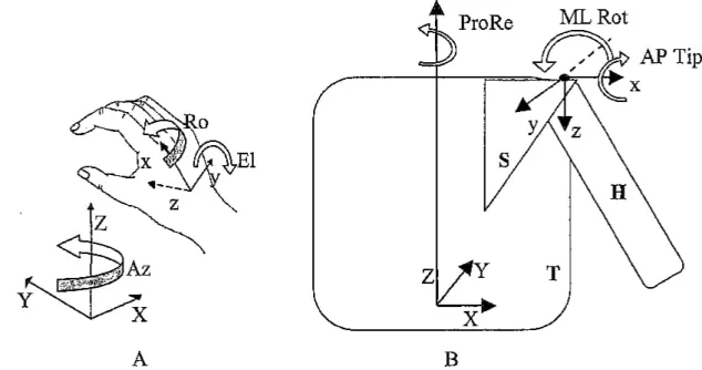
by the scapula and the clavicula, Thus, there are at least two distinct joints at the shoulder: the glenohumeral joint connecting the scapula and the upper arm, and the thoracoscapular joint connecting the thorax and the scapula. As the thorax was steady with regard to the system‘s reference, the receiver placed on the acromion (a bony landmark at the upper part of the scapula, above the shoulder) allowed precisely measuring the rotations of the scapula with regard to the thorax (thoracoscapular joint) (Karduna et al., 2001). Azimuth measured protraction (forward) — retraction (backward) of the scapula, elevation measured medial (upward) — lateral (downward) rotation, roll measured anterior — posterior tipping (Figure 2A). The definitions and order of rotations in the thoracoscapular joint were in keeping with the standards described in the literature (van der Helm, 1997; Zatsiorsky, 1998). The articular angles calculation method for the upper limb utilises a biomechanical model of the arm, consisting of a system of three segments (the hand, the forearm, the upper arm) with seven degrees of freedom (dof): the three dof between the scapula and the upper arm (the glenohumeral joint), flexion-extension at the elbow, pronation-supination, flexion-extension, and abduction-adduction at the wrist (Biryukova et al., 2000). The three dof at the glenohumeral joint were measured by the Euler's angles determining the orientation of receiver 3 placed on the upper arm with regard to receiver 4 placed on the acromion: they were horizontal abduction-adduction (rotation toward the internal-external part of the workspace), upward-downward elevation, and internal-external rotation of the upper arm (along the main axis of the upper arm) (van der Helm, 1997; Zatsiorsky, 1998). On the whole, we measured ten dof, which taken together provide a complete description of the kinematics of the upper limb. In addition, we studied the orientation of the flexion-extension axis of the elbow (FE axis) with regard to the horizontal plane. The orientation was calculated with regard to the receiver placed on the upper arm (Biryukova et al., 2000) and was then calculated with regard to the steady reference The data obtained for studies of the left arm were transformed by mathematical functions, in order to allow comparison with the data obtained for studies of the right arm.
Figure 2. Disposition of recording receivers and orientation of the coordinate axes. Coordinate frame X, Y, Z is the global frame of the steady STS reference, coordinate frames x, y, z are the local frames of the recording receivers. A: Euler’s angles measured by the receiver fixed on the hand are azimuth (Az), elevation (El) and roll (R0). B: At the acromion, azimuth
7
measures protraction-retraction (ProRe), elevation measures medial-lateral rotation (ML Rot) and roll measures anterior-posterior tipping (AP Tip) of the scapula with regard to the thorax. T: thorax, S: scapula, H: humerus.
Tangential hand velocity was calculated by deriving the x, y, z trajectories of the hand. For both the reaching and the return movements, the peak hand velocity value was determined. Observation of the hand velocity profile allowed characterizing the time course of the movement by using the following time marks: t0 is the onset of the reaching movement and allows synchronizing the different movement recordings; tl is the instant at which hand velocity during the reaching phase is maximal; t2 marks the end of the reaching movement: it is defined as the instant at which hand velocity value is 40% of the peak velocity value; t3 marks the first minimal hand velocity after the reaching movement; t4 is the onset of the return movement after the cone has been grasped; and t5 marks maximal hand velocity for the return movement The angles describing the arm’s configuration were measured at times £0, t2 and t4. This allowed calculating the amplitude of articular movements produced during reaching (between t0 and t2) and during grasping (between t2 and t4). We used t2 rather than t3 to indicate the end of reaching, since it corresponded more reliably to a reproducible point on the hand trajectory.
Measured values were analysed by means of an analysis of variance (ANOVA) using factors "subject group" and "movement direction". There were four subject groups: the five healthy subjects (healthy), the five patients with a C6 level lesion before surgery (C6 pre-op), the six C6 patients after tendon transfer (C6 post-op), and the three patients with a C6-C7 level lesion (C6-C7). Since group C6-C7 consisted of merely three subjects, it was excluded from some statistical analyses.
RESULTS
Hand trajectory and velocity profile
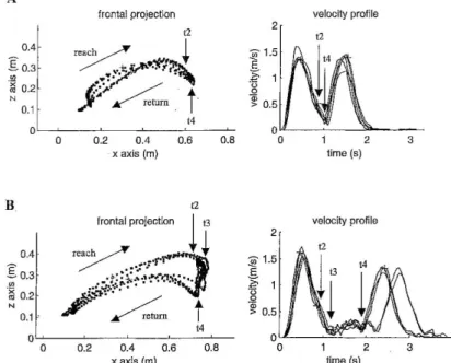
Figure 3 shows the trajectory (left) and the tangential velocity profile (right) of the hand during a series of five prehension movements made in the 0° direction, by a healthy subject (Figure 3A) and by
Figure 3. Projection of hand trajectory in the frontal plane and hand velocity profile. The recordings for the five trials have been superimposed. Arrows indicate reaching and return movements, as well as time marks. Left: hand tra- jectory. Right: velocity profile. A: Healthy subject ROB; time marks t3 and t4 are superimposed. B: C6 tetraplegic patient NAT before musculotendinous transfer.
8
a C6 tetraplegic patient before transfer (Figure 3B). For the healthy subject’s prehension movements, the hand trajectory was smooth and harmoniously curved. The velocity profile of the hand for each of the trials showed two bell-shaped phases that were contiguous in time: the first phase corresponded to the reaching movement, the second one to the return movement, which brought the cone back to the starting position on the table. The patient’s movements were more segmented than those of the healthy subject. During reaching, the hand moved upward, above the cone. It then slowly descended vertically in order to grasp the cone (t2 to t4), before returning to the starting position on the table. During the reaching phase, the patient raised his hand higher than the healthy subject. The patient’s hand velocity profile showed three phases. The first and the third phases were bell-shaped and corresponded to the reaching movement and the return movement, respectively, as it was the case for the healthy subject. The intermediate second phase corresponded to the grasping of the cone, during the hand’s movement down (t3 to t4). The presence of this supplementary phase indicates a difficulty for the patient to grasp the cone. Similar hand trajectories and velocity profiles were obtained for the other C6 patients, both before and after surgery. The mean value of the maximal velocity during reaching was 1.22 i 0.06 m/s for healthy subjects (mean i standard error), 1.07 1 0.05 m/s for C6 patients without transfer, 1.24 i 0.04 m/s for C6 patients with transfer and 1.14 i 0.08 m/s for C6-C7 patients. A statistical analysis by ANOVA, excluding C6-C7 patients, revealed a significant effect of group, F(94, 2) = 4.992, p = 0.0087, and direction, F(94, 5) = 22.279, p < 0.0001. The peak reaching velocity of C6 patients without transfer was significantly smaller than that of healthy subjects (12.47% smaller on an average) and that of C6 patients with transfer (13.74% smaller on an average), which was confirmed by a post-hoc Tukey-Kramer test.
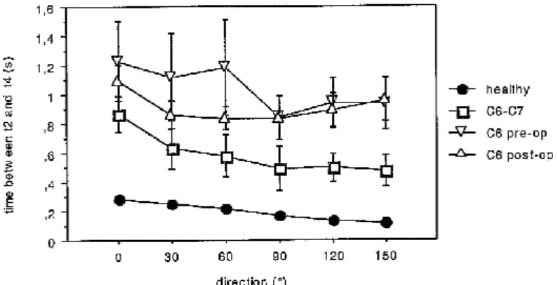
Time course of prehension
Figure 4 represents the time delay between the end of reaching (t2) and the end of grasping (t4), i.e., the time necessary to grasp the cone. The time between t2 and t4 was longer for all patients than for healthy subjects, for all of the six trial directions. The excess of time between t2 and t4 for the tetraplegic patients corresponded to the supplementary movement made by patients in order to grasp the cone and found expression in the intermediate second phase of the patients‘ three-phases hand velocity profile. An ANOVA excluding group C6-C7 showed a significant effect of group, F(94, 2) = 48.369, p < 0.0001, on the time delay between t2 and t4. A post-hoc Tukey-Kramer test revealed a significant difference between healthy subjects and C6 patients. On the other hand, there was no significant difference between C6 patients with transfer and C6 patients without transfer.
Figure 4. Time delay between the end of reaching (t2) and the end of grasping (t4) for each trial direction. Each point on the graph represents the mean value for the subject group ±the standard error
9
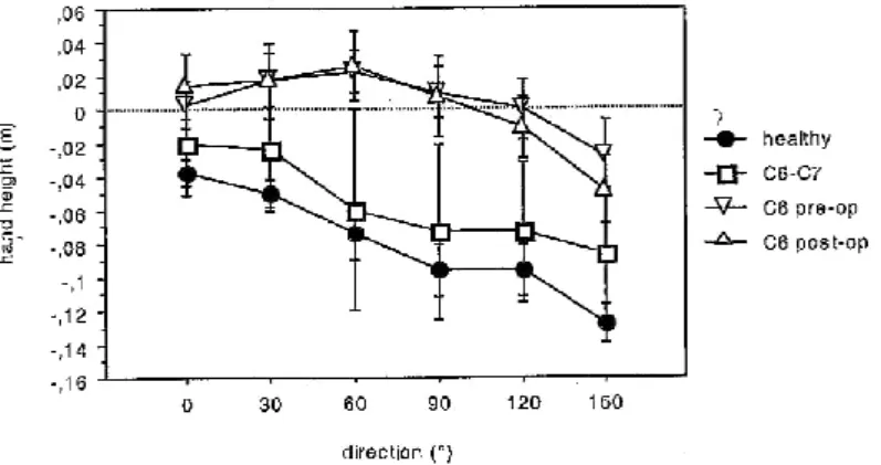
Hand height
Figure 5 shows the mean height of the hand, with regard to the height of the scapula, at the end of the reaching movement (t2). Considering the height of the hand with regard to the scapula, rather than the absolute value of the hand height with regard to the STS reference, allowed normalislng experimental conditions For healthy subjects, hand height at the end of reaching (t2) was higher for prehension movements made toward the lateral part of the workspace (directions 0°, 30°, and 60°)
than for those made forward (90°), or toward the inside of the workspace (l20° and 150°). For C6 patients both before and after transfer, hand height at t2 was higher than for healthy subjects, for all directions. Besides, patients’ hand height varied little according to the direction of the trial. An ANOVA excluding patients with C6-C7 lesions revealed a significant effect of group, F(94, 2) = 35 .629, p < 0.0001, and direction, F(94, 5) = 4.849, p = 0.0005. A post-hoc Tukey-Kramer test revealed a significant difference between healthy subjects and C6 patients, but not between C6 patients with transfer and C6 patients without transfer.
C6-C7 patients showed a high variability between subjects As a matter of fact, patients BAU and DRU behaved like healthy subjects (for example, for direction 150°, hand height was -0.12 and -0.13 m, respectively), whereas patient DAA behaved like C6 patients (for example, hand height for direction 150° was -0.01 m).
Joint rotation amplitudes
We examined the amplitudes of the rotations in all of the upper limb joints during prehension movements made in the different directions. As an example, Figures 6 and 7 show movement amplitudes for directions 0° and 150° during reaching (Figure 6) and during grasping (Figure 7). As expected, considering the experimental set-up, reaching movements made by healthy subjects mainly involved the shoulder and the elbow. For example, movements in the 0° direction, which moved the hand backward and to the right, involved horizontal abduction, elevation and external roll in the glenohumeral joint (Figure 6B), associated with an elbow extension (Figure 6C). The thoracoscapular joint also participated in the reaching movements, with a retraction and a posterior tipping of the scapula, but almost no medial-lateral rotation (Figure 6A). Healthy subjects also produced an extension and an adduction at the wrist, as well as a pronation. The rotations in the shoulder joints were sealed to target direction, but the elbow rotations were not (compare Figures 6A-6C and 6D-6F). The movements made by tetraplegic patients were roughly similar to those made by healthy subjects. However, the amplitudes of movements in some joints were significantly different, depending on trial
Figure 5. Hand Height with regards to the scapula (m) at t2 for each trial direction. Each point on the graph represents the mean value for the subject group ±the standard error
10
direction. Table 2 summarises the statistical results provided by an ANOVA assessing the effects of subject group and of trial direction. C6 patients produced a more important medial rotation of the scapula (Fig. 6A) and a greater elevation in the glenohumeral joint (Fig. 6B), especially for movements made in directions 0° to 90°. They also made a greater external roll in the glenohumeral joint, this difference occurring only for movements made in the 120° and 150° directions (Fig. 6E). The trajectory of the hand was higher in tetraplegic patients than in healthy subjects. Depending on the direction of the movement, the higher hand trajectory was due to different mechanisms. For movements toward the outside, it was due to a larger medial rotation in the thoracoscapular joint and a larger elevation in the glenohumeral joint. For movements toward the inside, it was due to a greater external roll in the glenohumeral joint. The amplitude of elbow extension was less important for patients than for healthy subjects, especially for movements made in the 120° and 150° directions (Fig. 6F). For all movement directions, patients produced a supination and a wrist flexion. Figure 7 shows the amount of joint rotations for grasping (between t2 and t4). During this short period, healthy subjects made few movements in the shoulder, i.e., in the thoracoscapular and glenohumeral joints (Figures 7A-B and 7D-E). Grasping involved a participation of the elbow (extension and supination), accompanied by a slight wrist flexion for movements toward the lateral part of the workspace (Fig. 7C) and a slight wrist extension for movements toward the inside (Fig. 7F). By opposition, the grasping phase in tetraplegic patients, which was longer in duration, involved an important participation of the thoracoscapular and glenohumeral joints. In particular, all the patients made a clear anterior scapula tipping, for all trial directions (Figs. 7A and 7D). They also made a large lateral rotation of the scapula for directions 0° to 90° (Fig. 7A) and a large internal roll of the glenohumeral joint for directions 120° and 150° (Fig. 7E). Finally, patients produced an important wrist extension for all trial directions (Figs. 7C and 7F).
Figure 6. Joint movement amplitudes (°) for reaching. Note the differences in scales between figures. Each bar on the graphs represents the mean value for the subject group ± the standard error.
A-C: movements made in the 0° direction. D~F: movements made in
the 150° direction. A and D: th0raco-scapular joint. ProRe: protraction-retraction; ML Rot: media!-lateral rotation; AP Tip: anterior-posterior tipping. B and E:
glenohumerai joint. Hor AA: horizontal abduction-adduction;
Ele: elevation; Ro: roll. C and F: elbow and wrist. FE e: elbow flexion-extension; PS: pronation-supination; FE w: wrist
flexion-extension; AA w: wrist abduction-adduction.
11
Table 2: Statistical analysis of the effect of subject group and trial direction on joint movement amplitudes, for reaching and for grasping
12
Figure 7. Joint movement amplitudes (°) for grasping. Note the differences in scales between figures. Each bar on the graphs represents the mean value for the subject group ± the standard error. A-C: movements made in the 0° direction. D-F: movements made in the 150° direction. A and D: thoracoscapular joint. B and E: glenohumeral joint. C and F : elbow and wrist. ,
Joint coordination
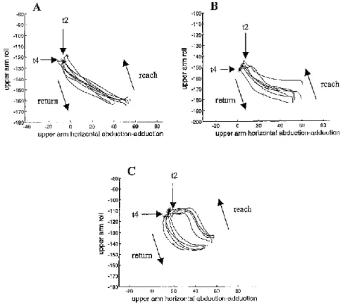
To further investigate how tetraplegic patients manage to successfully reach for and grasp the cone, we studied the temporal coordination of joint rotations (horizontal abduction-adduction and roll of the upper arm and flexion-extension at the wrist) and of the orientation of the flexion~ extension axis of the elbow, during the prehension movements. Figure 8 represents roll of the upper arm as a function of horizontal abduction-adduction of the upper arm, for a series of five movements made in the 0° direction, by a healthy subject (Fig. 8A) and by two tetraplegic patients (Figs. 8B and 8C). The reaching movement consisted of an abduction moving the arm in the direction of the cone, and bringing the hand in the vicinity of the object. This abduction was accompanied by an external roll. The return movement consisted of an adduction and an internal roll. For healthy subjects, there was a roughly linear temporal synchronisation between horizontal abduction-adduction and roll of the upper arm. For the patient illustrated by Figure SB, upper arm movements during reaching were similar to those of healthy subjects. This was the case for most of the patients (C6 patients AND, ELI, MAR both before and after transfer, MOS, NAT after transfer, SIM, C6-C7 patients DAA and DRU). For the other patients, the coordination between the two upper arm movements was markedly different from the healthy subjects, as the roll movement of the upper arm during reaching consisted of two successive phases. C6-C7 patient BAU and C6 patient NAT before transfer (Fig. 8C) produced the entire external roll during the early part of the abduction movement and did not produce any further roll of the upper arm until the end of reaching. C6 patients BER (left arm) and CAD, both before and after transfer, while abducing the upper arm, first produced an internal roll at the beginning of reaching, then an external roll until the end of reaching (not shown). For all the healthy subjects and for most of the patients, grasping occurred Without any noticeable abduction-adduction or external-internal roll of the upper arm, However, patients BER, CAD, and ELI grasped the cone by producing an internal roll between t2 and t4.
13
Figure 8. Graphical representation of roll of the upper am: (°) as a function of horizontal abduetiomadduction of the upper arm (°), for direction 0°. Arrows indicate time marks. A: healthy subject BAS. B: C6 patient SIM. C: C6 patient NA T before transfer.
In order to further investigate the coordination between shoulder and elbow, we studied the temporal coordination between elbow extension and the orientation of the elbow flexion-extension axis with regard to the horizontal plane (FE axis). On one hand, this orientation depends on roll and elevation movements of the upper arm, on ‘the other hand it conditions elbow extension. As a matter of fact, if the orientation of the elbow FE axis reaches a horizontal orientation, the forearm will extend passively because of the effect of gravity.
Figure 9 represents this coordination for movements made in the 90° direction, by a healthy subject (Fig. 9A) and by two tetraplegic patients (Figs. 9B and 9C). For healthy subjects, the elbow axis rotated toward the horizontal during reaching while the elbow extended. There was a close temporal relationship, illustrated by a roughly linear pattern, between elbow extension and the rotation of the elbow FE axis toward the horizontal (except for a small elbow flexion at the begirming of reaching in some cases). This also was the case during the return movement. Some tetraplegic patients showed similar coordination patterns as healthy subjects. This was the case in C6 patients AND, MAR, both before and after transfer, NAT after transfer and SIM, as well as C6-C7 patient DRU (Fig. 9B). However, most patients had a markedly different coordination pattern. As an example, Figure 9C shows movement recordings for C6-C7 patient DAA. This patient produced a large rotation of the FE axis of the elbow toward the horizontal throughout the entire movement. This rotation was first accompanied by an elbow flexion until just before t2. From this moment on, the elbow started to extend until the end of reaching, suggesting that the extension was due to gravity. A similar pattern was observed for C6 patients CAD 3 months after transfer, ELI and NAT before transfer, as well as for C6C7 patient BAU, Other patients (C6 patients BER, CAD before transfer and MOS) produced still another pattern, which
14
is not shown. For all patients, the temporal coordination for the return movement was linear and similar to that of healthy subjects.
Figure 9. Graphical representation of flexion-extension of the elbow (°) as a fimction of rotation of the elbow FE axis (°), for direction 90°. 0“ of rotation corresponds to a horizontal orientation of the flexion-extension axis of the elbow. A: healthy subject LAF. B." C6 patient MAR before transfer. C: C6-C7 patient DAA.
In order to study the temporal coordination between reaching and grasping, we plotted flexion-extension of the wrist as a function of horizontal abduction-adduction of the upper arm, as the glenohumeral joint is one of the main joints involved in reaching.
The temporal coordination of upper arm movements and wrist movements was markedly different between healthy subjects and all the tetraplegic patients. As an example, Figure 10 represents flexion-extension at the wrist as a function of horizontal abduction-adduction of the upper arm, for movements made in the 0° direction, by a healthy subject (Fig. 10A) and by a tetraplegic patient (Fig. 10B). For the healthy subject, movements of the upper arm and movements at the wrist were synchronised in time. The abduction of the upper arm during reaching was accompanied by a wrist extension that opened the hand to prepare for the grasping of the cone. The grasping movement was characterised by a short wrist flexion. During the return movement, the wrist returned to the same level of extension as before the grasping and stayed at this level until the end of the movement. For all the tetraplegic patients, movements of the wrist and movements of the upper arm were desy11chronised. At the beginning of the reaching movement, there was a wrist flexion accompanying the abduction of the upper arm. Wrist extension occurred after the end of reaching, when the upper arm did not move any more. Grasping consisted in a wrist extension. This extension continued after the beginning and during the early part of the return movement. The end of the return consisted in an adduction of the upper arm without any noticeable wrist movements.
For all of the articular coordinations studied, results obtained for the other movement directions showed similar patterns as those obtained for the 0° and 90° directions shown above.
15
Figure 10. Graphical representation of flexion-extension of the wrist (°) as a function of horizontal abduction-adduction of the upper arm (°), for direction 0°. A: healthy subject ROB. B: C6 patient NAT before transfer.
DISCUSSION
This pioneering study examines two main issues: firstly, the mechanisms allowing tetraplegic patients to perform prehension movements despite their extensive impairment, and secondly, the effect of a musculotendinous transfer. This clinical model is particularly relevant to investigate the current theories on motor control, since it shows a largescale adaptation of upper limb movements, which will be discussed in detail below.
Prehension movements in healthy subjects
The results obtained in healthy subjects, i.e., a smooth hand movement to reach and grasp the cone, are consistent with previous observations by Jeannerod and others (for a review, see Paulignan & Jeannerod, 1996). There was no delay due to grasping, probably because the opening of the hand was prepared during reaching (Jeannerod, 1984). Our recording method allows computation of the joint rotations, corresponding to ten degrees of freedom (dot), of the upper limb and the scapula. In agreement with Desmurget and Prablanc ( 1997), reaching to grasp movements involved most of the available dof, in the glenohumeral and elbow joints. Furthermore, we show that the thoracoscapular joint also participated in reaching, which has never been observed before. The rotations in most of the proximal joints were scaled to the direction of the movement. The rotations in the distal dof during reaching (prenation, wrist extension and adduction) suggest that healthy subjects prepared a hand posture for grasping that is close to the "function position" described in the literature (Kapandji, 1980).
Prehension performance in tetraplegic patients
The present work is a pioneering quantitative study of the surprising fact, although well known in clinics, that tetraplegic patients are able to successfully make prehension movements despite the extensive paralysis of their upper limb muscles including elbow extensor and hand muscles. This ability is not unexpected since they received intense occupational therapy during their rehabilitation treatment in hospital after their injury (Welch et al., 1986). The present study confirms previous ones since it shows that the hand velocity profile during the reaching phase was smooth and bell-shaped (Laffont et al., 2000). The efficiency of the control of the upper limb during reaching can be evaluated by means of the peak velocity value. Patients’ performance can be compared to that of healthy subjects, as both patients and healthy subjects self-paced their reaching movements. On an average, the peak velocity attained by patients with a triceps paralysis was only about 12% less than that attained by healthy subjects. This proves that the patients controlled their reaching movement in an
16
anticipated way, in order to compensate for the multiple perturbations due to the modifications of the limb dynamics resulting from muscle paralysis. However, tetraplegic patients had difficulty in grasping, because of paralysis of the hand. They had to make a supplementary movement in order to grasp the cone, which led to an intermediate phase in the velocity profile and to an important time delay between the end of reaching and the beginning of the return movement. This supplementary movement corresponds to an alternative strategy for grasping, which is known and taught as "tenodesis" in rehabilitation clinics (Smith, Mulcahey, & Betz, 1996). Patients have to place their hand above the object and to slide it from top to bottom down the object so that their fingers open passively. The closing of the hand for the actual grasping is obtained by an extension of the wrist, which passively flexes the fingers. Thus, the coordination of prehension in tetraplegic patients results from an adaptation at two different control levels. On the one hand, the planning and the timing of the action have to be modified in order to allow tenodesis. On the other hand, the coordination of the reaching movement has to be adapted in order to comply with two constraints: firstly, to prepare tenodesis, and secondly, to compensate for the paralysis or weakness of the triceps brachii. These issues will be successively examined, and we shall then confront these observations with regard to their possible interpretation in light of current theories of motor control.
Consequences of the tenodesis strategy
Jeannerod and co-workers showed that prehension consists of two components, namely, reaching and grasping, which are closely related in time for prehension, and that these components are controlled by distinct neuronal mechanisms or visuomotor channels occurring in parallel (Jeannerod, 1984; Paulignan & Jeannerod, 1996). Indeed, grasping is prepared during reaching so that hand aperture reaches a maximum before the end of reaching and that hand closure is achieved simultaneously with the end of reaching. The temporal coupling of the two prehension components suggests that at the CNS level, a higher-order program controls the entire prehension movement by coordinating its two components, each of them being controlled by a separate visuomotor channel (Hoff & Arbib, 1992). The method used in the present study does not involve a direct measure of the grip aperture during reaching. However, the timing of the grasping component can be analysed by observation of the hand trajectory and of the rotations in the distal joints. In particular, tetraplegic patients produced a large wrist extension for grasping, which induces the passive closure of the fingers by pulling on the tendons. Conversely, the wrist flexion made during reaching can be considered as preparatory to grasping since it relaxes the finger flexor tendons, thus facilitating the hand aperture. The delay between the reaching and the return movements (Figures 3B and 4) and the evolution of the wrist flexion-extension angle during reaching (Figure 10) confirm that tetraplegic patients executed the prehension movement in two successive stages. Thus, prehension movements made by tetraplegic patients lack the temporal coincidence between the reaching and grasping components, showing that the two components may be controlled in a rather sequential way. The insertion of the rotation of the joint mainly responsible for grasping, i.e., the wrist, within a relatively complex sequence of proximal joint rotations and mobilities (see Figure 10) suggests a subtle temporal coupling between reaching and grasping, rather than a mere rigid temporal succession of the components. The sequential prehension in tetraplegic patients is an example that shows the influence of the grasping component on the timing between reaching and grasping, which has been suggested in previous works (Castiello, Bennett, & Paulignan, 1992; Castiello, Bennett, & Stelmach, 1993; Gentilucci, Castiello, Corradini, Scarpa, Umilta, & Rizzolatti, 1991; Paulignan, Jeannerod, MacKenzie, & Marteniuk, 1991).
17
Reaching and grasping by tetraplegic patients can be considered as two sub-actions assembled in a sequential way to reach the goal. However, there is a dependence between these sub-actions, since the execution of first one, i.e., reaching, is influenced by specific constraints brought about by the execution of the second one, i.e., grasping. In other words, the need to use tenodesis for grasping has profound overall consequences on patients’ reaching to grasp movement. Two points will be examined: the trajectory of the hand during reaching and the joint coordination at the end of reaching. C6 tetraplegic patients produced a movement with a higher hand trajectory, regardless of the direction of the movement. By opposition, healthy subjects made high hand trajectories only to grasp objects in the external part of the workspace, where they have to move above the object before grasping. The height of the trajectory may be due to the necessity for the patients to position the hand above the cone for tenodesis, or to other constraints related to the paralysis of their triceps. Several arguments favour the first hypothesis. Firstly, a preliminary study has shown that pointing movements made by tetraplegic patients were not particularly higher than prehension movements (Laffont et al., 2000). Secondly, the height of the trajectory does not depend on the condition of the triceps muscle, since it was not different in C6 patients before and after surgery. Two out of three C6-C7 patients made hand trajectories that were close to those of healthy subjects. This fact is likely to be due to the relative preservation of their flexor carpi radialis (a Wrist flexor muscle), since an efficient wrist flexion during reaching may facilitate finger opening to prepare for grasping, and thus may reduce the need to move above the object. The influence of the constraints arising from a late sequence on the planning of the entire action, and consequently on the preceding sequences, has already been shown in other studies concerning prehension. Marteniuk, MacKenzie, Jeannerod, Athenes, and Dugas (1987) studied different prehension tasks within a complex action (grasping an object to throw it in a box or td place it on a support) and observed that the kinematics of the reaching movements differed according to the final result of the action. In a task associating two sub-tasks, i.e., grasping an object and then placing it in some place of the experimental set-up, Gentilucci, Negrotti, and Gangitano (1997) also observed that constraints concerning the sub-task of placing had an effect on the kinematics of reaching. Rosenbaum studied whether subjects picked up a bar with the thumb pointed toward one of its ends or toward the other, when they were asked to subsequently place either one of the ends on a disk (Rosenbaum & Jorgensen, 1992). Depending on the task demands, the handgrip used by the subjects was different, and the results strongly suggested that the way subjects produced their movement was such that it allowed them to complete the task with the hand and the arm in a comfortable posture. For prehension movements, the comfortable posture is the adequate posture for grasping, and it is the need to prepare this posture that constrains the joint rotations during reaching. As noted above, healthy subjects tended to prepare a grasping posture that Was similar to the classical "functional posture ". On the contrary, tetraplegic patients were constrained to grasp by tenodesis, which led to a profound reorganisation of the reaching movement (larger rotations in the thoracoscapular and glenohumeral shoulder joints in tetraplegic patients, as well as wrist flexion instead of extension). As a matter of fact, the constraint of grasping using tenodesis requires tetraplegic patients to position the hand above the object and to reduce finger stiffness.
Upper limb coordination for reaching
Reaching involved an elbow extension for all tetraplegic patients, even for those suffering from a triceps paralysis, although the amplitude of elbow extension was less important for patients than for healthy subjects. Elbow extension during reaching can be due to the active torque produced by the main elbow extensor, i.e., the triceps muscle, to a posture favouring the effect of gravity, and to
18
dynamic biomechanical interactions arising from the rotations in other joints. The production of an active extension torque is very unlikely in tetraplegic patients with a complete or almost complete paralysis of the triceps muscle. We have shown that the postural effect of gravity, when the orientation of the elbow flexion-extension axis was close to the horizontal, could partly explain elbow extension in some patients (see Figure 9C). As the rotation of the flexion-extension axis is due to rotations in the shoulder joints, in most of these tetraplegic patients, the elbow extension can be attributed to the effect of gravity and the dynamic interactions that are produced by rotation of the shoulder and that arise at the elbow. However, we were surprised to notice that interjoint coordination patterns made by tetraplegic patients often were similar to those made by healthy subjects. In addition, there was no unequivocal correspondence between the different interjoint coordination patterns produced by the patients and the different clinical conditions of the extensor muscle (paralysis, weakness, partial recovery of strength after tendon transfer). We therefore suggest that most of the patients mainly used the effect of gravity and the dynamic interactions arising from shoulder movements to extend the elbow during reaching. This hypothesis, which claims that the main effector of elbow movement during reaching is the shoulder, rather than the triceps muscle, seems to hold true for healthy subjects. It is supported by the recent observations by Dounskaia, Swinnen, Walter, Spaepen, and Verschueren (1998) who analysed cyclical movements of the elbow and wrist by an inverse dynamics computation. They concluded that the control of a movement involving ‘a couple of joints is hierarchical. The dominant or leading joint generates the torques responsible for its own movements, and imposes movements of the subordinate joint by producing interaction torques. The control of the subordinate joint intervenes to adjust the movements imposed by the interaction torques, according to the demands of the task. Similarly, it has been observed that the shoulder was the leading joint in a horizontal drawing task involving the shoulder and the elbow (Verschueren, Swinnen, Cordo, & Dounskaia, 1999). More recently, Sainburg and Kalakatiis (2000) performed inverse dynamic computations of horizontal planar reaching movements. They observed that elbow extension was due to the mechanical interactions generated by shoulder movements for the dominant arm, and to an active extensor torque for the non-dominant arm. Thus, the use of interaction torques to extend the elbow seems to be linked to a long-lasting skill acquisition. The overall interpretation of these and our data is that some tetraplegic patients are able to control their elbow via interaction torques in highly skilled manner, as do healthy subjects. Other patients made an increased use of the postural effect of gravity. Thus, the ability to perform an efficient reaching coordination seems to be related to skill acquisition as well as to the strength of the triceps.
In the light of these considerations, what is the role of the musculotendinous transfer? Musculotendinous transfers induce a clinical improvement of the reaching ability, which is often spontaneously reported by the patients and which can be evaluated by satisfaction scales (Revol et al., 1997). Accordingly, the present study shows that the peak velocity for reaching is higher in patients with some strength in the triceps (after transfer or because of a lower level lesion) than in patients with a triceps paralysis. The role of the triceps in reaching movements might be to increase the stiffness of the elbow joint by allowing co-contraction of the flexor muscles and of the transferred muscle. An increased stiffness can facilitate the correction and the stabilisation of the elbow extension induced by interaction torques from the shoulder. Thus, we suggest that the muscle transferred onto the triceps does not completely change (for the deltoid) or reverse (for the biceps) its function, but that it acts in co-contraction with elbow flexor muscles in order to stabilise the joint. This hypothesis is in agreement with previous studies of flexion-extension movements made in the horizontal plane and analysed by
19
EMG recordings. Such analyses failed to demonstrate an unambiguous reversal of function of the transferred muscle. Rather, the transferred muscle appeared to adopt various contraction patterns, corresponding to either the new or the original anatomical function, as well as intermediate patterns (Illert et al., 1986). The present hypothesis should be assessed by EMG recording during natural 3D reaching movements. Moreover, as the musculotendinous transfer at the elbow is merely the first step of the surgical program that is aimed at restoring the function of the upper limb, further information about the effect of the musculotendinous transfer may be provided by studying patients after completion of this program, which involves two more surgeries at the hand.
Mechanisms of motor control and adaptation
The summary of our results is that the tetraplegic patients showed a remarkable motor adaptation to their impairment. This adaptation occurred at two levels of motor control, since they developed a new grasping strategy called tenodesis and adapted their upper limb coordination in order to reach the goal of the task despite their impairment. In spite of the dynamic perturbations of the upper limb due to the muscular paralysis or weakness, the joint kinematics at the proximal joints were relatively preserved. The issue of how movements are controlled is still disputed, since some authors posit that the central nervous system controls muscles and torques (Gottlieb, 1998: Uno, Kawato, & Suzuki, 1989), while others posit that it controls joint rotations or postures (Desmurget 8c Prablanc, 1997; Feldman & Levin, 1995; Rosenbaum, Meulenbroek, Vaughan, & Jansen, 1999). The preservation of the joint kinematics in altered dynamic conditions that we observed in our study suggests that the control of the upper limb is more likely to be performed by control of the joint kinematics than of muscle dynamics. The results of the present study are consistent with the proposition that the CNS controls movements via reference postures related to the constraints of the task (Feldman & Levin, 1995; Rosenbaum & Jorgensen, 1992; Rosenbaum et al., 1999). In particular, our observations both in healthy subjects and in the various patient groups emphasise the importance of an intermediate "via posture" to be attained at the end of reaching in order to prepare for grasping. As we have shown, this posture, which is itself conditioned by the kind of the grasping action, determines the joint coordination for reaching.
The visuomotor channels hypothesis is commonly accepted for understanding the coordination between reaching and grasping (review in Paulignan & Jeannerod, 1996). However, the results obtained in tetraplegic patients are difficult to explain within the framework of this hypothesis. Indeed, we observed a profound reorganisation of both the timing and the coordination of the movement. Previous observations have shovsm that the use of different types of grasping by healthy subjects may delay the moment of maximal hand aperture (Castiello et al., 1992, 1993; Gentilucci et al., 1991). In the case of tetraplegic patients’ prehension movements, the timing of the two components is deeply altered, sinoe the components of prehension, which occurred simultaneously, are henceforth performed in a sequential way. This kind of behaviour cannot be explained directly by models of timing based on the visuomotor channels hypothesis (Hoff & Arbib, 1992). Similarly, the visuomotor channels hypothesis suggests that the module responsible for reaching controls proximal segments, whereas the grasping module controls distal segments. The results observed in tetraplegic patients contradict this suggestion, since movements of proximal segments, in particular displacement of the scapula, are involved in the grasping component. A similar observation was made by Wing and Fraser (1983), by studying a patient wearing a mechanical hand prosthesis, which was actively opened during reaching by a movement of the scapula acting on a harness. More likely, our result can be interpreted in the framework of dynamical systems in keeping with Bernstein’s (1967) pioneering work. The new motor
20
strategy developed by the patients takes into account the anatomo-functional constraints of the upper limb, including paralysis, in order to reach the goal of the task, which is to grasp the object. The adaptation evidenced by this particular task occurs at two levels: timing and load as defined by Schoner (1995). We observed both a new temporal synchronisation between joints to allow reaching for the object, and a temporal uncoupling of arm movements and wrist movements to allow grasping by tenodesis at the end of reaching. This finding suggests that the CNS has combined new task-related functional synergies in a time-ordered way. A similar spatiotemporal organisation of sub-actions, each of them corresponding to a synergistic coordination of several effectors, has been described in the field of language. The movements of the phonation effectors (lips, jaw, tongue, larynx) are coordinated with one another in a synergistic way in order to produce a particular sound, and the kinematic transitions between synergies depend on the constraints of the linguistic context in which the sound is produced (Gracco & Lofqvist, 1994). The mechanisms by which the CNS superposes and assembles these synergies in time, in order to combine action sequences, had until now never been described in the field of prehension.
CONCLUSION
The present study suggests that the CNS of tetraplegic patients adapts to the dynamic perturbation of the upper limb by taking into account the constraints that are due to the paralysis of some upper limb muscles and by consequently modifying both the planning and timing of the action of prehension and the movements of the upper limb joints that allow its physical execution, in order to compensate for the loss of function of the paralysed muscles. Tetraplegic patients are able to develop new motor strategies for prehension, which consist in a grasping using tenodesis and a production of a prehension in which the reaching movements and the grasping movements are uncoupled and occur in a sequential way. The results of the present study allow one to make suggestions as far as the issue of motor control of the upper limb is concerned. The production of elbow extension is likely to be due, both in tetraplegic patients and in healthy subjects, to the dynamical interactions between the shoulder and the elbow. During prehension, motor control of the upper limb by the CNS may be achieved by control of upper limb posture and of joint rotations.
ACKNOWLEDGEMENTS
We are grateful to the patients who participated in this study. Pr. Bussel and Pr. Dizien received us in the rehabilitation department and Michele Combeaud participated in the experiments. Pr. Marc Revol was in charge of the surgical procedures for the musculotendinous transfers. We also thank Sylvain Hanneton for fruitful discussions. Agnes Roby-Brami is supported by INSERM and AP-HP (France). Gilles Hoffmann receives a grant from the MCESR (Luxembourg). This work was supported by grants from the French ministry of research (Cognitique) and by the "Fondation EDF mécénat".
21
REFERENCES
Bernstein, N. (1967). The coordination and regulation of movements. Oxford: Pergamon.
Biryukova, E. V., Roby-Brami, A., Frolov, A. A., & Mokhtari, M. (2000). Kinematics of human arm reconstructed from spatial tracking system recordings. Journal ofBiomechanics, 33, 985-995.
Castiello, U., Bennett, K. M. B., & Paulignan, Y. (1992). Does the type of prehcnsion influence the kinematics of reaching? Behavioural Brain Research, 50, 7-15.
Castiello, U., Bennett, K. M. B., & Stelrnach, G. E. (1993). Reach to grasp: the natural response to perturbation of object size. Experimental Brain Research, 94, 163-178.
Desmurget, M., & Prablanc, C. (1997). Postural control of three-dimensional prehension movements. Journal of Neurophysiology, 77, 452-464.
Dounskaia, N. V., Swinnen, S. P., Walter, C. B., Spaepen, A. 1., & Verschueren, S. M. (1998). Hierarchical control of different elbow-wrist coordination patterns. Experimental Brain Research, I21 239-254. Feldman, A. G., & Levin, M. F. (1995). The origin and use of positional frames of reference in motor control. Behavioral and Brain Sciences, 18, 723-806.
Gentilucci, M., Castiello, U., Corradini, M. L., Scarpa, M., Umilta, C., 8:. Rizzolatti, G. (1991). Influence of different types of grasping on the transport component of prehension movements. Neuropsychologia, 29, 361-378.
Gentilucci, M., Negrotti, A., & Gangiiano, M. (1997). Planning an action. Experimental Brain Research, II5, 116-128.
Gottlieb, G. L. (1998). Muscle activation patterns during two types of voluntary single-joint movement. Journal of Neurophysiology, 80, 1860-1867. Gracco, V. L., & Lofqvist, A. (1994). Speech motor coordination and control: evidence frorn lip, jaw and laryngeal movements. Journal of Neuroscience, 14, 6585-6597.
Hoff, B., & Arbib, M. A. (1992). Models of trajectory formation and temporal interaction of reach and grasp. Journal of Motor Behavior, 25, 175192.
Illert, M., Trauner, M., Weller, E., & Wiedemann, E. (1986). Forearm muscles of man can reverse their function after tendon transfers: an electromyographic study. Neuroscience Letters, 67, 129-134. Jeannerod, M. (1984). The timing of natural prehension movements. Journal of Motor Behavior, I 6, 235-254.
Kapandji, I. A. (1980). Physiologie articulaire, Tome 1: le membre supérieur. Paris: Maloine. Karduna, A. R., McClure, P. W., Michener, L. A., & Sennett, B. (2001). Dynamic measurements of three-dimensional scapular kinematics: a validation study. Journal of Biomechanieal Engineering, I23, 184-190.
Laffont, 1., Briand, E., Dizien, 0., Combeaud, M., Bussel, B., Revol, M., & Roby-Brami, A. (2000). Kinematics of prehension and pointing movements in C6 quadriplegic patients. Spinal Cord, 38, 354-362.
Marteniuk, R. G., MacKenzie, C. L., Jeannerod, M., Athenes, S., & Dugas, C. (1987). Constraints on htunan arm movement trajectories. Canadian Journal of Psychology, 4], 365-378.
22
Paulignan, Y., & Jeannerod, M. (1996). The visuomotor channels hypothesis revisited. In A. M. Wing, P. Haggard, & J. R. Flanagan (Eds.), Hand and brain. The neurophysiology and psychology of hand movements (pp. 265282). San Diego, CA: Academic Press.
Paulignan, Y., Jeannerod, M., MacKenzie, C., & Marteniuk, R. (1991). Selective perturbation of visual input during prehension movements. 2. The effects of changing object size. Experimental Brain Research, 87, 407-420.
Revol, M., Briand, E., Cormerais, A., Pedelucq, J., Busnel, M., & Servant, J .-M. (1997). Rehabilitation fonctionnelle des membres supérieurs dons la tétraplégie traurnatique. Encyclopédie médica-chirurgicale. Techniques chirurgicales. Chirurgie plastique. Paris: Elsevier.
Rosenbaum, D. A., & Jorgensen. M. J. (1992). Planning macroscopic aspects of manual control. Human Movement Science, IJ, 61-69.
Rosenbaum, D. A., Meulenbroek, R. G., Vaughan, 1., & Jansen, C. (1999). Coordination of reaching and grasping by capitalizing on obstacle avoidance and other constraints. Experimental Brain Research, 128, 92-100.
Sainburg, R. L., & Kalakanis, D. (2000). Differences in control of limb dynamics during dominant and nondorninant arm reaching. Journal of Neurophysiology, 83, 2661-2675.
Schoner, G. (1995). Recent developments and problems in human movement science and their conceptual implications. Ecological Psychology, 7, 291314.
Smith, B. T., Mulcahey, M. J., & Betz, R. R. (1996). Quantitative comparison of grasp and release abilities with and without functional neuromuscular stimulation in adolescents with tetraplegia. Paraplegia, 34, 16-23.
Tobis, J. S., & I-long, C. Z. (1990). Muscle testing. In F. J. Kottke & I. F. Lehmann (Eds), Krusen’s handbook of physical medicine and rehabilitation (Fourth edition). Philadelphia, PA: Saunders Company.
Uno, Y., Kawato, M., & Suzuki. R. (1989). Formation and control of optimal trajectory in human multijoint arm movement. Minimum torque change model. Biological Cybernetics, 61, 89-101.
van der Helm, F. C. T. (1997). A standardized protocol for motion recordings of the shoulder. Proceedings of the First Conference of the ISG, http: _l/www.fbw.vu.nl/
Verschueren, S. M., Swinnen, S. P., Cordo, P. J., & Dounskaia, N. V. (1999). Proprioceptive control of multijoint movement: unimanual circle drawing. Experimental Brain Research, 127, 171-181.
Welch, R. D., Lobley, S. 1., O’Sullivan, S. B., & Freed, M. M. (1986). Functional independence in quadriplegia: critical levels. Archives of Physical Medicine and Rehabilitation, 67, 235-240.
Wing, A. M., & Fraser, C. (1983). The contribution of the thumb to reaching movements. Quarterly Journal of Experimental Psychology, 35A, 297-309.