HAL Id: hal-02478447
https://hal.archives-ouvertes.fr/hal-02478447
Submitted on 13 Feb 2020
HAL is a multi-disciplinary open access
archive for the deposit and dissemination of
sci-entific research documents, whether they are
pub-lished or not. The documents may come from
teaching and research institutions in France or
abroad, or from public or private research centers.
L’archive ouverte pluridisciplinaire HAL, est
destinée au dépôt et à la diffusion de documents
scientifiques de niveau recherche, publiés ou non,
émanant des établissements d’enseignement et de
recherche français ou étrangers, des laboratoires
publics ou privés.
Distributed under a Creative Commons Attribution| 4.0 International License
Investigation of Genetic Relationships Between
Hanseniaspora Species Found in Grape Musts Revealed
Interspecific Hybrids With Dynamic Genome Structures
Méline Saubin, Hugo Devillers, Lucas Proust, Cathy Brier, Cecile Grondin,
Martine Pradal, Jean Luc Legras, Cécile Neuvéglise
To cite this version:
Méline Saubin, Hugo Devillers, Lucas Proust, Cathy Brier, Cecile Grondin, et al.. Investigation of
Genetic Relationships Between Hanseniaspora Species Found in Grape Musts Revealed Interspecific
Hybrids With Dynamic Genome Structures. Frontiers in Microbiology, Frontiers Media, 2020, 10,
pp.1-21. �10.3389/fmicb.2019.02960�. �hal-02478447�
fmicb-10-02960 December 28, 2019 Time: 15:50 # 1 ORIGINAL RESEARCH published: 15 January 2020 doi: 10.3389/fmicb.2019.02960 Edited by: Matthias Sipiczki, University of Debrecen, Hungary Reviewed by: Lisa Solieri, University of Modena and Reggio Emilia, Italy Giuseppe Blaiotta, Università degli Studi di Napoli Federico II, Italy Anthony Borneman, The Australian Wine Research Institute, Australia *Correspondence: Cécile Neuvéglise Cecile.neuveglise@inra.fr
Specialty section: This article was submitted to Food Microbiology, a section of the journal Frontiers in Microbiology Received: 19 September 2019 Accepted: 09 December 2019 Published: 15 January 2020 Citation: Saubin M, Devillers H, Proust L, Brier C, Grondin C, Pradal M, Legras J-L and Neuvéglise C (2020) Investigation of Genetic Relationships Between Hanseniaspora Species Found in Grape Musts Revealed Interspecific Hybrids With Dynamic Genome Structures. Front. Microbiol. 10:2960. doi: 10.3389/fmicb.2019.02960
Investigation of Genetic
Relationships Between
Hanseniaspora Species Found in
Grape Musts Revealed Interspecific
Hybrids With Dynamic Genome
Structures
Méline Saubin1, Hugo Devillers1, Lucas Proust1, Cathy Brier1, Cécile Grondin2,
Martine Pradal3, Jean-Luc Legras3and Cécile Neuvéglise1*
1Micalis Institute, INRA, AgroParisTech, Université Paris-Saclay, Jouy-en-Josas, France,2Micalis Institute, INRA, AgroParisTech, CIRM-Levures, Université Paris-Saclay, Jouy-en-Josas, France,3SPO, Univ Montpellier, INRA, Montpellier SupAgro, Montpellier, France
Hanseniaspora, a predominant yeast genus of grape musts, includes sister species recently reported as fast evolving. The aim of this study was to investigate the genetic relationships between the four most closely related species, at the population level. A multi-locus sequence typing strategy based on five markers was applied on 107 strains, confirming the clear delineation of species H. uvarum, H. opuntiae, H. guilliermondii, and H. pseudoguilliermondii. Huge variations were observed in the level of intraspecific nucleotide diversity, and differences in heterozygosity between species indicate different life styles. No clear population structure was detected based on geographical or substrate origins. Instead, H. guilliermondii strains clustered into two distinct groups, which may reflect a recent step toward speciation. Interspecific hybrids were detected between H. opuntiae and H. pseudoguilliermondii. Their characterization using flow cytometry, karyotypes and genome sequencing showed different genome structures in different ploidy contexts: allodiploids, allotriploids, and allotetraploids. Subculturing of an allotriploid strain revealed chromosome loss equivalent to one chromosome set, followed by an diploidization event, whereas another auto-diploidized tetraploid showed a segmental duplication. Altogether, these results suggest that Hanseniaspora genomes are not only fast evolving but also highly dynamic.
Keywords: MLST, yeast, biodiversity, evolution, Hanseniaspora uvarum, Hanseniaspora guilliermondii
INTRODUCTION
Grape must is a complex ecosystem that combines grape and cellar micro-organisms. Many species
are interacting with each other, including yeasts and bacteria (Jolly et al., 2014; Capozzi et al.,
2015; Raymond Eder et al., 2017). It is now well recognized that natural microbial populations play an important role in winemaking, notably by increasing the complexity of wine aromas
fmicb-10-02960 December 28, 2019 Time: 15:50 # 2
Saubin et al. Hanseniaspora Interspecific Hybrids
(Gamero et al., 2016; Padilla et al., 2016; Bagheri et al., 2018;
Escribano et al., 2018). The occurrence of microorganisms in grapes is dependent on biotic and abiotic factors such as geographic location, soil, grapevine cultivar, viticultural practices,
and climate (Bokulich et al., 2014; Drumonde-Neves et al.,
2016). Yeast biodiversity in must is thus generally vintage
dependent (Vigentini et al., 2015). Whereas Saccharomyces
cerevisiae, already well known for its importance in diverse fermented food production, is the dominant species at the end of vinification, the yeast species commonly found on grapes and in musts at the beginning of spontaneous fermentations are rather Saccharomycotina than Basidiomycotina. They mostly belong to
genera Hanseniaspora, Lachancea, Metschnikowia, Pichia, and
Starmerella (Wang et al., 2015; Varela and Borneman, 2017;
Cioch-Skoneczny et al., 2018; Lorenzini and Zapparoli, 2019),
and the genusHanseniaspora is generally the most abundant at
the onset of the fermentation.
The Saccharomycotina genus Hanseniaspora belongs to the
Saccharomycodaceae family. Due to their lemon-shaped cell
structure, Hanseniaspora species have been called apiculate
yeasts, together with the closely related species of the genera Saccharomycodes and Nadsonia. The genus includes 21 described species, which can be separated into two lineages based on phylogenetic relationships deduced from D1D2 domains of
the 28S rRNA subunit (Boekhout et al., 1994; Ouoba et al.,
2015; Martin et al., 2018) or from the concatenation of
taxonomic markers (Cadez et al., 2019). Recently, based on
whole genome sequence comparison, Steenwyk et al. (2019)
confirmedHanseniaspora to be composed of two lineages, a
fast-evolving lineage (FEL) and a slow-fast-evolving lineage (SEL), which differ by their evolution rate and the extent of their gene loss. Species found on grapes and musts belong to both FEL (mostly H. uvarum) and SEL (H. vinae). Several studies report that they play an important role in wine fermentation by producing
flavors, modulating the growth and metabolism ofSaccharomyces
cerevisiae and affecting wine color (reviewed in Martin et al.,
2018). However, population structure and genetic relationships
betweenHanseniaspora species remain unclear. Nine species of
FEL are particularly close phylogenetically and some of them could be difficult to differentiate probably because they diverged quite recently (Cadez et al., 2002). D1D2 regions of the ribosomal subunit, which are generally used for taxonomic classification and phylogenetic trees, differ by less than seven nucleotides between pairs of species and only by two nucleotides between H. opuntiae and H. guilliermondii, and between H. meyeri
andH. clermontiae.
Four of these closely related species are associated with
grapes and wine environment: H. uvarum, H. opuntiae,
H. pseudoguilliermondii, and H. guilliermondii. Although frequently found in grapes, population studies have been
performed only inH. uvarum (Albertin et al., 2016). To clarify
the phylogenetic relationships within this species complex, and eventually to detect hidden sub-species clustering, we first designed multi locus sequence typing (MLST) markers to distinguish strains at the inter- and intraspecies level. MLST method allows comparison of variable sequences between strains or species inside highly conserved housekeeping genes.
This method, initially developed for the identification of
clones within populations of pathogenic bacteria (Maiden
et al., 1998), is progressively becoming classic for genomic diversity studies in yeasts from different environments, and
many markers have been reported (Bougnoux et al., 2002;
Ayoub et al., 2006; Odds and Jacobsen, 2008; Jacques et al., 2017; Tittarelli et al., 2018). In this study, MLST analysis
performed on Hanseniaspora strains was used to clarify the
complex of the four wine-growing species examined and allowed detection of inter-specific hybrids, which were then investigated by flow cytometry and karyotyping. Finally, genome sequencing and comparison to parental species genomes provided more details into the contribution of each of them to the genome of the hybrids.
MATERIALS AND METHODS
Hanseniaspora Strains
A total of 120Hanseniaspora strains were examined in this study,
35 of them were collected from French grape must in Occitanie region, France in 2015 and 2016 (Table 1). The other strains have been collected from grape musts or from other substrates in distant French regions and in countries mainly from Europe, South America and Africa. Yeast cells were cultivated on YPD medium (yeast extract 10 g/L, peptone 10 g/L, glucose 10 g/L) at 28◦
C.
DNA Extraction
DNA extractions were carried out on cells grown in complete medium to stationary phase with two different methods: extraction with the Masterpure yeast DNA purification kit (Epicentre, France), and with an in-house protocol involving a mechanical and chemical lysis. Briefly, cells were resuspended
in 200µL of lysis buffer (Tris 10 mM pH8, EDTA 1 mM, NaCl
100 mM, Triton 2% and SDS 1%), 200µL of phenol chloroform
isoamyl alcohol 25:24:1, and 0.3 g of glass beads. This step was followed by 4 min of vortexing and 5 min of centrifugation at 13,000 rpm. Then, the aqueous phase was mixed with ethanol and the precipitated DNA pellet washed twice with ethanol
70%, dried and resuspended in 100 µL of TE with 1 µL of
RNase A at 10 mg/mL.
Amplification and Sequencing of D1D2
and ITS Regions
All strains, which did not originate from international collections, were identified by amplification and sequencing of the D1D2 and ITSs regions of the ribosomal subunit. The identification of strains from international collections was verified likewise only when there was a doubt about the initial identification after MLST results. Sequences obtained were compared to reference strains by basic local alignment search tool
(BLAST) on the YeastIP server1. The primers used for these
amplifications were ITS1 (TCCGTAGGTGAACCTGCGG) and NL4 (GGTCCGTGTTTCAAGACGG). Amplifications were
1http://genome.jouy.inra.fr/yeastip/
fmic b-10-02960 De cember 28, 2019 T ime: 15:50 # 3 Saubin et al. Hanseniaspora Interspecific Hybrids
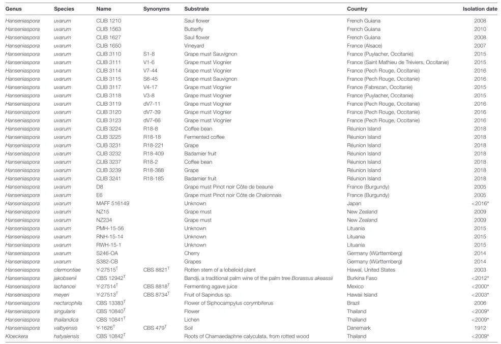
TABLE 1 | List of strains.
Genus Species Name Synonyms Substrate Country Isolation date
Hanseniaspora guilliermondii 11-1173 Toumodi Vin de rônier Ivory Coast 2016
Hanseniaspora guilliermondii 11-1176 Flower Ivory Coast 2016
Hanseniaspora guilliermondii CBS 1972 Grape juice Italy <1978∗
Hanseniaspora guilliermondii CBS 2591 Trachea of bee France <1978∗
Hanseniaspora guilliermondii CBS 95 Fermenting bottled tomatoes Netherlands <1978∗
Hanseniaspora guilliermondii CLIB 510T CBS 465T Infected nail South Africa 1978
Hanseniaspora guilliermondii CLIB 1559 Lemon French Guiana 2010
Hanseniaspora guilliermondii CLIB 3085 Toumodi Vin de rônier Ivory Coast 2016
Hanseniaspora guilliermondii CLIB 3092 V2-10 Grape must Viognier France (Sommières, Occitanie) 2015
Hanseniaspora guilliermondii CLIB 3093 S1-2 Grape must Sauvignon France (Puylacher, Occitanie) 2015
Hanseniaspora guilliermondii CLIB 3094 S1-3 Grape must Sauvignon France (Puylacher, Occitanie) 2015
Hanseniaspora guilliermondii CLIB 3095 S2-34 Grape must Sauvignon France (Saint Mathieu de Tréviers, Occitanie) 2015
Hanseniaspora guilliermondii CLIB 3096 S4-9 Grape must Sauvignon France (Ouveillan, Occitanie) 2015
Hanseniaspora guilliermondii CLIB 3097 S4-16 Grape must Sauvignon France (Ouveillan, Occitanie) 2015
Hanseniaspora guilliermondii CLIB 3098 V2-1 Grape must Viognier France (Sommières, Occitanie) 2015
Hanseniaspora guilliermondii CLIB 3099 V2-2 Grape must Viognier France (Sommières, Occitanie) 2015
Hanseniaspora guilliermondii CLIB 3100 V5-12 Grape must Viognier France (Pech Rouge, Occitanie) 2015
Hanseniaspora guilliermondii CLIB 3206 V1-9 Grape must Viognier France (Saint Mathieu de Tréviers, Occitanie) 2015
Hanseniaspora guilliermondii CLIB 3207 BIA-V1L15 Grape must Viognier France (Saint Mathieu de Tréviers, Occitanie) 2015
Hanseniaspora guilliermondii CLIB 3208 V5-13 Grape must Viognier France (Pech Rouge, Occitanie) 2015
Hanseniaspora guilliermondii CLIB 3209 V5-30 Grape must Viognier France (Pech Rouge, Occitanie) 2015
Hanseniaspora guilliermondii CLIB 3210 S4-54 Grape must Sauvignon France (Ouveillan, Occitanie) 2015
Hanseniaspora guilliermondii CLIB 3228 R18-212 Fermented juice of sugar cane (3 days) Réunion Island 2018
Hanseniaspora opuntiae x pseudoguilliermondii CCY46-1-3 Fruit; plum tree (Prunus domestica L. ‘Stanley’) Slovakia (Malé Leváre) 2009
Hanseniaspora opuntiae x pseudoguilliermondii CCY46-1-3a Single cell colony of CCY46-1-3 Lab strain 2019
Hanseniaspora opuntiae x pseudoguilliermondii CCY46-1-3b Single cell colony of CCY46-1-3 Lab strain 2019
Hanseniaspora opuntiae x pseudoguilliermondii CCY46-1-3c Single cell colony of CCY46-1-3 Lab strain 2019
Hanseniaspora opuntiae x pseudoguilliermondii CCY46-1-3d Single cell colony of CCY46-1-3 Lab strain 2019
Hanseniaspora opuntiae x pseudoguilliermondii CLIB 3101 V3-28 Grape must Viognier France (Puylacher, Occitanie) 2015
Hanseniaspora opuntiae x pseudoguilliermondii DBVPG 5828 Soil close to plum tree Algeria 2010
Hanseniaspora opuntiae x pseudoguilliermondii CLIB 3263 M18-204 Fermented pineapple Mayotte Island 2018
Hanseniaspora opuntiae x pseudoguilliermondii CLIB 3313 M18-207 Fermented pineapple Mayotte Island 2018
Hanseniaspora opuntiae x pseudoguilliermondii CLIB 3265 M18-215 Fermented pineapple Mayotte Island 2018
Hanseniaspora opuntiae 11-1102 Fruit papaya Guatemala (Flores) 2009
Hanseniaspora opuntiae 11-1139 Fallen fruit El Salvador (San Salvador) 2009
Hanseniaspora opuntiae 11-1184 Flower (Hybiscus) Palau (Ngerekebesang) 2010
Hanseniaspora opuntiae 11-1196 Rotten fruit (Syzygium malaccense) Palau (Koror) 2010
Hanseniaspora opuntiae CLIB 1203 Ivy French Guiana 2008
Hanseniaspora opuntiae CLIB 1208 Papaya French Guiana 2008
Hanseniaspora opuntiae CLIB 1557 Papaya French Guiana 2010
(Continued) Fr ontiers in Micr obiology | www .fr ontiersin.org 3 January 2020 | V olume 10 | Article 2960
fmic b-10-02960 De cember 28, 2019 T ime: 15:50 # 4 Saubin et al. Hanseniaspora Interspecific Hybrids TABLE 1 | Continued
Genus Species Name Synonyms Substrate Country Isolation date
Hanseniaspora opuntiae MUCL 49139T CBS 8733T Rot Hawaii Island <2003∗
Hanseniaspora opuntiae CLIB 1564 Ant French Guiana 2010
Hanseniaspora opuntiae CLIB 3102 V1-109 Grape must Viognier France (Saint Mathieu de Tréviers, Occitanie) 2015
Hanseniaspora opuntiae CLIB 3103 V3-16 Grape must Viognier France (Puylacher, Occitanie) 2015
Hanseniaspora opuntiae CLIB 3104 V4-31 Grape must Viognier France (Fabrezan, Occitanie) 2015
Hanseniaspora opuntiae CLIB 3105 S1-11 Grape must Sauvignon France (Puylacher, Occitanie) 2015
Hanseniaspora opuntiae CLIB 3106 S1-14 Grape must Sauvignon France (Puylacher, Occitanie) 2015
Hanseniaspora opuntiae CLIB 3107 BIA-S2L2 Grape must Sauvignon France (Saint Mathieu de Tréviers, Occitanie) 2015
Hanseniaspora opuntiae CLIB 3108 V1-1 Grape must Viognier France (Saint Mathieu de Tréviers, Occitanie) 2015
Hanseniaspora opuntiae CLIB 3109 V3-27 Grape must Viognier France (Puylacher, Occitanie) 2015
Hanseniaspora opuntiae CLIB 3203 V2-12 Grape must Viognier France (Sommières, Occitanie) 2015
Hanseniaspora opuntiae CLIB 3204 V2-31 Grape must Viognier France (Sommières, Occitanie) 2015
Hanseniaspora opuntiae CLIB 3205 S2-25 Grape must Sauvignon France (Saint Mathieu de Tréviers, Occitanie) 2015
Hanseniaspora opuntiae CLIB 3227 R18-143 "Grenadille" fruit Réunion Island 2018
Hanseniaspora opuntiae CLIB 3230 R18-219 Lemonade (3 days fermentation) Réunion Island 2018
Hanseniaspora opuntiae CLIB 3234 R18-484 Lemonade (3 days fermentation) Réunion Island 2018
Hanseniaspora pseudoguilliermondii 11-494 Fruit papaya The Philippines (Manila) 2011
Hanseniaspora pseudoguilliermondii CBS 8772T Orange juice concentrate unknown <2006∗
Hanseniaspora pseudoguilliermondii CLIB 1441 Orange French Guiana 2010
Hanseniaspora pseudoguilliermondii CLIB 3226 R18-113 Fruit Evi Réunion Island 2018
Hanseniaspora pseudoguilliermondii CLIB 3229 R18-218 Lemonade (3 days fermentation) Réunion Island 2018
Hanseniaspora pseudoguilliermondii CLIB 3233 Fruit French Guiana 2010
Hanseniaspora uvarum 10-1471 Grape Slovakia (Malá T`r ˇna) 2015
Hanseniaspora uvarum 11-1148 Flower Guatemala (Guatemala City) 2009
Hanseniaspora uvarum 11-1288 Flower (Leguminosae) Romania (Bucuresti) 2006
Hanseniaspora uvarum A1 Grape must Pinot noir Côte de nuit France (Burgundy) 2005
Hanseniaspora uvarum A4 Grape must Pinot noir Côte de nuit France (Burgundy) 2005
Hanseniaspora uvarum B2 Grape must Pinot noir Côte de nuit France (Burgundy) 2005
Hanseniaspora uvarum C4 Grape must Pinot noir Côte de beaune France (Burgundy) 2005
Hanseniaspora uvarum CBS 2583 Fermenting cucumber brine United States ?
Hanseniaspora uvarum CBS 2585 Sour dough Portugal 1978
Hanseniaspora uvarum CBS 2588 Tanning fluid France ?
Hanseniaspora uvarum CBS 286 Soil Indonesia <1934∗
Hanseniaspora uvarum CCY25-6-34 Grape must (7 days fermentation), variety Green Veltliner Slovakia (Strekov) 2008
Hanseniaspora uvarum CCY25-6-36 Soil adjacent to apricot tree Slovakia (Malé Zálužie) 2013
Hanseniaspora uvarum CCY46-1-2 Fresh-water lake Slovakia (Plaveck ˙y Štvrtok) 1987
Hanseniaspora uvarum CLIB 303T CBS 314T Muscatel grape Ukraine <1978∗
Hanseniaspora uvarum CLIB 512 Soil Danemark 1978
Hanseniaspora uvarum CLIB 979 Sylvaner start AF France (Alsace) 2001
Hanseniaspora uvarum CLIB 1207 Cacao Berry French Guiana 2008
Hanseniaspora uvarum CLIB 1209 Saul flower French Guiana 2008
(Continued) Fr ontiers in Micr obiology | www .fr ontiersin.org 4 January 2020 | V olume 10 | Article 2960
fmic b-10-02960 De cember 28, 2019 T ime: 15:50 # 5 Saubin et al. Hanseniaspora Interspecific Hybrids TABLE 1 | Continued
Genus Species Name Synonyms Substrate Country Isolation date
Hanseniaspora uvarum CLIB 1210 Saul flower French Guiana 2008
Hanseniaspora uvarum CLIB 1563 Butterfly French Guiana 2010
Hanseniaspora uvarum CLIB 1627 Saul flower French Guiana 2008
Hanseniaspora uvarum CLIB 1650 Vineyard France (Alsace) 2007
Hanseniaspora uvarum CLIB 3110 S1-8 Grape must Sauvignon France (Puylacher, Occitanie) 2015
Hanseniaspora uvarum CLIB 3111 V1-6 Grape must Viognier France (Saint Mathieu de Tréviers, Occitanie) 2015
Hanseniaspora uvarum CLIB 3114 V7-44 Grape must Viognier France (Pech Rouge, Occitanie) 2016
Hanseniaspora uvarum CLIB 3115 S6-45 Grape must Sauvignon France (Pech Rouge, Occitanie) 2016
Hanseniaspora uvarum CLIB 3117 V4-17 Grape must Viognier France (Fabrezan, Occitanie) 2015
Hanseniaspora uvarum CLIB 3118 V3-8 Grape must Viognier France (Puylacher, Occitanie) 2015
Hanseniaspora uvarum CLIB 3119 dV7-11 Grape must Viognier France (Pech Rouge, Occitanie) 2016
Hanseniaspora uvarum CLIB 3120 dV7-39 Grape must Viognier France (Pech Rouge, Occitanie) 2016
Hanseniaspora uvarum CLIB 3123 dV7-66 Grape must Viognier France (Pech Rouge, Occitanie) 2016
Hanseniaspora uvarum CLIB 3224 R18-8 Coffee bean Réunion Island 2018
Hanseniaspora uvarum CLIB 3225 R18-18 Fermented coffee Réunion Island 2018
Hanseniaspora uvarum CLIB 3231 R18-221 Grape Réunion Island 2018
Hanseniaspora uvarum CLIB 3232 R18-409 Badamier fruit Réunion Island 2018
Hanseniaspora uvarum CLIB 3237 R18-2 Coffee bean Réunion Island 2018
Hanseniaspora uvarum CLIB 3239 R18-388 Grape Réunion Island 2018
Hanseniaspora uvarum CLIB 3241 R18-185 Badamier fruit Réunion Island 2018
Hanseniaspora uvarum D8 Grape must Pinot noir Côte de beaune France (Burgundy) 2005
Hanseniaspora uvarum E6 Grape must Pinot noir Côte de Chalonnais France (Burgundy) 2005
Hanseniaspora uvarum MAFF 516149 Unknown Japan <2016∗
Hanseniaspora uvarum NZ15 Grape must New Zealand 2009
Hanseniaspora uvarum NZ234 Grape must New Zealand 2009
Hanseniaspora uvarum PMH-15-56 Unknown Lituania 2015
Hanseniaspora uvarum RNH-15-14 Unknown Lituania 2015
Hanseniaspora uvarum RWH-15-1 Unknown Lituania 2015
Hanseniaspora uvarum S246-OA Cherry Germany (Württemberg) 2014
Hanseniaspora uvarum S382-CB Grapes Germany (Württemberg) 2014
Hanseniaspora clermontiae Y-27515T CBS 8821T Rotten stem of a lobelioid plant Hawaï, United States 2003
Hanseniaspora jakobsenii CBS 12942T Bandji, a traditional palm wine of the palm tree Borassus akeassii Burkina Faso <2012∗
Hanseniaspora lachancei Y-27514T CBS 8818T Fermenting agave juice Mexico <2000∗
Hanseniaspora meyeri Y-27513T CBS 8734T Fruit of Sapindus sp. Hawaii Island <2003∗
Hanseniaspora nectarophila CBS 13383T Flower of Siphocampylus corymbiferus Brazil 2006
Hanseniaspora singularis CBS 10840T Flower Thailand <2009∗
Hanseniaspora thailandica CBS 10841T Lichen Thailand <2009∗
Hanseniaspora valbyensis Y-1626T CBS 479T Soil Danemark 1912
Kloeckera hatyaiensis CBS 10842T Roots of Chamaedaphne calyculata, from rotted wood Thailand <2009∗
∗
Isolation date is anterior to publication or sequence deposition related to this strain.
Fr ontiers in Micr obiology | www .fr ontiersin.org 5 January 2020 | V olume 10 | Article 2960
fmicb-10-02960 December 28, 2019 Time: 15:50 # 6
Saubin et al. Hanseniaspora Interspecific Hybrids
carried out by polymerase chain reaction (PCR) with the kit Taq
Mix (Dongsheng Biotech II) in a 40 µL reaction volume with
a first step of DNA denaturation at 94◦
C for 3 min, followed
by 30 cycles of DNA denaturation at 94◦
C for 30 s, primer hybridization at 55◦
C for 30 s, and elongation at 72◦
C for 1 min.
A final elongation was performed at 72◦
C for 3 min. All PCR
reaction were performed in a SimpliAmpTM thermal Cycler
(Applied BiosystemsTM).
Multilocus Sequence Typing
Five housekeeping genes previously used for MLST studies
in other yeast species (Bougnoux et al., 2002; Munoz et al.,
2009) were selected: ACC1, ADP1, GLN4, RPN2, and VSP13
(Supplementary Table S1). For each marker, homologous
sequences ofH. guilliermondii, H. opuntiae and H. uvarum were
retrieved from available genomes (Sternes et al., 2016; Seixas
et al., 2019) and aligned with Multalin (Corpet, 1988). A set of 11 primer pairs were designed in regions highly conserved surrounding variable sequences of 200–1100 nucleotides. Three additional markers were chosen in intergenic regions of
H. uvarum genome using Artemis (Rutherford et al., 2000) as
a visualization tool: between the homolog of MET5 and the upstream gene, between homologs of SKI2 and DUS3, and between RNR2 and CRM1. Then, homologous sequences of H. guilliermondii, and H. opuntiae were extracted from EMBL
files with Artemis and aligned with that of H. uvarum using
Multalin. Five primer pairs were designed in these regions the same way as for housekeeping genes (Supplementary Table S1). Selection of the best primer pairs was performed on an initial
set of four strains of Hanseniaspora: H. uvarum CLIB 303T,
H. opuntiae MUCL 49139T, H. guilliermondii CLIB 510T, and
H. pseudoguilliermondii CLIB 1441. Markers of these strains were amplified and sequenced with each pair of primers. The
PCR conditions in a 40 µL reaction volume were: 3 min
at 94◦
C followed by 30 cycles of 30 s at 94◦
C, 30 s at 55◦
C, and 1 min at 72◦
C, with a final step of 2 min at 72◦
C. Agarose gel electrophoresis was performed to select the best primer pairs. To this end, two criteria were used: (1) amplicon of good intensity in the four strains, (2) a single band per amplicon. Among the 16 primer pairs tested, five were retained and additional pairs of primers were designed specifically to amplify the selected markers in the divergent
strain Y-1626 of H. valbyensis, considered as the outgroup for
phylogenetic trees. In total, 107 strains of Hanseniaspora were
amplified and sequenced likewise with each of the five selected primer pairs, in order to detect heterozygous sites, to count polymorphic sites inside each species, and to build phylogenetic
trees. Sequences of homologous genes in H. guilliermondii
UTAD222 and H. uvarum AWRI 3580 were added to the
alignments. For each marker sequence, heterozygous sites were
looked for on Chromas (Technelysium)2, and replaced by
the corresponding degenerated base. Resulting sequences were aligned with clustal, manually cleaned for complex regions with gaps and highly variable positions, and then concatenated using Seaview (Gouy et al., 2010).
2http://technelysium.com.au/wp/
For phylogenetic analysis at the species level, concatenated sequences were converted into bi-allelic sequences using an in-house python script. Phylogenetic trees were constructed by
maximum likelihood using phyML (Guindon et al., 2009) with
a GTR substitution model. Robustness of the tree was assessed by the approximate likelihood ratio test approach (aLRT) and bootstrap of 100 replicates.
Population Structure Analysis
The concatenated sequence file for the five MLST markers
was converted into a STRUCTURE (Pritchard et al., 2000)
compatible format with an in-house Perl script where each base is encoded by two digits with values between 0 and 3, allowing consideration of bi-allelic positions. Then, population structure
was analyzed with STRUCTURE and InStruct (Gao et al., 2007).
Ten runs (STRUCTURE) and 15 runs (InStruct) were performed and the best partitioning was determined for each software output from different criteria: the best likelihood, the increase of likelihood, and the variation between the runs as proposed
by CLUMPAK (Kopelman et al., 2015), and using the best DIC
(Deviance information criterion) for Instruct. The most frequent consensus partition was then inferred at the optimal number of groups K with CLUMPAK.
Analysis of Genetic Diversity
Nucleotide diversity was compared between each marker at the
species level using the statistics5 and π (Nei and Li, 1979).
For that purpose, non-concatenated marker sequences were turned into bi-allelic sequences and analyzed with the R package
PopGenome (Pfeifer et al., 2014). In order to infer the mating
system, selfing rates were estimated for each species from the Fis obtained with the R package Genepop v1.13 after conversion of the structure data file to the genepop format using PGDspider (Lischer and Excoffier, 2012), and using the RMES software (David et al., 2007) from the heterozygosity profile obtained with a custom script.
Flow Cytometry
For flow cytometry analysis, cells were first grown overnight
in YPD medium at 28◦
C under 180 rpm agitation, and then
diluted to OD600 0.1 in 10 mL YPD and placed in the same
conditions during 5 h. After OD measuring, about 107 cells
were centrifuged 1 min at 10,000 rpm with 1 mL water. The cell pellets were then suspended in 1 mL water, added drop by drop in 8 mL of ethanol 75% with permanent vortexing and finally stored at 5◦
C. After one night at 5◦
C, the cells were centrifuged 5 min at 3,000 rpm, suspended in 1 mL of PBS buffer and centrifuged once again 1 min at 13,000 rpm. Cell
pellets were then suspended in 500µL of RNase A (2 mg/mL
in 10 mM Tris-Cl and 15 mM NaCl) and incubated 1 h at 37◦
C. Finally, cells were treated 1 h at 50◦
C with 200 µL of
1 mg/mL of proteinase K diluted in PBS buffer, centrifuged and
suspended in 500µL of PBS buffer before being sonicated 15 s
in a Branson Sonifier 250 sonicator at 10% of the maximum
power. About 106 cells were labeled with SYTOX R
green
(Invitrogen) in 250µL at a final concentration of 1 µM. The
DNA content was determined on a C6 Accuri (Ann Arbor, MI,
fmicb-10-02960 December 28, 2019 Time: 15:50 # 7
Saubin et al. Hanseniaspora Interspecific Hybrids
United States) spectrophotometer with an excitation wavelength of 488 nm and an emission wavelength of 530 ± 15 nm. Acquisition was performed on 30,000 events observed with a gating on forward scatter/side scatter signal. The flow rate was set to approximately 2,000 events per second (medium
flow, 35 µL/min; core, 16 µm). The Python (v. 2.7.13) module
FlowCytometryTools (v. 0.5.0) was used for data extraction and manipulation and an “in house” R (v. 3.3.3) script was developed for graphical representations.
Karyotyping
Yeast karyotyping was achieved by contour-clamped
homogeneous electric field (CHEF) gel electrophoresis.
Plugs of yeast chromosomes were prepared according to (Vezinhet et al., 1990). The CHEF-DR III pulsed-field gel electrophoresis system (Bio-Rad, Hercules, CA, United States) was set, for a first karyotype, to 3 V/cm with a switching time of 360 s for 23 h and then to 6 V/cm with pulses of
70–90 s for 20 h in 0.5X TBE buffer at 13.5◦
C in 1% Seakem GTG agarose (FMC) gel. A second karyotype was made to better separate the high molecular weight chromosomes by setting the apparatus to 3 V/cm with pulses of 480 s for 18.1 h, to 4 V/cm with pulses of 300 s for 24 h and to 6 V/cm
with pulses 90 s for 12 h in 0.5X TBE buffer at 13◦
C. We
used S. cerevisiae CLIB 112 (=YNN295) chromosomal DNA
as a marker. The agarose gels were stained with ethidium
bromide (0.5 µg/mL) and washed with water before being
visualized under UV.
Genome Sequencing and Analysis
The DNAs of strains CCY46-1-3, CLIB 3101, DBVPG 5828, CLIB 3263, CLIB 3313, CCY-46-1-3a, and S382-CB were used to construct shotgun 400-bp insert libraries, which were sequenced in paired-end (2 × 150 bp) using the Illumina HiSeq2000 platform (Supplementary Table S2). Sequencing
reads were cleaned using Fastp v. 0.19.4 (Chen et al., 2018)
and used for retrieving MLST marker sequences and whole genome analysis.
First, the cleaned reads of the six interspecific hybrids were used for mapping against the five amplified markers of strains
CLIB 1441 (H. pseudoguilliermondii) and MUCL 49139T(Type
strain ofH. opuntiae) using HiSat2 v. 2.0.4 (Kim et al., 2015) and the samtools package v. 1.9 (Li et al., 2009). SNPs and Indels were visualized using Artemis to determine manually the sequence of each allele. The different haplotypes obtained in this way were finally considered for the MLST analyses. Second, the cleaned reads were used to estimate the proportion of each parental
species using SppIDer (Langdon et al., 2018). This tool only
requires the genome sequences of each parental species, in FASTA format, and the sequencing reads of hybrid strains. It is rooted on read mapping and provides various statistics and graphical representations of read coverage against the considered parental
genomes. The genome assembly of H. pseudoguilliermondii
ZIM213 (Shen et al., 2018) andH. opuntiae AWRI 3578 (Sternes
et al., 2016) were used as references. In order to ease the reading and the interpretation of SppIDer analyses, the scaffolds of these two genomes were preliminary re-ordered to make
their sequences collinear as much as possible. To do so, we
used MUMmer tool suite v4,0,0beta (Marcais et al., 2018).
Maximal unique matches were retrieved with nucmer and the optimal scaffold order was obtained with the -layout option of mummerplot. The dotplot illustrating the re-ordered genome sequences is available in Supplementary Figure S1 on which we also reported the genomic position of the five MLST markers used in this study.
Genome-wide genotyping of hybrid strains was performed as follows. First, cleaned reads were separately aligned on both aforementioned reference genomes using HiSat2 under no-mixed and no-spliced-alignment options, leading to two SAM/BAM files per strain. Mapped read pairs were extracted from these files with samtools, and ambiguous reads that aligned on both reference genomes were removed using BBMap from the BBTools
suite3 (Supplementary Table S2). Optical and PCR duplicates
were then removed with the MarkDuplicates command from
Picard v. 2.9.04. Variant calling at the strain level was finally
performed using the HaplotypeCaller from GATK v. 3.7, the
Genome Analysis Toolkit (McKenna et al., 2010). At this step,
raw SNPs and Indels identified were outputted into genomic VCF files (gVCF).
In order to visualize the level of heterozygosity along both parental genomes, each individual gVCF file (two per strain) was first converted into a VCF file using GATK GenotypeGVCFs tool. Raw SNPs were extracted and submitted to a hard-filtering procedure. SNPs that matched the following criteria
were filtered out: QualByDepth (QD) < 5.0, FisherStrand
(FS)> 55, StrandOddsRation (SOR) > 2.0, RMSMappingQuality
(MQ) < 40, MappingQualityRankSumTest (MQRankSum)
< −5.0, and ReadPosRankSumTest (ReadPosRankSum) < −5.0. These cutoff thresholds were chosen after visual inspection of the respective distributions of these annotation fields. The rational here was to find a balance between a global transition/transversion ratio (Ts/Tv) close to 2 – an indicator of an effective false positive cleaning – and minimizing the loss of true biological SNPs. Following the hard-filtering procedure, the final call sets were constituted by retaining only biallelic positions. Heterozygous SNPs were then extracted using SnpSift v. 4.3 (Cingolani et al., 2012) and were quantified within 10-kb sliding windows along reference genomes. For that purpose, bedtools
v. 2.27.1 (Quinlan and Hall, 2010) was used for both genome
splitting into consecutive windows and SNP counting. The minor allele coverage (in percentage of the total read coverage) at biallelic positions was computed for each hybrid genome
on 2-kb non-overlapping sliding windows across H. opuntiae
reference genome.
Assessment of the genetic distance between hybrid strains relied on a similar genotyping approach as that described above, the only difference being that it was based on a joint variant analysis. Briefly, individual gVCF files obtained from one of the parental genome were first pooled together into a cohort gVCF file using the CombineGVCFs tool from GATK. This cohort file was then converted into a VCF file, and the final
3sourceforge.net/projects/bbmap 4https://broadinstitute.github.io/picard
fmicb-10-02960 December 28, 2019 Time: 15:50 # 8
Saubin et al. Hanseniaspora Interspecific Hybrids
SNP call set was constituted as previously described. Only an extra filtering step was added in order to remove all positions showing missing data. This cohort call set was first used to estimate nucleotide divergence between strains with the help
of Plink v. 1.9 (Purcell et al., 2007) in order to quantify the
number of SNPs sharing 0 and 1 Identity By State (IBS0 and IBS1, respectively) in pairwise comparisons for all strains. Principal component analyses were performed on cohort SNP data of each
sub-genome using the R package adegenet v. 2.1.1 (Jombart, 2008;
Jombart and Ahmed, 2011).
In order to estimate the level of heterozygosity inH. uvarum
triploid strain S382-CB, we measured the read coverage at each heterozygous position. For that purpose, we genotyped the strain
as described above, using H. uvarum strain AWRI 3580 as
reference genome (Sternes et al., 2016). Only the hard-filtering
procedure was applied to the individual VCF file so that all levels of ploidy were retained in the final individual-level call set. Bi- and tri-allelic positions were manually extracted according to the content of the GT and AD fields. Briefly, all positions enclosing either a 0/1- or a 1/2-GT field with less than 5% of total reads supporting the reference allele (AD field) were considered bi-allelic. Conversely, all positions containing a 1/2-GT field with reads supporting both reference and two alternative alleles were considered tri-allelic. Read coverage values were normalized in order to take into account variations of sequencing depth between positions.
Gene Annotation
As the genome annotation ofH. pseudoguilliermondii ZIM213
(Shen et al., 2018) was not available, we proceeded to the annotation of protein-coding genes of 576-kb corresponding to the region of CLIB 3263, which showed a 1.4 X coverage compared to the rest of the genome. Non-overlapping open reading frames larger than 180 nt, i.e., with a translated sequence larger than 60 amino acids were compared to S. cerevisiae proteome using blastp with an E-value threshold of 1.e-10. Sequences without any hit were compared to nr database limited to fungi at the NCBI using blastp. Sequences with positive matches were manually curated for initiator methionine (iMet) and introns. Amino acid sequences with an iMET and smaller than 100 aa without any homologs were discarded. Functional annotations, if any, were transferred from
their putative homologs. Similarly, the H. pseudoguilliermondii
homologous sequence of 126-kb corresponding to the region of H. opuntiae lost in CCY 46-1-3a was annotated the same way and
manually compared toH. opuntiae for synteny and gene model
prediction using artemis.
MAT Locus Analysis
The MAT loci of the reference genomes ZIM213 and AWRI 3578 were annotated (Supplementary Figure S2). Sequencing reads of the hybrid genomes were aligned on reference MAT loci using HiSat2 under no-mixed, no-discordant and
no-spliced-alignment options and k = 1 parameter. Mapped read pairs
were extracted with samtools view and bam2fastq. The recovered reads were then assembled with SPAdes with default parameters (Bankevich et al., 2012).
RESULTS
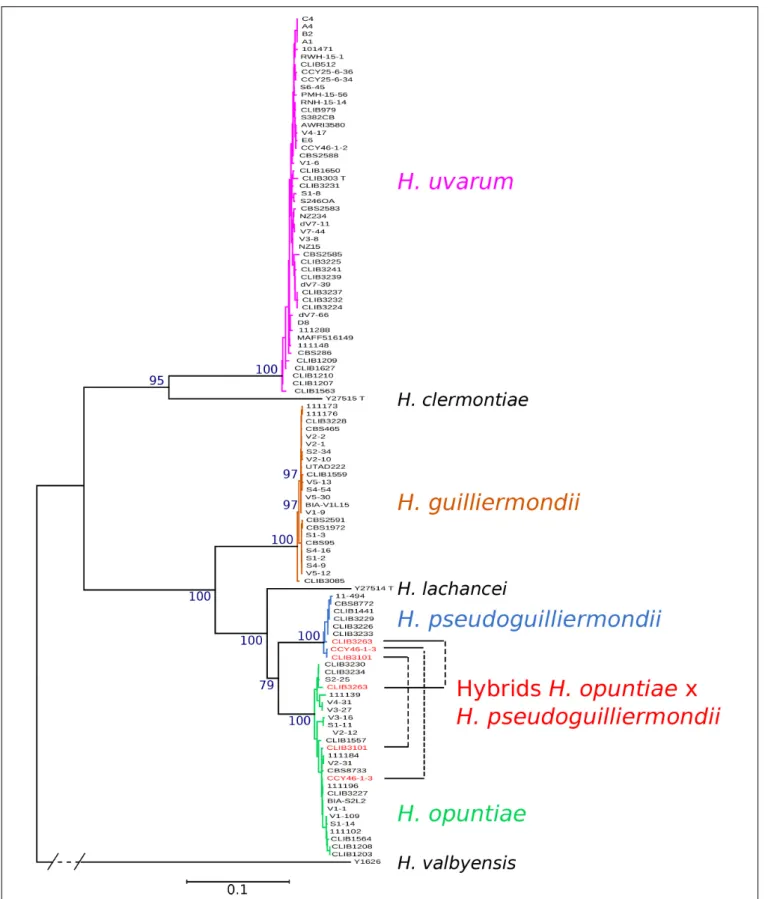
Species Delineation in Hanseniaspora
Among 16 primer pairs designed in eight markers, five were selected because they amplified and gave an amplicon of
the same size for H. uvarum, H. opuntiae, H. guilliermondii,
and H. pseudoguilliermondii, as estimated on agarose gel. The
selected loci and primer pairs were GLN4 Glutamine tRNA synthetase (GLN4F2/GLN4R2), ADP1 ATP-dependent permease (ADP1F2/ADP1R2), intergenic region upstream of MET5 sulfite reductase beta subunit (MET5F1/MET5R1), RPN2 subunit of the 26S proteasome (RPN2F2/RPN2R2) and VPS13 vacuolar protein sorting (VPS13F2/VPS13R2). 107 strains were successfully amplified and sequenced for MET5, ADP1, RPN2, VPS13
and GLN4, whereas strains of more divergent Hanseniaspora
species could not be amplified with some primer pairs (Supplementary Table S3). Among the 107 strains, three showed complex chromatograms, characteristic of hybrid markers. After unsuccessful attempts to clone the different alleles due to sequence recombination probably during the PCR step, we finally decided to sequence the whole genomes of CLIB 3101, CCY46-1-3 and DBVPG 5828. Their respective alleles were manually reconstructed by mapping the reads against the five markers of
both CLIB 1441 (H. pseudoguilliermondii) and MUCL 49139T
(H. opuntiae). Two to three divergent alleles per marker were obtained for each of the three strains, suggesting that the strains
are H. pseudoguilliermondii × H. opuntiae hybrids. The only
exception was an absence of H. pseudoguilliermondii MET5 in
DBVPG 5828, a region that may have been lost or rearranged. Phylogenetic trees were constructed from the concatenation of the five markers. Four clearly distinct groups of strains emerged from the tree, corresponding to the four studied species (Figure 1). The topology of the concatenated tree, which included H. valbyensis as outgroup and H. lachancei and H. clermontiae as internal references, was coherent with the topology based on concatenated datasets of actin, D1D2 and ITS gene sequences (Cadez et al., 2006) or on 1,034 orthologous proteins (Steenwyk et al., 2019). As expected, strains of H. pseudoguilliermondii appeared as a separate group, very close to the group of H. opuntiae strains. Alleles of CLIB 3101, CLIB3263 (its genome was also sequenced, see below) and CCY46-1-3, grouped with
bothH. pseudoguilliermondii and H. opuntiae strains, confirming
that these strains are hybrids that derive from a cross between
the two most closely related species, H. pseudoguilliermondii
andH. opuntiae.
Population Structure in H. uvarum,
H. guilliermondii, and H. opuntiae
In order to explore more thoroughly the phylogenetic relationships between strains of the same species, and to find putative links between strains of the same substrate or geographical origin, we drew trees relying on the concatenated
marker sequences, species by species for H. uvarum,
H. guillermondii and H. opuntiae. H. pseudoguilliermondii was excluded from this analysis due to the insufficient number
of strains. For H. uvarum (Supplementary Figure S3) and
fmicb-10-02960 December 28, 2019 Time: 15:50 # 9
Saubin et al. Hanseniaspora Interspecific Hybrids
FIGURE 1 | Phylogenetic tree of Hanseniaspora strains based on the five marker sequences ADP1, GLN4, RPN2, VPS13, and MET5. The tree was constructed with PhyML based on the concatenated sequence of 3220 sites. DBVPG 5828, which lost H. pseudoguilliermondii MET5 allele, was not included in the phylogenetic tree. H. valbyensis Y-1626Twas used as an outgroup. Branch lengths are proportional to the number of sites that differentiate each pair of strains. Branch support was
estimated by the approximate likelihood ratio test approach (aLRT) and bootstrap of 100 replicates.
fmicb-10-02960 December 28, 2019 Time: 15:50 # 10
Saubin et al. Hanseniaspora Interspecific Hybrids
H. opuntiae (Supplementary Figure S4), the tree topology was generally supported by low bootstrap values and no clear correlation to substrate origin emerged. Nevertheless, for H. uvarum, a somehow clear phylogenetic signal was obtained for a few strains isolated from the French Guyana and for a group enriched in strains from La Réunion Island, which separated from the other isolates. This signal was echoed by the population structure analysis performed with STRUCTURE and InStruct that provided the same clustering for K equal to
three. In contrast, the phylogeny of H. guillermondii presented
two distinct populations and a single separate strain, CLIB 3085 from Ivory Coast, which attests of the presence of a probable third population (Supplementary Figure S5A). STRUCTURE and InStruct analysis confirmed the partitioning of the strains into three distinct populations (Supplementary Figure S5B). Interestingly, three strains, which were isolated in the same grape must in Ouveillan (Occitanie, France), belong to population 1 (S4–54) or population 2 (S4–16 and S4–9). Similarly, V5–13 and V5–30 belong to population 1 and V5–12 to population 2; all of them were isolated in Pech Rouge (Occitanie, France). Last,
for H. opuntiae, STRUCTURE and Instruct analyses did not
provide any convergent population structure in agreement with the MLST data (Supplementary Figure S4).
Hanseniaspora Species Exhibit
Differences in Nucleotide Variability
Analysis of heterozygous and polymorphic sites permitted us to estimate the relative divergence of markers within each species (Table 2). Whereas the most conserved marker was GLN4 in all species, the intergenic region MET5 displayed the highest
diversity with up to 10.24% of polymorphic sites and aπ value
up to 40.55 nucleotides per site inH. opuntiae. In this species,
MET5 alleles showed two different sizes differing by an insertion of five nucleotides in the intergenic region; some strains showed the presence of both types. Insertions of 3–4 nt were also found
in MET5 fromH. guilliermondii CLIB 3085 and some strains of
H. pseudoguilliermondii. We also found an insertion of 3 nt in
the coding sequence of RPN2 inH. uvarum, which corresponds
to an additional amino acid. From these data, we could also
observe differences between species. H. opuntiae showed the
highest percentages of polymorphic sites and π values in all
markers but GLN4, even if the number of strains studied was
two times less than inH. uvarum. In contrast, H. guilliermondii
appeared the least variable species, with values quite similar to
that ofH. pseudoguilliermondii for which we studied three times
fewer strains.
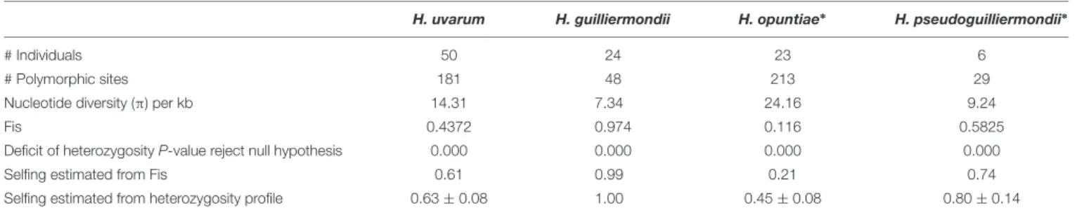
As the analysis of the genetic variation in each gene revealed high proportions of heterozygous loci variable across species, we wondered if these differences might originate from different life styles. The estimation of the different allelic frequencies revealed a highly significant deficit of heterozygosity for the four species. However, in relation with the Fis value of each population that varied from 0.1 to 1, the estimate of the selfing
rates s varied from 0.21 for H. opuntiae, 0.61 for H. uvarum,
0.74 for H. pseudoguilliermondii and 0.99 for H guillermondii
(Table 3). In order to avoid the impact of hidden population TABLE
2 | Divergence of markers in the four species of Hanseniaspora. H. uvarum H. guilliermondii H. opuntiae H. pseudoguilliermondii MLST locus ADP1 GLN4 MET5 RPN2 VPS13 ADP1 GLN4 MET5 RPN2 VPS13 ADP1 GLN4 MET5 RPN2 VPS13 ADP1 GLN4 MET5 RPN2 VPS13 # Sequences 50 50 50 50 50 24 24 24 24 24 26 26 26 26 26 9 9 9 9 9 # total sites 445 559 909 951 566 440 556 917 973 563 419 564 859 961 551 432 556 918 953 561 # heter ozygous sites 1 10 16 54 50 21 0 2 0 5 0 21 8 80 59 33 4 5 0 1 2 # polymorphic sites 2 17 21 59 60 24 7 4 21 11 5 22 8 88 60 35 5 5 16 14 5 % polymorphic sites 3.82 3.76 6.49 6.31 4.24 1.59 0.72 2.29 1.13 0.89 5.25 1.42 10.24 6.24 6.35 1.15 0.90 1.74 1.47 0.89 Nucleotide diversity (5 ) 3 4.35 2.87 14.52 17.33 10.00 2.52 1.79 11.12 6.53 3.37 4.62 2.21 40.55 16.9 16.76 3.39 4.11 9.89 10.89 3.33 1The number of heterozygous sites refers to the number of positions where at least one strain has a heterozygous position. 2The number of polymorphic sites refers to the number of positions where at least two alleles are present in the strain population, including heterozygous positions. 3Nucleotide diversity is measured by the average number of nucleotide differences between all pairs of strains in each alignment.
fmicb-10-02960 December 28, 2019 Time: 15:50 # 11
Saubin et al. Hanseniaspora Interspecific Hybrids
TABLE 3 | Statistics of genetic diversity applied to Hanseniaspora sequences.
H. uvarum H. guilliermondii H. opuntiae∗ H. pseudoguilliermondii∗
# Individuals 50 24 23 6
# Polymorphic sites 181 48 213 29
Nucleotide diversity (π) per kb 14.31 7.34 24.16 9.24
Fis 0.4372 0.974 0.116 0.5825
Deficit of heterozygosity P-value reject null hypothesis 0.000 0.000 0.000 0.000
Selfing estimated from Fis 0.61 0.99 0.21 0.74
Selfing estimated from heterozygosity profile 0.63 ± 0.08 1.00 0.45 ± 0.08 0.80 ± 0.14
∗
The three interspecific hybrids were removed from this analysis.
structure, we estimated Fis and s from a subset of H. uvarum
devoid of strains from Guyane or la Reunion and we obtained similar results indicating that population structure does not
explain Fis and s value forH uvarum. Last, selfing rates inferred
from the heterozygosity profile with RMES varied also in similar proportions with those obtained from Fis.
One major feature is the complete absence of heterozygous
sites in all strains ofH. guilliermondii but one, CLIB 3085, which
contrasts with the substantial heterozygosity of the other species. In order to check if these differences in heterozygosity might be due to differences in ploidy, we analyzed the ploidy of the strains of our strain set.
Variable Strain Ploidy in Hanseniaspora
Species
We used flow cytometry and compared the level of Sitox green
intensity in H. guilliermondii cells and in other Hanseniaspora
strains that were found heterozygous for the MLST markers, and are thus probably diploid. As shown in Figure 2A, the intensity
of all H. guilliermondii strains is of the same order as diploid
strains of other species, suggesting that these strains are also diploid. As for the hybrids, DBVPG 5828 (Figure 2B) and CLIB 3101 (Figure 2C) were clearly found triploid whereas it was more difficult to evaluate the ploidy of CCY46-1-3 (Supplementary
Figure S6). We also found another triploid strain, S382-CB,
for which only H. uvarum alleles were found in MLST. We
can suspect that this strain is not an interspecies hybrid but
rather aH. uvarum triploid (Figure 2D). Considering the quite
high number of hybrids that we found randomly, we decided to screen a collection of additional strains with MET5 as a selective marker. We found three candidate strains isolated from the same biological sample, fermented pineapple from Mayotte Island. All
of them have H. opuntiae and H. pseudoguilliermondii MET5
alleles. Two of them are diploid, CLIB 3313 and CLIB 3265, whereas the third one is tetraploid, CLIB 3263 (Figures 2A,E). As for the first hybrids, the genomes of strains CLIB 3313 and CLIB 3263 were sequenced and the deduced MLST marker sequences, identical for both strains, were added to the phylogenetic tree in Figure 1.
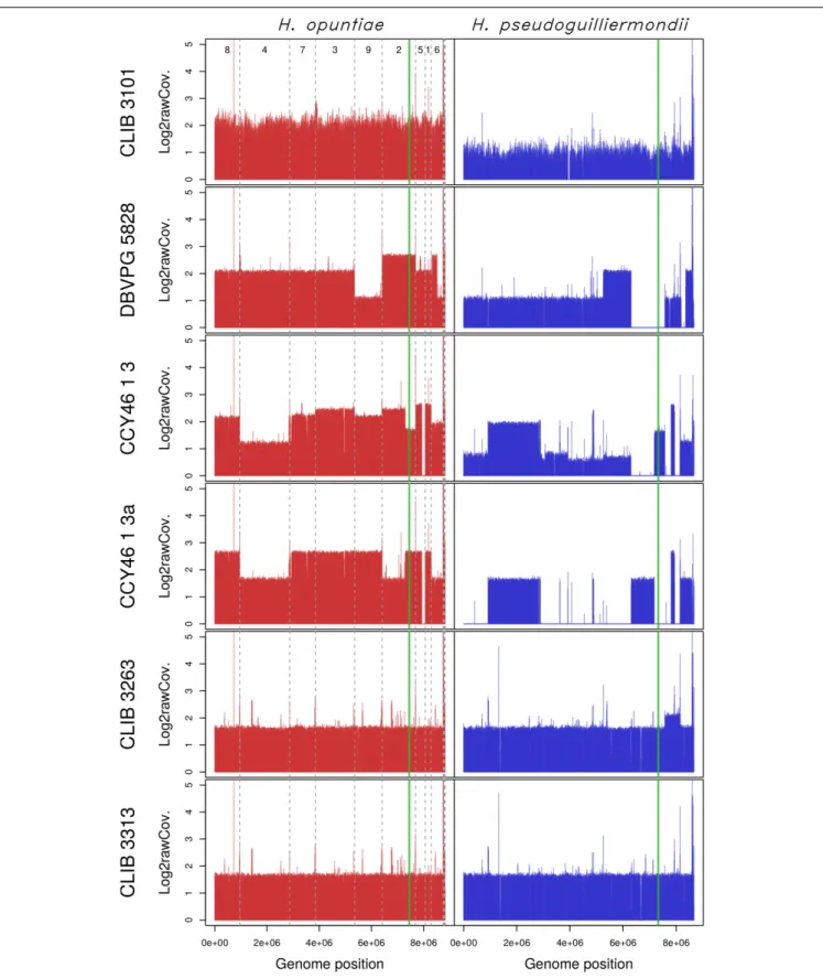
Genomic Structure of Hybrids
Considering that H. opuntiae and H. pseudoguilliermondii are
closely related but distinct species, we hypothesized that they might have different karyotypes as this is the case for sister
species in Saccharomyces (Fischer et al., 2000) and thus that
this could provide us with further clues about the genomic structure of hybrids. However, karyotypes of the type strains of parental species did not indicate any chromosome length polymorphism, and this was the case for the hybrids too (Supplementary Figure S7). We thus decided to sequence their genome with a shotgun strategy with Illumina sequencing chemistry, and to map the reads to reference genomes with
SppIDer. To this end, the genome of H. pseudoguilliermondii
ZIM213 (Shen et al., 2018) andH. opuntiae AWRI 3578 (Sternes
et al., 2016) were used as references. A preliminary analysis with H. guillermondii UTA222, H. uvarum AWRI 3580, H. osmophila
AWRI 3579 andH. vineae T02/19AF suggested that the hybrids
did not have a third parent (Supplementary Figure S8). The genomes of CLIB 3101, DBVPG 5828 and CCY46-1-3 showed an overall proportion of 2:1, suggesting that these strains are
allotriploid H. opuntiae (2n) × H. pseudoguilliermondii (1n),
with the nomenclature proposed by Nguyen and Boekhout (Nguyen and Boekhout, 2017). In CLIB 3101, the reads were homogeneously distributed along the chromosomes whereas in DBVPG5828, differences in parental genome contribution was observed along the chromosomes, accounting for numerous chromosomal rearrangements leading, in some cases, to losses
of H. pseudoguilliermondii genomic regions, which were
counterbalanced by a triploidization ofH. opuntiae homologous
regions (Figure 3). One of the lost regions contained the MET5
allele ofH. pseudoguilliermondii, which could not be amplified
in the MLST approach. The pattern of CCY46-1-3 was much more difficult to analyze as the read coverage sometimes showed intermediate values to 1n, 2n, or 3n. Together with the flow cytometry pattern, this result suggested that CCY46-1-3 might be a population of cells with different genomic contents. We thus selected four individual colonies from CCY46-1-3. They had the same pattern in flow cytometry, suggesting that they were all tetraploid (Supplementary Figure S5). Sequence analysis of the genome of one of them, CCY46-1-3a, showed large H. opuntiae genomic regions of probable ploidy 2n and 4n if we consider CCY46-1-3a is a tetraploid. Homologous regions
originated from the H. pseudoguilliermondii parent were 2n or
lost, respectively. A region of 126.5 kb long was lost inH. opuntiae
and is 4n in H. pseudoguilliermondii. One extremity of this
region corresponds to the extremity of the scaffolds in both reference genomes (PPNX01000020 in ZIM213; LPNL01000005 in AWRI 3578), which might be a subtelomeric region. The other extremity is internal to the scaffolds, at the locus of
fmicb-10-02960 December 28, 2019 Time: 15:50 # 12
Saubin et al. Hanseniaspora Interspecific Hybrids
FIGURE 2 | DNA content of Hanseniaspora strains as measured by flow cytometry. (A) Mean intensity of cell DNA at G1 (blue dots) and G2 (red dots) phases, normalized by the intensity of CBS 8772. Species name are colored in blue (H. opuntiae), orange (H. guilliermondii), turquoise (H. uvarum), green (hybrids), dark red (H. pseudoguilliermondii). (B,C,E) Intensity curve of H. opuntiae × H. pseudoguilliermondii hybrids (red) compared to the type strain of the parental species H. opuntiae in blue and H. pseudoguilliermondii in green.(D) Intensity curve of two H. uvarum strains, the type strain CLIB 303 and the triploid strain S382CB.
fmicb-10-02960 December 28, 2019 Time: 15:50 # 13
Saubin et al. Hanseniaspora Interspecific Hybrids
FIGURE 3 | Read coverage from the six hybrid strains along the genomes of the two parental species H. opuntiae (in red) and H. pseudoguilliermondii (in blue). Reference scaffolds were reordered beforehand so that the genome relative position (x-axis) is directly comparable between both parental species. Mean coverage values were computed with SppIDer tool with a sliding-window of 1700 nucleotides (without overlap) and normalized by the mean coverage of Hanseniaspora values present in Supplementary Figure S8. Values are expressed in a log2 scale. Dashed gray lines indicate scaffold boundaries. The vertical green line indicates the position of the MAT locus in each subgenome.
fmicb-10-02960 December 28, 2019 Time: 15:50 # 14
Saubin et al. Hanseniaspora Interspecific Hybrids
homologs of the tandem genesCSH2 and CSH3. Other changes
in read coverage occurred internal to H. opuntiae scaffolds
(e.g., in LPNL01000002, LPNL01000007, and LPNL01000008), which may suggest chromosomal rearrangements rather than chromosome loss and gain.
Sequencing of strains CLIB 3263 and CLIB 3313 showed
the same proportion of sequences from H. opuntiae and
H. pseudoguilliermondii parents, with the exception of a H. pseudoguilliermondii region of about 576 kb in CLIB3263, which had a coverage ratio of about 1.4 compared to the rest of the genome. Knowing that CLIB 3263 is tetraploid, this suggests that either a segmental or a chromosome duplication occurred
in only one of the two H. pseudoguilliermondii homologous
chromosomes – but in that case the ratio should be 1.5, not 1.4 – or that CLIB 3263 is a population of heterogeneous cells having undergone or not an event of segmental or chromosome duplication.
Genetic Diversity of Hybrids
To get clues about the event that led to the hybrid formation, we first investigated their MAT loci by comparison to those
of reference genomes. H. pseudoguilliermondii ZIM 213 has
a single MAT locus with only MATalpha1 gene. To recover the MATa locus we used CBS 8772, which is diploid and
possesses both MAT loci. The reference genome ofH. opuntiae
AWRI 3578 contains a MAT locus with both MATalpha1 and MATa2 genes (Supplementary Figure S2A). Mapping and assembly of the mapped reads of the six hybrids revealed that CCY46-1-3, CCY46-1-3a and DBVPG 5828 had both MATa
and MATalpha from H. opuntiae, suggesting that the parental
diploids were MATa/MATalpha, whereas CLIB 3101 had only a MATa locus (Supplementary Figure S2B). CCY46-1-3 and
CLIB 3101 had also aH. pseudoguilliermondii MATalpha locus
whereas CCY46-1-3a and DBVPG 5828 have lost this genomic
region, which has been replaced by itsH. opuntiae counterpart
(Figure 3). CLIB 3263 and CLIB 3313 had a single MAT locus
per subgenome, MATa from H. opuntiae and MATalpha from
H. pseudoguilliermondii.
Then, we investigated the level of heterozygosity in both subgenomes of each hybrid. A high level of heterozygosity,
up to 0.503%, was observed for H. opuntiae subgenome in
strains CLIB 3101, DBVPG 5828, CCY46-1-3 and its derivative
CCY46-1-3a, which suggests that H. opuntiae parental strains
were heterozygous diploids in each case (Table 4). This level is probably under-estimated, as only bi-allelic positions in the
TABLE 4 | Percentage of heterozygosity in H. opuntiae and H. pseudoguilliermondii subgenomes of hybrids.
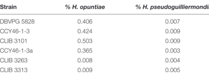
Strain % H. opuntiae % H. pseudoguilliermondii
DBVPG 5828 0.406 0.007 CCY46-1-3 0.424 0.009 CLIB 3101 0.503 0.009 CCY46-1-3a 0.365 0.003 CLIB 3263 0.008 0.004 CLIB 3313 0.009 0.005
population were considered. The distribution of heterozygous positions was not homogeneous along the chromosomes (Figure 4 and Supplementary Figure S9), and showed some regions of loss of heterozygosity (LOH). Interestingly, the largest LOH in CLIB 3101 and DBVPG 5828 covers the same region. In contrast, in these four strains a very low level of heterozygosity
was observed for theH. pseudoguilliermondii subgenomes, even
in the duplicated or triplicated chromosomal regions, which may correspond to false positive, sequencing errors or single nucleotide mutations after duplication. In strains CLIB 3263 and CLIB 3313, very few heterozygous sites were observed, neither
forH. opuntiae, nor for H. pseudoguilliermondii subgenomes.
This is coherent with the fact that CLIB 3313 is a diploid with 1n chromosomes from each parent and this suggests that the tetraploid strain CLIB 3263 derives from an auto-diploidization event. This also corroborates the subsequent 576-kb segmental duplication event in CLIB 3263.
As MLST marker sequences were almost identical in CLIB 3101 and DBVPG 5828 and as their regions of LOH were at the same genomic position, we suspected that both strains could derive from the same hybridization event. To address this hypothesis, we examined the pairwise allele sharing between strains across the genomic regions present in all hybrids, which
represents more than 95% of H. opuntiae and about 30% of
H. pseudoguilliermondii reference genomes. It appeared that CLIB 3101 and DBVPG 5828 showed a major divergence
regarding their respective H. opuntiae subgenomes. There are
7648 positions with no nucleotide in common (IBS0), and 49,107 positions (IBS1) where only one allele is in common (Figure 5A). These data suggest that the parental diploids were different and consequently that CLIB 3101 and DBVPG 5828 may derive from
two distinct hybridization events. TheH. pseudoguilliermondii
part is much less divergent with only 282 different positions between the two strains, which may be linked to the low level of
divergence observed inH. pseudoguilliermondii with the MLST
analysis. The same analysis performed on the other interspecific hybrid genomes showed that the three European strains are
different between them at least forH. opuntiae subgenomes and
very distant from the two strains from Mayotte Island for both subgenomes (Figure 5B).
Assessment of the heterozygosity inH. uvarum triploid strain
S382-CB revealed 55,355 bi-allelic positions with one allele in common with the reference strain AWRI 3580, 160 bi-allelic positions with both alleles being alternative to the reference allele, and 20 tri-allelic positions. Bi-allelic positions presented a distribution of read coverage of 1/3-2/3, which confirm the triploid nature of the strain (Supplementary Figure S10).
DISCUSSION
The aim of this study was to characterize and to clarify the genetic relationships between strains of a species complex with
a method that would facilitate further studies ofHanseniaspora
strains, especially strains isolated from vineyards. The results obtained from the MLST approach showed that even if five loci are not representative of the whole genome, our
fmicb-10-02960 December 28, 2019 Time: 15:50 # 15
Saubin et al. Hanseniaspora Interspecific Hybrids
FIGURE 4 | Density of heterozygous SNPs along H. opuntiae and H. pseudoguilliermondii collinearized parental genomes in hybrid strains. Each dot represents the total number of heterozygous SNPs enclosed within 10-kb sliding windows. Scaffold order of reference genomes is the same as in Figure 3. Dashed gray lines indicate scaffold boundaries.
markers are pertinent to discriminate species and therefore hybrids. Indeed, the topology of our multi-species tree is
consistent with that of Steenwyk et al. (2019) based on
1,034 orthologous groups, with strong bootstrap values. In comparison, trees based on classical taxonomic markers harbor variable topologies sometimes poorly supported. For instance,
the trees ofCadez et al. (2006, 2019)showed different topologies
due to the additional use of EF-1α in the latter tree. It
is therefore essential to have relevant markers that allow resolving accurately species delineation. Our combination of markers is also sufficiently divergent to unveil the presence
of subpopulations, as this is the case for H. guilliermondii
populations. However, we failed to establish a clear population structure related to substrate origin or geographical localization
fmicb-10-02960 December 28, 2019 Time: 15:50 # 16
Saubin et al. Hanseniaspora Interspecific Hybrids
FIGURE 5 | Analysis of individual-based genetic distance between hybrid strains. (A) Heatmap representations of the number of differentiating SNPs between strain pairs. The divergence between strains was assessed here using the number of IBS0 and IBS1 (Identity By State) SNPs. A SNP is defined as IBS0 when no allele is shared within the considered pair, and IBS1 when one allele is found in common. Therefore, as hybrids are haploid on their H. pseudoguilliermondii subgenome part (or auto-diploid in the case of CLIB 3263), only IBS0 SNPs were analyzed in that context. (B) Principal Component Analyses of the hybrid strain total SNP data. The principal components were constructed as linear combinations of 117,191 and 23,189 total SNPs identified following a joint variant analysis of H. opuntiae and H. pseudoguilliermondii reference genomes, respectively. Only the two first components are displayed, the total variance supported by both axes is indicated within brackets.
except for H. uvarum for which a group of strains isolated
from Guyana and a group enriched in strains from La Réunion Island separated from the other strains. Using microsatellite
analysis, Albertin et al. (2016) reported some population
structure for H. uvarum oenological strains according to
geographic origin, i.e., South Africa versus other origins, primarily from France and New Zealand, and to sampling year. However, this clustering might be a fuzzy rule, as some Bordeaux isolates such as strain CRB1430 were identical to South Africa isolates.
Another aspect of our MLST analysis is the assessment of the level of genetic diversity according to species and markers. As expected, the four markers designed in exons of housekeeping genes (GLN4, ADP1, RPN2 and VPS13) were generally less divergent than MET5, which includes an intergenic region, known to be highly variable. It was, however, surprising to reach almost 10% of polymorphic sites
inH. opuntiae MET5 with only 26 studied strains, whereas it is
only 2.29% inH. guilliermondii with nearly the same number of
strains. This clearly denotes a species-specific variability, with a significant difference even in the two most closely related
species, H. opuntiae and H. pseudoguilliermondii. Another
interesting result emerged from the level of heterozygosity. No
heterozygous sites were observed in strains ofH. guilliermondii,
except in CLIB 3085. This finding does not result from a
difference of ploidy, as all of the H. guilliermondii strains
were found diploid by flow cytometry, like most of the strains from the other species. This rather indicates a major difference in life style compared to the other species. While we observed a high genetic diversity, a high heterozigosity for most species and some population structure with admixture
for H. uvarum, the absence of heterozygosity suggests
an absence of random mating in both population 1 and
population 2 ofH. guilliermondii. A whole genome sequencing
strategy at the population level may provide clues to address this hypothesis.
The use of MLST markers compatible with multiple species amplification allowed us to detect interspecific hybrids
fmicb-10-02960 December 28, 2019 Time: 15:50 # 17
Saubin et al. Hanseniaspora Interspecific Hybrids
with a surprisingly high frequency, i.e., six strains among 107 studied strains. Yeast hybrids have been extensively
studied in Saccharomyces sister species. They appeared
to occur rarely in nature but much more frequently in anthropogenic environments, where they present a great interest for their biotechnological potentials in winemaking
and lager brewing (Sipiczki, 2018). With the development
of second and third generation sequencing technologies, interspecies hybrids have been reported for a number of
other Saccharomycotina species, such as Pichia sorbitophila
(Louis et al., 2012),Zygosaccharomyces bailii (Mira et al., 2014;
Ortiz-Merino et al., 2017),Zygosaccharomyces parabailii ( Braun-Galleani et al., 2018), Zygosaccharomyces rouxii (Gordon and Wolfe, 2008; Solieri et al., 2008;Bizzarri et al., 2019), Dekkera
bruxellensis (Borneman et al., 2014), Candida orthopsilosis
(Schroder et al., 2016), or Saccharomycopsis fibuligera (Farh et al., 2017).
We exclusively found hybrids between the two closest related
species H. opuntiae and H. pseudoguilliermondii. They showed
different genomic structures. CLIB 3101, DBVPG 5828 and CCY46-1-3 are allotriploids with different degrees of chimerism. CCY46-1-3a, a single colony of CCY46-1-3, is an allotetraploid. CLIB3313 and CLIB 3263 are allodiploid and allotetraploid, respectively. We propose different scenarios of hybrid creation
in Figure 6. Genome sequencing of recently isolated strains CLIB 3101, CLIB 3313 and CLIB 3263 showed an absence of mosaicism between the parental strains. They probably derive from a recent rare mating between a diploid cell of H. opuntiae and a haploid cell of H. pseudoguilliermondii for CLIB 3101 and from a mating between two haploid cells for CLIB 3313 and CLIB 3263. These scenarios are in agreement with the organization of their MAT loci. Indeed, CLIB 3313 and
CLIB 3263 areH. opuntiae MATa and H. pseudoguilliermondii
MATalpha. CLIB 3101 is homozygous MATa/MATa for the
mating type of H. opuntiae subgenome and MATalpha in
H. pseudoguilliermondii part, which enables the mating program (Sipiczki, 2018). As there are no silent cassettes inH. opuntiae, there are only two ways to become MATa/MATa, i.e., the conversion of MATalpha to MATa in a diploid cell, or auto-diploidization of a haploid MATa strain. As heterozygosity
was observed in H. opuntiae subgenome, we favored the
conversion hypothesis. In contrast, DBVPG 5828 and CCY46-1-3 have complex genomes with multiple chimeric chromosomes and unexpectedly they possess both MATa and MATalpha
loci in the H. opuntiae subgenome, which suggests more
intricate creation and evolution scenarios than in CLIB 3101. Genome sequences and karyotypes of hybrids, which are similar to those of parental species strains, showed that at least
FIGURE 6 | Scenario for hybrid formation. Each Hanseniaspora species is depicted by color coded budding cells: blue (H. opuntiae), yellow
(H. pseudoguilliermondii), orange (H. uvarum), and different shades of green for the hybrids, depending on the proportion of each parental species. The number of vertical lines inside the mother cells represents ploidy: one line for haploids, two for diploids, three for triploids and four for tetraploids. Lines with multiple colors represent Mosaic chromosomes.