HAL Id: tel-01635233
https://tel.archives-ouvertes.fr/tel-01635233
Submitted on 14 Nov 2017
HAL is a multi-disciplinary open access
archive for the deposit and dissemination of sci-entific research documents, whether they are pub-lished or not. The documents may come from teaching and research institutions in France or abroad, or from public or private research centers.
L’archive ouverte pluridisciplinaire HAL, est destinée au dépôt et à la diffusion de documents scientifiques de niveau recherche, publiés ou non, émanant des établissements d’enseignement et de recherche français ou étrangers, des laboratoires publics ou privés.
Rhenium tricarbonyl complexes for the labelling and
multimodal imaging of peptides and proteins
Sarah Hostachy
To cite this version:
Sarah Hostachy. Rhenium tricarbonyl complexes for the labelling and multimodal imaging of peptides and proteins. Organic chemistry. Université Pierre et Marie Curie - Paris VI, 2015. English. �NNT : 2015PA066426�. �tel-01635233�
THÈSE DE DOCTORAT DE
l’UNIVERSITÉ PIERRE ET MARIE CURIE Spécialité
Chimie
École doctorale 406 Chimie Moléculaire de Paris-Centre
Présentée par
Sarah HOSTACHY
Pour obtenir le grade de
DOCTEUR de l’UNIVERSITÉ PIERRE ET MARIE CURIE
Sujet de la thèse :
Rhenium tricarbonyl complexes for the labelling and multimodal
imaging of peptides and proteins
soutenue le 12 octobre 2015 devant un jury composé de :
M. Nils METZLER-NOLTE Rapporteur
M. Anthony ROMIEU Rapporteur
Mme Anna PROUST Examinatrice
M. Andrew THOMPSON Examinateur
M. Nicolas DELSUC Encadrant de thèse
Remerciements
Une thèse ne s’effectue pas en solitaire, et je souhaiterais remercier ici toutes les personnes qui m’ont accompagnée, aidée et soutenue tout au long de ce projet.
Je tiens tout d’abord à remercier le Pr. Solange Lavielle, le Dr. Sandrine Sagan et le Dr. Jean-Maurice Mallet de m’avoir accueillie au sein de l’équipe Peptides, Glycoconjugués et Métaux en Biologie du Laboratoire des Biomolécules.
Je suis également très reconnaissante au Pr. Nils Metzler-Nolte et au Pr. Anthony Romieu d’avoir accepté d’être les rapporteurs de ce manuscrit, ainsi qu’au Dr. Andrew Thompson et au Pr. Anna Proust, qui ont également accepté de participer au jury qui examine ce travail de thèse.
Je remercie le Dr. Florence Volatron et le Pr. Christophe Thomas, membres de mon comité de suivi, pour leur regard critiques sur ce travail, leurs conseils et leurs encouragements.
Je souhaiterais évidemment remercier ma directrice de thèse, le Pr. Clotilde Policar, dont le dynamisme, l’esprit critique et la capacité à engager des collaborations à l’interface chimie-physique-biologie m’ont accompagnée, déjà bien avant le début de cette thèse. Merci de m’avoir fait confiance, conseillée et soutenue tout au long de ces années.
Un grand, grand merci également au Dr. Nicolas Delsuc, qui a co-encadré ce travail de thèse. Son dynamisme, son optimisme, sa disponibilité, ses conseils et ses encouragements ont été une aide et un soutien précieux pour ce travail, tant scientifiquement qu’humainement. Domo arigato gozaimashita!
J’ai eu la chance de travailler au sein d’un laboratoire interdisciplinaire et en collabora-tion avec des chimistes, des biologistes et des (bio)physiciens, que je souhaiterais remercier chaleureusement pour tout le temps qu’ils m’ont consacré et l’aide qu’ils m’ont apportée.
Je remercie le Pr. Olivier Lequin pour sa disponibilité, sa gentillesse (et sa patience!) lors des productions et purifications de protéines, ainsi qu’à toute l’équipe "Structure et Dynamique des Biomolécules" (en particulier Ludovic, Bruno, Shahid, Sébastien et Cyril), pour leur accueil et pour leur aide lors de mes passages dans leurs locaux.
Je souhaiterais exprimer ma gratitude à Françoise Illien, Emilie Mathieu et au Dr. Elodie Quevrain pour leur aide, leur gentillesse (et leur patience, encore une fois!) pour la culture cellulaire. Merci également aux équipes "Analyse, Interactions Moléculaires et Cellulaires" et "Micro-organismes et physiopathologie intestinale" de m’avoir accueillie dans leurs locaux.
Je suis reconnaissante au Dr Mayeul Collot de nous avoir fourni le dérivé rhodamine-piperazine qui nous a permis de synthétiser le dérivé rhodamine maléimide pour le marquage de l’homéodomaine.
Je remercie également les membres de la plateforme de spectrométrie de masse et pro-téomique de Jussieu pour leur accueil souriant.
Ce travail a également bénéficié du regard critique et de discussions fructueuses avec les Prs. Solange Lavielle et Olivier Lequin et les Drs. Fabienne Burlina, Gérard Chassaing, Sandrine Sagan et Jean-Marie Swiecicki, et je souhaiterais les en remercier.
Je remercie très chaleureusement le Dr Zoher Gueroui et son équipe de m’avoir permis d’utiliser leurs microscopes de fluorescence. Je suis également très reconnaissante au Synchrotron Soleil de m’avoir permis l’accès à ses locaux lors de projets avec les lignes SMIS et Nanoscopium. J’adresse tous mes remerciements aux équipes de ces deux lignes pour leur accueil chaleureux. Merci en particulier aux Dr. Christophe Sandt, Andrea Somogyi, Kadda Madjoubi ainsi qu’à Antoine Bergamashi de leur accueil, du temps qu’ils m’ont consacrée et des discussions que nous avons eues.
Je remercie la Japanese Society for the Promotion of Science de m’avoir donné l’opportunité de participer au JSPS Summer Program 2014, ainsi que le Pr. Itaru Hamachi de m’avoir accueillie au sein de son laboratoire à l’université de Kyoto, dans le cadre de ce programme. J’ajoute à ces remerciements l’ensemble de son équipe, qui m’a réservé un accueil chaleureux et dynamique, et en particulier Tatsuyuki Miki, Matsuda Marie et le Dr. Kiyonaka Shigeki qui m’ont aidée au quotidien lors de mon séjour dans leur laboratoire.
Je tiens bien sûr à remercier tous les membres présents et passés du Laboratoire des Biomolécules, sans qui ces années de thèse n’auraient certainement pas été les mêmes... Un grand merci donc à Cécile, Rodrigue, Jean-Marie, Hélène, Paul, Vincent, Héloise, Margherita, Cillian, Guillaume, May Lee, Sylvain, Anne-Sophie, Roba, Julien, Laurent, Alex, Clara, Mehdi, Géraldine, François, Stéphane, Karin et à tous ceux qui ont partagé, de près ou de loin, mon quotidien au laboratoire. Merci pour votre bonne humeur, votre enthousiasme et pour nos dis-cussions, j’ai beaucoup appris - et ri - à vos côtés!
Pour finir, je veux remercier ma famille et mes amis de m’avoir soutenue (et supportée!) tout au long ces années de thèse. Merci en particulier à Madeleine et Daniel pour leur amitié depuis déjà fort fort longtemps, à Mélanie et Natacha pour le stress et les rires partagés. Je remercie mes parents, mes soeurs et mon frère du soutien et de la force qu’ils ont représentés pendant toutes mes études, et qu’ils me donnent encore aujourd’hui. Enfin, je ne remercierai jamais assez Clément pour sa présence et son soutien sans faille.
Contents
I
Results and Discussion
1
Introduction: Re(I) tricarbonyl complexes for the covalent labelling of
pro-teins 3
1 Luminescent Re(CO)3 complexes for bio-imaging . . . 4
1.1 Spectroscopic properties of luminescent Re(I) tricarbonyl complexes 4 1.2 Re(I) tricarbonyl complexes with specific cell localization. . . 5
1.3 Labelling of amino-acids and peptides with luminescent Re(CO)3 complexes . . . 9
1.4 Labelling of proteins . . . 12
2 Re(CO)3 complexes for vibrational bio-imaging . . . 16
2.1 Carbonyl Metallo-ImmunoAssay (CMIA) . . . 16
2.2 Labelling of proteins with Re(CO)3 moieties for Infrared studies . . 17
2.3 Infrared Imaging and Metal Carbonyl complexes . . . 18
3 Re(CO)3 as surrogates for 99mTc(CO) 3 . . . 20
4 Use of Re(CO)3 as multimodal probes for correlative imaging: S ingle Core Multimodal Probes for I maging (SCoMPIs) . . . 21
1 Labelling of peptides with SCoMPIs 25 1.1 Introduction: Cell-Penetrating Peptides . . . 25
1.1.1 Cellular uptake . . . 25
1.1.2 Methods for studying the internalization mechanisms . . . 26
1.1.3 Methods for quantifying the internalization efficiency . . . 27
1.2 Luminescence study of SCoMPI-labelled CPPs. . . 29
1.2.1 Design and synthesis of Re(I) tricarbonyl derivatives for N -terminal labelling of peptides . . . 29
1.2.2 Spectroscopic properties of the SCoMPIs in solution . . . 31
1.2.3 Spectroscopic properties of the labelled peptides in solution . . . 31
1.2.4 Spectroscopic properties of the labelled peptide in presence of model membranes . . . 32
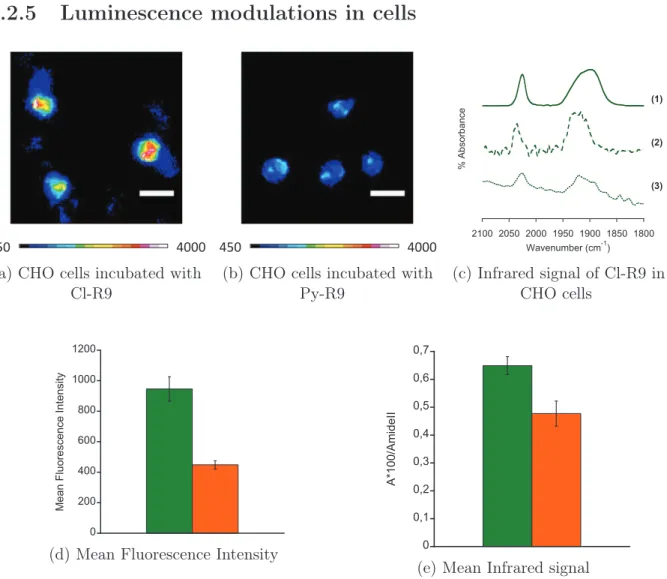
1.2.5 Luminescence modulations in cells . . . 37
CONTENTS
2 In vitro labelling of Engrailed-2 Homeodomain 43
2.1 Homeoproteins and homeodomains . . . 43
2.2 Homeodomain production . . . 46
2.2.1 Plasmid construction . . . 47
2.2.2 Protein expression and purification . . . 48
2.3 Synthesis of SCoMPIs for thiol-maleimide labelling . . . 49
2.4 Labelling of Homeodomain . . . 50
2.5 Imaging of Homeodomain . . . 53
2.6 Conclusion . . . 55
3 Labelling of endogenous Carbonic Anhydrases in cells 57 3.1 Strategies for the covalent labelling of proteins in living cells . . . 58
3.1.1 From genetic engineering to synthetic chemistry: strategies to label proteins in living cells . . . 58
3.1.2 Strategies for traceless affinity-guided labelling of endogenous proteins 61 3.2 Synthesis of the LDAI-SCoMPI reagents . . . 64
3.3 Determination of in vitro labelling efficiency . . . 68
3.4 Labelling and imaging of Carbonic Anhydrases IX and XII in cells . . . 72
Conclusion and perspectives 75
II
Experimental section
77
4 N -terminal labelling of peptides with SCoMPIs 81 4.1 Peptide synthesis . . . 814.2 Synthesis of [Re(CO)3(X)(PytaCOOH)] and labelling of CPP . . . 82
4.3 Preparation of Large Unilamellar Vesicles (LUV) . . . 89
4.4 Fluorescence measurement methodology . . . 89
4.5 Imaging on CHO and HaCaT cells . . . 90
4.5.1 Internalization of Cl-R9 and Py-R9 in CHO cells . . . 90
4.5.2 Internalization of R9Cl in synchronized HaCaT cells. . . 91
5 In vitro labelling of Engrailed-2 Homeodomain 93 5.1 Procedures for the synthesis of Re(I) tricarbonyl complexes for thiol-maleimide labelling . . . 93
5.2 Production of Engrailed Homeodomain Cys-HD and Cys-NLS-HD . . . 99
5.3 Labelling of Cys-HD and Cys-NLS-HD with SCoMPIs . . . 100
5.4 Imaging of SCoMPI-labelled homeodomains on CHO cells . . . 101
5.4.1 Stock solutions . . . 101
CONTENTS
5.4.3 Fluorescence and IR imaging . . . 101
5.4.4 Synchrotron X-ray Fluorescence microspectroscopy . . . 101
6 Labelling of Carbonic Anhydrases with SCoMPIs in living cells 103 6.1 Procedures for the synthesis of LDAI-SCoMPIs for the labelling of Carbonic Anhydrases . . . 103
6.2 Labelling of Carbonic Anhydrases with LDAI-SCoMPI . . . 117
6.2.1 Stock solutions . . . 117
6.2.2 In vitro labelling of human Carbonic Anhydrase 1 (hCA1) . . . 117
6.2.3 Labelling of CA-IX and CA-XII in cells for multimodal imaging . . . 118
6.2.4 Fluorescence and IR imaging . . . 118
6.2.5 Synchrotron X-ray Fluorescence microspectroscopy . . . 118
A Measurements of quantum yields 119 B Publication 121 C Résumé 129 C.1 Marquage et imagerie multimodale de CPPs. . . 132
C.2 Marquage et imagerie de l’homéodomaine Engrailed-2 . . . 138
C.3 Marquage et imagerie des Anhydrases Carboniques . . . 142
List of Figures 153
List of Tables 157
List of Abbreviations 159
Part I
Introduction: Re(I) tricarbonyl
complexes for the labelling of proteins
Fluorescence microscopy is one of the most widely used techniques for the visualization and study of biological processes. Its good spatial resolution (∼ 102nm) makes it well-suited for cell imaging, since typical cell diameters are in the range of a few tens of microme-ters. In addition, it is possible to perform fluorescence imaging on live cells. A large set of fluorescent probes with high quantum yields and diverse excitation and emission wave-lengths are available, thus enabling sensitive and multi-color imaging. Although highly valuable, fluorescence techniques also suffer from some drawbacks, such as photobleach-ing and photodamage of cells. Some techniques, like FRAP (Fluorescence Recovery After Photobleaching) or FLIP (Fluorescence Loss in Photobleaching), take advantage of photo-bleaching to investigate diffusion processes in cell membranes, but generally speaking this effect is undesirable. Moreover, fluorescence intensity of a probe is often very sensitive to its environment, which often makes rigorous quantification difficult.
On the other hand, complementary methods for bio-imaging are emerging. Vibrational techniques such as infrared (IR) and Raman microspectroscopies have raised a particular interest in the past few years. They involve vibrational levels of energy (instead of electronic transitions for fluorescence), thus no photobleaching occurs. In addition, information on the local chemical environment may be collected. However, infrared microspectroscopy generally displays lower spatial resolution than fluorescence microscopy. The spatial res-olution of optical microscopy is limited by ∼ λ/2 (Abbe diffraction limit), where λ is the light wavelength. For infrared microscopy, typical spatial resolution is thus in the 1–10 µm range.
Multimodal probes are thus attractive, as they would combine the advantages of the different methods of imaging on a single molecule. To reach that goal, a well-documented strategy consists of combining several modalities by covalently linking a probe for each modality to a template. This leads to high molecular weight compounds which require multi-step synthesis and whose cell penetration can be altered in a large extent. Alterna-tively, metal complexes may be particularly adapted to obtain multimodality, as some of them display unique spectroscopic properties and can be easily functionalized by ligand modification. In the following chapter, we will particularly focus on luminescent Re(I)
tri-INTRODUCTION: RE(I) TRICARBONYL COMPLEXES FOR THE LABELLING OF PROTEINS
carbonyl complexes, since they display interesting luminescence properties (see Section 1) and intense absorption bands (1800–2200 cm−1) in the IR transparency window of biologi-cal samples. Our group has recently been interested in the development of such complexes as S ingle Core M ultimodal P robe for I maging (SCoMPI) [1–4]. We will describe exam-ples of utilization of Re(I) tricarbonyl complexes in a biological context, used for their lu-minescence (Section 1) or for their infrared properties (Section 2). We will finally describe examples of Re(I) tricarbonyl complexes as mutimodal probes. The use of Re(CO)3 com-plexes for biological applications based either on their luminescence or infrared properties has been extensively reviewed [2, 5–15], and we will attempt here to give an overview of their applications for the labelling and imaging of biomolecules, with a particular interest in peptide and protein labelling.
1
Luminescent Re(CO)
3complexes for bio-imaging
1.1
Spectroscopic properties of luminescent Re(I) tricarbonyl
com-plexes
Re(I) fac-tricarbonyl complexes bearing low energy π∗orbitals (e.g. α-diimine or dipicolylamine-derived ligands) display attractive luminescence properties and biocompatibility features. Similarly to other luminescent d6complexes (including luminescent Ir(III) and Ru(II) com-plexes), low-spin, 18 electron Re(I) tricarbonyl complexes are kinetically inert, which lim-its ligand exchange and the toxicity associated with heavy metal ions [15]. Luminescence properties of [Re(CO)3(N^N)X] complexes (L = α-diimine, X = halide, pyridine, ...), e.g. large Stokes shift and long emission lifetimes, are generally associated with3MLCT excited state (Figure 1): upon irradiation, one electron is promoted from a metal-centered dπ or-bital to a ligand-centered π∗ orbital (1MLCT excited state); inter-system crossing (ISC) then leads to the emissive3MLCT excited state. However, halide-to-ligand charge-transfer (3XLCT) or ligand-to-ligand charge-transfer (3LLCT) may also occur, as well as mixing with intraligand3IL (π → π∗) states [8, 16]. Although the nature of their emission is thus phosphorescence, these complexes are widely referred to as luminescent in the literature, and we will use this naming in the following manuscript. Finally, emission properties of Re(I) complexes are often sensitive to the presence of triplet quenchers like3O2. Although interaction with3O2 results in emission intensity loss (quenching), this mechanism do not chemically alter the complex, and is thus distinct from photobleaching1 [15].
Due to their photophysical features, Re(I) tricarbonyl have raised an increasing in-terest for fluorescence bioimaging applications.2 Several cellular and biomolecular probes
1Photobleaching may be defined as the photochemical destruction of the luminescent probe during
the fluorescence microscopy experiment [15].
2
The term "fluorescence microscopy" is historically used to design this imaging technique, although it may involve fluorescent as well as phosphorescent probes.
1. LUMINESCENT RE(CO)3 COMPLEXES FOR BIO-IMAGING !"#$%& '"#$%& ()*+,-&./0/1& -!" 23$& 4#" 4#!" 5,)& 5)&
Figure 1: Jablonski diagram for [Re(CO)3(diimine)(X)] complexes (MLCT model) Adapted from reference [16]
based on these complexes have thus been developed. In particular, efforts were - and still are - made to improve the spectroscopic features of these complexes, i.e. to obtain higher quantum yields and excitation at longer wavelength. This is generally done by tuning the structure of the bidentate N^N ligand, since it is involved in the MLCT. Two families of ligand have been particularly explored: (i) polypyridyl ligands, mostly derived from 2,2’-bipyridine and 1,10-phenanthroline (Figure 2) [8–10, 12–15, 17–28] and (ii) bis-quinoline ligand and its derivatives, e.g. bis(phenanthridinylmethyl)amine (bpm) [11, 29–31]. How-ever, other polyazaheterocycles have also been developed, such as 1-R-4-(2-pyridyl)-1,2,3-triazole (pyta, R = linker, functional groups, etc.) and its derivatives [32, 33]. More re-cently, Re(I) tricarbonyl complexes incorporating 5,5’-disubstituted 3,3’-bisisoxazole have also been described [34]. Modifying the X ligand may also alter the luminescence proper-ties of the complex. For instance, for [Re(CO)3(bpy)(X)] complex, replacing the chloride ligand for a pyridine derivative has an impact on both emission wavelength and quantum yield (Table 1).
Spectroscopic properties of some rhenium complexes are summarized in Table 1. It can be noted that varying the bidentate and ancillary ligands may have a strong influence on quantum yield or emission wavelength. Excitation wavelength, on the other hand, is gen-erally in the 340–360 nm range. Interestingly, though, Re(I) tricarbonyl complexes bearing a bis-quinoline ligand could be imaged in cells by fluorescence microscopy with excitation at 488 nm [35, 36].
1.2
Re(I) tricarbonyl complexes with specific cell localization.
Re(I) tricarbonyl complexes targeting the different cell compartments have been designed. A thiol-reactive chloromethyl group was for instance appended to a Re(I) tricarbonyl com-plex (1, R=Cl) in order to target mitochondria [27]. Indeed, compound 1 (R=Cl) was found to co-localize with TMRE (tetramethylrhodamine ethyl ester), a known marker of mitochondria, in MCF-7 cells (Figure 3). Interestingly, a similar complex bearing a
hy-INTRODUCTION: RE(I) TRICARBONYL COMPLEXES FOR THE LABELLING OF PROTEINS Re OC X N CO N CO [Re(CO)3(N^N)(X)]n+ N N N N N NN O N N O N N N N R R R R N N N N R N N NN R N N N N N N Ph Ph
phen Me2-phen Me4-phen Ph2-phen
pyta quinta tapy taquin
3,3'-bisisoxazole bpy N N N R bisquinoline N N R Cl Br Py Py-3-R X = N^N = N N N R bpm
Figure 2: Changing the ligands to tune the spectroscopic properties of Re(I) tricarbonyl complexes [8, 30, 33, 34]
Figure 3: Colocalisation of 1 (R=Cl, left) with TMRE (center) in MCF-7 cell Reproduced from [27] with permission of The Royal Society of Chemistry, on behalf of the European Society for Photobiology, the European Photochemistry Association and the RSC.
1. LUMINESCENT RE(CO)3 COMPLEXES FOR BIO-IMAGING CO Re OC OC N N N O N H H N O N H O S NH HN O H H O H N N H O H N O S HN NH O H H n n HN R1 R1 R2 R2 N N N CO CO Re CO S H N N H O H N O S NH HN O H H n = 0,1
Golgi apparatus, Endoplasmic Reticulum
R1 = R2 = H; R1 = R2 = CH3; R1 = H, R2 = C6H5 N N N NH O N H N N H N CO CO Re CO N N N O N C OC OC Re CO O COOH N 2+ Nucleus CO Re OC OC N N N SO3 -SO3 -OH Membrane CO Re OC OC N N N N H O Dead/damaged cells R = Cl, OH Mitochondria CO Re OC OC N N N R 1 2 3 4 5 6 7
Scheme 1: Examples of luminescent Re(I) tricarbonyl complexes targeting the different cell compartments [27, 28, 38, 39]
INTRODUCTION: RE(I) TRICARBONYL COMPLEXES FOR THE LABELLING OF PROTEINS
Table 1: Examples of photophysical properties of Re(I) tricarbonyl complexes in acetonitrile at 298 K
N^N ligand X ligand λexc (nm) λem (nm) Φem (%) Reference bisquinoline1 – 366 425, 580 0.44 [29] bpm – 350 570 – 575 N.A. [30] bpy Cl 355 633 0.27 [32] bpy Py−3-C(O)NH2 343 551 15.6 [17] phen Py−3-C(O)NH2 368 546 12.2 [17] phen Py-3-C(O)NHEt 355 548 33 [24] Me2−phen Py-3-C(O)NHEt 355 536 30 [24] Me4−phen Py-3-C(O)NHEt 355 515 54 [24] Ph2−phen Py-3-C(O)NHEt 355 560 34 [24] pyta (R = Bn) Cl 355 538 0.33 [32]
pyta (R = Alkyl chain) Cl 332 522 0.1 [33]
quinta2 Br N.E.2 N.E.2 – [33]
tapy Cl 360 569 0.19 [33]
tapy Br 360 564 0.17 [33]
taquin Br 380 617 0.06 [33]
1 Measurements were made in ethylene glycol instead of acetonitrile for this example.
2
[Re(CO)3(Quinta)(Br)] complexes are non emissive (N.E.) in acetonitrile [33] but display typical MLCT
features in aqueous solution (containing DMSO). Typical photophysical data for those complexes are λex∼335–345 nm, λem∼580–600 nm, and Φ ∼ 0.2–0.6 % [33, 37].
droxymethyl group (1, R = OH) displayed very similar localization pattern in MCF-7 cells, despite the apparent lack of a thiol-reactive group [28]. To explain this mitochondrial local-ization, it was postulated that phosphorylation of the internalized complex could lead to a thiol-reactive moiety, although no experimental evidence for this hypothesis was obtained. Coogan and co-workers synthesized a range of Re(I) tricarbonyl polypyridyl complexes varying in charge, size and lipophilicity in a first attempt to rationalize the cell localization of such compounds [28]. Although rationalization proved sometimes difficult, they could observe for instance that compound 2 was localized at the plasma membrane of MCF-7 cells, probably interacting with the cationic residues of the glyocoprotein layers (glycoca-lyx). Additionnally, 3 was found to be internalized only in dead or damaged cells, being thus a potential cell-death marker. Examples of biotinylated Re(CO)3 polypyridyl com-plexes (4, 5) were found to localize in lipophilic compartments like Golgi apparatus or endoplasmic reticulum [8, 25]. Finally, a few examples of nucleus localization (compounds 6, 7) can also be found [38, 39].
On the other hand, targeting specific biomolecules rather than cell compartments is also desirable, and various strategies have been adopted to label (and image) molecules of biological interest (e.g. ions, drugs, sugars, PNA, peptides, proteins,...) with Re(I) tricar-bonyl complexes. In the following parts, we will focus on the strategies for the incorporation
1. LUMINESCENT RE(CO)3 COMPLEXES FOR BIO-IMAGING of a Re(CO)3 moiety into peptides and proteins.
1.3
Labelling of amino-acids and peptides with luminescent Re(CO)
3complexes
CO Re OC OC N N N O CO Re OC OC N N N R O R H N COOMe H N COOMe H N COOMe R = CO Re OC OC C N N N O R N O R N P CO CO Re CO H N COOH R = H N COOH OH Ph Ph 8 9 10 11Scheme 2: Examples of Re-appended amino acids. [40, 41]
Single amino-acids have been derivatized with luminescent Re(CO)3 complexes [11, 29–31, 40–43]. Complexes 8 and 9 (Scheme 2), for instance, have been appended with phenylalanine and tyrosine, and their photophysical properties studied [40]. The study particularly focused on the use of the rhenium complex as photo-oxidant to generate tyrosyl radicals. Indeed, MLCT excited states may be used to study redox biological processes, in particular electron transfer (see 1.4).
Gimeno et al. synthesized two series of amino-acids appended with [Re(CO)3(bpy)(py)]+ complexes (compounds 10 and 11, Scheme 2) [41]. The amino acids were linked to the com-plex through the pyridine ligand, at either the meta (10) or para (11) position of pyridine. They studied the cellular uptake of both series and could observe that this minor change in structure had major consequences on the properties of the complex: the para derivatives proved highly cytotoxic and displayed high photobleaching, whereas the cells incubated with the meta derivatives remained healthy and displayed localization patterns typical for monocationic, lipophilic Re(I) tricarbonyl complexes (i.e. cytoplasmic staining, with some concentration in mitochondria). Although no explanation could be given as certain, the authors hypothesized that unidirectional electron shuttling from the metal centre to the
INTRODUCTION: RE(I) TRICARBONYL COMPLEXES FOR THE LABELLING OF PROTEINS
acid was easier with the para than with the meta derivatives, generating an amino-acid radical. N N N Re OC CO CO FmocHN OH O N N N M OC CO CO H N N H O OH O O N H O H N O N H H O S M = Re, 99mTc N N N Re OC CO CO COOH N N N Re OC CO CO N H O H N O HN H2N NH O N O H N N H O NH NH2 HN OH H N O OH O N N N FmocHN OH O 12 13 14 15 16
Scheme 3: Re(I) tricarbonyl complexes for the labelling of peptides [29, 30, 43] The single amino-acid chelate (SAAC) strategy was developed in the early 2000s to in-corporate Re/99mTc complexes into peptides by solid-phase peptide synthesis (using Fmoc strategy). Valliant, Zubieta et al. modified a lysine with a bis-quinoline ligand to form the SAAC ligand 12, which could then be reacted with a Re(CO)3precursor to form the SAAC-Re complex 13 [29, 43]. Both SAAC and SAAC-SAAC-Re could be incorporated by automated peptide synthesis into fMLF peptide (fMLF(SAAC-Re)G, 14), a peptide sequence target-ing the formyl peptide receptor (FPR). FPR is expressed on neutrophils and is a target for the (radio)imaging of trafficking of white blood cells. The Re(CO)3-labelled peptide could be observed by fluorescence microscopy in human leukocytes, and its localization was consistent with previous reports on chemotactic peptides. Interestingly, a 99mTc(CO)3 analogue could be easily prepared by reacting the precursor [99mTc(CO)3(H2O)3]+with the Re-free peptide fMLF(SAAC)G, thus enabling correlative studies with radioimaging. This strategy was later applied to peptides such as HIV-Tat basic domain or β-breaker pep-tides3 [11]. Other biomolecules could also be labelled and imaged, e.g. biotin, folic acid or thymidine derivatives [11].
A Re(I) tricarbonyl complex (15), suitable for N -terminal labelling of peptides, could
3
β-breaker peptides are short peptide fragments from the amyloid-β peptide. They can bind to amyloid-β plaques and inhibit fibril formation.
1. LUMINESCENT RE(CO)3 COMPLEXES FOR BIO-IMAGING be coupled to neurotensin (8-13) fragment on solid-phase [30]. The complex 15 and the labelled peptide 16 were both internalized in various cell lines, and displayed cytosolic localization. Uptake of the unconjugated complex seemed more efficient than uptake of the labelled peptide. However, more pronounced differences between cell lines could be observed for the peptide, suggesting some selectivity in its uptake mechanism.
N N N Re OC CO CO COOH N N N Re OC CO CO NH O HS N H O NH HN NH2 H N O NH HN NH2 O NH HN NH2 H N O NH HN NH2 N H O NH2 NH2 O H N N H NH NH H N N H H N N H H N N H H N NH2 O O SH O O H2N O HN O O O O N NH O O O N N N Re OC CO CO O CO Re OC OC N N N N N N H N O N H O YGRKKRRQRRR CO Re OC OC N C O O O HN WAVGHLM N N NMe2 Me2N 17 18 19 20 21
Scheme 4: Examples of Re(I) tricarbonyl complexes conjugated to peptides for specific cell of organelle targeting [39, 44, 45]
On the other hand, peptides may also be conjugated to Re(I) tricarbonyl complexes to target the complex to specific subcellular compartments or to enhance their uptake by (spe-cific) cells, generally for therapeutic purposes. For instance, Gasser et al. conjugated Re(I) tricarbonyl bisquinoline compound 17 with a Nuclear Localization Signal (NLS) sequence (18) and to a derivative of the Bombesin neuropeptide (19) [44]. Bombesin is often used to selectively target cancer cells over healthy cells, since its receptor is overexpressed in some types of cancers. The aim of the study was to evaluate the potential of Re(I) tricarbonyl
INTRODUCTION: RE(I) TRICARBONYL COMPLEXES FOR THE LABELLING OF PROTEINS
complexes as photodynamic therapy (PDT) photosensitizers, namely their ability to gen-erate the reactive singlet oxygen (1O2) upon light irradiation. Although Ru(II) polypyridyl species have been shown to be efficient photosensitizers for1O2, there are only scarce exam-ples of use of Re(I) complexes for this application [46–48]. Fluorescence microscopy showed that the NLS-conjugated complex 18 accumulated in nucleoli of HeLa cells, whereas its par-ent complex was homogeneously distributed in the cells. The Bombesin conjugate (19), however, displayed only weak fluorescence in those cells, which was explained by either low uptake of the conjugate or quenching of the luminescence of the complex in the cell. Con-jugation to NLS sequence greatly increased the cytotoxicity of the compound 18, whereas neither the parent complex 17 nor the Bombesin conjugate 19 displayed cytotoxicity in the dark. However, irradiation of the cells incubated with 17 or 19 resulted in increased cytotoxicity (up to 20-fold for Re-Bombesin 19), which is encouraging for further studies on Re(I) tricarbonyl complexes as photosensitizers. 1O2 production involves non-radiative quenching of the3MLCT excited state, and thus decreases the effective quantum yield of the complex. However, all derivatives could be detected by fluorescence microscopy in live and fixed cells, with the exception of the Bombesin derivative.
The same group reported the conjugation of a Re(I) tricarbonyl bipyridine complex to a lipopeptide (namely, myristoylated HIV Tat peptide) known for its cell-penetration properties [45]. The conjugate compound 20 displayed enhanced uptake in HeLa cells (Fig-ure 4), as well as increased cytotoxicity. Finally, Alberto et al. reported the synthesis of an other Bombesin-conjugated Re(I) tricarbonyl complex, compound 21 [39]. The goal of the study was to design Re/Tc tricarbonyl complexes that could be addressed selectively to the nucleus of cancer cells. The acridine moiety was appended for nucleus targeting, and the bombesin derivative for cancer cell selectivity. Interestingly, compound 21 could be ob-served by fluorescence microscopy in PC-3 (human prostate adenocarcinoma) cells, which express the GRP receptor (Gastrin releasing peptide receptor) targeted by bombesin, but was not detected in B16-F1 cells (mouse melanoma cell line) which do not express this re-ceptor. As mentioned above, the complex 7 missing the bombesin sequence accumulated in the nucleus. However, the bombesin conjugate displayed homogenous distribution and no nuclear uptake. It was thus hypothesized that the bombesin sequence prevented the nuclear uptake of 21.
1.4
Labelling of proteins
In vitro covalent labelling of proteins through functionalization of Re(I) tri-carbonyl complexes
Luminescent Re(I) tricarbonyl complexes have been functionalized with thiol- or
amine-reactive groups in order to label peptides and proteins in vitro. For instance, N -hydroxysuccinimide ester activated complex 22 was used to label human serum albumin (HSA) and
Immunoglob-1. LUMINESCENT RE(CO)3 COMPLEXES FOR BIO-IMAGING
Figure 4: Enhanced cellular uptake of compound 20
Reprinted with permission from [45]. Copyright 2014 American Chemical Society. Hela cells were fixed after 2h incubation. (A) Control cells; (B) Cells incubated with 100 µM of parent complex; (C) Cells incubated with 20 µM of peptide conjugate 20.
ulin G (IgG) [49]. The authors later used Re-labelled HSA in a Fluorescence Polariza-tion Immunoassay [50]. The isothiocyanate-funcPolariza-tionalized complexes 23, bearing various bidentate ligands, were coupled to HSA [8]. Similarly, thiol-reactive groups such as iodoac-etamide (24, Scheme 6) and maleimide (25) have been appended to Re(I) tricarbonyl com-plexes. Compound 24 was successfully used to label HSA, with a final dye:protein ratio of 0.7 [51]. Similarly, compound 25 was used to label HSA and BSA (Bovine Serum Albu-min) [23]. Between four and five cystein residues were labelled, over the 35 cystein residues BSA and HSA contain. A luminescent Re(I) complex bearing an epoxide-functionalized bidentate ligand (26) was used to label various cystein mutants of cytochrome P450 BM3 heme domain [52]. Interestingly, the 5,6-epoxy-5,6-dihydro-[1,10]phenanthroline bidentate ligand could be coordinated to other metal centres (e.g. IrIII, RuII, OsII). Very recently, Lo and co-workers published a Re(I) tricarbonyl complex functionalized with a dibenzocy-clooctyne (DIBO) (compound 27) for labelling of azide-modified biomolecules by copper-free azide-alkyne cycloaddition [18]. They could modify BSA and HSA with an azide func-tion at N -terminal posifunc-tion, and then label to azide-modified proteins with the DIBO-appended rhenium complex. The same group also designed tetrazine-DIBO-appended Re(I) tri-carbonyl complexes (28, 29) for the labelling of alkyne or alkene-modified protein [53]. They could label efficiently BCN4-modified apo-transferrin (aTf) as well as BSA and HSA. Interestingly, the luminescence of the tetrazine-appended Re(I) complexes was quenched due to energy transfer to the tetrazine. Upon reaction with dienophile-labelled proteins, the tetrazine moiety was reacted to a non-quenching moiety and the luminescence was re-stored. This is thus an interesting example of phosphorogenic Re(I) complexes.
In vitro labelling of proteins by reaction with aqua Re(I) tricarbonyl
com-plexes.
Aqua Re(I) complexes may also react with amino-acids of proteins, in particular with his-tidines (Scheme 8) [54–57]. A luminescent aqua complex [Re(CO)3(phen)(H2O)]+ (phen = 1,10-phenanthroline, compound 30) and its derivate [Re(CO)3(dmp)(H2O)]+ (dmp =
4
INTRODUCTION: RE(I) TRICARBONYL COMPLEXES FOR THE LABELLING OF PROTEINS CO Re OC OC N N N O O N O O CO Re OC OC N N N N C S N N N N = N N N N N N N N N N N N N N Cl N N N N N N N N N N 22 23
Scheme 5: Examples of luminescent Re(I) tricarbonyl complexes for amine labelling [49, 50] (adapted from [8]).
4,7-dimethyl-1,10-phenanthroline) were shown to label His residues of Azurin from Pseu-domonas aeruginosa and mutants by substitution of the aqua ligand by the imidazole ring of histidine (see 31) [54–56]. Photoexcitation of [Re(CO)3(N^N)(His)] in the labelled Azurins led to the oxidation of the copper center of the protein. Interestingly, the car-bonyl ligands were used in these studies to sense the environment of the complex by Time-Resolved Infrared (TRIR) spectroscopy, thus enabling the author to study structural changes in the structure of the protein, as well as electron transfer processes. It might thus be con-sidered as an example of the use of the multimodality of Re(I) tricarbonyl complexes.
Non covalent labelling of proteins
The protein labelling methods described above, although useful for proof of concept and for in vitro studies of purified proteins, are difficult to apply for the selective labelling of proteins in a complex cell environment. To our knowledge, fluorescence cell imaging of proteins with Re(I) tricarbonyl has only been performed using non-covalent methods, i.e. by labelling a specific ligand of the protein of interest with a luminescent Re(I) tricarbonyl complex. For instance, biotinylated complexes 4 and 5 were designed as cross-linkers for
1. LUMINESCENT RE(CO)3 COMPLEXES FOR BIO-IMAGING CO Re OC OC Cl N N CO Re OC OC N N N N H O I N O O CO Re OC OC N N N O 24 25 26
Scheme 6: Examples of luminescent Re(I) tricarbonyl complexes for thiol labelling [51, 52] (adapted from [8])
CO Re OC OC N N N O N H H N O O CO Re OC OC N N N N N N N CO Re OC OC N N N N N N N N N = Me2-phen, Ph2-phen 27 28 29
Scheme 7: DIBO and tetrazine-functionalized Re(I) tricarbonyl complexes for bio-orthogonal protein labelling [18, 53]
CO Re OC OC H2O N N CO Re OC OC N N N HN 30 31
INTRODUCTION: RE(I) TRICARBONYL COMPLEXES FOR THE LABELLING OF PROTEINS
avidin. Similarly, Re(I) tricarbonyl complexes were appended to estrogen derivatives to target estrogen receptors [3, 9] or to indoles to target indole-binding proteins [9]. Doyle et al. conjugated luminescent Re(I) bisquinoline coomplexes to folic acid and to vitamin B12 to target folate receptor and cubilin receptor, respectively [35,42]. We also described above conjugation of Re(I) complexes to bombesin to target GPR receptor.
2
Re(CO)
3complexes for vibrational bio-imaging
2.1
Carbonyl Metallo-ImmunoAssay (CMIA)
Co2(CO)6 Mn Cr Co2(CO)6 HO O OH Cr(CO)3 HO OH Co2(CO)6 Fe OC N CO N H O O Ph Ph N HO O OH HO O N H O N N H O H N O Ph Ph O O N N H O O CO OC CO HN H N O O O N H O CO OC CO Carbamazepine (Antiepileptics) Phenobarbital (Antiepileptics) Diphenylhydantoin (DPH) (Antiepileptics) Cortisol (Hormone) Estradiol (Hormone) 32 33 34 35 36 37 38
Scheme 9: Examples of molecules labelled with metal-carbonyl complexes for Carbonyl MetalloImmunoAssay (reviewed in [5])
As mentioned in the preamble, metal-carbonyl complexes present attractive infrared properties, with intense absorption bands in the 1800–2200 cm−1 range. The first example of utilization of these unique properties in a biological context was a Carbonyl Metallo-ImmunoAssay (CMIA) developed by Jaouen’s group. CMIAs are non-isotopic (the tracer is not a radiolabel), competitive (a known quantity of labelled analyte and a known, lesser quantity of the corresponding antibody are introduced in the sample to be analyzed) and heterogenous (a separation step of bound and unbound fractions is required prior to quan-tification) immuno-assays, where the tracer used for quantifications is a metal-carbonyl moiety. They were able to design mono-immunoassays for various molecules of clinical in-terest, such as antiepileptics and hormones (Scheme 9), or of environmental interest
(pes-2. RE(CO)3 COMPLEXES FOR VIBRATIONAL BIO-IMAGING ticides) [5]. Interestingly, when the infrared bands of different M(CO)x do not overlap (Figure 5), simultaneous immunoassay (multi-CMIA) can be envisioned. Multi-CMIA of compounds 32, 33 and 34 was reported [58]. Although this principle was applied to in vitro assays, it can be envisioned to use it for multi-color infrared imaging.
Figure 5: Principle of multi-CMIA: infrared spectra of 32 (A), 33 (B) and 34 (C). Reprinted from reference [58], Copyright (1999), with permission from Elsevier.
2.2
Labelling of proteins with Re(CO)
3moieties for Infrared
stud-ies
CO OC OC Re Re O O N O O OC CO CO Re O OC CO CO O O N O O H N O O N O O O (OC)5Re O Re OC CO CO S S O O n n = 1,3,4 39 40 41 42 43Scheme 10: [Re(CO)3(Cp)] complexes for specific labelling of amines and thiols [59–61] Due to the sensitivity of vibrational modes to their environment, vibrational
spectro-INTRODUCTION: RE(I) TRICARBONYL COMPLEXES FOR THE LABELLING OF PROTEINS
scopies may be used to probe the structure and dynamics of biomolecules [62]. The posi-tion, width, lifetimes of signal of IR probes may for instance give information on the local conformation and dynamics of a protein or on the local electric field near a catalytic site. Although other IR probes (nitrile, azide, etc.) can be used, Re(CO)3 display more intense signals [61], thus allowing a better sensitivity. In vitro labelling of proteins with Re(CO)3 complexes may thus be of use not only for further vibrational imaging, but also to study intrinsic properties of the protein. Other metal carbonyl complexes have been used for the labelling of proteins [6], but in this part we will focus on examples with [Re(CO)3(Cp)].
In addition to the examples described in 1, non-luminescent Re(CO)3 complexes have also been appended to proteins. As for luminescent Re(CO)3, amine-specific (compounds 39, 40, 41, 42) and thiol-specific (43) groups have been appended to the cyclopentadi-enyl rhenium tricarbonyl complex. NHS-activated esters 39, 40, 41 and pyrilium ion 42 were synthesized and used to label BSA, for instance [59, 60]. Compound 43 bears a thiol-reactive group frequently used for nitroxide spin labelling in EPR [61]. Upon reaction with a cystein side chain, the methanethiosulfonate group leaves and a disulfide bond is formed between the label and the protein. This complex was used to label cystein mutants of ubi-quitin (K6C and K63C) and α-synuclein (V71C). The authors used 2D IR spectroscopy to measure lifetime and frequency of symetric stretching band in various solvents and when bound to the proteins. They could relate these parameters to solvation and electrostatic field, which can then give insights into the structure and dynamics of the proteins. As mentioned in Section 1, structural changes and electron tunneling of Re-labelled Azurin mutants had also been studied using the carbonyl ligands of the Re(CO)3 moiety.
2.3
Infrared Imaging and Metal Carbonyl complexes
Although examples of infrared bioimaging of proteins do not exist to our knowledge, other biomolecules have been successfully labelled with metal-carbonyl units and image by IR or Raman microscopy. In 2007, Leong and co-workers reported the first example of a biomolecule labelled with a M(CO)x moiety and imaged in cells by infrared microspec-troscopy [63]. They labelled a fatty acid and an analogue of phosphatidylcholine with os-mium clusters (compounds 44, 45) and were able to detect these compounds in mucosa cells.
Synchrotron sources display much higher brightness5 than thermal sources (∼ 100-1000 fold) [2, 67], since they focus photons on a smaller area: typical illumination areas are 100 µm for a conventional thermal source, as compared to 10–20 µm for a synchrotron source [67]. A higher flux of photons thus reaches the IR detector, which improves signal-to-noise ratio. As a consequence, with a synchrotron source, it is possible to reduce the aperture size while keeping an acceptable signal-to-noise ratio. It is thus well adapted for
5
Briefly, brightness is the photon flux per unit area and per unit solid angle at the source, or photon flux density.
2. RE(CO)3 COMPLEXES FOR VIBRATIONAL BIO-IMAGING HO O (CH2)10 S Os (CO)3 (CO)3 Os H Os(CO)4 O O (CH2)10 S Os(CO)3 (CO)3 Os H Os(CO)4 (CH2)10 S Os(CO)3 (CO)3 Os H Os(CO)4 O O O P O O Me3N Mn N N CO CO N CO N N N H + OH H N O NH2 N H O H2N N O N H O OH H N O N O H N O SH O Mn CO OC OC Re OC CO CO OH OH 44 45 46 47 48
Scheme 11: Metal carbonyl complexes for vibrational imaging [63–66]
sub-cellular imaging of small cells (< 20 µm) [68]. A rhenium carbonyl tamoxifen derivative (compound 46) was imaged in MDA-MB-231 breast cancer cells using this technique [64]. Raman microscopy involves higher energies, but displays a better spatial resolution [2] than IR microscopy. The uptake and distribution of the CO-releasing molecule (CORM) 47 in HT29 cells was investigated using Raman microspectroscopy by Havenith, Metzler-Nolte, Schatzschneider and co-workers [65]. Interestingly, surface-enhanced Raman spec-troscopy (SERS) makes use of the local amplification of Raman signals due to surface plas-mon resonance of metallic nanostructure, thus enhancing sensitivity of detection. Olivo, Leong et al. successfully applied this method to the detection and imaging of an epider-mal growth factor receptor (EGFR) in OSCC cells (oral squamous carcinoma cells) over-expressing EGFR: they functionalized PEGylated gold nanoparticles with an anti-EGFR antibody (for selectivity) and with osmium carbonyl clusters for detection (see compounds 44, 45) [69]. Coupling of osmium clusters to nanoparticules resulted in better compa-tibility and enhanced signal. Incubation concentrations were in the picomolar range for nanoparticles, which is equivalent to a ∼ 340 µM cluster concentration: this is thus an im-provement as compared to the millimolar concentrations often needed for Raman imaging. More recently, they developed an assay for glucose detection based on SERS [70].
Spatial resolution of infrared imaging may also be enhanced by coupling infrared mea-surement to scanning probe microscopy, e.g. atomic force microscopy (AFM). A cystein-modified, gold-binding peptide derived from neurotensin was labelled with a cymantrene moiety (compound 48) for detection by scattering scanning near-field infrared microscopy
INTRODUCTION: RE(I) TRICARBONYL COMPLEXES FOR THE LABELLING OF PROTEINS
(IR s-SNOM) [66]. The Mn(CO)3-labelled peptide was used to map patterned self-assembled monolayer (SAM) gold surfaces, with a good spatial resolution of 90 × 90 nm2. Compound 46 was mapped in MDA-MB-231 cells using Photothermal Induced Resonance (PTIR) [71], which couples excitation of a tunable infrared laser with detection of the photothermal re-sponse by AFM. In this set-up, also called AFMIR, spatial resolution is the one of the AFM tip, i.e. 20–50 nm.
Finally, development of 3D-IR imaging would provide additional information on sub-cellular structures and labelled biomolecule localization, and a few examples of tomographic infrared imaging of biological samples are emerging [72, 73].
3
Re(CO)
3as surrogates for
99mTc(CO)
3N N N Re OC CO CO SO2NH2 N N N Re OC CO CO SO2NH2 N N N N N Re OC CO CO SO2NH2 N N COOH HOOC N N N Re OC CO CO SO2NH2 N N O N OH O HO O N O HO O O HO N N N Re OC CO CO SO2NH2 N N HN O NH O O HO HO O O HO O OH O OH OH O 49 50 51 52 53
Scheme 12: Examples of Re(CO)3-labelled inhibitors of Carbonic Anhydrase IX [74] Radiotracers and radiopharmaceuticals are widely used in nuclear medicine for di-agnosis and treatment. Among radionuclides, 99mTc displays attractive features, includ-ing availability from commercial generators, and well-suited properties (t1/2 = 6 h, 140 keV) [11,74–77]. Moreover, the use of [99mTc(H2O)3(CO)3]+reagent, developed by Alberto and Schibli et al. , enable efficient preparation of the radiotracer from the ligand [78]. Finally, non radioactive isostructural Re(I) tricarbonyl complexes can be prepared and characterized without handling radioactive materials. As a consequence, a large number of target-specific Re(CO)3 complexes have been developed with the intent to replace Re by its 99mTc counterpart for the final application. Various proteins have been targeted,
4. USE OF RE(CO)3 AS MULTIMODAL PROBES FOR CORRELATIVE IMAGING: S INGLE CORE MULTIMODAL P ROBES FOR I MAGING (SCOMPIS) including Translocator Protein TSPO [79], metallothioneins [80], G Protein-Coupled Es-trogen Receptor GPER/GPR30 [81] or Carbonic Anhydrase IX [74]. As an example, the synthesis of a series of M(CO)3-based (M = Re, Tc) Carbonic Anhydrase IX inhibitors was developed using Re(CO)3 (Scheme 12), while the affinity for CA-IX was evaluated using the final 99mTc complexes [74]. Peptides such as bombesin were also labelled with Re/Tc tricarbonyl complexes, generally in order to target specific cell lines or events [57, 82–84]. In most of these examples, the rhenium complex is simply used for its similarity with the technetium. However, as mentioned in Section 1, efforts are made to use the potential of Re(CO)3 complexes as luminescent probes, and to develop isostructural nuclear and op-tical probes based on 99mTc and Re, respectively [11, 29, 31, 83, 85]. Nevertheless, these complexes, luminescent or not, may be inspirational for further development of Re(CO)3 complexes as multimodal imaging probes.
4
Use of Re(CO)
3as multimodal probes for
correla-tive imaging: S ingle Core Multimodal P robes for
I
maging (SCoMPIs)
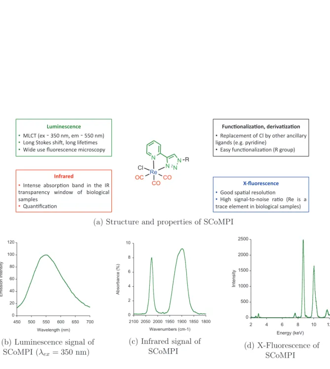
As described in the preamble and in this chapter, luminescent Re(I) tricarbonyl complexes have a potential for multimodal imaging. A few years ago, Policar et al. developed such complexes for the labelling of biomolecules, and their imaging by infrared and lumines-cence microscopies. This led to the concept of S ingle Core Multimodal P robes for I maging (SCoMPIs) [4], for which a general structure is given in Figure 6. Although most of the ex-amples of luminescent Re(I) complexes presented in this chapter bear polypyridine-derived ligands, other polyazaheterocycle might be used to form luminescent Re(I) tricarbonyl com-plexes [33,86–89]. In particular, 4-(2-pyridyl)-1,2,3-triazole (Pyta) ligand is attractive due to its ease of preparation and functionalization, and to the interesting luminescence prop-erties of the related Re(I) tricarbonyl complexes. As a proof of concept, a Re(I)
tricar-N Cl N Re OC CO CO N N ReC12N3 N3 12 N Cl N Re OC CO CO N N N N N O HO N Cl N Re OC CO CO N N HN O R9NH2 SCoMPI-R9 (SR9) 54 55 56
Scheme 13: SCoMPIs used for multimodal imaging in biological samples (cells and skin) [1, 3, 4]
INTRODUCTION: RE(I) TRICARBONYL COMPLEXES FOR THE LABELLING OF PROTEINS ! ! ! ! ! "# $% %& %$ %$ !"#$%&'(&%(&) !!!"#$%&'(%!%)*+%,-.%'-%!%**+%,-/% !!"0,1%2304'5%5678.%90,1%97:';-'5% !!<7='%>5'%?>0@'5A',A'%-7A@05A0BC%) *"%(+,%-.$/-+,%0)1&2$3-+/-+,%) !! D'B9EA'-',3%0:%#9%FC%036'@%E,A799E@C% 971E,=5%&'G1G%BC@7=7,'/% !! HE5C%:>,A;0,E97IE;0,%&D%1@0>B/) 456",2&'(&%(&) !!J00=%5BE;E9%@'509>;0,%
!!K716% 571,E9L30L,075'% @E;0% &D'% 75% E% 3@EA'%'9'-',3%7,%F709017AE9%5E-B9'5/%) 7%82-2&1) !!M,3',5'% EF50@B;0,% FE,=% 7,% 36'% MD% 3@E,5BE@',AC% N7,=0N% 0:% F709017AE9% 5E-B9'5% !!O>E,;PAE;0,)
(a) Structure and properties of SCoMPI
! "! #! $! %! &!! &"! #'! '!! ''! $!! $'! (!! )*+,,+-./0.12.,+13 456272.819/:.*; (b) Luminescence signal of SCoMPI (λex= 350 nm) ! " # $ % &! &%!! &%'! &(!! &('! "!!! "!'! "&!! ) *+,-*./012345 6.71/89*1-+2309:&5 (c) Infrared signal of SCoMPI ! "!! #!!! #"!! $!!! $"!! $ % & ' #! #$ ()*+),-*. /)+01.234+56 (d) X-Fluorescence of SCoMPI
Figure 6: General structure and features of S ingle Core Multimodal P robes for I maging (SCoMPIs)
4. USE OF RE(CO)3 AS MULTIMODAL PROBES FOR CORRELATIVE IMAGING: S INGLE CORE MULTIMODAL P ROBES FOR I MAGING (SCOMPIS) bonyl complex bearing a pyta ligand appended with a long alkyl chain (compound 54) was synthesized, and incubated in MDA-MB-231 cells [4]. The azide moiety was used for com-parison of the infrared signals from N3 and CO groups inside cells. The compound could be detected by both fluorescence and infrared microspectroscopies, with consistent perin-uclear location in both microscopies. It was possible to assign the location of the SCoMPI to the Golgi apparatus through colocalization with a fluorescent marker of this organelle. Besides, it was shown on similar compounds that the FTIR signal of the SCoMPI can be used to quantify reliably the cellular uptake of a SCoMPI-labelled compound in a collection of cells [90].
The mestranol derivative 55 could be imaged as well in MDA-MB-231 and MCF-7 breast cancer cell lines [3]. Correlative imaging could be performed using a set of lunescence and vibrational microscopies, including synchrotron radiation UV and FTIR mi-crospectroscopies (SR-UV-MS and SR-FTIR-MS, respectively), wide field and confocal flu-orescence microscopies, AFMIR and confocal Raman microscopy.
Recently, the group was also interested in detection of SCoMPIs in skin samples. In collaboration with the group of O. Torres in Barcelona, we used a ReC12 SCoMPI (similar to compound 54 without an azide group) to follow the penetration of lipid assemblies into skin by SR-FTIR-MS [91]. A SCoMPI was also conjugated to a nona-arginine peptide (56), and its penetration into skin samples was imaged correlatively by SR-FTIR-MS and fluorescence microscopy [1].
All these biomolecules are small, exogenous molecules, and we were interested in the feasibility to label endogenous molecules, in particular proteins, at the cellular level. To reach that goal, we proceeded by steps of increasing difficulty in terms of labelling and detection. We first investigated the cellular uptake of SCoMPI-labelled cell-penetrating peptides (CPP) (Chapter 1). This led us to study the luminescence properties of various SCoMPIs in solution or in presence of lipid membranes (Section 1.2) as well as the evo-lution of cellular uptake with cell cycle (Section 1.3). We then designed a SCoMPI for in vitro thiol-maleimide labelling of cystein-containing proteins. Since the aim of the study was to perform correlative imaging of proteins in cells, labelling a protein that could be internalized in cells was preferable. In collaboration with O. Lequin and S. Sagan, we thus decided to label the homeodomain of Engrailed-2, which is known to be internalized into cells (Chapter 2). Finally, we tested the possibility to label and image endogenous proteins with SCoMPI. To do so, we used one of the so-called Traceless Affinity Labelling strategies developed by the group of I. Hamachi for the labelling of endogenous proteins. We targeted Carbonic Anhydrases IX and XII (CA-IX and CA-XII, respectively), two membrane-bound isoforms of Carbonic Anhydrases that are over-expressed in solid tumor cancers.
Notably, the possibility to use SCoMPI as probe for synchrotron-based X-ray cence (SXRF) microspectroscopy (SXRF) was also explored. X-ray absorption or fluores-cence methods are valuable for the detection of metal centers and sensing of their chemical
INTRODUCTION: RE(I) TRICARBONYL COMPLEXES FOR THE LABELLING OF PROTEINS
environment, in particular in biological samples [92–95]. SXRF, for instance, enables simul-taneous collection of data for multiple elements, provided their edge energy is lower than the incident energy. Information on the chemical environment of the studied metal center is thus immediately available. Moreover, the spatial resolution of this technique is in the nanometer - micrometer range, which is relevant for biological applications. Interestingly, the natural abundance of Re is very low, which would make possible to detect SCoMPI-labelled molecules with a good signal-to-noise ratio. We thus investigated the potential of Re as X-ray fluorescence probe (Figure 6) for the detection and subcellular imaging of proteins. The preliminary results will be discussed in Chapters 2 and 3.
Chapter 1
Labelling of peptides with SCoMPIs
1.1
Introduction: Cell-Penetrating Peptides
1.1.1
Cellular uptake
Cell-penetrating peptides (CPPs) are small peptides (generally 8 to 30 amino acids) that are efficiently internalized into cells. Their ability to deliver various conjugated cargos in-side cells makes them potential (drug) delivery or diagnostic agents [96]. Over the past two decades, several cell-penetrating peptides have been discovered or designed [96–101], and numerous examples of intracellular delivery of various cargos (including drugs, nucleic acids or nanoparticles) have been reported [96, 102]. Despite high variability in their se-quences, CPPs present physico-chemical similarities. In particular, most CPPs present a high density of positive charges [97, 99]. Actually, the arginine-rich CPPs such as TAT peptides and oligoarginines form an important and extensively studied class of CPPs.
Understanding the mechanism(s) of cellular uptake of CPPs is essential to rationally design new CPPs and to use them efficiently for cargo delivery. Described internalization pathways of CPPs are summarized in Figure 1.1. Cellular uptake may occur through di-rect translocation or endocytosis (pinocytosis). Energy-dependent endocytotic pathways involve macropinocytosis as well as clathrin-dependent, caveolae/lipid raft-dependent or clathrin/caveolae independent uptake. After endocytosis, the CPP and its cargo must then escape the endosomes to reach cytosol. Direct translocation may also occur, and various model have been suggested: formation of inverted micelles, pore formation, and the car-pet model, in which the peptide accumulates at the membrane and induces its disruption above a concentration threshold. Several parameters may impact the efficiency and mech-anism of internalization of CPP, among which the nature of the peptide itself (sequence, structure, charge,etc), the nature and size of the cargo, the incubation conditions (concen-tration, temperature), or the cell type [96, 102, 103]. Due to this variability, determination of the exact mechanism of internalization is sometimes difficult or even controversial, and it is likely that different pathways coexist, even for the same CPP, depending on the
experi-CHAPTER 1. LABELLING OF PEPTIDES WITH SCOMPIS !"#$%&$'()*+$,,$-( !!(.,/&0%*"1'$2$"'$"&( !!(./#$3,/$1)$'*/&$'( !!(.,/&0%*"4+/#$3,/$(*"'$2$"'$"& 5/+%32*"3+6&3-*-(
!"#$%&'()*
./%2$&( 73%3*'/,4 8/%%$,1-&/#$( 23%$()3'$,(9!:;.7(7:<=>?@.<7!@=(
;=9@.A7@>!>(
!"##$%"%&'()"$
!**$
Figure 1.1: Mechanisms for the internalization of Cell-Penetrating Peptides. Inspired by reference [102]
mental conditions. Nevertheless, understanding which parameters are important to favour one pathway or another is potentially important for predicting the uptake and delivery effi-ciency of a CPP. Biophysical, chemical and biological approaches have thus been developed to address these questions.
1.1.2
Methods for studying the internalization mechanisms
Large and Giant Unilamellar Vesicles (LUVs and GUVs, respectively) are often used as models for cell membrane to study direct translocation of CPP. They enable fine tuning of experimental parameters (lipid composition, temperature, pH, concentration, etc) as well as the use of physico-chemical methods that would be difficult to set up or interpret in living cells (fluorescence spectroscopy, isothermal titration calorimetry, NMR, etc) [104– 108]. Such studies showed for instance the importance of membrane fluidity and of the electrostatic interactions of arginine-rich CPPs with anionic lipids for internalization [109]. Mechanisms and efficiency of internalization inside cells have also been intensively in-vestigated for the past decades. One of the most popular techniques to study the CPP distribution is fluorescence microscopy. Although powerful, possible artefacts should be kept in mind when using this tool. For instance, it was shown in the early 2000s that cell fixation could cause redistribution of CPP from endosomes to the cytoplasm and nu-cleus, thus questioning previous studies [110, 111]. Quantification from fluorescence im-ages is also difficult, as highly cationic CPP tend to adsorb on the plastic or glass slides, and, more importantly, remains bound to cell membrane even after washings.
Quantifica-1.1. INTRODUCTION: CELL-PENETRATING PEPTIDES tion of the internalized peptide thus require to remove the signal of the non-internalized, membrane-bound peptide, for instance using trypsin digestion or chemical methods (e.g. quenching by dithionite of non-internalized CPP labelled with nitrobenzofurazan (NBD) fluorophore) [112]. Finally, fluorescence emission of a given fluorophore may (strongly) de-pend on its local environment. In fact, a recent study by our laboratory showed that CPPs labelled with common fluorophores (rhodamin and fluorescein) (Lavielle et al. , submit-ted) could remain unseen by fluorescence microscopy in the areas the most concentrated in peptide, because of fluorescence quenching. An elegant dilution protocol of the labelled peptide with "cold" acetylated peptide enabled them to unravel the "hidden" areas where the peptide was the most concentrated. Non-quenching fluorophores such as NBD could be likely to circumvent this problem.
1.1.3
Methods for quantifying the internalization efficiency
Figure 1.2: Protocol for the quantification of internalization of CPP and CPP-Cargo constructions using MALDI-TOF mass spectrometry. Adapted by permission from
Macmillan Publishers Ltd: Nature Protocols (reference [113]), copyright (2006). A method based on mass spectrometry was previously designed in our laboratory [113]
CHAPTER 1. LABELLING OF PEPTIDES WITH SCOMPIS
in order to reliably quantify cell internalization of CPPs (Figure 1.2). Quantification by MALDI-TOF MS relies on the use of an isotope-labelled internal standard having the same sequence as the peptide of interest. To do so, a tag made of either four non-deuterated glycine for the analyte or four bi-deuterated glycines for the internal standard was ap-pended to the peptide sequence. Moreover, both peptides were biotinylated to allow ex-traction from cell lysate and isolation steps using streptavidin-coated magnetic beads. The internal standard, having the same sequence as the peptide of interest, will be recovered with the same efficiency and give the same intensity response during MALDI-TOF anal-ysis, but with distinguishable spectra (δm/z = 8). In this protocol, the analyte (non-deuterated [1H]CPP) is incubated on adherent cells, and the excess of peptide is washed after incubation. Membrane-bound peptide was removed by a trypsin digestion step of the membrane-bound peptide (trypsin may be replaced by an other protease, if more appro-priate). Cells are then transferred in a microtube, and a known amount of the internal standard ([2H]CPP) is added. After cell lysis, both peptides are captured on streptavidin-coated magnetic beads and undergo several washing steps. Finally, the sample is analyzed by MALDI-TOF MS, the ratio of [M+H] signal areas of both peptides measured, and the quantity of internalized [1H]CPP calculated from this ratio.
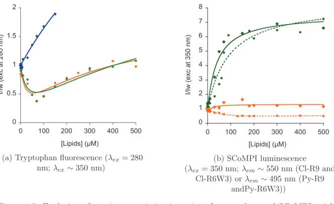
Although this method allows to accurately and robustly quantify the global amount of internalized peptide, it does not allow to distinguish between cell compartments. This information on localization can be obtained through fluorescence imaging. However, as de-scribed above, self-quenching of some fluorescent probes may lead to misinterpretations on peptide location, and generally speaking, fluorescence dependance on environment results in the impossibility reliably quantify the amount of peptide in a given area. On the other hand, the intensity and area of CO elongation signal from M(CO)n do not strongly depend on the environment of the molecule. These infrared signals can thus be used for quantifi-cation purposes. We wanted to use the bimodality of SCoMPIs in order to detect CPP by fluorescence microscopy (for higher spatial resolution) and by infrared (for quantification). In this chapter, we will first focus on the effect of environment on the luminescence properties of two SCoMPIs. We designed and synthesized two SCoMPI suitable for N -terminal labelling of peptides, and labelled two arginine-rich peptides with them. We studied the spectroscopic properties of the SCoMPI-labelled peptides in solution and in presence of lipid model membranes, to see if self-quenching could occur for these SCoMPIs when locally accumulated.
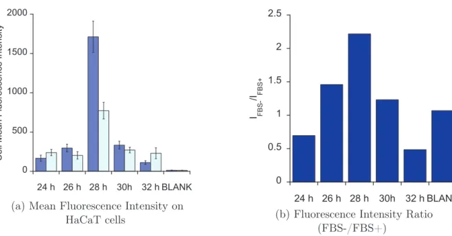
Besides, our group recently published correlative imaging of SCoMPI-labelled nona-arginine peptide in skin samples [1]. Complementary experiments on keratinocytes high-lighted an inhomogeneous fluorescence among cells. Nucleus staining with DAPI then hinted a possible correlation between internalization of the labelled peptide and cell cy-cle. In a preliminary work, we thus looked at internalization of SCoMPI-labelled CPP depending on the cell division cycle by fluorescence microscopy.
1.2. LUMINESCENCE STUDY OF SCOMPI-LABELLED CPPS.
1.2
Luminescence study of SCoMPI-labelled CPPs.
1.2.1
Design and synthesis of Re(I) tricarbonyl derivatives for
N
-terminal labelling of peptides
Cl Cl O Cl H N O OMe O N3 H N O OMe O N H N O OMe O N N N NH OH O O N N N N Re OC Py CO CO NH OMe O O N N N N Re OC Py CO CO BF4 BF4 NH OH O O N N N N Re OC Cl CO CO NH OMe O O N N N N Re OC Cl CO CO NH OMe O O N N N N Re OC Br CO CO a b c e f g d d' 57 58 59 60 61 62 63 64
Scheme 1.1: Synthesis of the SCoMPI derivatives for N -terminal labelling of peptides. Reaction conditions: (a) β-alanine methyl ester hydrochloride salt, DIEA, dry DCM, 1h, 0◦C to RT, 66%,
(b) NaN3, NaI, acetone:water (3:1 v:v), 16h, 35◦C, 79%, (c) 2-ethynylpyridine, CuSO4, sodium ascorbate,
acetone:water (2:1 v:v), 2h, RT, 70%, (d) Re(CO)5Cl, toluene, 6h, reflux, 100%, (d’) Re(CO)5Br, toluene,
6h, reflux, 100%, (e) LiOH · H2O, THF:water (2:1 v:v), 45 min, RT, 80%, (f) AgBF4, acetonitrile, 5h,
reflux, Ar; pyridine, THF, 20h, reflux, 79%, (g) LiOH · H2O, THF:water (2:1 v:v), 1h, RT, 87 %.
Modifying the rhenium coordination sphere may have an impact on its spectroscopic properties. Our group recently investigated the effects of structural modifications of the bidentate ligand on the luminescence properties of Re(I) tricarbonyl complexes [33]. Here we focused on the effect of the monodentate ancillary ligand (chloride or pyridine) on the spectroscopic and physico-chemical properties of the SCoMPI. Luminescent rhenium tri-carbonyl complexes have initially been developed with 2,2’-bipyridine derivatives, and sev-eral of these polypyridine complexes, bearing either a halide or a pyridine derivative as monodentate ancillary ligand, have been designed for the labelling of biomolecules and for bioimaging [8]. In addition, luminescent Re(I) complexes presenting other polyaza-heterocyclic ligands have also been developed. 4-(2-pyridyl)-1,2,3-triazole (Pyta) deriva-tives have raised particular interest due to their ease of synthesis and functionalisation [32, 37, 88, 114–116]. [Re(CO)3(Pyta)(L)]n+complexes for biomolecule labelling have been widely developed with a halide ligand [1, 32, 37, 88, 114, 115] (L= Cl, Br; n=0), but
ex-CHAPTER 1. LABELLING OF PEPTIDES WITH SCOMPIS
amples of such complexes with pyridine (L = Py; n=1) are also described [116]. We thus designed two Re(I) tricarbonyl probes incorporating a Pyta ligand suitable for grafting to the N -terminus of peptides, and bearing either a chloride or pyridine as monodentate lig-and. The halide ligands are potentially more readily exchanged than the pyridine; besides, it should be noted that the positive charge of the [Re(CO)3(Pyta)(Py)]+complex may have an impact on the properties of the probe, including interactions with membranes.
The methyl ester protected 4-(2-pyridyl)-1H -1,2,3-triazole ligand (Pyta-COOMe) was easily obtained in three steps and in good yield starting from β-alanine methyl ester (Scheme 1.1). Reaction of chloroacetyl chloride with β-alanine methyl ester hydrochloride salt in dry DCM led to the chloride derivative 57. The nucleophilic substitution of the chloride by an azido group was then performed with sodium azide in presence of sodium iodide in a ace-tone:water (2:1 v:v) mixture. The resulting azide derivative (58) was finally reacted with 2-ethynylpyridine according to standard CuAAC procedures to lead to the Pyta ligand (59). The ligand was then refluxed in toluene in presence of the rhenium precursor Re(CO)5X (X = Cl, Br), leading to the formation and precipitation of [Re(CO)3(X)(PytaCOOMe)] complexes (X = Cl, Br).
The chloride derivative (60) was then saponified by lithium hydroxide in a mixture of THF and water (2:1 v:v) to obtain 61. Although exchange of the chloride ligand with carboxylates has been described for rhenium tricarbonyl chloride complexes [117], in our hands this undesired exchange did not occur during saponification. This might be due to different experimental conditions (temperature, base, reaction time).
For the Re(I) bromide complex, on the other hand, ligand exchange was performed using the methyl ester form of the complex (63). This bromide derivative of the complex (62) was refluxed in acetonitrile in presence of silver tetrafluoroborate, then in THF in presence of an excess of pyridine, to obtain the ester form of the rhenium tricarbonyl py-ridine complex (63) in good yields. Saponification was then performed on the complex to obtain compound 64. In both synthetic pathways, the complexes were obtained as a racemic mixture of fac isomers.
Due to the harshness of conditions of peptide cleavage from the resin (HF cleavage), labelling of R9 and R6W3 with the rhenium complexes could not be realized on solid sup-port and was rather performed in solution. To do so, the complex was first activated either with EDC or with DCC/NHS, then reacted with the desired peptide. The reaction mix-ture was then directly purified by HPLC. Particular attention should be paid to the chloride derivative, as we could observe in some cases chloride exchange under HPLC conditions in-volving 0.1% of trifluoroacetic acid (data not shown). In order to avoid this exchange, the labelled peptide solutions were freeze-dried immediately after purification. Structures of the studied peptides are summarized in Scheme 1.2.
![Figure 2: Changing the ligands to tune the spectroscopic properties of Re(I) tricarbonyl complexes [8, 30, 33, 34]](https://thumb-eu.123doks.com/thumbv2/123doknet/14493762.526401/15.892.176.687.139.741/figure-changing-ligands-tune-spectroscopic-properties-tricarbonyl-complexes.webp)

![Figure 1.1: Mechanisms for the internalization of Cell-Penetrating Peptides. Inspired by reference [102]](https://thumb-eu.123doks.com/thumbv2/123doknet/14493762.526401/35.892.192.665.112.485/figure-mechanisms-internalization-cell-penetrating-peptides-inspired-reference.webp)