HAL Id: hal-02975685
https://hal.archives-ouvertes.fr/hal-02975685
Submitted on 26 Oct 2020
HAL is a multi-disciplinary open access
archive for the deposit and dissemination of
sci-entific research documents, whether they are
pub-lished or not. The documents may come from
teaching and research institutions in France or
abroad, or from public or private research centers.
L’archive ouverte pluridisciplinaire HAL, est
destinée au dépôt et à la diffusion de documents
scientifiques de niveau recherche, publiés ou non,
émanant des établissements d’enseignement et de
recherche français ou étrangers, des laboratoires
publics ou privés.
Direct inhibition by nitric oxide of the transcriptional
ferric uptake regulation protein via nitrosylation of the
iron
Benoit d’Autreaux, Daniè Le Touati, Beate Bersch, Jean-Marc Latour,
Isabelle Michaud-Soret
To cite this version:
Benoit d’Autreaux, Daniè Le Touati, Beate Bersch, Jean-Marc Latour, Isabelle Michaud-Soret. Direct
inhibition by nitric oxide of the transcriptional ferric uptake regulation protein via nitrosylation of
the iron. Proceedings of the National Academy of Sciences of the United States of America , National
Academy of Sciences, 2002, �10.1073/pnas.252591299�. �hal-02975685�
Direct inhibition by nitric oxide of the transcriptional
ferric uptake regulation protein via nitrosylation of
the iron
Benoıˆt D’Autre´aux*, Danie`le Touati†, Beate Bersch‡, Jean-Marc Latour*, and Isabelle Michaud-Soret*§
*Laboratoire de Physicochimie des Me´taux en Biologie (Formation de Recherche en Evolution–Universite´ Joseph Fourier–Commissariat a` l’Energie
Atomique–Centre National de la Recherche Scientifique no. 2427), Grenoble, F-38054 Grenoble Cedex 9, France;†Laboratoire de Ge´ne´tique
Mole´culaire des Re´ponses Adaptatives (Unite´ Mixte de Recherche 7592, Institut Jacques Monod, Universite´ Paris 6 et 7), F-75251 Paris
Cedex 5, France; and‡Laboratoire de Re´sonance Magne´tique Nucle´aire, Institut de Biologie Structurale, Jean Pierre Ebel (Unite´
Mixte de Recherche Commissariat a` l’Energie Atomique–Centre National de la Recherche Scientifique–Universite´ Joseph Fourier 5075), F-38027 Grenoble Cedex 1, France
Communicated by Irwin Fridovich, Duke University Medical Center, Durham, NC, October 1, 2002 (received for review June 7, 2002)
Ferric uptake regulation protein (Fur) is a bacterial global regulator that uses iron as a cofactor to bind to specific DNA sequences. The function of Fur is not limited to iron homeostasis. A wide variety of genes involved in various mechanisms such as oxidative and acid stresses are under Fur control. Flavohemoglobin (Hmp) is an NO-detoxifying enzyme induced by NO and nitrosothiol com-pounds. Fur recently was found to regulate hmp in Salmonella
typhimurium, and in Escherichia coli, the iron-chelating agent
2,2ⴕ-dipyridyl induces hmp expression. We now establish direct
inhibition of E. coli Fur activity by NO. By using chromosomal Fur-regulated lacZ reporter fusion in E. coli, Fur activity is switched off by NO at micromolar concentration. In vitro Fur DNA-binding activity, as measured by protection of restriction site in aerobactin promoter, is directly sensitive to NO. NO reacts with FeIIin purified
FeFur protein to form a Sⴝ 1兾2 low-spin FeFur–NO complex with
a g ⴝ 2.03 EPR signal. Appearance of the same EPR signal in
NO-treated cells links nitrosylation of the iron with Fur inhibition. The nitrosylated Fur protein is still a dimer and is stable in anaerobiosis but slowly decays in air. This inhibition probably arises from a conformational switch, leading to an inactive dimeric protein. These data establish a link between control of iron metabolism and the response to NO effects.
M
icroorganisms have developed several mechanisms to sur-vive in their hosts’ environments. These include compe-tition with their hosts for metal acquisition (1) and resistance to host defenses such as nitric oxide (NO), a cytotoxic weapon generated by macrophages (2). In eukaryotic cells, NO is met-abolically produced by NO synthase from arginine, O2, andNADPH (3). In macrophages, an inducible NO synthase is produced after activation by endotoxins or cytokines and gen-erates copious amounts of NO to poison pathogens (2). A few examples of bacterial NO synthases have been described (4, 5). An endogenous source of NO also may be found in some denitrifying bacteria (6). Because they use nitrate in place of oxygen for energy production, NO is a metabolic intermediate in the denitrification pathway. Denitrifiers possess an improved NO reductase that catalyzes conversion to N2O, keeping NO at
a nontoxic level inside the cell (1–50 nM). NO, which is uncharged and nonpolar, can cross membranes to trigger its target responses (7). NO can injure cells by attacking the iron centers (8) in various key proteins such as nitrogenase (9) and ribonucleotide reductase (10). NO also induces modification of thiol-containing proteins, yielding nitrosothiol groups (11). Spe-cific responses to NO are found in bacteria to prevent NO or NO-mediated damages. NO induces the expression of specific NO-detoxifying enzymes, the flavohemoglobin (Hmp) (12) and the flavorubredoxin (13). Hmp is an O2-nitroxylase in aerobic
condition that catalyzes the reaction of NO⫺with O2to give
NO3⫺(14, 15). In anaerobic conditions, the flavorubredoxin has
an O2-sensitive NO reductase activity in Escherichia coli (13).
NO also induces the expression of enzymes involved in the oxidative stress response such as the manganese-containing superoxide dismutase (SodA). At the level of gene expression, the regulation occurs through SoxR activation (7). Indeed, evidence has been provided that SoxR may be an NO sensor (7). NO activates the SoxR protein by direct formation of a dini-trosyliron species, leading to the induction of the oxidative stress response (16). Concerning the Hmp, NO has been shown to induce 19-fold the hmp gene expression, in aerobic conditions, independently of the SoxRS regulon (12). Moreover, the same level of activation of hmp expression was observed after treat-ment with the iron-chelating agent 2,2⬘-dipyridyl in aerobic and anaerobic conditions, suggesting the involvement of an iron-dependent regulatory protein (12). Furthermore, in E. coli, this activation cannot be explained by the other described regulators of hmp: fumarate nitrate reductase, an anaerobic repressor, and the MetR protein (methionine biosynthetic pathway regulation). Indeed, a mutation in fumarate nitrate reductase stimulates only 4-fold hmp gene expression (12), and MetR activates hmp gene expression in response to nitrosothiol but not to NO (17). Thus, in E. coli, there is a missing link between NO stress and Hmp expression. Results obtained in other bacteria suggest that the ferric uptake regulation protein (Fur) may be the link. Indeed, in Salmonella typhimurium, expression of hmp also is induced after NO treatment (18), and its control is independent of the SoxS and OxyR transcription factors but relies on the iron-dependent Fur repressor (18). The control of hmp expression by Fur in S. typhimurium suggests a link between the control of iron metabolism and NO detoxification.
Fur is a global regulator ubiquitous in Gram-negative bacteria that controls the expression of⬎90 genes in E. coli (19). This dimeric protein (2 ⫻ 17 kDa) first was described as being involved in iron-uptake regulation. Fur is the key protein for the control of the intracellular iron concentration. The active form of the Fur protein, FeFur, contains a nonheme ferrous iron site with oxygen and nitrogen donor ligands (20, 21). When the cellular iron level becomes too low, the active Fur repressor looses Fe2⫹, its corepressor, and is no longer able to bind to
specific DNA sequences. Fur links iron metabolism and the regulation of oxidative stress defenses. It regulates the expres-sion of the superoxide dismutases (represexpres-sion of the manganese superoxide dismutase and activation of the iron one) (22, 23), and fur expression is under control of OxyR and SoxRS (24).
Abbreviations: Fur, ferric uptake regulation protein; FeFur, Fe(II)–Fur complex; Hmp, flavohemoglobin; hmp, flavohemoglobin gene; NONOate, 2,2⬘-(hydroxynitrosohy-drazino); DEANO, diethylamine NONOate;-gal, -galactosidase.
§To whom correspondence should be addressed at: Laboratoire de Physicochimie des
Me´taux en Biologie, CEA–Grenoble, 17 avenue des Martyrs, F-38054 Grenoble Cedex 9, France. E-mail: imichaud@cea.fr.
In this paper, we present investigations of NO action on the Fur protein from E. coli by using in vivo and in vitro assays of Fur activity as well as spectroscopic characterization of nitrosyl adduct of pure, reconstituted FeFur. We find that the FeFur protein reacts with NO to form an inactive and stable iron nitrosyl adduct with a g⫽ 2.03 EPR signal. Nitrosylation of Fur also occurs in vivo because the same EPR signal is observed in intact Fur-overproducing cells treated with NO. We show that NO inhibits Fur repressor activity in vivo and Fur DNA-binding activity in vitro. These data establish a link between the control of iron metabolism and the response to NO effects.
Materials and Methods
Chemicals. Mohr’s salt Fe(SO4)2(NH4)2䡠6 H2O, diethylamine,
1,3-bis[tris(hydroxymethyl)methylamino]propane, EDTA,14NO
gas (98.5% purity), and15NO gas (98%15N) were obtained from
Sigma, and DEANO [diethylamine 2,2⬘-(hydroxynitrosohy-drazino) (NONOate)] was obtained from Cayman Chemicals, Ann Arbor, MI. DEANO solutions were prepared in 10 mM NaOH; they decompose to 1.5 NO molecules in acidic solu-tion (25).
Construction of the Mutant Strains.Bacterial strains used in the
study were all⌬lac E. coli K-12 [QC 2461 (23)] derivatives of MG1655 (wild type, E. coli Genetic Stock Center). ( fhuF::lacZ) and ( fiu::lacZ) fusions (26) and ⌬fur::cat mutation (23) were introduced by P1 transduction as described (23), giving QC2949 (⌬lac fhuF::lacZ), QC2950 (⌬lac fiu::lacZ), QC6009 (⌬lac ⌬fur::cat fhuF::lacZ), and QC6008 (⌬lac ⌬fur::cat fiu::lacZ).
Media, Growth Conditions, and-Galactosidase (-Gal) Assays.Cells were grown in anaerobiosis (Forma Scientific anaerobic cham-ber) in LB adjusted at pH 7.0 containing 1% glucose and 40 g兾ml kanamycin.
For cultures in the presence of NONOate, 0.8 ml was inocu-lated in 20 ml of medium at 37°C with an anaerobic culture to OD600of 0.05–0.1. After exponential growth recovery,
NONO-ate at 50 mM was added. Various times after inoculation, concentrations (10–100 ⌴) of DEANO (see Fig. 1) were assayed. Samples for measuring optical density and-gal activity were taken at intervals. -Gal assays were performed as de-scribed according to Miller and others (23, 27).
Overproduction and Purification of Fur.The T7 RNA polymerase兾
promoter system was used to overproduce Fur. The correspond-ing codcorrespond-ing sequence was cloned into the NdeI兾XhoI sites of a pET-30c expression vector (Novagen). The resulting plasmid, called pFur1, was transformed into BL21 (DE3) E. coli strain. Freshly transformed bacteria were plated on LB (0.1% agar) containing 50g兾ml kanamycin and grown at 37°C. Cells were grown at 37°C from an overnight culture in 300 ml of LB containing 50g兾ml kanamycin. Fur expression was induced at OD600⫽ 0.7–0.8 by isopropyl-D-thiogalactoside (200 mM, 600
l) during 2 h and 30 min. Dimeric Fur protein was purified as described (20). The buffer was exchanged for 100 mM 1,3-bis[tris(hydroxymethyl)methylamino]propane兾100 mM KCl, pH 7.5. The purity was checked from SDS兾PAGE. Protein concen-trations were calculated by using an absorption coefficient at 275 nm of 0.4 mg⫺1䡠ml䡠cm⫺1for the monomer of pure apo-Fur (20).
NO Treatment on Intact Cells.Fur expression was induced in 200 ml
of culture, as described in the previous section, during 2 h and 30 min. DEANO (200 mM) then was added to a final concen-tration of 10M. Aliquots (200 l) were taken after 10, 60, and 180 min and transferred to an EPR tube. To increase signal intensity, 50 ml of bacterial culture immediately was concen-trated 100-fold (by centrifugation at 900⫻ g and suspension of
the pellet in 300l of LB) and 200 l was transferred to an EPR tube. The supernatant after centrifugation also was checked by EPR.
Preparation of FeFur.Fur apoprotein samples (1 mM, 500l) have
been reconstituted anaerobically with 0.95 equivalent of Fe2⫹as
described (21). In the case of isotopically enriched57Fe samples,
the following modifications were used. Solid 57Fe Mohr’s salt
from Chemgas (100 mg, 255 mol; Boulogne, France) was dissolved in water (5 ml), and the iron concentration was titrated by ferrozine (28). Fe2⫹is incorporated efficiently in Fur as shown
by the absence of the iron in the filtrate (checked with the ferrozine assay) of a 2-fold concentrated sample by using 10-kDa Ultrafree 0.5 (Millipore).
NO Complex Preparation. For the FeFur EPR sample without
isotopic labeling, FeFur (500l, 1 mM) was exposed for 1 h and 30 min at 20°C to 1 equivalent of NO using DEANO solution (240 mM, 1.3l), under gentle stirring in a container with limited free volume above the sample to minimize NO equilibration with the gas phase. The sample finally was concentrated to 2.5 mM by using 10-kDa Ultrafree 0.5. For the EPR quantification sample, 250l of a solution of57FeFur at 2.6 mM was exposed to three
successive additions of DEANO (7l at 240 mM) followed each time by 1 h and 30 min of incubation at 20°C. To remove unbound material from NO-treated FeFur, the sample was loaded on NAP-5 column (Amersham Pharmacia) followed by concentra-tion to 200l at 3.25 mM on 10-kDa Ultrafree 0.5.
For the addition of14NO and15NO via NO gas, NO gas first
was purified by passage through 5 M KOH to remove NO2. Then,
an NO-saturated solution (1–2 mM, 250 l) was prepared by bubbling purified NO gas for 30 min through a buffer solution deaerated by equilibration with argon. The solution was frozen in liquid nitrogen before it was added to concentrated FeFur solution (6 mM, 50l) in anaerobic conditions. A change from colorless to yellow-green was observed immediately. Samples were concentrated to 200l (1.5 mM). Controls by UV-visible and EPR spectroscopies of the filtrate as well as ferrozine assay showed the absence of Fe2⫹and Fe–NO complex.
Activity Assay. The capacity of metal-substituted Fur to bind
DNA was investigated by using the method developed by Bagg and Neilands (29), which is based on the protection by the activated Fur protein of a hinfI site located in a Fur box inserted in a plasmid. pDT10 is a pUC19 derivative carrying the aer-obactin promoter region: a 165-bp DNA fragment from the iuc (aerobactin gene) promoter region (⫺143 to ⫹32), encompass-ing the Fur-box, was amplified by PCR by usencompass-ing primers carryencompass-ing
BamHI and HindIII restriction sites at their 5⬘ extremities. The
resulting fragment was purified, digested with BamHI and
HindIII, and inserted between the corresponding sites of pUC19,
giving a 2,834-bp plasmid. pDT10 was transformed into a DH5␣
E. coli strain (Novagen). Plasmid DNA purification was
per-formed according to the protocol of the Flexiprep kit (Amer-sham Pharmacia) and yielded 1.8 mg兾ml plasmid DNA. pDT10 concentrations were obtained by using an absorption coefficient at 260 nm of 20 mg⫺1䡠ml䡠cm⫺1and a molecular weight of 1.87⫻
106g䡠mol⫺1.
All buffers were treated on chelex resin (Bio-Rad) (30) to get reliable assays. pDT10 and apoFur (Fur protein without activating metal) samples were dialyzed for 2 h against metal-free buffer (100 mM 1,3-bis[tris(hydroxymethyl)methyl-amino]propane兾100 mM KCl, pH 7.5). Protein (1l at various concentrations) and pDT10 (500 nM, 1l) were mixed under anaerobic conditions to a final volume of 10l in the metal-free buffer containing MgSO4at 1 mM and incubated for 30 min at
room temperature. Several conditions were assayed varying the final FeFur concentration from 200 nM to 200M. For assays
in the presence of NO, the FeFur–pDT10 complex first was prepared with FeFur (2 and 20M) before the addition of two equivalents of DEANO (1 l of solution at 4 and 40 M, respectively) and incubation for 1 h. Digestion was carried out at 37°C by adding 1 unit of HinfI (1l). The reaction was stopped after 1 h by the addition of EDTA (0.25 M, 1l).
Gel-Exclusion Chromatography.Samples containing 200M
Apo-Fur, FeApo-Fur, and FeFur–NO complex in 100 mM 1,3-bis[tris(hydroxymethyl)methylamino]propane兾100 mM KCl, pH 7.5, were loaded on analytical Superdex 75 10兾30 (Amersham Pharmacia FPLC system) equilibrated with the same buffer and eluted at a flow rate of 1 ml兾min at 4°C.
UV-Visible and EPR Spectroscopies.UV-visible spectra were
re-corded on a Hewlett–Packard 8453 diode array spectrophotom-eter. X-band EPR spectra were recorded on a Varian E109 spectrometer equipped with an ESR-9 continuous-flow liquid helium cryostat (Oxford). Spin concentrations were measured by double-integration of the first-derivative EPR spectra. The resulting areas were compared with the signal from 1 mM aqueous Cu(H2O)6recorded with identical instrument settings.
A standard was prepared from 10 mM stock solution as follows: solid CuSO4(100 mg) and NaClO4(11.24 g) were dissolved in
HCl (10 mM, 40 ml). EPR simulations were obtained with Frank Neese’s program named EPR V.1.0 (University of Konstanz,
Konstanz, Germany). The axis systems of the [g] and [A] tensors of Fur-57Fe-NO were assumed to be colinear, in the absence of
any further information.
Results
In Vivo, the Activity of Fur Repressor Is Sensitive to NO.To probe the
influence of NO on Fur activity in vivo, we used an E. coli strain harboring a single copy of a fhuF::lacZ operon fusion. The fhuF gene (ferrioxiamin B utilization), a Fur-regulated gene, was chosen as reporter because its expression is very sensitive to small changes in Fur activity (31). To generate NO in the medium, DEANO, an NO donor, has been used. Each DEANO molecule liberates 1.5 NO, and t1/2 ⫽ 2 min at 37°C (pH 7.5).
These experiments were done in strict anaerobic conditions to avoid the reaction of NO with O2. In the absence of NO, only a
basic level of -gal activity was observed as a result of Fur repressor activity. After adding DEANO at 10M, a growth delay of ⬇50 min was observed. This delay increased with DEANO concentration and approached 1 h and 30 min at 25M (Fig. 1A). The growth delay is caused by the cytostatic action of NO. As cells recovered growth ability,-gal was induced (Fig. 1B). The induction was not persistent, and the maximum of-gal expressed was not proportional to the concentration of DEANO but rather to the amount of bacteria initially present (OD600)
before DEANO decomposition. These effects mirrored irrevers-ible modifications after the short burst of NO. Moreover, the slope of the curves were almost identical (1,700 Miller units) to that obtained with the⌬fur mutant (Fig. 1B). This indicates that 15M NO (10 M DEANO) was sufficient to lead to a complete derepression of the fhuF::lacZ fusion. In the same experiment, the⌬fur mutant treated with 200M DEANO showed a large growth delay but no alteration in the-gal induction (Fig. 1B). In contrast, by using a fiu::lacZ fusion that needs a severe iron deficiency to be derepressed (32), no induction was observed by NO treatment (data not shown) even at 200M DEANO. This suggests that this NO concentration was not sufficient to avoid Fur binding at the fiu promoter.
Although these experiments showed inhibition of Fur activity, indirect Fur inactivation mediated by NO could not be ruled out; thus, in vitro assays were performed.
FeFur Is Sensitive to NOin Vitro.The Fur repressor acts by binding
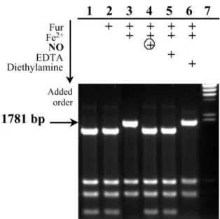
to a specific sequence located upstream of the regulated genes, thereby inhibiting RNA polymerase binding. In the restriction site protection assay, Fur protects a hinfI site from digestion, located between the ⫺10 and ⫺35 regions of the aerobactin promoter. According to Bindereif and Neilands (33), a 152-bp fragment of the aerobactin promoter is sufficient to provide regulation of a downstream iucA::lacZ fusion. Therefore, the protection of the hinfI site in pDT10 containing a 166-bp fragment of this promoter would indicate that the active Fur is bound to the promoter as shown in Fig. 2. Total HinfI digestion of pDT10 gave six fragments, including the 1,530- and 251-bp fragments, which came from the cleaved aerobactin promoter. A 1,781-bp fragment was expected when active Fur had protected the hinfI site. The Fur apoprotein did not bind the aerobactin promoter, but required a metal, Fe2⫹in our case, to protect the hinfI site within the aerobactin promoter. FeFur was able to bind
the aerobactin promoter in the whole range of molar ratios [FeFur dimer]兾[pDT10] superior to 20 (not shown). When EDTA, a strong iron chelator, was added, protection was not observed anymore.
Fig. 1. Effect of NO on growth and-gal expression in strains carrying
fhuF::lacZ fusion. Strains fhuF::lacZ and⌬-Fur fhuF::lacZ were grown in the presence or absence of DEANO in anaerobiosis at 37°C (A) and assayed for -gal (B). The differential rate of -gal synthesis is represented as the total -gal synthesized by milliliter of culture: -gal activity expressed in Miller
units⫻ A600(-gal units兾ml) in function of the absorbance at 600 nm (A600).
Arrows indicate time of addition of DEANO.E, Strains fhuF::lacZ with 25M
DEANO;‚and*, with 10M DEANO; andF, no DEANO;䊐,⌬-Fur fhuF::lacZ
with 200M DEANO;■, no DEANO.
When FeFur, first incubated with pDT10, was exposed to three equivalents of NO via DEANO, the 1,781-bp fragment was totally cleaved and generated the 1,530- and 251-bp fragments, suggesting direct inactivation of FeFur by NO. A control exper-iment showed that the aerobactin promoter remained fully protected in the presence of diethylamine, the other product of DEANO decomposition.
NO Binds to FeII in FeFur to Form a S ⴝ 1兾2 Dimeric Species.
Spectroscopic studies on NO-modified FeFur were performed to characterize the interaction between NO and FeFur. The addi-tion of NO gas to an anaerobic soluaddi-tion of FeFur resulted in immediate change from colorless to yellow-green, associated to the appearance of absorption bands in the UV-visible spectra as well as a g⫽ 2.03 EPR signal. Both features are characteristics of NO–iron complexes (34–38). The rate of NO complex for-mation was controlled by the decomposition of DEANO (t1/2⫽
15 min at 20°C, pH 7.5). The NO-complex formation developed ⬎1 h and 30 min after exposure to one equivalent of NO. There was no difference in the EPR spectra when using NO gas instead of DEANO.
The X-band EPR spectrum of the FeFur–NO complex was characterized by an anisotropic signal around g⫽ 2, with an isotropic g factor of giso⫽ 2.03. This signal arose from a species
with a S⫽ 1兾2 ground state (Fig. 3A). When the apoprotein was treated with NO in the same conditions, no EPR-detectable species appeared.
Furthermore, the57Fe-labeled FeFur–NO complex displayed
a distinct X-band EPR spectrum, especially when a shoulder was visible in the low-field region (Fig. 3B). This feature was due to the hyperfine contribution arising from the interaction between the electronic spin S⫽ 1兾2 and the nuclear spin of57Fe (I⫽ 1兾2).
This interaction demonstrates that iron is directly involved in the S⫽ 1兾2 species.
The double integration of this signal indicated that the cor-responding species represented 85% of the iron in the sample, which was in agreement with Mo¨ssbauer quantification analysis
(not shown). This proportion remained unchanged after new additions of NO as well as after gel-exclusion chromatography to remove any adventitious element bound to the protein.
The use of15NO in place of the unlabeled NO gas to generate
the FeFur–NO complex yielded a narrower EPR spectrum, especially in the low-field region (Fig. 3C). These changes arose from the distinct contribution of the15N nucleus of NO to the
hyperfine interactions. As expected for the contribution of the
15N nuclear spin I⫽ 1兾2, compared with the14N nuclear spin I⫽
1, the linewidths were smaller than the linewidths associated to the unlabeled NO. The 15N labeling of NO demonstrates the
binding of NO to the iron.
UV-visible spectra (not shown) revealed three bands at 310, 365, and 400 nm, characteristic of charge-transfer bands ob-served with small NO–iron complexes (35–38). The appearance of these bands grew in parallel with the increase of the EPR signal intensity. This links the S⫽ 1兾2 species and the UV-visible spectrum. The spectrum remained unchanged during 24 h in anaerobic conditions at 20°C. In air, the NO complex was stable for 1 h. Nevertheless, the UV-visible spectrum disappeared over several hours.
NO-Treated FeFur Is Still a Dimer.We used analytical gel filtration
to study the oligomeric state of the protein after the treatment by NO. FeFur treated with NO was eluted at the same volume as apo-Fur and FeFur species, which were both dimers (not shown), showing that NO-modified FeFur was still a dimer.
Fig. 2. In vitro assay of NO effect on Fur DNA binding. Fifty nanomolar
plasmid pDT10 was cleaved by HinfI in the absence or presence of active Fur and after NO treatment. Reaction mixtures were analyzed on 1.5% agarose
gel electrophoresis. Lanes: 1, no addition; 2, 20M apoFur; 3, 20 M FeFur; 4,
20M FeFur ⫹ 40 M DEANO after 1 h at 20°C; 5, 20 M FeFur ⫹ 50 M EDTA;
6, 20M FeFur ⫹ 40 M diethylamine after 1 h at 20°C; 7, ladder DNA HindIII
digest (arrow indicates the restriction fragment (1,781 bp) carrying the Fur box
that was not cleaved (into 1,530-bp⫹ 251-bp fragments) in the presence of
active Fur.
Fig. 3. EPR spectrum of isotopically labeled FurFe–NO complex. (A) FurFe– NO at 2.5 mM. The simulation is obtained with the g values (2.042; 2.032;
2.015) by using the linewidths 7.9, 7.0, and 4.0 G. (B) Fur57Fe-NO at 3.25 mM.
The simulation is achieved by using the [g] values obtained previously with
FurFe-NO and the hfs constants A兾h (45.6, 35.9, and 3.8 MHz) with the
respective linewidths (8.3, 7.5, and 4.6 G). (C) Comparison of Fe(II)-Fur–NO
complex at 1.5 mM generated with unlabeled NO and15NO. The simulations
are achieved with the same set of g values (2.042; 2.032; 2.015) and the same linewidths as in A for unlabeled species, but with the respective linewidths
(6.7, 7.2, and 3.5 G) for15NO complex. Nonsaturating EPR conditions:
micro-wave frequency, 9.655 GHz; power, 5W; modulation amplitude, 4 G;
mod-ulation frequency, 100 kHz; temperature, 30 K.
Fur Is Nitrosylated by NO Inside the Cell.To analyze EPR species
resulting from the interaction between NO and Fur inside cells, bacteria overexpressing the Fur protein were used. The cells treated by NO via DEANO exhibited a strong EPR signal in the
g⫽ 2.03 region (Fig. 4A). The signal was absent in untreated cells (Fig. 4B) or in treated cells in which Fur was not overexpressed (Fig. 4C). The signal also was observed when cultures were done in anaerobic conditions (data not shown). This S⫽ 1兾2 species revealed [g] values, linewidths, and saturation properties iden-tical to those of the FeFur–NO complex. This EPR signal is associated to the presence of the FeFur–NO complex inside the cells.
The concentration of this NO complex estimated by EPR quantification was⬇0.3 M in the bacterial culture in aerobic conditions. Because 50 mg of Fur兾liter of culture usually was expressed, we expected a maximum Fur concentration of 3M in the cell suspension. Nevertheless, no iron has been added to the medium, so the concentration of active Fur protein, FeFur, is probably ⬍3 M. Moreover, these experiments have been done in aerobic conditions, which reduce the NO concentration because of its oxidation. Thus, the quantification of the EPR signal shows the possibility to complex large amounts of FeFur with NO inside cells. The intensity of the EPR signal was the same when bacterial culture was sampled 2 h after the addition of NO.
Discussion
The mechanism by which bacteria sense NO and protect them-selves against NO is still poorly understood. Several studies established that Hmp, inducible by NO and nitrosothiol com-pounds, protects against nitrosative stress. In contrast to several NO-inducible genes involved in oxidative stress protection (7), induction of hmp expression by NO was not soxRS-dependent (12). Membrillo-Hernandez et al. (17) showed that induction by nitrosothiol depended on the MetR regulatory protein and proceeded via the nitrosation of its homocysteine cofactor. The mechanism of hmp induction by NO seems different. In E. coli, treatment with the iron chelator 2,2⬘-dipyridyl greatly induced
hmp expression, but the underlying mechanism was unclear (12).
In S. typhimurium, hmp expression was induced in a fur mutant
and induction by NO was Fur-dependent, but it remained unclear whether this was a direct or an indirect effect (18).
Here, we establish, both in vitro and in vivo, that the active iron-containing form of Fur from E. coli, FeFur, is sensitive to NO at micromolar concentrations. The induction of the fur regulon thus appears as a new pathway for the cell response to NO stress in bacteria.
Fur-regulated lacZ fusions were used to probe Fur activity against NO in vivo. Fusions with different Fur-regulated genes were chosen because of their different inductions in response to iron chelators that presumably reflect specific Fur affinity for promoter regions. The fiu::lacZ fusion requires seven times more ferrozine to yield half of the derepression compared with
fhuF::lacZ (32). Consistently, four overlapping Fur boxes were
found in the promoter region of the fiu operon (39), and only two were found in the fhuF promoter (40). In our normal growing conditions, Fur was active. To assay Fur activity against NO, a range of NO concentrations close to the estimated intracellular Fur concentration was used. A value of⬇5,000 Fur copies per cell in exponentially growing E. coli cultures has been measured (24). Considering a cell volume 2⫻ 10⫺15liters in the
exponential growth phase (41), the intracellular Fur concentra-tion would be⬇5M. Addition of 15 M of NO, using DEANO, to anaerobic cell cultures led to complete derepression of the
fhuF::lacZ fusion. Comparatively, hmp::lacZ fusions were
dere-pressed in aerobic or anaerobic cultures by 20M NO gas in E.
coli (12) and by 1 mM SperNO, another NO donor (smaller
concentrations were not tested), in S. typhimurium (18). In contrast, the fiu::lacZ fusion still was repressed at 300M NO. The promoter-dependent expression reflected the specific affinity of Fur for each promoter and then clearly involved inhibition of Fur activity. These data suggested that Fur was able to modulate the genes’ expression in response to NO, depending on Fur affinity for the promoter.
The interaction between NO and Fur suggested by in vivo assays was investigated on the purified Fur protein. The DNA-binding ability of FeFur was assayed in vitro by using the aerobactin promoter region. In agreement with in vivo assays, we observed that a 3-fold excess of NO was sufficient to switch off Fur binding to the aerobactin promoter. According to the in vitro assays, Fur was inactivated directly by a small excess of NO. To specify the interaction between NO and FeFur, we recorded EPR and UV-visible spectra.
The addition of NO by using NO gas resulted in the immediate complexation of NO with FeFur, yielding a species stable in anaerobic conditions that stood during⬎1 h in aerobiosis. EPR spectroscopy showed that NO interacts directly with FeII in
FeFur, inducing a low spin configuration characterized by an S⫽ 1兾2 ground state. The same characteristic EPR signal was recorded from Fur-overexpressing cells treated with NO in aerobic and anaerobic conditions. This result indicated that NO was able to cross the cell membrane and to target the active Fur protein. Furthermore, the stability of the S⫽ 1兾2 EPR signal in the cell was compatible with the timing of the Fur inhibition observed in the in vivo activity assay. It showed the link between the formation of the S ⫽ 1兾2 FeFur–NO complex and Fur inhibition reflected by the derepression of gene expression under Fur control.
EPR investigations on57Fe- and15N-labeled FurFe–NO
com-plex established that iron and NO were involved in a nitrosyliron unit. The EPR signal had an isotropic g value of g ⫽ 2.03, commonly associated with bis-cysteine dinitrosyl iron complexes of the type Fe(NO)2(RS)2(34, 37, 42). They are referred to as
the ‘‘g⫽ 2.03’’ complexes because of their characteristic isotropic
g factor. These kinds of complexes have been proposed in
iron–sulfur proteins such as soxR and aconitase after reaction with NO only by means of their EPR properties (16, 43). However, the first, direct structural insight concerning the g⫽
Fig. 4. EPR spectrum of intact cells treated with NO. Bacterial strains containing pFur1 were grown at 37°C in aerobiosis from the same preculture and sampled at the same time. (A) Fur synthesis was induced by isopropyl
-D-thiogalactoside during 2 h and 30 min and a sample was taken 60 min after
the addition of DEANO (10M). (B) A sample was taken after Fur synthesis was
induced during 3 h and 30 min without any addition of DEANO. (C) A sample
was taken 60 min after the addition of DEANO (10M) in noninduced culture.
EPR conditions: microwave frequency, 9.655 GHz; power, 5W; modulation
amplitude, 10 G; modulation frequency, 100 kHz; temperature, 30 K.
2.03 family has been obtained with a bis-imidazole-coordinated complex mimicking histidine residues (34). In addition, some mononitrosyl–iron complexes containing nitrogen donor ligands also exhibit these types of EPR features (35, 36). Recent data concerning the nature of the ligands coordinated to iron in FeFur are much more in agreement with the environments in these compounds. Indeed, spectroscopic data on cobalt- and iron-substituted Fur indicated that the metal is hexacoordinated with one ligand at a longer distance and only nitrogen and oxygen donor ligands including two (or three) histidines and one (or two) aspartate or glutamate (20, 21). In conclusion, the nature of the EPR signal of the nitrosyl iron species in the FeFur–NO complex cannot be assigned without further spec-troscopic characterizations. On this purpose, the studies by Mo¨ssbauer, x-ray absorption, electron nuclear double resonance, and Fourier transform–IR spectroscopies currently are in progress.
To fully explain in vivo observations, we wondered how the interaction of NO with FeFur could lead to the inactivation of Fur. It has been proposed that coordination of Fe2⫹induces a
conformational change of Fur dimer. Recent work has estab-lished that this conformational change enhances solvent acces-sibility of the DNA-binding region of Fur (44). Coordination of a divalent metal and a dimeric structure both are essential for DNA-binding activity. Gel filtration showed that the dimeric structure was not broken in the FurFe–NO complex. As NO binds to the iron, it may remove one of the six ligands. This change in the metal environment could alter the conformation of the protein and, therefore, modify the ability of Fur to bind to DNA. On this purpose, site-directed mutagenesis experiments have shown that a single mutation of residue likely involved in Fe2⫹ coordination yields an inactive Fur protein (45, 46).
Mutation especially of His-90, a highly conserved amino acid in
Fur sequences, leads to an inactive protein still able to dimer-ize (45).
Taken together, in vivo as well as in vitro assays and spectro-scopic studies suggested that a fast and irreversible inhibition of Fur occurs by nitrosylation of FeII. It is the first time that
reactivity of Fur iron site with an exogenous molecule is ob-served. We demonstrate that Fur is not only a sensor of iron, but through reactivity with an exogenous molecule at the iron site, provides a fine tuning of transcriptional control.
The modulation of transcription control via Fur in response to NO stress may provide protection against NO by several ways. The coupled protection against oxidative stress via regulation of superoxide dismutases by Fur minimizes the formation of ex-tremely deleterious peroxynitrite. The activation of hmp expres-sion through Fur inhibition in S. typhimurium directly detoxifies NO. E. coli (148 aa) and S. typhimurium (150 aa) proteins share 99% identity (or 96% identity and 2% similarity if the compar-ison is done the other way) with a strict conservation of the iron putative ligands. This suggests strongly that the mechanism of induction by NO of hmp expression in S. typhimurium is done by inactivation of Fur, via nitrosylation of the iron of the Fur protein, as demonstrated in this paper for Fur of E. coli. In E.
coli, the mechanism of hmp regulation appears more subtle and
depends on many regulators. However, a slight effect of Fur (2-fold induction in fur mutant; unpublished data) also is seen, coupling iron metabolism and defense against NO. Stimulation of iron metabolism as shown by the derepression of fhuF and iuc expression in E. coli (this work) and derepression of ircA and ircC in Salmonella (18) presumably favors the reconstitution of the iron proteins damaged by NO.
We are indebted to Sarah Dubrac for her assistance with the in vivo experiments, and Jacques Gaillard and Laurent Le Pape for their help with the EPR experiments.
1. Neilands, J. B. (1972) Struct. Bonding (Berlin) 11, 145–170.
2. Hibbs, J. B., Jr., Taintor, R. R., Vavrin, Z. & Rachlin, E. M. (1988) Biochem.
Biophys. Res. Commun. 157, 87–94.
3. Marletta, M. A., Yoon, P. S., Iyengar, R., Leaf, C. D. & Wishnok, J. S. (1988)
Biochemistry 27, 8706–8711.
4. Sari, M.-A., Moali, C., Boucher, J. L., Jaouen, M. & Mansuy, D. (1998)
Biochem. Biophys. Res. Commun. 250, 364–368.
5. Adak, S., Aulak, K. S. & Stuehr, D. J. (2002) J. Biol. Chem. 277, 16167–16171. 6. Goretski, J., Zafiriou, O. C. & Hollocher, T. C. (1990) J. Biol. Chem. 265,
11535–11538.
7. Nunoshiba, T., DeRojas-Walker, T., Wishnok, J. S., Tannenbaum, S. R. & Demple, B. (1993) Proc. Natl. Acad. Sci. USA 90, 9993–9997.
8. Lancaster, J. R., Jr., & Hibbs, J. B., Jr. (1990) Proc. Natl. Acad. Sci. USA 87, 1223–1227.
9. Michalski, W. P. & Nicholas, D. J. D. (1987) Arch. Microbiol. 147, 304–308. 10. Lepoivre, M., Flaman, J. M. & Henry, Y. (1992) J. Biol. Chem. 267, 22994–
23000.
11. Hausladen, A., Privalle, C. T., Keng, T., DeAngelo, J. & Stamler, J. S. (1996)
Cell 86, 719–729.
12. Poole, R. K., Anjum, M. F., Membrillo-Hernandez, J., Kim, S. O., Hughes, M. N. & Stewart, V. (1996) J. Bacteriol. 178, 5487–5492.
13. Gardner, A. M., Helmick, R. A. & Gardner, P. R. (2002) J. Biol. Chem. 277, 8172–8177.
14. Gardner, A. M. & Gardner, P. R. (2002) J. Biol. Chem. 277, 8166–8171. 15. Hausladen, A., Gow, A. & Stamler, J. S. (2001) Proc. Natl. Acad. Sci. USA 98,
10108–10112.
16. Ding, H. & Demple, B. (2000) Proc. Natl. Acad. Sci. USA 97, 5146–5150. 17. Membrillo-Hernandez, J., Coopamah, M. D., Channa, A., Hughes, M. N. &
Poole, R. K. (1998) Mol. Microbiol. 29, 1101–1112.
18. Crawford, M. J. & Goldberg, D. E. (1998) J. Biol. Chem. 273, 34028–34032. 19. Hantke, K. (2001) Curr. Opin. Microbiol. 4, 172–177.
20. Adrait, A., Jacquamet, L., Le Pape, L., Gonzalez de Peredo, A., Aberdam, D., Hazemann, J. L., Latour, J. M. & Michaud-Soret, I. (1999) Biochemistry 38, 6248–6260.
21. Jacquamet, L., Dole, F., Jeandey, C., Oddou, J. L., Perret, E., Le Pape, L., Aberdam, D., Hazemann, J. L., Michaud-Soret, I. & Latour, J. M. (2000) J. Am.
Chem. Soc. 122, 394–395.
22. Tardat, B. & Touati, D. (1991) Mol. Microbiol. 5, 455–465.
23. Dubrac, S. & Touati, D. (2000) J. Bacteriol. 182, 3802–3808.
24. Zheng, M., Doan, B., Schneider, T. D. & Storz, G. (1999) J. Bacteriol. 181, 4639–4643.
25. Maragos, C. M., Morley, D., Wink, D. A., Dunams, T. M., Saavedra, J. E., Hoffman, A., Bove, A. A., Isaac, L., Hrabie, J. A. & Keefer, L. K. (1991) J. Med.
Chem. 34, 3242–3247.
26. Hantke, K. (1984) Mol. Gen. Genet. 197, 337–341.
27. Miller, J. H. (1992) in A Short Course in Bacterial Genetics (Cold Spring Harbor Lab. Press, Plainview, NY), pp. 352–355.
28. Stookey, L. L. (1970) Anal. Chem. 42, 779–781.
29. Bagg, A. & Neilands, J. B. (1987) Biochemistry 26, 5471–5477. 30. Holmquist, B. (1988) Methods Enzymol. 158, 6–12.
31. Stojiljkovic, I., Ba¨umler, A. J. & Hantke, K. (1994) J. Mol. Biol. 236, 531–545. 32. Niehaus, F., Hantke, K. & Unden, G. (1991) FEMS Microbiol. Lett. 68,
319–323.
33. Bindereif, A. & Neilands, J. B. (1985) J. Bacteriol. 162, 1039–1046. 34. Reginato, N., McCrory, C. T. C., Pervitsky, D. & Li, L. (1999) J. Am. Chem.
Soc. 121, 10217–10218.
35. Franz, K. J. & Lippard, S. J. (1999) J. Am. Chem. Soc. 121, 10504–10512. 36. Hauser, C., Glaser, T., Bill, E., Weyhermu¨ller, T. & Wieghardt, K. (2000) J. Am.
Chem. Soc. 122, 4352–4365.
37. Costanzo, S., Me´nage, S., Purrello, R., Bonomo, R. P. & Fontecave, M. (2001)
Inorg. Chim. Acta 318, 1–7.
38. Jo, D.-H., Chiou, Y.-M. & Que, L., Jr. (2001) Inorg. Chem. 40, 3181–3190. 39. Newman, D. L. & Shapiro, J. A. (1999) Mol. Microbiol. 33, 18–32. 40. Zheng, M., Wang, X., Doan, B., Lewis, K. A., Schneider, T. D. & Storz, G.
(2001) J. Bacteriol. 183, 4571–4579.
41. Ali Azam, T., Iwata, A., Nishimura, A., Ueda, S. & Ishihama, A. (1999)
J. Bacteriol. 181, 6361–6370.
42. Boese, M., Mordvintcev, P. I., Vanin, A. F., Busse, R. & Mu¨lsch, A. (1995)
J. Biol. Chem. 270, 29244–29249.
43. Kennedy, M. C., Antholine, W. E. & Beinert, H. (1997) J. Biol. Chem. 272, 20340–20347.
44. Gonzalez de Peredo, A., Saint-Pierre, C., Latour, J. M., Michaud-Soret, I. & Forest, E. (2001) J. Mol. Biol. 310, 83–91.
45. Braun, V., Scha¨ffer, S., Hantke, K. & Tro¨ger, W. (1990) in 41. Colloquium
Mosbach 1990 (Springer, Berlin), pp. 164–179.
46. Coy, M., Doyle, C., Besser, J. & Neilands, J. B. (1994) Biol. Metals 7, 292–298.