HAL Id: tel-02927784
https://tel.archives-ouvertes.fr/tel-02927784
Submitted on 2 Sep 2020HAL is a multi-disciplinary open access archive for the deposit and dissemination of sci-entific research documents, whether they are pub-lished or not. The documents may come from teaching and research institutions in France or abroad, or from public or private research centers.
L’archive ouverte pluridisciplinaire HAL, est destinée au dépôt et à la diffusion de documents scientifiques de niveau recherche, publiés ou non, émanant des établissements d’enseignement et de recherche français ou étrangers, des laboratoires publics ou privés.
Mechanisms and function of mitophagy in adaptation to
heat stress during development of C. elegans
Yanfang Chen
To cite this version:
Yanfang Chen. Mechanisms and function of mitophagy in adaptation to heat stress during develop-ment of C. elegans. Subcellular Processes [q-bio.SC]. Université Paris Saclay (COmUE), 2019. English. �NNT : 2019SACLS217�. �tel-02927784�
Mechanisms and function of mitophagy
in adaptation to heat stress during
development of C. elegans
Thèse de doctorat de l'Université Paris-Saclay Préparée à l’Université Paris-Sud École doctorale n°577 : Structure et dynamique des systèmes vivants (SDSV) Spécialité de doctorat: Sciences de la vie et de la santé
Thèse présentée et soutenue à Gif-sur-Yvette, le 23 juillet 2019, par
Yanfang CHEN
Composition du Jury : Audrey ESCLATINE
Professeure, Université Paris-Sud (CNRS UMR 9198) Présidente Nadine CAMOUGRAND
DR, Université de Bordeaux (CNRS UMR 5095) Rapporteuse Kathrin GIESELER
Professeure, Université Lyon 1 (CNRS UMR 5310, INSERM
U1217) Rapporteuse
Fabrizia STAVRU
CR, Institut Pasteur-UMR3528 Examinatrice Renaud LEGOUIS
DR, INSERM (CNRS UMR 9198) Co-Directeur de thèse Agnès DELAHODDE
DR, CNRS (CNRS UMR 9198) Co-Directrice de thèse
Equipe "Autophagie et développement" de Renaud Legouis NNT 2 01 9S AC LS 21 7
Acknowledge
At the beginning, I would like to thank the jury. These reporters: Nadine CAMOUGRAND and Kathrin GIESELER, who have red my thesis and provided me suggestions for better construct my thesis. These examinants: Audrey ESCLATINE and Fabrizia STAVRU, who come to attend my defense. Thank you all for your time!
The profound gratitude should go firstly to my directors: Reanud LEGOUIS and Agnès DELAHODDE. Four years ago, it was Renaud and Agnès who accepted my PhD application and offered me this opportunity to come here to start my PhD career. During the four years study in Renaud’s lab, Renaud offered me instructive advices and useful suggestions for my project. My sincere gratitude to Renaud also for his constant encourage. It is not easy for me at the beginning of my PhD, I met many difficulties and always doubt myself. During this harsh time, Renaud never blamed me; on the contrary, he supported me and taught me to positively solve the problems. MERCI beaucoup, Renaud! It is a pity that I did not spend lots time with my another supervisor, Agnès DELAHODDE. But Agnès always cares about my work and spent lots of time to guide my project, correct my thesis. Each time I asked Agnès for help, she assisted me with her best.
Secondly, I want to express my heartfelt thanks to Emmanuel CULETTO. When I came to the lab, I knew nothing about C. elegans. It was Emmanuel who taught me hand by hand to deal with worms. Emmanuel is my teacher not only on experiments, but also on English. Although he was quite busy as a lecture in Paris-Sud, he helped me to correct my English writing for my each committee, international meeting and this thesis. I want to tell Emmanuel that I am not always a good student, but you are really an excellent teacher! I would also thanks to all our “autophagy and development team”: Céline LARGEAU, Siham ZENTOUT (M2 student), Vincent SCARCELLI, Romane LEBOUTET, Christophe LEFEBVRE, Céline JENZER. Especially Céline LARGEAU and Siham ZENTOUT, who have participate in my project and contributed to the figures in my manuscript. Without your excellent work, the manuscript would not be complete. I would like to thank Céline JENZER and Christophe LEFEBVRE for their help with some experiments. I would also thank Vincent and Romane, for your generous help in experiments as well as in life. Thank you for your patient translation and help me to call the doctor, the CAF, the bank, the FREE and so on. All your kindness makes me feel not so lonely in France which is far from my hometown. You mean friends and even families to me in France. I will miss you all……
I also owe a specific gratitude to the people in I2BC, SDSV and Gif. I am so sorry that finally I am not able to learn French, and my English is not good, neither. The communications between us is not so smooth, but I can feel your kindness to me.
I am so deeply indebted to China Scholarship Council (CSC), for their financial support during my four years PhD study.
Last but not least, my thanks would go to my beloved families and my friends who have always been helping me out of difficulties and supporting without a word of complaint. Thank you all, I have spent a nice period in France. I will always keep this beautiful memories in my mind.
1
LIST OF TABLES AND FIGURES………5
ABBREVIATIONS……….…9
I INTRODUCTION………19
1. Autophagy………..21
1.1 Three types of autophagy………21
1.1.1. Microautophagy and endosomal microautophagy………22
1.1.2. Chaperon-mediated autophagy (CMA)……….23
1.1.3. Macroautophagy………..24
1.1.4. Core machinery for autophagosome formation……….25
1.1.5. Fusion of autophagosome with lysosome………..27
1.1.6. Autophagic lysosome reformation ………30
1.2 Non-selective and selective autophagy………30
1.3 Physiological roles of autophagy………33
1.3.1. Developmental cell death and autophagy………..34
1.3.2. Aging and autophagy……….35
1.3.3. Cancer and autophagy………..36
2. C. elegans, a model animal for autophagy studies………36
2.1 General knowledge of C. elegans………36
2.1.1. C. elegans maintenance and life cycle………..37
2.1.2. C. elegans tissues……….40
2.1.2.1 Epidermis……….41
2.1.2.2 Muscles………..………..42
2.1.2.3 The digestive system………..……….42
2.1.2.4 Reproductive tissue………..………43
2.1.2.5 The nervous system………..………44
2.2 C. elegans is an ideal model animal for autophagy study………44
2.2.1 Allophagy………46
2.2.2 Aggrephagy………49
2.2.2.1 Degradation of germline P granule………..……….49
2.2.2.2 Degradation of SQST-1 postive aggrephagy during embryogenesis………..………51
2.2.3 Autophagy in stress conditions………..53
2.2.3.1 Autophagy in starvation condition………..………..53
2.2.3.2 Autophagy in heat stress condition………54
2.2.3.3 Autophagy and dauer formation………..……….55
2.2.4 Autophagy and longevity……….56
2.2.5 Autophagy and neurodegenerative diseases………..57
2.2.6 Other examples of autophagy associated process………59
2.2.6.1 Lipophagy……….………..59
2.2.6.2 Autophagy mediated apoptotic cell death………..………..59
2
3.1 Mitophagy concept………60
3.2 Mitophagy in yeast……….61
3.3 Mitophagy in mammals………63
3.3.1 PINK1/Parkin mediated mitophagy………64
3.3.1.1 PINK1, a sensor for mitophagy initiation and Parkin activation……….………64
3.3.1.2 Receptors involved in Parkin-mediated mitophagy………66
3.3.2 FUNDC1 mediated mitophagy……….68
3.3.2.1 Regulation of FUNDC1 by phosphorylation………..………..69
3.3.2.2 The ubiquitin regulation of FUNDC1………..………..70
3.3.3 NIX (BNIP3L) mediated mitophagy………70
3.3.4 Other mitophagy mediators……….71
3.3.4.1 NDP52 and TBK1 cooperate during mitophagy……….71
3.3.4.2 MUL1, an OMM E3 ligase………..………..72
3.3.4.3 Bcl2L13/Bcl-Rambo………73
3.3.5 Lipid-mediated mitophagy……….73
3.3.6 Mitophagy related diseases………..74
3.4 Mitophagy in C. elegans……….75
3.4.1 Basal or induced mitophagy affects worm lifespan……….75
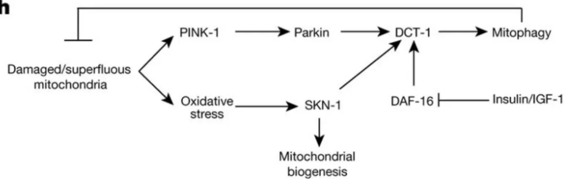
3.4.1.1 DCT-1 mediated mitophagy in aging and stress conditions………..……..76
3.4.1.2 Tomatidine induced mitophagy………78
3.4.1.3 Urolithin A induced mitophagy……….79
3.4.1.4 Iron-starvation-induced mitophagy………..………80
3.4.2 The role of mitophagy in neurodegenerative diseases………..81
4. The relationship between mitochondrial dynamics and mitophagy………83
4.1 Mitochondrial fusion……….84 4.2 Mitochondrial fission……….86 4.2.1 Drp1………89 4.2.2 Drp1 adaptors……….89 4.2.3 Drp1 posttranscriptional modifications………92 4.2.4 Dnm2……….94
4.3 Mitochondrial dynamics in mitophagy process……….95
4.3.1 Drp1-dependent mitophagy……….95
4.3.2 Drp1-independent mitophagy……….98
4.4 ER and mitochondria contact sites………101
4.4.1 The concept and functions of contact sites………101
4.4.2 The role of contact sites in mitochondrial morphology regulation………102
4.4.3 The contact sites are also involved in autophagy and mitophagy………105
4.5 The roles of Drp1………107
4.5.1 Drp1 in mitophagy………107
4.5.2 Other function of Drp1………108
4.5.2.1 Drp1 is involved in peroxisomal fission………..………109
3
II RESULTS………111
1. How C. elegans responds to acute heat stress?...113
1.1 Summary………113 1.2 Manuscript………117 1.3 Extended data………151 1.3.1 Phenotypic characterizations……….151 1.3.2 Autophagy/Mitophagy………..152 1.3.3 Drp1……….157 1.3.4 Discarded tools………162
2. A preliminary exploration of MAM candidates in C. elegans……….165
2.1 Summary……….165
2.2 Results………168
2.2.1 The potential MAM proteins in C. elegans………..168
2.2.2 The role of ER-mitochondria contact sites in heat stress-induced autophagy…………171
2.2.3 Autophagy/mitophagy is altered by fzo-1 RNAi………172
2.2.4 New tools for studying MAM……….174
3. Ether lipid and autophagy (collaborative project) ……….179
3.1 Summary……….179
3.2 Results………..181
III DISCUSSION AND PERSPECTIVES………183
1. The role of autophagy during heat stress………185
1.1 Heat stress studies………...185
1.2 How autophagy affects worm recovery from heat stress? ...188
1.3 Mitophagy mediators………..189
2. ER-mitochondria contact sites in C. elegans……….191
2.1 The study of ER-mitochondria is an emerging field in C. elegans……….191
2.2 Tools for studying ER-mitochondria contact sites………..191
3. The autophagy defect in drp-1 mutant worms ……….192
IV ANNEXES………197
V BIBLIOGRAPHY……….……….219
5
LIST OF TABLES AND
FIGURES
7
Table 1. The Sequence alignment and binding partner of the reported
autophagy receptors
Table 2. Developmental timing of C. elegans at different growth temperatures.
Table 3. Autophagy genes in C. elegans, and homologs
Table 4.
Proteins regulating mitochondrial fission and fusion in yeast, mammals
and C. elegans
Table 5. Functional roles of the MAM proteins
Table 6. Autophagy responds differently in muscle and epidermis
Table 7. The potential MAM proteins in C. elegans and their homologs in
mammals
Table 8. A comparison of different heat stress in the study of autophagy in C.
elegans
Figure 1. Three types of autophagy coexist within the eukaryotic cells
Figure 2. Autophagy process and its core machinery in mammalian cells
Figure 3. Functions of autophagy receptors and adaptors in selective
autophagy
Figure 4. C. elegans life cycle at 22°C
Figure 5. C. elegans anatomy
Figure 6. The autophagy is essential for various physiological processes during
whole life
Figure7. The process of allophagy
Figure 8. The hierarchical order of autophagy genes in the aggrephagy pathway
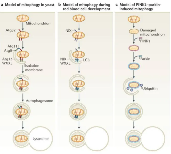
Figure 9. Different types of mitophagy in yeast and mammals
Figure 10. The process of Parkin activation
8
Figure12. Model for mitochondrial fission in animal cells
Figure13.
Models for mitochondrial division during mitophagy
Figure14. Function of Arf1 in mitochondria-related processes
Figure 15. Autophagosome forming at the ER (endoplasmic
reticulum)-mitochondria contact sites
Figure 16. Heat stress impairs the tissue morphologies during development
Figure 17. Heat stress also induces autophagy in the muscle and germline
Figure 18. Lipophagy is not induced after heat stress
Figure 19. drp-1 deletion predisposes mitochondria to damage upon heat
stress
Figure 20. drp-1 RNAi also induces autophagic clusters in germline and early
embryo
Figure 21. DRP-1 responds to heat stress
Figure 22. The discarded tools for monitoring mitophagy in C. elegans muscle
Figure 23. The roles of MAM candidates in mitochondrial morphology
regulation and heat stress-induced autophagy
Figure 24. Autophagy/mitophagy is altered by fzo-1 RNAi
Figure 25. Tools for studying ER-mitochondria contact sites
9
11
Aβ: β-amyloid peptide
AC: actinonin
acl7: acyltransferase-like
AD: Alzheimer's disease
ads1:
Alkyldihydroxyacetonephosphate
synthase
AIM: Atg8-family interacting motif
AHA-1: Aryl hydrocarbon receptor
nuclear translocator homolog
aHS: acute heat stress
ALLO-1: Allophagy receptor -1
ALR: autophagic lysosome
reformation
ALS: amyotrophic lateral sclerosis
Ambra1: autophagy and beclin 1
regulator 1
Ams1p: α-mannosidase 1
Apf: after puparium formation
APG6: autophagy protein 6
ARF1: ADP-ribosylation factor 1
ARIH1: Ariadne RBR E3 Ubiquitin
Protein Ligase 1
ACS4: acyl-CoA synthetase long
chain 4
ATG: autophagy related gene
Atg1: autophagy related gene 1
Atg2: autophagy related gene 2
Atg3: autophagy related gene 3
Atg4: autophagy related gene 4
Atg5: autophagy related gene 5
Atg7: autophagy related gene 7
Atg8: autophagy related gene 8
Atg9: autophagy related gene 9
Atg12: autophagy related gene 12
Atg13: autophagy related gene 13
Atg14L: Autophagy-Related Protein
14-Like Protein
Atg16L: autophagy related 16-like 1
Atg18: autophagy related gene 18
Atg19: autophagy related gene 19
Atg30: autophagy related gene 30
Atg32: autophagy related gene 32
Atg36: autophagy related gene 36
Atg101: Autophagy Related 101
12
ATP: Adenosine triphosphate
Aup1p: autophagy-related protein
phosphatase
BAG1: Bcl2-associated athanogene
1 protein
BAK1: Bcl-2 Homologous
Antagonist/Killer
BAX: Bcl-2-associated X protein
Bcl-2: B-cell lymphoma 2
BCL2L1: BCL2-like 1
Bcl2L13: Bcl-2-like protein 13
BECN1: Beclin 1
BNIP3: BCL2/adenovirus E1B 19 kDa
protein-interacting protein 3
BNIP3L: BCL2/adenovirus E1B 19
kDa protein-interacting protein
3-like
BP: 2,2’-dipyridyl
BWM: body-wall muscle cell
CAF4: CCR4-associated factor 4
CAMK-II: Ca
2+-calmoduline
dependent kinase II
CCCP:
2[2(3Chlorophenyl)hydrazinylyidene
]propanedinitrile
CDK-1: Cyclin-dependent kinase 1
CED-3/4: Cell death protein 3/4
CerS1: ceramide synthase 1
CK2: Casein kinase 2
CLS: chronological life span
CMA: Chaperon-mediated
autophagy
CNX1: Calnexin
COPII: coat protein complex II
CORVET: class C core
vacuole/endosome tethering
CPS6: Ced-3 protease suppressor 6
CRISPR: clustered regularly
interspaced short palindromic
repeats
Cvt: cytoplasm-to-vacuole targeting
DAF-2/16: Abnormal dauer
formation protein 2/16
DCT-1: Daf-16/FOXO controlled
germline tumor affecting-1
DFCP-1: Double FYVE-Containing
Protein 1
DFP: deferiprone
DHAP: Dihydroxyacetone phosphate
DIC: Differential interference
constrast
13
DNM1L: Dynamin-1-like protein
Dnm2: Dynamin-2
DNA: deoxyribonucleic acid
DNC: dorsal nerve cord
Drp1: dynamin-related protein 1
Dyn2: dynamin 2
ECM: extracellular matrix
EM : electron microscopy
eMi: endosomal microautophagy
Endo G: endonuclease G
EPG-2/3/4/5/8 : Ectopic P granules
protein 2/3/4/5/8
ER: endoplasmic reticulum
ERMES: ER-mitochondria encounter
structure
ESCRT: endosomal Sorting
Complexes required for transport
FACL4: Fatty-Acid-Coenzyme A
Ligase, Long-Chain 4
fard-1: Fatty acyl-CoA reductase
FCCP: Carbonyl cyanide
4-(trifluoromethoxy)phenylhydrazone
FIP200: focal adhesion kinase (FAK)
family interacting protein of 200 kD
Fis1: fission 1
FOXO: Forkhead box O
FRDA: Friedreich’s ataxia
frh1: Frataxin
FUNDC1: FUN14 Domain Containing
1
FYCO1: FYVE and coiled-coil
domain-containing protein 1
GABA: gamma-amino butyric acid
GABARAP: Gamma-aminobutyric
acid receptor-associated protein
GAP: GTPase-activating protein
GBF1: Golgi-specific brefeldin
A-resistance guanine nucleotide
exchange factor 1
GDAP1: Ganglioside-induced
differentiation associated protein 1
GDP: guanosine di-phosphate
GED: GTPase effector domain
GFP: green fluorescent protein
gld-1: germ line developmen-1
GLP1: Glucagon-like peptide-1
Gpb2: Guanine nucleotide-binding
protein subunit beta 2
GSK3β: Glycogen synthase kinase-3
beta
14
HD: Huntington's disease
HIF: hypoxia-inducible factors
Hip: Hsp70- interacting protein
HLH30: Helix Loop Helix
Hog1: High osmolarity glycerol
response protein 1
Hop: Hsp70-Hsp90 organizing
protein
HOPS: homotypic fusion and
vacuole protein sorting
HRP: horseradish peroxidase
HSC70: heat stress cognate protein
of 70 kDa
HSE: heat shock element
HSF1: Heat shock factor 1
Hsp40/70/90: heat shock protein
40/70/90
HSPA8: heat shock protein family A
member 8
Htt: huntingtin
IGF1: Insulin-like growth factor 1
IKKE-1: Inhibitor of nuclear factor
kappa-B kinase epsilon subunit
homolog 1
ILV: internal lumenal vesicles
IM: isolation membrane
IMM: inner mitochondrial
membrane
IMS: intermembrane space
INF2: Inverted Formin-2
ISC: iron-sulfur-cluster
KO: knock out
LAMP2: lysosomal-associated
membrane protein 2
LAMP2A: lysosomal-associated
membrane protein type2A
LAP: LC3-associated phagocytosis
LD: lipid droplet
LIPL4: Lipase-Like Abhydrolase
Domain-Containing Protein 4
LIR: LC3-interacting region
LC3: microtubule-associated protein
light chain 3
LLPS: liquid-liquid phase separation
lrk1: Leucine-rich repeat
serine/threonine protein kinase 1
LRRK2: Leucine-rich repeat kinase 2
LRS: LC3 recognition sequence
MAM: mitochondria-associated
membranes
MAPK: Mitogen-activated protein
kinase
15
MAPKK: Mitogen-activated protein
kinase kinase
MARCH5: membrane-associated
ring finger (C3HC4) 5
MCU: mitochondrial calcium
uniporter
Mdm10/12/34: Mitochondrial
distribution and morphology
protein 10/12/34
MDV1/2: Mitochondrial division
protein 1/2
MEF: mouse embryonic fibroblasts
Mff: mitochondrial fission factor
Mfn1/2: mitofusin 1/2
Mgm1 : Mitochondrial genome
maintenance protein 1
Mi: microautophagy
MiD49/51: Mitochondrial Dynamics
Protein Of 49 KDa/51 kDa
MIRO1: Mitochondrial Rho GTPase
1
Mmm1: Maintenance of
mitochondrial morphology protein 1
MMP: mitochondrial membrane
potential
MOs: membranous organelles
MPP+ :
1-methyl-4-phenylpyridinium
MPTP
:
1-methyl-4phenyl-1,2,3,6-tetrahydropyridine
mtDNA: mitochondrial DNA
mTOR: mechanistic target of
rapamycin
MUL1 : Mitochondrial E3 Ubiquitin
Protein Ligase 1
MVB: multivesicular body
MXL3: MaX-Like
Nbr1: neighbor of BRAC1 gene
NDP52: nuclear dot protein 52 kDa
NGM: Nematode growth media
NIX: NIP-3-Like Protein X
NMN: nicotinamide
mononucleotide
NO: nitric oxide
NRF2/NFE2L2: NF-E2-Related Factor
2 / Nuclear Factor Erythroid
2-Related Factor 2
NS: no significance
NVT: Nbr1-mediated vacuolar
targeting
OCR: oxygen consumption rate
OMM: outer mitochondrial
membrane
16
OMMAD: outer mitochondrial
membrane associated degradation
OPA1: Optic Atrophy 1
OPTN:
Optineurin
OXPHOS: Oxidative phosphorylation
p62/SQST1: Sequestosome 1
PACS2: Phosphofurin Acidic Cluster
Sorting Protein 2
PARL: Presenilins-associated
rhomboid-like protein
PAS: phagophore assembly site
PB1: Phox and Bem1
Pbs2: Polymyxin B resistance
protein 2
PD: Parkinson's disease
PE: phosphatidylethanolamine
PGAM5: phosphoglycerate mutase
family member 5
PGL-1/3: P granule abnormality
protein 1/3
PH: pleckstrin homology
PHB2: Prohibitin-2
PI3K: phosphatidylinositol 3 kinase
PI3KC3C1: phosphatidylinositol
3-kinase catalytic subunit type 3
complex I
PINK1: PTEN-induced kinase 1
PKA: Protein kinase A
PKC: Protein kinase C
PKM1: pyruvate kinase M1
PLEKHM1: pleckstrin homology and
RUN domain containing M1
PME: paternal mitochondrial
elimination
polyQ: Polyglutamine
PP1: Protein phosphatase 1
PP2A: Protein phosphatase 2
PS: phosphatidylserine
Ptdlns(3)P: phosphatidylinositol
3-phosphate
PTPIP51: Protein tyrosine
phosphatase interacting protein 51
Rab5: Ras-related protein Rab-5A
Rab7: Ras-related protein Rab-7A
Rab9: Ras-related protein Rab-9A
RET-1: Reticulon-like protein
RILP: Rab-interacting lysosomal
protein
RNA: Ribonucleic acid
RNS: reactive nitrogen species
ROS: reactive oxygen species
17
SEPA-1: suppressor of ectopic P
granules in autophagy
SIAH1: Seven In Absentia Homolog
1
SKN-1: Protein skinhead-1
Slt2: Suppressor of the LyTic
phenotype
SM: Sec1/Munc18-like protein
SMURF1: Smad Ubiquitination
Regulatory Factor 1
SNAP-29: Synaptosomal-associated
protein 29
SNARE: Soluble
N-ethylmaleimide-sensitive factor activating protein
receptor
SNX18: Sorting nexin-18
STX17: syntaxin 17
SV: synaptic vesicles
TBC1D15: TBC1 Domain Family
Member 15
TBK1: TANK-Binding Kinas 1
TEM: transmission electron
microscopy
TFEB: Transcription factor EB
TIM: Translocase of the inner
membrane
TMD: trans-membrane domain
TMRM: tetramethylrhodamine
TOM: translocase of the outer
membrane
UA: Urolithin A
Ub: ubiquitin
UBA: ubiquitin-associated
Uba1: Ubiquitin-like modifier
activating enzyme 1
UbI: ubiquitin-like domain
ULK1/2: unc-51-like kinase 1/2
UPR: unfolded protein response
UTH1: Youth protein 1
VAMP7/8: Vesicle-associated
membrane protein 7/8
VAPB: VAMP Associated Protein B
VDAC1: Voltage-dependent
anion-selective channel 1
VMP1: Vacuole membrane protein
1
VNC: ventral nerve cord
Vps
3/8/11/15/16/18/30/33/34/39/41:
Vacuolar protein sorting-associated
protein
3/8/11/15/16/18/30/33/34/39/41
Whi2: Whiskey 2
18
WIPI2: WD repeat domain
phosphoinositide-interacting
protein 2
WT: wild type
YFP: Yellow fluorescent protein
Yme1: Yeast mitochondrial escape
protein 1
19
I INTRODUCTION
1. Autophagy
2. C . elegans, a model animal for autophagy study
3. Mitophagy
21
1. Autophagy
Autophagy (from the Greek words auto (meaning self) and phagein (meaning to eat) is a degradation process of intracellular components through lysosome. It was first termed by De Duve in 1963(De Duve 1963) to describe the cell degradation process taking place in the double membraned organelle containing parts of cytosol and bits of organelle, seen under Electron Microscope. In the following years, studies in the field of autophagy were mostly descriptive: (1) showing that glucagon, insulin, amino acid deprivation and pharmacological agents such as methyl adenine and worthmannine could regulate autophagy and (2) describing autophagosome morphology and dynamic during its formation, fusion with either lysosome or endosome and their degradation. Molecular understanding of the autophagy process started when Yoshinori Ohsumi lab identified a series of genes directly involved in the autophagy process. These genes were initially named apg for AutoPhaGy related genes then further renamed atg for “AuTophaGy related” gene or protein. Atg genes were first cloned from the yeast Saccharomyces cerevisiae after many S.cerevisiae autophagy defective mutants were obtained by genetic screening for mutants that survive in rich medium but died when subjected to prolonged period of nitrogen starvation (Tsukada and
Ohsumi 1993). Both autophagy process and ATGs are highly conserved from yeast to
mammalian system. This degradative process has been widely shown to play important roles in many physiological and pathological processes, such as development, growth, aging, neurodegeneration, cancer. Ohsumi was awarded the Nobel Prize in physiology or medicine in 2016 for his key contributions to the research field of autophagy.
1.1. Three types of autophagy
Based on the mechanisms of delivery of cytoplasmic components to the lysosome, autophagy has been classified in three types: microautophagy, chaperone-mediated autophagy and macroautophagy (Boya, Reggiori et al. 2013). Basic features charaterizing these three types of autophagy are presented on Figure1.
22
Figure 1. Three types of autophagy coexist within the eukaryotic cells (a) Macroautophagy is characterized by
the cargo sequestration within a double membrane structure, which is named autophagosome. (b) Chaperone-mediated autophagy degrades proteins that containing KFERQ motif, which is recognized by chaperone Hsp70. During chaperone-mediated autophagy, LAMP2 mediates the fusion of Hsp70-protein aggregates with lysosome. Microautophagy refers to the direct sequestration of cargoes into lysosome for degradation. (c) Microautophagy directly engulfs cytoplasmic components by lysosomes.(Boya, Reggiori et al. 2013)
1.1.1. Microautophagy and endosomal microautophagy
Microautophagy (Mi) is defined as the engulfment of cytosolic cargoes directly via invagination of the lysosomal/vacuolar membrane. It is the least studied form of autophagy. It can degrade soluble proteins or even bigger cargoes such as portion of the nucleus, lipid droplets, mitochondria and endosomes (Veenhuis, Douma et al. 1983, Takahashi,
Murayama et al. 1993, Tuttle and Dunn 1995, Kissova, Salin et al. 2007) . This
microautophagy depends on the activity of the Endosomal Sorting Complexes required for transport (ESCRT) machinery. ESCRT components are present on the vacuolar membrane
23
and induced membrane dynamics in a similar way to that observed on endosome during intraluminal vesicles formation.
Additionally, it has been recently proposed to formerly distinguich a second type of microautophagy which relies on lysosomal protrusion and not its invagination (Oku and
Sakai 2018). This type of microautophagy is observed in the yeast Pichia pastoris when
lysosome sequesters peroxisome. This mechanism seems to be Atg18 dependent.
Another type of microautophagy, named endosomal microautophagy (eMi), shares very similar features to that of microautophagy described above but involves endosome (Sahu,
Kaushik et al. 2011, Uytterhoeven, Lauwers et al. 2015, Mukherjee, Patel et al. 2016).
Notably, endosomal microautophagy could selectively degrade proteins containing KFERQ motif via binding to heat shock protein family A member 8 (HSPA8), also known as heat stress cognate protein of 70 kDa (HSC70), which directs the cargo to the endosomal membrane for internalization into multivesicular body (MVB) in an ESCRT-I and -III dependent mechanism (Sahu, Kaushik et al. 2011, Uytterhoeven, Lauwers et al. 2015,
Mukherjee, Patel et al. 2016). Interestingly, two additional microautophagy like processes
have been recently discovered. One has been identified in mouse embryonic fibroblasts (MEF) and shows that organelle such as mitochondria could be guided to the Rab5 positive early endosome in a ESCRT-0, -I and -II dependent mechanism (Hammerling, Shires et al.
2017). Additionally, in the fission yeast Schizosaccharomyces pombe, two hydrolytic
enzymes and the autophagic adaptor Nbr1, are delivered into ILV inside the MVB in an ubiquitination and ESCRT-dependent mechanism. The authors named this pathway NVT (Nbr1-mediated vacuolar targeting) (Liu and Du 2015).
1.1.2. Chaperon-mediated autophagy (CMA)
Chaperon-mediated autophagy (CMA) is a type of autophagy that directly imports substrates to lysosome for degradation (Majeski and Dice 2004, Massey, Kiffin et al. 2004). In a similar way to eMi, CMA degrades proteins which interact with HSPA8 through KFERQ like motif. However, different from endosomal microautophagy, CMA is independent of ESCRT but relies on lysosomal-associated membrane protein type2A (LAMP2A) for the cargo translocation into lysosome (Cuervo and Dice 1996).
The substrate-chaperone complex binds to LAMP2A at its cytosolic side, triggering the translocation of the complex into lysosomal lumen (Cuervo and Dice 2000). As soon as the
24
translocation is finished, the degradation of the complex is rapid (Kon and Cuervo 2010). Although, HSPA8 is the main chaperone for CMA substrates, HSPA8-cargo interaction can also be regulated by other chaperons and co-chaperones including Hsp90, Hsp40, the Hsp70-Hsp90 organizing protein (Hop), the Hsp70-interacting protein (Hip), and the Bcl2-associated athanogene 1 protein (BAG-1)(Agarraberes and Dice 2001).
CMA is an important way for cellular protein degradation besides the ubiquitin system. Analysis of the cytosolic proteome predicted that nearly 30% of its component could be degraded via CMA (Dice 1990). This degradative mechanism probably acts in a coordinated way with other types of autophagy, to adapt cells to starvation and many othertress conditions (Kon and Cuervo 2010).
1.1.3. Macroautophagy
Unlike Mi and CMA, macroautophagy (hereafter referred to as autophagy) is characterized by the formation of double-membrane vesicle named autophagosome, which is dedicated to cytoplasmic materials sequestration and delivery either directly or after fusion with endosome, to lysosome for degradation. The cellular process and molecular mechanism of autophagy underlying autophagosome formation are conserved from yeast, C. elegans,
Drosophila to mammalian cells. I decided to present a general overview of the molecular
machinery of autophagy mainly based on data coming from studies in mammalian and yeast systems.
The process of autophagy is continuous and dynamic, but for the convenience of this presentation, I followed Klionsky et al (Yang and Klionsky 2010) proposition to separate the autophagosome formation mechanism into five distinct and consecutive steps: the initiation of autophagosome genesis through forming an isolation membrane (IM) also named phagophore, elongation and expansion of the phagophore, closure and completion of a double-membrane autophagosome, autophagosome maturation and fusion with lysosome, breakdown and degradation of the cargoes (Figure 2). Genes whose products are involved in autophagy process are termed Atg (Autophagy-relaTed) (Klionsky 2012).The first ATG gene, ATG1, was identified in 1997 by Oshumi lab in yeast (Matsuura, Tsukada et al. 1997). Now, more than 40 ATGs have been revealed in yeast, many of them have homologues in mammalian and other model systems. Additionally, several metazoan specific Atg genes have been isolated by Zhang Hong (Tian, Li et al. 2010).
25
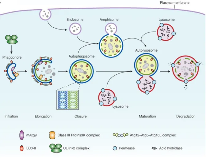
Figure 2. Autophagy process and its core machinery in mammalian cells Mammalian autophagy starts with
phagophore formation, and then the double membrane structure elongates until its closure. Then, the sealed autophagosome fused with lysosome to form autolysosome. Alternatively, autophagosome can fuse with endosome to form amphisome, followed by its final fusion with lysosome. Consequently, autophagy cargoes are degraded within autolysosome due to the lysosomal acid enzymes. The core autophagy machinery which consists in four different complexes is also depicted. ULK1/2 and the PI3KC3-C1 complexes are required for phagophore initiation, while two ubiquitin-like conjugation systems Atg12-Atg5-Atg16L and Phosphatidylethanolamine Atg8/LC3 (Atg8/LC3/GABARAP-Atg4-Atg7) as well as Atg9, function downstream and are involved in the phagophore elongation. (Yang and Klionsky 2010)
1.1.4. Core machinery for autophagosome formation
Atg proteins that are part of the core autophagy machinery, assemble and interact with each other to make four major complexes. Atg proteins contained in the core autophagy machinery are critical for autophagosome formation and are conserved from yeast to mammalian. These four complexes are: the Atg1/Ulk1 complex (Atg1/Ulk1/2, Atg13, FIP200 and Atg101); the class III phosphatidylinositol 3 kinase (PI3K) complex I (PI3KC3-C1) which is composed of Beclin 1, Atg14L, Vps34, Vps15 and Ambra1; two ubiquitin-like conjugation systems:Atg12-Atg5-Atg16L and Phosphatidylethanolamine Atg8/LC3
(Atg8/LC3/GABARAP-26
Atg4-Atg7) (Yang and Klionsky 2010). These complexes are displayed along with the autophagosome formation and maturation processes in Figure 2.
The Phagophore (or isolation membrane, IM) identified as crescent-shaped double membrane (Suzuki, Kirisako et al. 2001), is the pre-structure of autophagosome. Autophagosome nucleate at a single or several pre-autophagosomal sites named phagophore assembly site (PAS) and omegasome in yeast and higher eukaryotic organisms, respectely. In yeast, the PAS is located next to the vacuole whereas omegasomes were initially identified as a specific zone on the endoplasmic reticulum membrane which could serve as a cradle for phagophore initiation. In both locations, Phagophore formation requires the Atg1/Atg13 and PI3K complexes for its induction and nucleation. Pre-autophagosomal structure received lipids input from various vesicles including vesicles containing the unique autophagy transmembrane protein Atg9 (Noda, Kim et al. 2000), coming from the Golgi apparatus. Additionally, recycling endosomes labelled with Atg16L and probably other sources such as vesicles coming from COPII dependent vesicles from the ERGIC are recuited to the phagophore and participate to its extension (Carlsson and
Simonsen 2015). The retrieval of Atg9 is dependent on Atg1/13 complex and other Atg
proteins (Reggiori, Tucker et al. 2004).
Atg8/LC3/GABARAP is another important protein probably involved in phagophore expansion, and its role in autophagy process mainly relies on the modification of its C-terminal glycine by phosphatidylethanolamine (PE). This post-translational mechanism is dependent on key ubiquitine-like conjugation systems. This process starts with the Atg4 protease which cleaves the C-terminal part of the Atg8 precursor, to expose its C-terminal glycine residue. Then, this precursor form is activated by an E1-like enzyme Atg7, and then conjugated to the phagophore membrane phosphatidylethanolamine (PE) by E2-like enzyme Atg3. This conjugation activity requires Atg12-Atg5 system, which is also dependent on Atg7 (Mizushima, Noda et al. 1998, Tanida, Tanida-Miyake et al. 2002). Atg12-Atg5 further forms complex with Atg16L, and then is recruited to phagophore via the interaction with WIPI2 (Dooley, Razi et al. 2014), where Atg12-Atg5 cooperates with Atg7-Atg3 for phagophore expansion and autophagosome formation.
Atg8/LC3/GABARAP has a key role during phagophore expansion. Additionally, this protein, which is the only one to be constantly associated to autophagosome, is also involved in the
27
recognition and recruitment of autophagy substrates. The latter role is further discussed in chapter 1.2.
Although, it is considered that the core machinery described above is essential for general autophagy process, some specific autophagy events do take place without some of these Atg genes. For instance, a type of Atg5/Atg7-independent macroautophagy takes place in mouse embryonic fibroblast (MEF). In these cells, after etoposide exposure or starvation induction, autophagosomes were still found in Atg5-/- or Atg7-/- genetic backgrounds. Cells which lack the lipidation of LC3 (homologue of Atg8 in mammalian cells) make autophagosome by a Rab9-dependent fusion between isolation membrane with vesicles from late endosome and trans-Golgi (Nishida, Arakawa et al. 2009). Another study shows that the autophagy mediated cell size reduction of the Drosophila intestine, requires neither Atg7 nor Atg3 but a novel E1 enzyme Uba1 (Chang, Shravage et al. 2013). These works enlighten the complexity and variety of autophagy pathways.
1.1.5. Fusion of autophagosome with lysosome
Once the autophagosome is sealed, it can fuse either directly with lysosome for degradation or with endosome to form an intermediate organelle termed amphisome before its fusion with lysosome takes place (Tooze, Hollinshead et al. 1990, Mullock, Bright
et al. 1998). Since endocytosis and autophagy deliver macromolecules to the lysosome, both
pathways share many proteins involved in the regulation of fusion with lysosome. There are four important proteins/families mediating the fusion step: the small GTPase of the Rab family Rab7, the membrane containing rab7 tethering complex HOPS that may help to recruit SNARE proteins and ESCRT. The details are as followings:
Rab7 is not required for the initiation or formation of late autophagic vacuoles (Gutierrez,
Munafo et al. 2004, Jager, Bucci et al. 2004). Gutierrez et al. (2004) studied the role of Rab7
in CHO cells, by overexpressing the dominant negative form Rab7T22N, which can not exchange GDP for GTP. They analyzed the autophagosome maturation, using either monodansylcadaverine or indirect immunolocalisation of c-myc-LC3, and fusion events with endosome and/or lysosome, marked by internalized rhodamine dextran and lysosomal enzyme cathepsine D antibody, respectively. They showed that Rab7 is recruited to autophagosomes directly from the cytoplasm but not from the fusion with vesicles containing Rab7. The authors clearly showed that the final fusion of autophagosome with
28
lysosome is largely impeded by the expression of Rab7T22N and results in an accumulation of larger autophagosomes and a defect in degradation of long-lived protein (Gutierrez,
Munafo et al. 2004).
These results were confirmed and developed by Ganley et al (2011). The authors have shown in various mammalian cells that thapsigargin, an inhibitor of the Endoplasmic Reticulum Calcium ATPase pump, also blocks the autophagic flux by inhibiting the fusion of autophagosomes with endocytic compartments. Using both confocal analysis and EM coupled to immunogold detection of GFP-LC3, the authors showed that there is indeed an accumulation of autophagosomes in cells that have been starved and then treated with thapsigargin. Thapsigargin blocks the autophagic flux and led to the accumulation of autophagosomes that could not fused anymore with endosomes to give amphisomes, nor directly with lysosomes (Ganley, Wong et al. 2011). Interestingly, under thapsigargin treatment Rab7 is no more localized to autophagosome membrane, meanwhile it is still recruited to endosomes. Because endocytosis was not affected by thapsigargin, the authors suggested that a thapsigargin sensitive factor could be involved in Rab7 recruitment at the membrane of autophagosomes to promote their final maturation.
However, the small GTPase Rab7 controls the transport of late endosomes and lysosomes
(Guerra and Bucci 2016). One of the key roles played by Rab7 is to cluster the autophagic
structure next to lysosome for subsequent fusion. Rab7 interacts with RILP and FYCO1 to control autophagosome dynamic along the microtubule cytosqueleton, which is essential for the later degradation within lysosomes.
Additionally, Rab7 interacts with a number of key proteins required for the autophagosome-lysosome fusion: epg-5, PLEKHM1 which participates to recruit HOPS and also interacts with Atg8/LC3. Moreover, Rab7 has an additional role in Parkin-mediated autophagy compared with starvation-induced autophagy. It has been shown that the GAP protein TBC1D15 acts as an inhibitor for Rab7, governing autophagosome biogenesis in the process of mitophagy (Yamano, Fogel et al. 2014).
Most importantly, several data have indicated that Rab7 could recruit tethering complexes. The initial interactions, between vesicles before they fused, are mediated by dedicated protein complexes that act as tethering factors. Those proteins are capable to establish a physical link between membranes (Epp, Rethmeier et al. 2011). Briefly, tethering factors could be divided in two main groups, the dimeric proteins which are mainly involved in the
29
secretory pathway and the multimeric complexes, mainly inplicated in Golgi vesicles-endosome and vesicles-endosome-lysosome interactions. Specifically, two multimeric complexes, the CORVET and HOPS (Homotypic vacuolar fusion and Protein Sorting), are involved in regulating the machinery of fusion of early and late endosomes respectively. These complexes are made of a number of common subunits, Vps11, Vps16, Vps18 and Vps33, and the specific subunits Vps39, Vps41, and Vps3, Vps8 for HOPS and CORVET, respectively. Rab7 is more specifically involved in tethering by recruiting the HOPS complex.
Subunits of the HOPS complex have also a critical role to mediate membrane fusion by interacting with SNARE proteins. Homotypic fusion and vacuole protein sorting (HOPS) can recruit SNAREs to the late endosomes (Balderhaar and Ungermann 2013). Soluble N-ethylmaleimide-sensitive factor activating protein receptor (SNARE) is a protein family involved in endosome-lysosome fusion. Most of intracellular membrane fusion events (excluding mitochondria fusion) required two groups of proteins: the SNARE (Soluble N-ethylmaleimide-sensitive factor Attachment protein Receptor) and the SM (Sec1/Munc18-like) proteins (Sudhof and Rothman 2009). The Q/t- and R/v- SNARE proteins are localized respectively on two different vesicles and interact progressively to form a quadruple helix called the trans-SNARE. The trans-SNARE complex forms a zipper that progressively closes and creates a pulling force on both membranes allowing them to fuse. The SM proteins, among which the Vps33 subunit, have been found to interact with both Q/t- and R/v-SNAREs but also binds the trans-SNARE complex. The hairpin-type tail-anchored SNARE syntaxin 17 (STX17) has been identified to be present on enclosed autophagosome and facilitate its fusion with endosome or lysosome, via the interaction with SNAP-29 (Itakura,
Kishi-Itakura et al. 2012). Moreover, STX17–SNAP29 binary t-SNARE complex further binds
to VMP8 to mediate an Atg14-dependent fusion (Diao, Liu et al. 2015). STX17 is recruited once the autophagosome formation is achieved and therefore could constitute a checkpoint to avoid any fusion of the lysosome with unclosed phagophores. Interestingly, STX17 could be used as a new autophagosome marker.
Homotypic fusion and vacuole protein sorting (HOPS) can recruit SNAREs to the late endosomes (Balderhaar and Ungermann 2013) and Vps39, a subunit of HOPS, additionally functions as a co-factor for Rab7, increases the active Rab7 pools at the endosomal membrane (Wurmser, Sato et al. 2000). LC3 probably also participates in the fusion step. Interestingly, in C. elegans, LGG-2 (LC3 homologue), but not LGG-1 (GABARAP homologue),
30
directly interacts with VPS-39 HOPS complex subunit and controles autophagosome-lysosome fusion (Manil-Segalen, Lefebvre et al. 2014).
The endosomal sorting complex required for transport (ESCRT) is a key factor for the fusion of autophagosomes with lysosomes. ESCRT complexes are needed to mature endosome into multivesicular bodies. Subsequently, MVB fuse with autophagosome to generate amphisomes. Probably, this fusion brings to autophagosomes key membrane molecules necessary for the subsequent fusion with lysosomes. Interestingly, ESCRT proteins could be involved in non-canonical functions such as vesicle tethering. A review has described the details of the roles of ESCRT in autophagy (Lefebvre, Legouis et al. 2018). Other proteins are also shown to be involved in the autophagosome fusion process, for instance, the ectopic P-granules autophagy protein 5 (EPG-5) in C. elegans (Tian, Li et al. 2010). More information about EPG-5 protein is discussed in chapter 2.2.2. aggrephagy.
1.1.6. Autophagic lysosome reformation, ALR
Once autophagosome fused with lysosome, the autophagic cargoes are degraded by lysosomal acidic hydrolases, and the degraded materials are transported out to the cytosol via specific catabolite transporters for recycling. However, these transports do not completely end the autophagy process. In some stress conditions such as starvation, a mechanism named autophagic lysosome reformation (ALR) is triggered, immediately after the degradation step, to restore exhausted lysosomes during the autophagy process. In starved condition, mTOR is inhibited at autophagy initiation step. However it could be activated back by catabolites released from autolysosome after a prolonged starvation period, to trigger the ALR via an unknown mechanism. Moreover, the mTOR reactivation inhibits autophagy initiation and provides a negative feedback for avoiding excess autophagy (Yu, McPhee et al. 2010). Once ALR is triggered, tubules extruded from autolysosomes, named proto-lysosomes, and buds away from the lysosome to finally form tubules which will eventually mature into independent functional lysosomes.
1.2. Non-selective and selective autophagy
Initially, autophagy was considered to be a non-selective degradation pathway. This term means that autophagosome isolates and degrades bulk part of the cytoplasm without any specificity. Probably, it is because research activities focused on starvation-induced
31
autophagy for which autophagosome enrolled indistinctly bulk part of the cytoplasm containing proteins and organelles. However, a growing number of experimental results demonstrated that cells could trigger selective autophagy, i.e. an autophagy-dependent elimination (selective sequestration) of distinct cargoes which uses series of specific autophagy receptors. Autophagy receptors can specifically recognize the cargoes that need to be degraded and bridge them with autophagosome by interacting with Atg8/LC3
(Komatsu and Ichimura 2010). Based on the types of cargo engulfed for degradation,
selective autophagy can be further classified into mitophagy (mitochondria), pexophagy (peroxisome), nucleophagy (nucleus), ER-phagy (ER), lipophagy (lipid droplets), aggrephagy (protein aggregates), xenophagy (pathogens) (Galluzzi, Baehrecke et al. 2017). Different cargoes for selective autophagic degradation are shown in figure3. Among them, the selective elimination of mitochondria by autophagy, i.e. mitophagy, is the most studied one. I will present more information on molecular mechanisms underlying receptor-mediated mitophagy in chapter 3 mitophagy. I would like to emphasize the fact that one should be carefull to distinguish the autophagy receptors from the autophagy adaptors. Although autophagy adaptors participate in the substrates recognition during autophagy, they are not degraded; even they can interact with Atg8/LC3 as well as autophagy receptors. Autophagy receptors participate to the substrates recognition and interact with Atg8/LC3 during autophagy, and could be degraded, whereas autophagy adaptors serve as anchor points for the autophagy machinery (see Figure3).(Galluzzi, Baehrecke et al. 2017) (Stolz, Ernst et al.
32
Figure 3. Functions of autophagy receptors and adaptors in selective autophagy (a) Aggregated proteins,
cellular organelles such as ER, mitochondria, and ribosome, or pathogens (bacteria and viruses) can be selectively recognized by autophagy through the interaction with distinct autophagy receptors that are recognized by LC3/Atg8 via their LIR motif. (b,c) Autophagy adaptors role during autophagosome formation(b) and trafficking(c). Autophagy adaptors are involved in membrane elongation (ULK1, Beclin1, FIP200 and VPS34), LC3 lipidation (ATG16, ATG5-ATG12), autophagosome trafficking (FYCO1) and the autophagosome-lysosome fusion (SNX18). Autophagy receptors as well as cargos are engulfed by autophagosomes for degradation whereas, autophagy adaptors are only present on autophagosome/phagophore surface.(Stolz, Ernst et al. 2014)
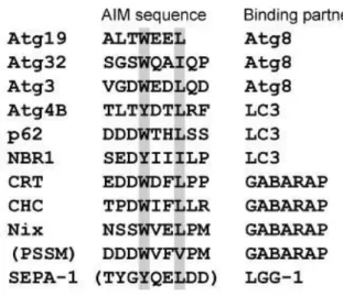
Most autophagy receptors are characterized by the LC3-interacting region (LIR) in mammals or Atg8-family interacting motif (AIM) in yeast. LIR (or AIM) consists of a hydrophobic amino acid motif WxxL, which is necessary for the interaction with two hydrophobic pockets (W and L pockets) present in LC3 (Ichimura, Kumanomidou et al. 2008), and is conserved from yeast to mammalian (Noda, Ohsumi et al. 2010). This applies to the
33
identified autophagy receptors: Atg19 (Cvt pathway), Atg36 and Atg30 (pexophagy) and Atg32 (mitophagy) in yeast (Meijer, van der Klei et al. 2007, Stolz, Ernst et al. 2014); SEPA-1 in C. elegans ; p62, BNIP3L, BNIP3 NDP52 in mammalian. Several of the LC3 binding proteins which containing the WxxL motif are listed in Table1
Table 1. The Sequence alignment and binding partner of the reported autophagy receptors (Noda, Ohsumi et al. 2010)
A specific type of selective autophagy in S. cerevisiae is the cytoplasm-to-vacuole targeting (Cvt) pathway. The Cvt pathway selectively transports vacuolar hydrolases (Ape1, Ams1, etc…) into the vacuole in budding yeast cells. Although Cvt pathway shares the similar core machinery with autophagy (Scott, Hefner-Gravink et al. 1996, Xie and Klionsky 2007), Cvt vesicles (150nm) are much smaller than autophagosome (300nm-1µm). Atg19 functions as the Ape1 receptor during Cvt pathway. It binds to both CVT-Ams1p-complex and Atg8, results in the preference formation of double membrane vesicles around Cvt complex
(Komatsu and Ichimura 2010). Cvt can not be considered as a type of autophagy since this
pathway is biosynthetic (it helps vacuole to be efficient), rather than degradative. 1.3. Physiological roles of autophagy
Autophagy is an important way for the degradation of cellular proteins and organelles to maintain the homeostasis. It clears the damaged or redundant components as well as recycles materials in stress conditions. Autophagy plays multiple roles at the cell level which when integrated at the level of an organism are responsible for key physiological functions. Autophagy is involved in many physiological processes, such as cell death, infection,
34
adaptive immune response and aging. Moreover, autophagy dysfunction is tightly linked with many human diseases, like cancer, cardiomyopathies, Paget’s disease, neurodegenerative diseases (Choi, Ryter et al. 2013). For example, in mammalians, autophagy stimulation is critical at birth because it supplies newborn cells with nutrients and energy while maternal nutrients supply has ceased (Kuma, Hatano et al. 2004). Considering about the pages limitation, I hereafter focus on the physiological roles of autophagy that have been studied in C. elegans rather than going into a detailed description of the many physiological roles of autophagy. Therefore, I present how autophagy controls or participates to: (1) developmental cell death; (2) aging and (3) cancer.
1.3.1. Developmental cell death and autophagy
In some instances, cell dies with accumulating autophagosome and autolysosome within their cytoplasm. These dying cells are therefore very different from the ones that die from the type I cell death/apoptosis or necrosis and therefore this cell death mechanism has been named type II cell death or autophagic cell death (Schweichel and Merker 1973, Clarke 1990). The first evidence of autophagic cell death was observed in Drosophila. As early as 1960s, researchers had observed the booming of autophagosome/autolysosomes in salivary glands and midgut during Drosophila metamorphosis (Schin and Clever 1965, Nopanitaya and
Misch 1974). Further studies confirmed that autophagy mediated developmental cell death
is required for both salivary glands and midgut degradation after puparium formation (apf), although the mechanisms may vary from tissue to tissue (Berry and Baehrecke 2007, Denton,
Chang et al. 2012).
Besides its role in type II cell death, autophagy and/or key genes involved in the general autophagy machinery are involved in apoptosis. In mammalian system the autophagy protein LC3 could facilitate dead cell corpse phagocytosis for lysosomal degradation. This mechanism has been named LC3-associated phagocytosis (LAP) (Sanjuan, Dillon et al. 2007,
Florey, Kim et al. 2011, Martinez, Almendinger et al. 2011). Recently, Jenzer et al showed
that autophagy is required in the cell destined to apoptosis to produce the energy required to induced their phagocytosis by neighbouring cells (Jenzer, Simionato et al. 2019). This work extended in C. elegans the observations that apoptotic cells clearance is autophagy dependent that have also been observed during either the mouse or bird development (Qu,
35
and LGG-2 are not involved in LAP but are rather required differently during an autophagy process used to mature phagosome.
1.3.2. Aging and autophagy
One common character in aging cells is the accumulation of misfolded proteins and damaged organelles. Autophagy is one of the cellular pathways involved in eliminating these harmful components and therefore it is not surprising that autophagy dysregulation may influence aging. Many data indicate that autophagy induction leads to a lifespan extension in many organisms. Whereas it has been revealed that autophagy, the major pathway for intracellular degradation of proteins and organelles, is also down-regulated in aging cells
(Donati, Cavallini et al. 2001, Del Roso, Vittorini et al. 2003, Nakamura, Oba et al. 2019). This
may indicate the anti-aging function of autophagy. Indeed, the autophagy-deficient mutant,
Atg7-/- Drosophila is short-lived; while by contrast, the lifespan is extended by promoting autophagy function (Juhasz, Erdi et al. 2007, Simonsen, Cumming et al. 2008). In
Saccharomyces cerevisiae, rapamycin extends yeast chronological life span (CLS) by inducing
autophagy (Alvers, Wood et al. 2009).
Specifically, in C. elegans, autophagy activity is required for life-span extension in daf-2 (the insulin-like receptor) mutant, as well as for feeding defect induced longevity (Melendez,
Talloczy et al. 2003, Jia and Levine 2007, Hansen, Chandra et al. 2008). Moreover, atg-7
RNAi and most other Atg gene down-regulation or mutant shortens lifespan in wild type worms (Hars, Qi et al. 2007).
The roles of autophagy in slowing aging and extending life span have been strongly established, whereas the mechanisms are not clear yet. Insulin-like growth factor (Melendez,
Talloczy et al. 2003) and TOR (Jia and Levine 2007, Hansen, Chandra et al. 2008, Ramos, Kaeberlein et al. 2013)pathways are the main signalling pathways involved in this process. The clearance of damaged mitochondria via autophagy is also thought to contribute to reduce aging, since many genes for mitochondrial function affect longevity (Lee, Lee et al.
2003, Oh, Mukhopadhyay et al. 2006). The extraordinary high ROS level in dysfunctional
mitochondria that are not cleared by autophagy is considered as one of the triggers for aging. Whereas another study demonstrates that mitochondrial permeability uncouples elevates autophagy and extend C. elegans lifespan (Zhou, Kreuzer et al. 2019). Spermidine induced lifespan extension is also autophagy-dependent; it may motivate the clinic
36
exploration of anti-aging drugs (Eisenberg, Knauer et al. 2009). Spermidine is a natural polyamine that can induce a modification of the chromatine structure by inhibiting histone acetyltransferases. This altered status of the chromatine leads to Atg genes transcription increase which triggers autophagy in yeast, flies, worms and human cell (Morselli, Marino
et al. 2011).
1.3.3. Cancer and autophagy
Cancer is one of the main threatens for human health. The molecular mechanisms in cancer are extremely complicated and infinite, and the role of autophagy in cancer is debating. Here I will only give a brief introduction about the autophagy studies in cancer. Many evidences demonstrate autophagy as tumour suppressive machinery. Autophagy has been widely recognized as a main biological event involved in both the regulation of cancer cell proliferation and in the response of several anticancer drugs.An early study found Beclin1 is mono-allelically deleted in 40–75% breast cancers and ovarian cancers
(Aita, Liang et al. 1999). In addition, the deficiency of other ATGs also exhibits an
increased tumour growth (Marino, Salvador-Montoliu et al. 2007, Takamura, Komatsu et
al. 2011). The mutant of p53, a well-known tumour suppresor, facilitates tumorigenesis
by inhibiting autophagy activity (Tasdemir, Chiara Maiuri et al. 2008).
There are also evidences suggesting that autophagy offers survival advantages for tumour growth, especially in metabolic stress conditions. Cancer cells are always exposed in hypoxia condition due to the insufficient vascularization, leading to an increased autophagy through glycolytic enzyme pyruvate kinase M1 (PKM1). PKM1-Atg7 knockout mice exhibit a decreased tumour growth, indicating autophagy helps cancer cells to resist metabolic stress (Morita, Sato et al. 2018). Autophagy knockdown also contributes to the inhibition of tumorigenesis during tumour therapy (White 2012).
Taken together, autophagy is a tool to suppress tumour growth; on the other hand, autophagy may be used by cancer cells to resist harsh stress.
2. C. elegans, a model animal for autophagy studies
2.1. General knowledge of C. elegans37
Caenorhabditis elegans (C. elegans), is a nematode found on all continents (thought rarely in Asia) as well as on isolated islands such as Hawaii. It does not have a specific natural habitat but seems to be present in humid temperate region. C. elegans is mostly found on rooting fruit and stalk, on substrate containing many microbes decomposing vegetal material and barely in soil sample (Schulenburg and Felix 2017).
Historically, there were a number of studies made in the early time of the twentieth century that reported the use of C. elegans to address basic biological questions. These studies were mainly done or headed by a few researchers; most of them were French people: Emile Maupas (France), Hikokura Honda (USA), Victor Nigon (France), Ellsworth Dougherty (USA) and Jean-Louis Brun (France) (Nigon and Felix 2017). Later on, in the late 1960s, Sydney Brenner who participated in the discovery of mRNA and to the elucidation of the genetic code, was looking for a model organism with which he will address questions of developmental biology and neurobiology. He used to present biochemistry as “-an insurmountable barrier to the study of metazoans” and genetics as “a tunnel through this barrier”. In his mind this model organism should be a genetically tractable organism with a simple anatomy that could be reconstructed from electron micrographs. It should be easily culture in the lab having a small size and a rapid life cycle. Finally, he has chosen C.
elegans for all these reasons. C. elegans became model organism as important as the yeast
and Drosophila models. In the past 50 years, C. elegans has been used by many labs to understand a large variety of molecular, cellular, developmental, physiological, and behavioral biology questions and related molecular mechanism, as well as to modelize human diseases (Miguel-Aliaga, Culetto et al. 1999). Indeed, the worm has plenty of advantages making it a powerful model animal, for instance, tiny size, rapid life cycle, self-fertilizing, easy rising, transparency and so on.
2.1.1. C. elegans maintenance and life cycle
C. elegans has a rapid life cycle of around 3 and half days at 20°C. Its optimized growth
temperature is 12°C-25°C. Although lower or higher temperature leads to sterility, lifespan modulation or even death of animals, C. elegans is capable of cold acclimatation. Moreover, temperature acts differently on lifespan depending on the time window it is applied. Its life cycle consists of 3 main steps: embryogenesis, larval development (L1-L4), maturation to adulthood and adulthood. It is noteworthy, C. elegans is capable to go to an alternative
38
larval development stage called dauer larvae when it experiences harsh condition such as starvation, high population density or elevated temperature during a time window encompassing L1 and L2 stages (Figure 4).
Figure 4. C. elegans life cycle at 22°C A life cycle for C. elegans consists of embryogenesis, larva development
(L1-L4), adult maturation (Young Adult to adult) and sometimes an additional dauer stage. After fertilization, embryos firstly develop within the mother for 150 minutes until eggs are laid out. Blue words indicate the time it takes for the development of each stage. (from WormAtlas)
Following fertilization, the embryogenesis can be subdivided into stages as: fertilization, proliferation, gastrulation, morphogenesis, mlongation, quickening and hatching. C. elegans is an ectotherm/poikilotherms animal and therefore its time development is dependent of the temperature; it takes longer for worm to develop from embryo to adult at lower temperature, as shown in Table2.