Volume 2013, Article ID 129645,9pages http://dx.doi.org/10.1155/2013/129645
Research Article
A Novel Sit4 Phosphatase Complex Is Involved in the
Response to Ceramide Stress in Yeast
Alexandra Woodacre,
1Museer A. Lone,
2Daniel Jablonowski,
1,3Roger Schneiter,
2Flaviano Giorgini,
1and Raffael Schaffrath
1,31Department of Genetics, University of Leicester, Leicester, LE1 7RH, UK
2Division of Biochemistry, Department of Biology, University of Fribourg, CH-1700 Fribourg, Switzerland 3Institut f¨ur Biologie, FG Mikrobiologie, Universit¨at Kassel, 34132 Kassel, Germany
Correspondence should be addressed to Flaviano Giorgini; [email protected] and Raffael Schaffrath; [email protected] Received 10 May 2013; Revised 28 June 2013; Accepted 25 July 2013
Academic Editor: Joris Winderickx
Copyright © 2013 Alexandra Woodacre et al. This is an open access article distributed under the Creative Commons Attribution License, which permits unrestricted use, distribution, and reproduction in any medium, provided the original work is properly cited.
Ceramide is a building block for complex sphingolipids in the plasma membrane, but it also plays a significant role in secondary signalling pathways regulating cell proliferation and apoptosis in response to stress. Ceramide activated protein phosphatase activity has been previously observed in association with the Sit4 protein phosphatase. Here we find that sit4Δ mutants have decreased ceramide levels and display resistance to exogenous ceramides and phytosphingosine. Mutants lacking SIT4 or KTI12 display a shift towards nonhydroxylated forms of long chain bases and sphingolipids, suggesting regulation of hydroxylase (SUR2) or ceramide synthase by Sit4p. We have identified novel subunits of the Sit4 complex and have also shown that known Sit4 regulatory subunits— SAP proteins—are not involved in the ceramide response. This is the first observation of separation of function between Sit4 and SAP proteins. We also find that the Sit4p target Elongator is not involved in the ceramide response but that cells deficient in Kti12p— an accessory protein with an undefined regulatory role—have similar ceramide phenotypes to sit4Δ mutants. Therefore, Kti12p may play a similar secondary role in the ceramide response. This evidence points to a novel Sit4-dependent regulatory mechanism in response to ceramide stress.
1. Introduction
Ceramide is a building block for complex sphingolipids which comprise an important structural component of the plasma membrane. It is also a secondary signalling molecule that accumulates in response to stresses such as heat shock
[1]. It is therefore important for sphingolipid metabolism
to be tightly regulated, and the damaging effects of dys-regulation are apparent in patients with Tay-Sachs disease, Fabry disease, and other inherited sphingolipidosis disorders
[2]. Ceramide mediates controlled cell death by triggering
several signalling cascades to initiate caspase-dependent and
independent apoptosis [3]. In contrast, the phosphorylated
ceramide precursors dihydrosphingosine (DHSP) and phy-tosphingosine (PHSP) are signals for pathways that promote
cell proliferation [4].
Although it is known that the cellular response to ceramide is important for the regulation of cell proliferation and cell death pathways, the precise molecular mechanisms for this regulation still remain elusive. It is vital to further understand the way cells respond to stress in order to develop strategies to modify them, either to accelerate cell death using targeted anticancer drugs or to prevent accumulation of toxic
products in sphingolipidoses [5–7].
Saccharomyces cerevisiae has been used effectively as a
model to study sphingolipid metabolism, andFigure 1shows
a detailed summary of the sphingolipid biosynthetic pathway
in yeast [8]. Many of the genes involved are conserved from
yeast to higher eukaryotes, with diversion in the synthesis of complex sphingolipids occurring only after the production of ceramides, resulting in the production of different end prod-ucts in the pathway. The addition of inositol to ceramide in
Phytoceramide Dihydroceramide DHSP LCB1, LCB2, TSC3 TSC10 CSG1, CSG2, CSH1 IPT1/KTI6 ISC1 LCB3, YSR3 LCB3, YSR3 LCB4, LCB5 LCB4, LCB5
LAG1, LAC1, LIP1 LAG1, LAC1, LIP1
ELO1, FEN1, YBR159w, PHS1, TSC13 YPC1 ceramidase Palmitoyl-CoA+ serine
Serine palmitoyl transferase (SPT) 3-Ketodihydrosphingosine ceramide synthase ceramide synthase C26-CoA C26-CoA PHSP
Inositol phosphoceramide (IPC)
Mannose-inositol phosphoceramide (MIPC) Complex sphingolipids
Mannose-(inositol-P)2-ceramide (M(IP)2C)
Dihydrosphingosine (DHS) Phytosphingosine (PHS) hydroxylaseSUR2 hydroxylaseSUR2 AUR1 ceramidaseYDC1
Figure 1: Biosynthesis of sphingolipids in Saccharomyces cerevisiae. Key enzymes discussed in the text are highlighted and genes encoding all relevant parts of the pathway are included. The directions of arrows indicate the end products of enzymatic reactions.
yeast forms inositol phosphoceramide, and glucose, galactose or phosphorylcholine is added to ceramide in mammalian cells to generate glycosphingolipids and sphingomyelin
[9].
Early work by Nickels and Broach showed that a cera-mide-activated phosphatase activity was present in Saccha-romyces cerevisiae, which is separate from the activity of
the major PP2A phosphatases Pph21p and Pph22p [10]. The
ceramide resistance of a sit4Δ mutant strain suggested that the PP2A-like phosphatase Sit4p is responsible for this activity. SIT4 is an essential gene in the absence of the suppressor allele SSD1-v and has important roles in the progression of the cell cycle, cell integrity, nutrient responses via TORC1, drug resistance via efflux pumps, and tRNA modification
[11–16]. Diverse regulatory subunits of Sit4p are partially
responsible for the different specificities of Sit4p; for
exam-ple, Tap42p is phosphorylated by Tor and binds Sit4p [17]
and Sap185p and Sap190p subunits are essential for correct
phosphoregulation of Elongator and tRNA modification [18].
Mutation of the SAPs (Sit4 associated proteins) can confer different specificities on Sit4p but a deletion of all four SAPs always results in the same phenotypes as deletion of SIT4, for example resistance to the tRNAse toxin zymocin, cell cycle
arrest, and sensitivity to rapamycin [19,20]. The accessory
protein Kti12p is also essential for the phosphoregulation of the Elongator subunit Elp1p by the Sit4p/Sap185p/Sap190p complex. Kti12p interacts with the casein kinase Hrr25p
in an Elongator-dependent manner but the mechanism by which Kti12p regulates phosphorylation remains unclear
[21].
The aim of this study was to further investigate the role of Sit4 as the ceramide-activated protein phosphatase (CAPP) in yeast. We identify KTI12 as an important gene mediating ceramide toxicity and show that ceramide toxicity is independent of Elongator function. Mutants lacking SIT4 or KTI12 have decreased levels of ceramide and the balance of hydroxylated and nonhydroxylated sphingolipids is altered. The confirmation that Tpd3p and Cdc55p can interact with Sit4p and a separation of function between SIT4 and the reg-ulatory SAP subunits indicates that the CAPP is likely to be an alternative Sit4 complex operating via a novel mechanism.
2. Methods
2.1. Yeast Strains and Media. Yeast were routinely grown in yeast extract peptone dextrose medium (YPD; 1% yeast
extract, 1% peptone, 2% glucose) at 30∘C with shaking.
Glucose was replaced with 2% galactose to induce expression of SIT4 and PPH21 from the GAL1 promoter. Synthetic defined medium without inositol (0.67% yeast nitrogen base, 2% glucose, supplemented with essential amino acids) was
used for labelling with[3H]myo-inositol. Yeast strains used
Table 1: Yeast strains used in this study.
Strain Genotype Reference
CY4029 Mat a ade2-1 his3-11,15 leu2-3,112 trp1-1 ura3-1 can1-100 SSD1-v1 gal+ [20]
CY3938 CY4029, sit4Δ::HIS3 [20]
CY5236 CY4029, sap4Δ::LEU2 sap155Δ::HIS3 sap185Δ::ADE2 sap190Δ::TRP1 [20]
CY5220 CY4029, sap4Δ::LEU2 sap155Δ::HIS3 [20]
CY5224 CY4029, sap185Δ::ADE2 sap190Δ::TRP1 [20]
CY4917 CY4029, sap185Δ::ADE2 [20]
CY4380 CY4029, sap190Δ::TRP1 [20]
DJY101 CY4029, sit4Δ::HIS3 kti12ΔKlLEU2 [18]
LFY3 Mat a ade2-1 his3-11,15 leu2-3,112, ura3-1 can1-100, elp1Δ::TRP1 [18]
LFY4 Mat a ade2-1 his3-11,15 leu2-3,112, ura3-1 can1-100, elp2Δ::TRP1 [18]
LFY5 Mat a ade2-1 his3-11,15 leu2-3,112, ura3-1 can1-100, elp3Δ::TRP1 [22]
LFY6 Mat a ade2-1 his3-11,15 leu2-3,112, ura3-1 can1-100, kti12Δ::TRP1 [22]
AWY1 CY4029 TRP1::GAL1::(HA)3-SIT4 CDC55-(c-myc)3::HIS3MX6 This study
AWY2 CY4029, kanMX6::PGAL1::(HA)3-PPH21, CDC55-(c-myc)3::HIS3MX6 This study
AWY3 CY4029, CDC55-(c-myc)3::HIS3MX6 This study
2.2. Growth Tests Using Ceramide and Long Chain Bases. C2-ceramide, C2-phytoC2-ceramide, dihydrosphingosine (DHS), and phytosphingosine (PHS) powders were purchased from Enzo Life Sciences and resuspended in 100% ethanol. Stock
solutions (5 mg/mL) were stored at−20∘C for a maximum
of 1 week. Yeast cultures were diluted from a starter culture
to 5 × 10³ cells/mL in YPD containing ceramides/long
chain bases or an equal volume of ethanol as an untreated control. Cultures were grown until the untreated control reached exponential phase (from 15–36 hours depending
on the strain) and the OD600 measured for both treated
and untreated cultures. After 24 hours, an additional dose of ceramide/long chain base was added to counteract the effects of compound degradation. The amount of growth in each concentration of ceramide/long chain base was then expressed as a percentage of the growth in untreated media. This method of standardising growth enables the comparison of slow-growing mutants such as sit4Δ to a
faster-growing wild-type strain. Raw OD600 data is provided in
Supplementary Tables 1 and 2 (see Supplementary Material
available online athttp://dx.doi.org/10.1155/2013/129645). A
minimum of three biological replicates were performed for each strain and a one-way ANOVA with Bonferroni post-test was used to determine if growth was significantly different from the wild type (CY4029).
2.3. Immunoprecipitation. Dynabeads (Invitrogen) were
cou-pled with 5𝜇g of anti-HA antibody per mg of beads, following
the manufacturer’s instructions. Total protein extracts were prepared from 50 mL cultures grown for 8 hours in YPD supplemented with galactose. Cell pellets were resuspended
in 400𝜇L B60 buffer (50 mM HEPES pH 7.3, 60 mM sodium
acetate, 5 mM magnesium acetate, 0.1% Triton X-100, 10% glycerol, 1 mM sodium fluoride, 20 mM glycerophosphate, 1 mM DTT, 1X Complete Mini Protease Inhibitor Cocktail (Roche)). An equal volume of glass beads was added and cells disrupted using a bead beater for 1 minute, followed by
centrifugation at 15700 g, 4∘C for 5 minutes. The supernatant
was transferred to a new tube and centrifuged at 15700 g, 4∘C
for 20 minutes. The cleared protein extract was quantified using spectrophotometry and 3.5 mg of total protein extract was incubated with 1.5 mg of antibody-coated beads for 30
minutes at 4∘C. Unbound proteins were removed by three
washes with 1 mL B60 buffer, and antibody-bound proteins
were eluted with 50𝜇L of 10% (v/v) SDS for 10 minutes at
room temperature. The beads were then removed with a magnet and the supernatant used for Western blot analysis.
SDS-PAGE of 100𝜇g of total protein from each strain and
immunoprecipitation supernatants (equal volumes) was car-ried out using 12% acrylamide gels and then Western blotted at 100 V for 1 hour. Blots were probed with anti-HA (F7 Santa Cruz), anti-c-myc (A14 Santa Cruz), or anti-Tpd3 (Y. Jiang, University of Pittsburg School of Medicine, USA) and secondary antibodies conjugated to horseradish peroxidase (Roche Diagnostics) were detected by chemiluminescence and exposed to X-ray film.
2.4. Sphingolipid Analysis by ESI-MS. Overnight cultures
grown at 24∘C in YPD media were diluted to OD600 0.2
and grown until they reached OD600 of 2. A total of 10 OD
units of cells were collected and washed once with sterile water. Lipid extraction was performed by a two-step lipid
extraction method [23]. Cells were resuspended in 1 mL of
150 mM ammonium bicarbonate (NH4HCO3) and 600𝜇L
of glass beads were added. After cell lysis using a Precellys 24 homogenizer ((Bertin technologies) 5000 rpm, 3x 30 sec on 30 sec off), lysates were diluted in 5 mL of 150 mM
NH4HCO3 solution and internal standards were added.
Long chain bases and ceramides were quantified relative to respective lipid standards, and inositol phosphoceramides were measured relative to a phosphoinositol standard. Lipid standards were purchased from Avanti Polar Lipids. ESI-MS analysis was performed using a Bruker Esquire HCT ion trap mass spectrometer in positive or negative ion mode. Peaks
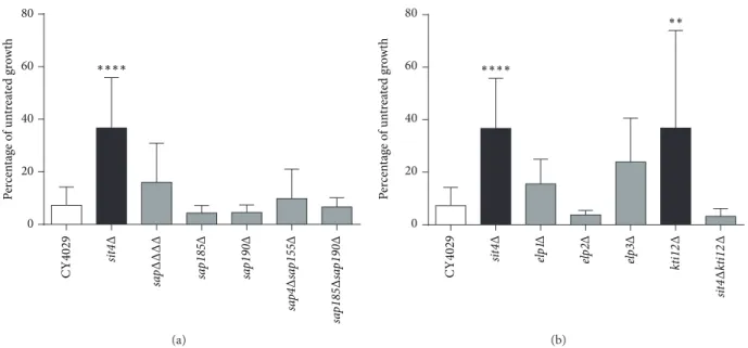
sa pΔΔΔ Δ sa p185 Δ sa p190 Δ sa p4 Δ sa p155 Δ sa p185 Δ sa p190 Δ 0 20 40 60 80 P er cen ta ge o f un tr ea te d gr o w th ∗∗∗∗ sit 4Δ C Y 4 029 (a) 0 20 40 60 80 ∗∗∗∗ ∗∗ kt i12 Δ sit 4Δ kt i12 Δ sit 4Δ C Y 4 029 elp 1Δ elp 2Δ elp 3Δ P er cen ta ge o f un tr ea te d gr o w th (b)
Figure 2: Deletion of SIT4 or KTI12 confers resistance to excess dihydroceramide. (a) Ceramide growth tests in Sit4 associated protein (SAP) mutants. (b) Ceramide growth tests in Elongator-associated mutants. Yeast cultures were diluted to 5× 10³cells/mL in YPD with the addition of either 15𝜇M C-2 dihydroceramide or an equal volume of ethanol. Cells were grown until the untreated culture reached exponential phase and the OD600of all cultures was determined. Growth in 15𝜇M dihydroceramide is expressed as a percentage of untreated growth. Raw OD600 values are given in Supplementary Table 1. A minimum of three replicates are shown and error bars represent the standard deviation above and below the mean. A one-way ANOVA with a Bonferroni post-test was used to determine if mutants showed a significant difference in growth compared to the wild type (CY4029) (∗∗𝑃 < 0.01, ∗∗∗𝑃 < 0.001, and∗∗∗∗𝑃 < 0.0001).
were identified based on their fragmentation pattern and by comparison to commercially available standards. Three biological replicates were included in each analysis.
2.5. Incorporation of [3H]-Labelled Inositol. Overnight
cul-tures grown at 24∘C in YPD were diluted to OD6001.0 in
syn-thetic defined media containing 40𝜇Ci [3H] myo-inositol
(American Radiolabelled Chemicals, MO, USA) and grown
at 24∘C for 4 h until they reached OD600of approximately 2. A
total of 10 OD units were harvested, and lipids were extracted using chloroform/methanol/water (10 : 10 : 3) and analysed
as previously described [24] by thin layer chromatography
with or without mild-base treatment. Mild-base treatment to remove inositol phosphate and leave only N-acetylated sphingolipids was performed by incubating lipids in 0.1 M
NaOH at 30∘C for 1 hour. Radioactivity was detected using
a phosphorimager (Typhoon FLA9500, GE Healthcare) and a representative image of two biological replicates is shown.
3. Results
3.1. Deletion of Sit4-Associated Proteins (SAPs) Does Not Confer Resistance to Exogenous Dihydroceramide. As
previ-ously described [10], deletion of SIT4 leads to significant
resistance to 15𝜇M dihydroceramide (Figure 2,𝑃 < 0.0001).
However, mutation of the four SAP regulatory proteins, either individually or in combination, does not confer resistance
to dihydroceramide (Figure 2(a)). In previous studies, the
phenotype of the quadruple sap mutant has been
indistin-guishable from that of sit4Δ [19, 20]. Thus, the ceramide
sensitivity of the sap mutant is the first observed separation of function between sit4Δ and sapΔΔΔΔ.
3.2. Kti12p Appears to Be the Only Elongator-Associated Protein Involved in the Ceramide Response. As Sit4p plays a major role in the phosphoregulation of the Elongator
com-plex [18, 21] and previous studies suggested that Elongator
mutants were resistant to ceramide, Elongator components were investigated as potential targets of Sit4p in the response to excess dihydroceramide. Although deletion of Elongator subunits did not confer statistically significant resistance to
15𝜇M dihydroceramide, deletion of the Elongator
acces-sory protein Kti12p did confer resistance (Figure 2(b)) to
some extent, though the obtained data were rather variable (Supplementary Table 1). Interestingly, deletion of SIT4 and KTI12 in tandem restored sensitivity to dihydroceramide, whereas in a previous study mutants lacking one or both of these genes had the same phenotype that resulted in
hyperphosphorylation of Elp1p and zymocin resistance [18].
In addition, phosphorylation of Elp1p was unchanged in the presence of dihydroceramide, and this was not affected by deletion of SIT4 and/or KTI12 (data not shown). Therefore, our data suggest that Kti12p might play a regulatory role in the ceramide response that is independent of Elongator. 3.3. PHS Resistance of sit4Δ Indicates Separation of Func-tion from kti12Δ. Growth in phytoceramide decreases in a concentration-dependent manner in both wild-type and mutant strains; however, sit4Δ and kti12Δ mutants show significantly more growth (𝑃 < 0.005) than the parental
5 10 15 20 0 50 100 150 0 Phytoceramide (𝜇M) sit4Δ kti12Δ CY4029 P er cen ta ge o f un tr ea te d gr o w th (a) 0 50 100 150 DHS (𝜇M) 0.0 0.2 0.4 0.6 sit4Δ kti12Δ CY4029 P er cen ta ge o f un tr ea te d gr o w th (b) 0 8 0 50 100 150 sit4Δ kti12Δ 2 4 6 PHS (𝜇M) CY4029 P er cen ta ge o f un tr ea te d gr o w th (c)
Figure 3: Response of sit4Δ and kti12Δ mutants to phytoceramide and long chain bases. Yeast cultures were diluted to 5 × 10³cells/mL in
YPD with the addition of the indicated concentrations of (a) phytoceramide, (b) dihydrosphingosine (DHS), (c) phytosphingosine (PHS), or an equal volume of ethanol. Cells were grown until the untreated culture reached exponential phase and then the OD600of both treated and untreated cells was measured and plotted as a percentage of untreated growth. Raw OD600values are given in Supplementary Table 2. A minimum of three replicates are shown and error bars represent the standard error above and below the mean. A Student’s t-test was used to determine if the mutants showed a significant difference in growth compared to the wild type (CY4029) at each concentration shown.
CY4029 strain at concentrations of 10–15𝜇M (Figure 3(a)).
In contrast, growth of sit4Δ and kti12Δ mutants in excess dihydrosphingosine (DHS) is indistinguishable from CY4029 (Figure 3(b)). The most striking result is that while kti12Δ
is also sensitive to phytosphingosine (PHS), sit4Δ shows
significant (𝑃 < 0.05) resistance to 3–6 𝜇M PHS (Figure 3(c)),
suggesting divergence of function between Kti12p and Sit4p in the response to long chain bases.
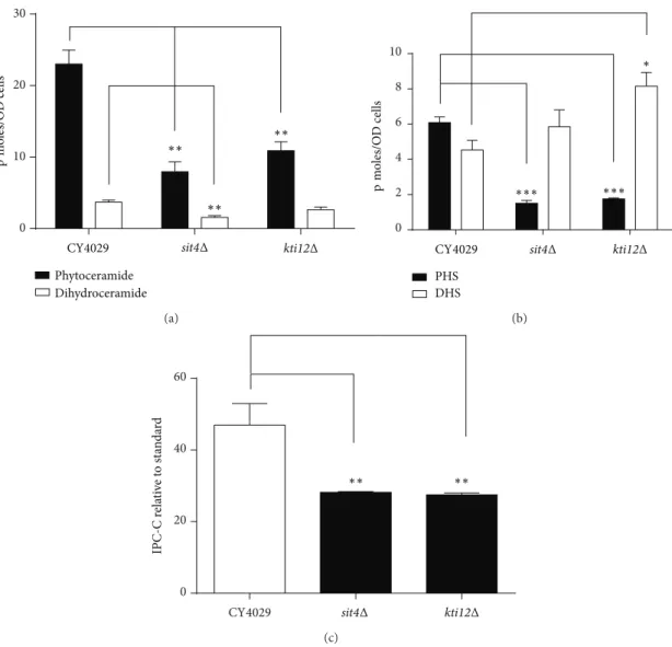
3.4. Ceramide and Long Chain Base Levels Are Reduced in sit4Δ and kti12Δ Mutants. To investigate the possibility that Sit4p and Kti12p regulate the sphingolipid biosynthesis
pathway, we measured steady state levels of ceramides, long chain bases, and inositol phosphate in sit4Δ and kti12Δ strains relative to wild-type yeast cells. Deletion of SIT4 or KTI12 reduces the intracellular levels of phytoceramide by
approximately 50% (Figure 4(a)). Levels of dihydroceramide
are also reduced in both mutants, but the decrease is only
statistically significant in sit4Δ (Figure 4(a)). This suggests
that the mutants may be able to tolerate otherwise toxic levels of exogenous ceramides due to the constitutively lower levels present within the cell. The reduction of PHS levels by approximately two-thirds in the sit4Δ mutant could permit the strain to survive excess concentrations of PHS
0 10 20 30 p m oles/O D cells ∗∗ ∗∗ ∗∗ CY4029 Phytoceramide Dihydroceramide sit4Δ kti12Δ (a) 0 2 4 6 8 10 PHS DHS ∗∗∗ ∗∗∗ ∗ p m oles/O D cells
CY4029 sit4Δ kti12Δ
(b) 0 20 40 60 ∗∗ ∗∗ IPC-C r ela ti ve to st an da rd sit4Δ kti12Δ CY4029 (c)
Figure 4: Mass spectrometric analysis of sphingolipid species. Yeast cultures were diluted to an OD600of 0.2 in YPD and grown for 8 hours at 24∘C. A total of 10 OD600units of cells were removed and lipids extracted for mass spectrometry analysis. (a) Ceramides, (b) long chain bases phytosphingosine (PHS) and dihydrosphingosine (DHS), and (c) Inositol phosphoceramide-C (IPC-C) were quantified using relevant internal standards. Average values for a minimum of three biological replicates are shown and error bars represent the standard error above and below the mean. A Student’s t-test was used to determine if the mutants showed a significant difference from the wild type CY4029. (∗𝑃 < 0.05,∗∗𝑃 < 0.01, and ∗∗∗𝑃 < 0.005).
(Figure 4(b)). However, a similar decrease in PHS levels in the kti12Δ mutant does not correlate with resistance to
exogenous PHS (Figures 4(b) and 3(c)), suggesting that a
more complex mechanism underlies PHS resistance. There is a small increase in the levels of DHS in sit4Δ and kti12Δ
mutants (Figure 4(b)) which is unlikely to affect the toxicity
of DHS seen inFigure 3(b).
3.5. Sit4 Mutants Show an Increase in the Proportion of Dihydro Sphingolipids and a Corresponding Decrease in the Proportion of Hydroxylated Sphingolipids. Tritium labelled inositol incorporation was used to analyse the matura-tion of complex sphingolipid species formed from both
dihydroceramide and phytoceramide. Dihydroceramide B
(18:0;2/26:0;0) and phytoceramide C (18:0;3/26:0;0) form
inositol phosphoceramide B (IPC-B) and inositol phosphoce-ramide C (IPC-C) respectively. IPC-C and the corresponding MIPC-C generated from it form the relatively more abundant
species of their sphingolipid class in the wild type (Figure 5).
Interestingly, the sit4Δ mutant contains increased levels of IPC-B and MIPC-B compared to the wild type, with a
decrease in the levels of IPC-C and MIPC-C (Figure 5). The
kti12Δ mutant also shows a similar trend in the relative levels of sphingolipid species, but the differences from the wild type are less pronounced than those for sit4Δ. This indicates a shift towards more sphingolipids being synthesised from dihydroceramide/DHS precursors than from the
hydroxy-lated phytoceramide/PHS precursors. Figure 4(c) provides
additional evidence for this shift and quantification of IPC-C levels shows a significant decrease in both sit4Δ and kti12Δ mutants. This also correlates with the increase in DHS and
MIPC } MIPC WT WT PI { IPC-B IPC-C IPC-B IPC-C −NaOH +NaOH
sit4Δ kti12Δ sit4Δ kti12Δ
M[IP]2C
M[IP]2C
Figure 5: Tritium labelled inositol incorporation into yeast cells. CY4029 (WT), sit4Δ, and kti12Δ were incubated with [3H]-inositol
for 4 hours, and lipids were extracted and analysed by thin layer chromatography, before and after mild-base treatment to remove inositol phosphate (PI). Equal CPMs were loaded for all the samples. A representative image of two biological replicates is shown.
decrease in PHS levels observed in sit4Δ and kti12Δ mutants (Figure 4(b)).
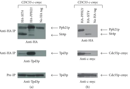
3.6. Novel Interactions of Sit4p with Tpd3p and Cdc55p Suggest a Role for an Alternative Phosphatase Complex in the Response to Ceramide. Previous studies suggested that Tpd3p and Cdc55p could be part of the CAPP complex as
deletion of these genes conferred resistance to ceramide [10].
Indeed, we found via immunoprecipitation experiments with HA-labelled Sit4p that Tpd3p and Cdc55p-c-myc interact with Sit4p, forming a minor complex compared to the
Pph21p/Tpd3p/Cdc55p complex (Figure 6). As this novel
Sit4p/Tpd3p/Cdc55p trimer is formed constitutively and is not induced by the presence of ceramide (data not shown), the mechanism by which the phosphatase is activated in response to an increase in ceramide levels remains unclear.
4. Discussion
The aim of this study was to further investigate the role of the Sit4 phosphatase in response to ceramide and to determine if this signalling pathway is directly related to the biosynthesis of ceramides and sphingolipids. The phosphorylation status of Orm1p regulates the activity of serine palmitoyltransferase and therefore the production of all downstream products of the sphingolipid pathway. Orm1p is phosphorylated by Ypk1p and evidence suggests that dephosphorylation may involve Sit4p and/or its TOR-dependent subunit Tap42p
[25, 26]. However, Orm1p is unlikely to be the direct
sub-strate for the Sit4p or Tap42p phosphatases as mutation of these genes leads to decreased phosphorylation of Orm1p
[25].
The Sit4 phosphatase is a well-characterised regulator of
tRNA modification via the Elongator complex [18,21,27–29].
However, here we show that the role of Sit4p in the ceramide response is independent of Elongator yet still involves the multifunctional and Elongator-related accessory protein Kti12p. Although previous work suggested that Elongator
may be involved in ceramide resistance [18], more detailed
analysis in this current study indicates that Elongator mutants are sensitive to ceramide. Although Kti12p is essential for the phosphoregulation of Elongator, its precise role remains
unclear [18,21]. Kti12p also has diverse roles in other cellular
processes including the cell cycle [30] and transcription [31],
and regulation of the ceramide response can now be added to this list.
The Sit4 phosphatase has multiple regulatory subunits including the Sit4 associated proteins (SAPs) Sap4p, Sap155p, Sap185p, and Sap190p. A quadruple deletion of all SAPs is sensitive to excess ceramide, in contrast to the resistant sit4Δ mutant. Importantly, this is the first separation of function observed between the sit4Δ and sapΔΔΔΔ mutants. This suggests that an alternative Sit4 phosphatase complex is involved in the regulation of the ceramide response, support-ing the idea that this process is independent of Elongator functions that require Sit4/Sap complexes. The identification of a Sit4p/Tpd3p/Cdc55p trimer also supports the theory that the ceramide activated protein phosphatase could be acting via a previously unknown mechanism.
The alteration of the sphingolipid makeup in sit4Δ and kti12Δ mutants and the decreased levels of ceramide and long chain bases indicate that there is regulation of the biosynthetic pathway at some level by Sit4p and/or Kti12p. The decreased level of endogenous ceramide and PHS in the mutants presumably enables them to survive an otherwise toxic concentration of these compounds. This suggests that deletion of SIT4 and KTI12 mediates a downregulation or partial inactivation of ceramide synthesis rather than a complete block, as there are clearly sufficient precursors available for effective biosynthesis of sphingolipids. The pres-ence of multiple genes encoding enzymes for synthesis and degradation of ceramides is a key way in which sphingolipid metabolism can be maintained when the pathway is partially blocked. Phosphoregulation of ceramide synthases has not been previously observed, but three phosphorylated serine residues are conserved in both Lag1p and Lac1p ceramide synthases and could be potential targets for
dephosphory-lation by Sit4p [32, 33]. In common with sit4Δ and kti12Δ
mutants, lac1Δlag1Δ mutants are resistant to the tRNase toxin zymocin, but the mechanism of action is due to a defect in plasma membrane integrity caused by decreased levels of the
sphingolipid M(IP)2C and not via Elongator [34].
In sit4Δ and kti12Δ mutants, the relative proportion of lipids synthesised from dihydroceramides/DHS is higher than those synthesised from phytoceramides/PHS, suggest-ing that there could be a defect in the Sur2 hydroxylase which
hydroxylates both long chain bases and ceramides [35]. This
is also reflected in the increased levels of DHS seen in the mutants and is therefore unlikely to simply be a defect in the synthesis of ceramide or downregulation at an earlier stage in the pathway, as not all components of the pathway are downregulated. The ceramidases Ypc1p and Ydc1p also have a minor ceramide synthase activity and show specificity for
Sit4p Pph21p Tpd3p Tpd3p Anti-HA IP Anti-HA IP Pre-IP (a) HA -SIT4 HA -PPH 21 N o H A t ag Anti-Tpd3p Anti-Tpd3p Anti-HA CDC55-c-cmyc (b) Sit4p Pph21p N o H A t ag Anti-HA Cdc55p-cmyc Cdc55p-cmyc Anti-c-myc Anti-c-myc HA -SIT4 HA -PPH 21 CDC55-c-cmyc
Figure 6: Immunoprecipitation of HA-Sit4p reveals novel interactors. Equal amounts of protein extracts were immunoprecipitated with magnetic beads coated with anti-HA antibodies, and the precipitates were then subjected to Western blotting and probed with anti-HA and anti-Tpd3 (a) or anti-c-myc (b) antibodies. Equal amounts of total protein extracts (without immunoprecipitation) were also probed with anti-Tpd3 or anti-c-myc antibodies.
hydroxylated and nonhydroxylated forms of long chain bases,
respectively. [36] Dysregulation in sit4Δ could cause a shift
towards the synthesis of nonhydroxylated sphingolipids by these enzymes. However, these enzymes contribute a minor level of ceramide synthase activity compared to Lag1p, Lac1p,
and Lip1p [37], so a change in their regulation is unlikely
to have any detrimental effects on the overall sphingolipid composition of the plasma membrane, even if the balance of individual components is altered.
These new insights into the novel ceramide-associated functions of Sit4p and Kti12p are helpful in understanding the diverse roles these proteins play in the cell and expand our knowledge of their importance beyond their association with the Elongator complex. Relatively little is known about the human orthologues of Sit4p and Kti12p, and thus yeast studies are vital in unravelling the essential role they play in regulating cell proliferation and cell death in both healthy and malignant cells.
5. Conclusions
This study indicates that the roles of Sit4p and Kti12p in the ceramide response are distinct from their roles in the regula-tion of the Elongator complex and are therefore likely to be mediated via a novel mechanism. The separation of function between sit4Δ and sapΔΔΔΔ mutants and the interaction of Sit4p with the alternative regulatory subunits Cdc55p and Tpd3p also support this theory. Alterations in the levels of ceramides, long chain bases, and complex sphingolipids in
sit4Δ and kti12Δ mutants indicate that these proteins are
also likely to regulate the sphingolipid biosynthesis pathway. Future work will be targeted at delineating the underlying mechanism(s) underlying these observations.
Acknowledgments
This work was funded by The Wellcome Trust (Grant no. WT088104MA). The authors would like to thank Yu Jiang for the donation of anti-Tpd3p antibody and Michael Stark and Robert Mason for helpful discussions.
References
[1] G. M. Jenkins and Y. A. Hannun, “Role for de Novo sphingoid base biosynthesis in the heat-induced transient cell cycle arrest of Saccharomyces cerevisiae,” Journal of Biological Chemistry, vol. 276, no. 11, pp. 8574–8581, 2001.
[2] T. Kolter, “A view on sphingolipids and disease,” Chemistry and
Physics of Lipids, vol. 164, no. 6, pp. 590–606, 2011.
[3] P. P. Ruvolo, “Intracellular signal transduction pathways activated by ceramide and its metabolites,” Pharmacological
Research, vol. 47, no. 5, pp. 383–392, 2003.
[4] S. A. Saddoughi, P. Song, and B. Ogretmen, “Roles of bioactive sphingolipids in cancer biology and therapeutics,” Sub-Cellular
Biochemistry, vol. 49, pp. 413–440, 2008.
[5] S. Gatt and A. Dagan, “Cancer and sphingolipid storage disease therapy using novel synthetic analogs of sphingolipids,”
Chem-istry and Physics of Lipids, vol. 165, no. 4, pp. 462–474, 2012.
[6] S. Ponnusamy, M. Meyers-Needham, C. E. Senkal et al., “Sph-ingolipids and cancer: ceramide and sphingosine-1-phosphate in the regulation of cell death and drug resistance,” Future
Oncology, vol. 6, no. 10, pp. 1603–1624, 2010.
[7] L. K. Ryland, T. E. Fox, X. Liu, T. P. Loughran, and M. Kester, “Dysregulation of sphingolipid metabolism in cancer,” Cancer
Biology and Therapy, vol. 11, no. 2, pp. 138–149, 2011.
[8] R. C. Dickson, “New insights into sphingolipid metabolism and function in budding yeast,” Journal of Lipid Research, vol. 49, no. 5, pp. 909–921, 2008.
[9] N. Bartke and Y. A. Hannun, “Bioactive sphingolipids: metabolism and function,” Journal of Lipid Research, vol. 50, supplement, pp. S91–96, 2009.
[10] J. T. Nickels and J. R. Broach, “A ceramide-activated protein phosphatase mediates ceramide-induced G1 arrest of
Saccha-romyces cerevisiae,” Genes and Development, vol. 10, no. 4, pp.
382–394, 1996.
[11] M. A. de la Torre-Ruiz, J. Torres, J. Ari˜no, and E. Herrero, “Sit4 is required for proper modulation of the biological functions mediated by Pkc1 and the cell integrity pathway in
Saccharomyces cerevisiae,” Journal of Biological Chemistry, vol.
277, no. 36, pp. 33468–33476, 2002.
[12] A. R. Butler, R. W. O’Donnell, V. J. Martin, G. W. Gooday, and M. J. R. Stark, “Kluyveromyces lactis toxin has an essential chitinase activity,” European Journal of Biochemistry, vol. 199, no. 2, pp. 483–488, 1991.
[13] B. Huang, J. Lu, and A. S. Bystr¨om, “A genome-wide screen identifies genes required for formation of the wobble nucleo-side 5-methoxycarbonylmethyl-2-thiouridine in Saccharomyces
cerevisiae,” RNA, vol. 14, no. 10, pp. 2183–2194, 2008.
[14] Y. Jiang and J. R. Broach, “Tor proteins and protein phosphatase 2A reciprocally regulate Tap42 in controlling cell growth in yeast,” EMBO Journal, vol. 18, no. 10, pp. 2782–2792, 1999. [15] M. N. Miranda, C. A. Masuda, A. Ferreira-Pereira, E. Carvajal,
M. Ghislain, and M. Montero-Lomel´ı, “The serine/threonine protein phosphatase Sit4p activates multidrug resistance in
Saccharomyces cerevisiae,” FEMS Yeast Research, vol. 10, no. 6,
pp. 674–686, 2010.
[16] A. Sutton, D. Immanuel, and K. T. Arndt, “The SIT4 protein phosphatase functions in late G1 for progression into S phase,”
Molecular and Cellular Biology, vol. 11, no. 4, pp. 2133–2148, 1991.
[17] C. J. Di Como and K. T. Arndt, “Nutrients, via the Tor proteins, stimulate the association of Tap42 with type 2A phosphatases,”
Genes and Development, vol. 10, no. 15, pp. 1904–1916, 1996.
[18] D. Jablonowski, L. Fichtner, M. J. R. Stark, and R. Schaffrath, “The yeast elongator histone acetylase requires Sit4-dependent dephosphorylation for toxin-target capacity,” Molecular Biology
of the Cell, vol. 15, no. 3, pp. 1459–1469, 2004.
[19] D. Jablonowski, J.-E. T¨aubert, C. B¨ar, M. J. R. Stark, and R. Schaffrath, “Distinct subsets of Sit4 holophosphatases are required for inhibition of Saccharomyces cerevisiae growth by rapamycin and zymocin,” Eukaryotic Cell, vol. 8, no. 11, pp. 1637– 1647, 2009.
[20] M. M. Luke, F. D. Seta, C. J. Di Como, H. Sugimoto, R. Kobayashi, and K. T. Arndt, “The SAPs, a new family of proteins, associate and function positively with the SIT4 phosphatase,”
Molecular and Cellular Biology, vol. 16, no. 6, pp. 2744–2755,
1996.
[21] C. Mehlgarten, D. Jablonowski, K. D. Breunig, M. J. R. Stark, and R. Schaffrath, “Elongator function depends on antagonistic regulation by casein kinase Hrr25 and protein phosphatase Sit4,” Molecular Microbiology, vol. 73, no. 5, pp. 869–881, 2009. [22] D. Jablonowski, F. Frohloff, L. Fichtner, M. J. R. Stark, and R.
Schaffrath, “Kluyveromyces lactis zymocin mode of action is linked to RNA polymerase II function via Elongator,” Molecular
Microbiology, vol. 42, no. 4, pp. 1095–1105, 2001.
[23] S. Han, M. A. Lone, R. Schneiter, and A. Chang, “Orm1 and Orm2 are conserved endoplasmic reticulum membrane pro-teins regulating lipid homeostasis and protein quality control,”
Proceedings of the National Academy of Sciences of the United States of America, vol. 107, no. 13, pp. 5851–5856, 2010.
[24] F. Reggiori, E. Canivenc-Gansel, and A. Conzelmann, “Lipid remodeling leads to the introduction and exchange of defined ceramides on GPI proteins in the ER and Golgi of
Saccha-romyces cerevisiae,” EMBO Journal, vol. 16, no. 12, pp. 3506–3518,
1997.
[25] M. Liu, C. Huang, S. R. Polu, R. Schneiter, and A. Chang, “Regulation of sphingolipid synthesis through Orm1 and Orm2 in yeast,” Journal of Cell Science, vol. 125, no. 10, pp. 2428–2435, 2012.
[26] F. M. Roelants, D. K. Breslow, A. Muir, J. S. Weissman, and J. Thorner, “Protein kinase Ypk1 phosphorylates regulatory pro-teins Orm1 and Orm2 to control sphingolipid homeostasis in
Saccharomyces cerevisiae,” Proceedings of the National Academy of Sciences of the United States of America, vol. 108, no. 48, pp.
19222–19227, 2011.
[27] B. Huang, M. J. O. Johansson, and A. S. Bystr¨om, “An early step in wobble uridine tRNA modification requires the Elongator complex,” RNA, vol. 11, no. 4, pp. 424–436, 2005.
[28] D. Jablonowski, S. Zink, C. Mehlgarten, G. Daum, and R. Schaffrath, “tRNAGlu wobble uridine methylation by Trm9 identifies Elongator’s key role for zymocin-induced cell death in yeast,” Molecular Microbiology, vol. 59, no. 2, pp. 677–688, 2006. [29] C. Mehlgarten, D. Jablonowski, U. Wrackmeyer et al., “Elonga-tor function in tRNA wobble uridine modification is conserved between yeast and plants,” Molecular Microbiology, vol. 76, no. 5, pp. 1082–1094, 2010.
[30] A. R. Butler, J. H. White, Y. Folawiyo, A. Edlin, D. Gardiner, and M. J. R. Stark, “Two Saccharomyces cerevisiae genes which control sensitivity to G1 arrest induced by Kluyveromyces lactis toxin,” Molecular and Cellular Biology, vol. 14, no. 9, pp. 6306– 6316, 1994.
[31] T. G. Petrakis, T. M. M. Søgaard, H. Erdjument-Bromage, P. Tempst, and J. Q. Svejstrup, “Physical and functional interaction between elongator and the chromatin-associated Kti12 protein,”
Journal of Biological Chemistry, vol. 280, no. 20, pp. 19454–
19460, 2005.
[32] B. Bodenmiller, D. Campbell, B. Gerrits et al., “PhosphoPep: a database of protein phosphorylation sites in model organisms,”
Nature Biotechnology, vol. 26, no. 12, pp. 1339–1340, 2008.
[33] A. Huber, B. Bodenmiller, A. Uotila et al., “Characterization of the rapamycin-sensitive phosphoproteome reveals that Sch9 is a central coordinator of protein synthesis,” Genes and
Develop-ment, vol. 23, no. 16, pp. 1929–1943, 2009.
[34] S. Zink, C. Mehlgarten, H. K. Kitamoto et al., “Mannosyl-diinositolphospho-ceramide, the major yeast plasma mem-brane sphingolipid, governs toxicity of Kluyveromyces lactis zymocin,” Eukaryotic Cell, vol. 4, no. 5, pp. 879–889, 2005. [35] M. M. Grilley, S. D. Stock, R. C. Dickson, R. L. Lester, and
J. Y. Takemoto, “Syringomycin action gene SYR2 is essential for sphingolipid 4- hydroxylation in Saccharomyces cerevisiae,”
Journal of Biological Chemistry, vol. 273, no. 18, pp. 11062–11068,
1998.
[36] C. Mao, R. Xu, A. Bielawska, and L. M. Obeid, “Cloning of an alkaline ceramidase from Saccharomyces cerevisiae. An enzyme with reverse (CoA-independent) ceramide synthase activity,”
Journal of Biological Chemistry, vol. 275, no. 10, pp. 6876–6884,
2000.
[37] C. Mao, R. Xu, A. Bielawska, Z. M. Szulc, and L. M. Obeid, “Cloning and characterization of a Saccharomyces cerevisiae alkaline ceramidase with specificity for dihydroceramide,”
Jour-nal of Biological Chemistry, vol. 275, no. 40, pp. 31369–31378,
Submit your manuscripts at
http://www.hindawi.com
Hindawi Publishing Corporation
http://www.hindawi.com Volume 2013
Oxidative Medicine and Cellular Longevity
Hindawi Publishing Corporation
http://www.hindawi.com Volume 2013 Hindawi Publishing Corporation
http://www.hindawi.com Volume 2013
The Scientific
World Journal
International Journal of
Endocrinology
Hindawi Publishing Corporation
http://www.hindawi.com Volume 2013
ISRN
Anesthesiology Hindawi Publishing Corporation
http://www.hindawi.com Volume 2013
Oncology
Journal of Hindawi Publishing Corporationhttp://www.hindawi.com Volume 2013
PPAR
R e s e a r c h
Hindawi Publishing Corporation
http://www.hindawi.com Volume 2013
Ophthalmology
Journal ofHindawi Publishing Corporation
http://www.hindawi.com Volume 2013
ISRN
Allergy Hindawi Publishing Corporation
http://www.hindawi.com Volume 2013
BioMed Research International
Hindawi Publishing Corporation
http://www.hindawi.com Volume 2013
Hindawi Publishing Corporation
http://www.hindawi.com Volume 2013
ISRN
Addiction Hindawi Publishing Corporation
http://www.hindawi.com Volume 2013
Hindawi Publishing Corporation
http://www.hindawi.com Volume 2013 Computational and Mathematical Methods in Medicine ISRN AIDS Hindawi Publishing Corporation
http://www.hindawi.com Volume 2013 Clinical & Developmental Immunology
Hindawi Publishing Corporation
http://www.hindawi.com Volume 2013
Diabetes ResearchJournal of
Hindawi Publishing Corporation
http://www.hindawi.com Volume 2013
Evidence-Based Complementary and Alternative Medicine
Volume 2013 Hindawi Publishing Corporation
http://www.hindawi.com
Hindawi Publishing Corporation
http://www.hindawi.com Volume 2013
Gastroenterology Research and Practice
Hindawi Publishing Corporation
http://www.hindawi.com Volume 2013
ISRN
Biomarkers
Hindawi Publishing Corporation
http://www.hindawi.com Volume 2013