HAL Id: inserm-00369777
https://www.hal.inserm.fr/inserm-00369777
Submitted on 21 Mar 2009
HAL is a multi-disciplinary open access
archive for the deposit and dissemination of
sci-entific research documents, whether they are
pub-lished or not. The documents may come from
teaching and research institutions in France or
abroad, or from public or private research centers.
L’archive ouverte pluridisciplinaire HAL, est
destinée au dépôt et à la diffusion de documents
scientifiques de niveau recherche, publiés ou non,
émanant des établissements d’enseignement et de
recherche français ou étrangers, des laboratoires
publics ou privés.
oocytes and embryonic stem cells.
Said Assou, Doris Cerecedo, Sylvie Tondeur, Véronique Pantesco, Outi
Hovatta, Bernard Klein, Samir Hamamah, John de Vos
To cite this version:
Said Assou, Doris Cerecedo, Sylvie Tondeur, Véronique Pantesco, Outi Hovatta, et al.. A gene
expres-sion signature shared by human mature oocytes and embryonic stem cells.. BMC Genomics, BioMed
Central, 2009, 10, pp.10. �10.1186/1471-2164-10-10�. �inserm-00369777�
Open Access
Research article
A gene expression signature shared by human mature oocytes and
embryonic stem cells
Said Assou
1,2,3,4, Doris Cerecedo
1,2, Sylvie Tondeur
1,2,3,
Véronique Pantesco
1,2, Outi Hovatta
5, Bernard Klein
1,2,3,
Samir Hamamah*
1,2,3,6and John De Vos*
1,2,3Address: 1CHU Montpellier, Institute for Research in Biotherapy, Hôpital Saint-Eloi, Montpellier, F-34000 France, 2INSERM, U847, Montpellier, F-34000 France, 3Université MONTPELLIER1, UFR de médecine, Montpellier, F-34000 France, 4MacoPharma, Tourcoing, F-59200 France, 5Department of Obstetrics and Gynecology, CLINTEC, Karolinska Institutet, Karolinska University Hospital, Huddinge, Stockholm, Sweden and 6CHU Montpellier, Unité biologie clinique d'AMP – DPI, Hôpital Arnaud de Villeneuve, Montpellier, F-34000 France
Email: Said Assou - said.assou@inserm.fr; Doris Cerecedo - dcereced@prodigy.net.mx; Sylvie Tondeur - sylvie.tondeur@inserm.fr; Véronique Pantesco - veronique.pantesco@inserm.fr; Outi Hovatta - outi.hovatta@ki.se; Bernard Klein - bernard.klein@inserm.fr; Samir Hamamah* - s-hamamah@chu-montpellier.fr; John De Vos* - john.devos@inserm.fr
* Corresponding authors
Abstract
Background: The first week of human pre-embryo development is characterized by the induction of totipotency and then pluripotency. The understanding of this delicate process will have far reaching implication for in vitro fertilization and regenerative medicine. Human mature MII oocytes and embryonic stem (ES) cells are both able to achieve the feat of cell reprogramming towards pluripotency, either by somatic cell nuclear transfer or by cell fusion, respectively. Comparison of the transcriptome of these two cell types may highlight genes that are involved in pluripotency initiation.
Results: Based on a microarray compendium of 205 samples, we compared the gene expression profile of mature MII oocytes and human ES cells (hESC) to that of somatic tissues. We identified a common oocyte/hESC gene expression profile, which included a strong cell cycle signature, genes associated with pluripotency such as LIN28 and TDGF1, a large chromatin remodelling network (TOP2A, DNMT3B, JARID2, SMARCA5, CBX1, CBX5), 18 different zinc finger transcription factors, including ZNF84, and several still poorly annotated genes such as KLHL7, MRS2, or the Selenophosphate synthetase 1 (SEPHS1). Interestingly, a large set of genes was also found to code for proteins involved in the ubiquitination and proteasome pathway. Upon hESC differentiation into embryoid bodies, the transcription of this pathway declined. In vitro, we observed a selective sensitivity of hESC to the inhibition of the activity of the proteasome.
Conclusion: These results shed light on the gene networks that are concurrently overexpressed by the two human cell types with somatic cell reprogramming properties.
Published: 8 January 2009
BMC Genomics 2009, 10:10 doi:10.1186/1471-2164-10-10
Received: 3 June 2008 Accepted: 8 January 2009
This article is available from: http://www.biomedcentral.com/1471-2164/10/10 © 2009 Assou et al; licensee BioMed Central Ltd.
This is an Open Access article distributed under the terms of the Creative Commons Attribution License (http://creativecommons.org/licenses/by/2.0), which permits unrestricted use, distribution, and reproduction in any medium, provided the original work is properly cited.
Background
Oocytes have the unique ability to remodel the chromatin of the germinal nuclei into a totipotent state. These mod-ifications are particularly striking for the male pro-nuclei: upon fertilization, the sperm chromatin packaging pro-tamines are stripped off and replaced by histones, the DNA is demethylated within 4 hours of fertilization, and the amino terminal tails of histones are modified includ-ing methylation of arginin 9 and phosphorylation of serin 10 of histone H3 (H3K9 and PhH3S10, respectively) [1,2]. Remarkably, the reprogramming properties of oocytes are not restricted to the very specialized germinal nuclei. Indisputably, the cloning of Dolly has shown that the oocyte cytoplasm is able to extensively reverse the chro-matin modifications associated with a differentiated state [3,4]. Somatic cell nuclear transplantation (SCNT) has since been extended to other species, including human cells, and to many cell types, including terminally differ-entiated cells such as granulocytes [5,6]. Thus differentia-tion is not anymore considered as an irreversible process, but rather as modifications of the cellular epigenome and transcriptome, that are amenable to complete reversal. In addition to oocytes, other cell types can reprogram somatic cells towards pluripotency. For example, using cell fusion strategies, it has been shown that hybrid cell clones obtained by fusion of a differentiated cell with either teratocarcinoma cells or embryonic stem cells dis-play features of pluripotent, undifferentiated cells with concomitant loss of the markers associated with differen-tiation [7,8]. More recently, and quite unexpectedly, Taka-hashi and Yamanaka have shown that the expression of only four selected transcription factors, OCT3/4, SOX2, CMYC and KLF4, is sufficient to drive a mouse fibroblast into an induced pluripotent stem cell (iPS) with all the features of embryonic stem cells, including a high growth rate and the ability to form a variety of tissues from all three germ layers in vitro and in vivo [9]. These results have been confirmed by other studies, extended to human cells, and applied to non-fibroblastic cells such as mesen-chymal stem cells (MSCs), gastric epithelial cells or hepa-tocytes [10-12]. At the center of cellular reprogramming lies the activation of the pluripotency transcriptional reg-ulatory circuitry involving POU5F1/OCT4, NANOG and SOX2 [13] and extensive chromatin-remodeling. How-ever, the details of this process, such as the exact media-tors of the chromatin modifications, remain ill defined. Data from xenopus egg experiments point to nucleosomal ATPases, but these findings await confirmation using mammalian oocytes [14,15].
As oocytes and ES cells are two cell types able to repro-gram a somatic cell such as fibroblasts into pluripotent cells, the comparison of the gene expression program of these two cell types could contribute to the understanding of these cell reprogramming properties. Therefore, we
generated a transcriptome compendium of 205 samples by collecting public microarray data and compared the gene expression profile of oocytes and hESC to that of somatic tissues. We defined a common oocyte/hESC sig-nature, which comprised many cell cycle genes, but also several biological pathways not associated with cell growth. Strikingly, a large set of genes is coding for genes involved in protein ubiquitination and the proteasome pathway. Upon hESC differentiation into embryoid bod-ies, the transcription of this pathway declines. In agree-ment with this preferential expression in pluripotent cells, we observed a selective sensitivity of hESC to the pharma-cological inhibition of the proteasome activity, suggesting a role for this machinery in the maintenance of pluripo-tency.
Results
Human oocytes and hESC share a common transcriptome signature
To identify a gene expression signature shared by mature oocytes and hESC but not by somatic cells, we confronted 9 oocyte and 29 hESC expression profiles to a transcrip-tome collection of 167 samples spanning a wide variety of fetal and adult somatic cell samples (see Table 1; see Addi-tional file 1). This microarray compendium quantified the expression of 13 279 unique Refseq transcripts in a total of 205 samples (see Methods and Additional file 2). A non-supervised analysis using principal component anal-ysis (PCA) mapped the samples in a two dimensional space with a differentiation gradient ranging from undif-ferentiated hESC samples to highly specialized tissues such as hematopoietic cell samples or nervous system tis-sue samples (Figure 1A). The hESC samples grouped together very tightly, even though the transcriptomes were obtained from six different studies and included 15 differ-ent hESC cell lines, in agreemdiffer-ent with the low variation between hESC cell lines [16] and the robustness of the Affymetrix microarrays [17]. Strikingly, the human mature oocyte samples were situated in the vicinity of the hESC samples, distantly located from most somatic tis-sues samples. A hierarchical clustering confirmed these findings, showing that MII oocytes and hESC clustered together, sharing a signature of overexpressed genes, dem-onstrating a close gene expression (Figure 1B; see Addi-tional file 3). We computed an oocyte and a hESC signature by comparing each of these two categories to somatic cells. Using Significance Analysis of Microarrays (SAM) software with a false discovery rated (FDR) of 0.05% and a ratio between groups of at least 2, we deter-mined that oocytes overexpressed 2622 probesets (PS) (2097 different Refseq transcripts) compared to somatic samples, whereas hESC overexpressed 1792 PS (1436 dif-ferent Refseq transcripts) (see Additional file 4). The "oocytes signature" comprised DAZL, SOX30, ZP2, GDF9, AURKC, PTTG3, etc. (Figure 1C) which have previously
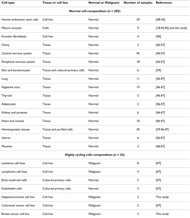
Table 1: Samples of the microarray compendium
Cell type Tissue or cell line Normal or Malignant Number of samples References
Normal cell compendium (n = 205)
Human embryonic stem cells Cell line Normal 29 [38-43]
Mature oocytes Cells Normal 9 [18,44,45] and this study
Foreskin fibroblasts Cell line Normal 4 [40]
Ovary Tissue Normal 3 [46,47]
Central nervous system Tissue Normal 44 [46,47]
Peripheral nervous system Tissue Normal 18 [46,47]
Skin and keratinocytes Tissue and cultured primary cells Normal 6 [39]
Lung Tissue Normal 11 [46,47]
Digestive tract Tissue Normal 19 [46,47]
Thyroid Tissue Normal 5 [46,47]
Adipocytes Tissue Normal 2 [46,47]
Kidney and prostate Tissue Normal 6 [46,47]
Heart and muscle Tissue Normal 10 [46,47]
Hematopoietic tissues Tissue and purified cells Normal 30 [39,46,47]
Uterus Tissue Normal 6 [46,47]
Placenta Tissue Normal 3 [46,47]
Highly cycling cells compendium (n = 22)
Leukemia cell lines Cell line Malignant 8 [47]
Lymphoma cell lines Cell line Malignant 4 [47]
Early erythroid cells Cultured primary cells Normal 2 [47]
Endothelial cells Cultured primary cells Normal 2 [47]
Hepatocarcinoma cell line Cell line Malignant 2 This study
Colorectal cancer cell line Cell line Malignant 2 [47]
Breast cancer cell line Cell line Malignant 2 This study
been identified as overexpressed by female germinal cells by our group and others [18,19]. Similarly, the "hESC sig-nature" displayed numerous genes known to be specifi-cally overexpressed in hESC such as POU5F1/OCT4, NANOG, DPPA4, TDGF1, CD24, PODXL, HELLS, etc. [20]. These findings validated the biological relevance of our compendium and the signatures specific to oocytes and hESC. We intersected these two lists of genes and established a "oocyte/hESC signature", composed of 652 PS (558 different Refseq transcripts) (Figures 1D; see Additional file 4).
A strong cell cycle signature
To get an insight into the oocyte/hESC signature, we searched for overrepresented gene ontology (GO) func-tional annotations. As expected from our previous studies on oocytes transcriptome or hESC transcriptome [18,20], the oocytes/hESC signature was highly enriched in genes associated with intra-cellular localization, DNA and RNA binding, and conversely, it was significantly depleted in genes which encode for secreted proteins or proteins implicated in signal transduction (Figure 2A and 2B). Remarkably, we observed that the biological process
Human mature MII oocytes and hESC display a common transcriptome signature
Figure 1
Human mature MII oocytes and hESC display a common transcriptome signature. Principal component analysis (PCA) (A). A PCA of 9 mature oocytes (MII), 29 hESCs and 167 somatic tissue samples. CNS, central nervous system; PNS, peripheral nervous system. Hierarchical clustering (B). Each horizontal line represents a PS and each column represents a single sample. PS up and down-regulated were colored in red and purple, respectively. In white, PS which were neither over nor under-expressed. Clusters: a: hESCs genes; b: MII oocytes genes; c: the oocytes/hESC signature; d: genes expressed in CNS; e: genes expressed in hematopoietic cells. AD: adipocytes; hFF: human foreskin fibroblasts; SK: skin and keratinocytes; DT: diges-tive tract; Hemato: hematopoietic cells; PL: placenta; H: heart; LU: lung; TH: thyroid; UT: uterus; OV: ovary; M: muscle. Expression bar charts of selected genes (C). Expression bar charts of three oocytes specific genes (DAZL, SOX30, ZP2), three hESCs specific genes (POU5F1/OCT4, NANOG, and DPPA4), three genes up-regulated in hESC and oocytes MII (DNMT3B, TOP2A and ZNF84) and one ubiquitously expressed gene (RPL4) using our on-line expression atlas Amazonia!
http://amazo-nia.montp.inserm.fr. Diff: non-specific differentiated hESC; MCF7: breast cancer cell line; HEPG2: hepatocarcinoma cell line. Venn diagram detailing shared and distinct gene expression among hESCs and MII human oocytes (D). The oocyte/hESC signa-ture was defined as the intersection of the hESC signasigna-ture (genes overexpressed in hESC compared to somatic tissues (ST)) and the MII oocytes signature (genes overexpressed in MII oocytes compared to ST). The oocytes/hESC signature was then further parted into a fraction sharing a cell cycle signature and a non-cell cycle part.
annotations related to cell cycle such as "mitotic cell cycle", "cell cycle progress", "nucleobase, nucleoside, nucleotide synthesis" were among the most highly enriched in the oocyte/hESC signature (P < 1. 10-3). These
findings were in line with the very short cell cycle duration of primate ESC [21] and with the fact that MII oocytes samples are pure populations of cells undergoing the sec-ond meiotic division, which has many features of mitosis. Thus, these two tissue types highly express genes involved in the process of cell division. To delineate more clearly
the cell cycle contribution to the oocyte/hESC signature, we defined a cell cycle signature independently from the oocytes and hESC samples. We compared samples charac-terized by a high proliferation index to our somatic sam-ples series. Proliferating samsam-ples included normal cells such as rapidly dividing CD71+ early erythroid progeni-tors and CD105+ endothelial cells, as well as cell lines originating from haematological, hepatic, breast and colorectal tumors (See Table 1 and Additional file 1 for the list of additional samples). A SAM analysis with a FDR
Biological annotations of genes overexpressed in human mature MII oocytes and hESC
Figure 2
Biological annotations of genes overexpressed in human mature MII oocytes and hESC. Cellular compartment localisation according to Gene Ontology (GO) annotations (A). Statistical comparison of the distribution of GO annotations in the oocytes/hESC signature with the genes underexpressed in oocytes and hESC (B). Gene Ontology categories which differed significantly (p value ≤ 0.01) between oocytes and hESC are shown. Oocyte/hESC gene networks (C). We computed interac-tion networks from the oocyte/hESC signature. Genes included in the oocyte/hESC signature are in red (the color intensity is proportional to the oocyte/hESC to somatic samples fold change). Genes not found in the signature are in white. In each net-work, edge types are indicatives: a plain line indicates direct interaction, a dashed line indicates indirect interaction; a line with-out arrowhead indicates binding only; a line with an arrowhead indicates "acts on". Node types represent different types of molecules: diamond, enzyme; square, cytokine; triangle, phosphatase; and circle, other. Double line edge represents a group or a complex. Validation of microarray data (D). Gene expression of DNMT3A, SMARCA5, PSMA5, PSMA2 were assayed with quantitative RT-PCR (QRT-PCR). The expression of these four selected genes was compared in mature oocytes, hESC and various somatic samples: human fibroblasts (hFF), human breast adenocarcinoma cell line (MCF7), human hepatocellular liver carcinoma cell line (HepG2), endometrial cells (End7) and T-lymphocyte cells (TL) using QRT-PCR. All measurements were performed in duplicate in two separate runs. The relative levels of gene expression of target mRNA was normalized against GAPDH expression. Fold change values are plotted on a log10 scale.
of 0.05% identified 818 PS (682 different Refseq tran-scripts) overexpressed in proliferating somatic samples, composing a "cell cycle signature". Intersection with the oocyte/hESC signatures revealed that 220 PS (33.7%) were shared with the cell cycle signature (Figure 1D; see Additional file 4). This cell cycle part of the oocytes/hESC signature included enzymes involved in general cell metabolism (METAP2, SHMT2, etc.), nucleoside synthe-sis (DHFR, TYMS, RRM2, PPAT, etc.), DNA repair includ-ing mismatch repair (MSH2 and MSH6) or base excision repair (UNG, PCNA), main components of the cell cycle regulatory machinery (CCNB1 and 2, CCNA, CCNE, etc.), regulator of the topologic state of DNA (TOP1, TOP2A) and components of the mitotic spindle assembly check-point (the centromer constituents CENPE, the securin PTTG1, and MAD2L1, BUB1B, BUB3) (Figure 2C). How-ever, it must be noted that 36 genes from the oocyte/hESC signature that are functionally annotated "cell cycle" by Gene Ontology were not included in the cell cycle signa-ture. These are cell cycle genes that are preferentially expressed in mature oocytes and hESC as compared to other cycling cell types such as malignant cell lines or pro-liferating primary hematopoietic cells, and included the spindle checkpoint gene CHEK1, FBXO5/EMI1, the cyclin dependent kinase-activating kinase CDK7 and a compo-nent of transcription factor IIH, CDK8.
Human oocytes and hESC share a large chromatin remodelling network
We focused the second part of our analysis on the non-cell cycle part of the oocyte/hESC signature. Removing the 220 cell cycles PS led to the definition of a "non-cell cycle oocyte/hESC signature" that retained 432 PS (384 tran-scripts). This signature contained many transcripts involved in DNA and histone modifications. One of these transcripts was DNMT3B, involved in DNA methylation. The high fold change of DNMT3B in hESC and oocytes compared to somatic samples (43.4 and 9.4, respectively) suggests a central role of this DNA methyltransferase in the control of the epigenome of these cells. In addition, several transcripts, comprising JARID2, SMARCA5, CBX5, CHAF1A and CBX1, were involved in histone modifica-tion processes (Figure 2C). We selected two chromatin remodelling genes, DNMT3B and SMARCA5, and vali-dated by QRT-PCR their preferential expression in oocytes and hESC compared to somatic samples (Figure 2D).
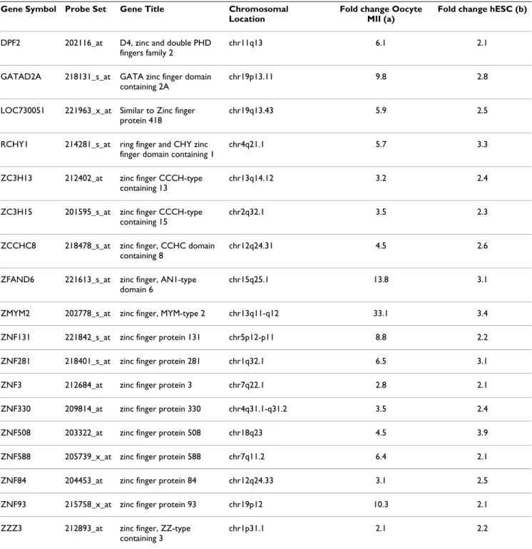
Zinc finger genes
Mature MII oocytes and hESC overexpressed numerous zinc finger domain genes. The zinc finger motif is a DNA binding domain dependent on a zinc ion, frequently found in transcription factors. The non-cell cycle oocyte/ hESC signature was significantly enriched in zinc finger PS : 20 (18 Refseq transcripts) out of 432 PS (4.6%) in the signature compared to 611 out of 22215 PS (2.7%) in the
complete list of PS (P = 0.018) (Table 2). The expression bar charts for the zinc finger domain gene ZNF84 is shown in Figure 1C and that of all 18 zinc finger domain genes from the non-cell cycle oocyte/hESC list is available as Additional file 5.
Ubiquitination and proteasome
Surprisingly, we found that a highly significant propor-tion of genes of the non-cycle oocytes/hESC signature was involved in the protein ubiquitination and proteasome canonical pathway (P-value ≤ 1.93E-06, using Ingenuity software) (Figure 3A). Enzymes from the three E1/E2/E3 ubiquitination classes were found significantly overex-pressed in the functional regulatory network: the E1 ubiq-uitin-activating enzyme UBE1C, the E2 ubiquitin-conjugating enzymes UBE2G1, UBE2V1 and UBE2V2, and the E3 ubiquitin protein ligases UBE3B and breast cancer 1, early onset (BRCA1) (Figure 3A). It was recently showed that BRCA1 is part of a holoenzyme complex containing BRCA1, BRCA2, BARD1 and RAD51 which is called the BRCA1- and BRCA2-containing complex (BRCC) that dis-plays an ubiquitin E3 ligase activity [22]. We found that three out of the four components of BRCC, namely BARD1, BRCA1 and RAD51 are overexpressed in MII oocytes and hESC whereas BRCA2 is simply expressed. One key consequence of protein ubiquitination is to tar-get proteins for degradation by the 26S proteasome. In line with the biased high expression of ubiquitination pathway components, we also found many subunits of the proteasome significantly overexpressed in MII oocytes and hESC (Figure 3B). As expected for a ubiquitous cell machinery complex, the expression of most subunits of the 26S proteasome is detected by microarrays in oocytes and hESC. Among these, four catalytic alpha proteasome subunits (PSMA2, PSMA3, PSMA4 and PSMA5) and three regulatory subunits, the ATPase PSMC6 and the non-ATPase PSMD10 and PSMD11, are significantly upregu-lated in MII oocytes and hESC as compared to somatic tis-sues (P < 0.001 for each cited proteasome subunit) (Figure 3B), in line with the overexpression of the ubiquitin path-way. In addition, three ubiquitin-specific proteases (USP1, USP7 and USP9X), that are deubiquitinating enzymes, were also found highly expressed in the oocytes and hESC. Remarkably, microarray analysis showed that upon differentiation, the expression level of proteasome components that are overexpressed in hESC decreased to a level similar to that of somatic cells (Figure 3C).
High sensitivity of human embryonic stem cells to proteasome inhibition
The high expression level of the proteasome machinery in hESC and its decrease during hESC differentiation sug-gested that this pathway could play an important role in pluripotent cells. To determine the consequence of func-tional blocking of the proteasome activity in hESC, we
tested the effect of MG132, a specific proteasome inhibi-tor, on embryonic stem cells [23]. Increasing doses of MG132 were added to the culture media of HS181 hES cells. At 250 nM the morphology of the hESC colonies clearly showed large patches of differentiation and at 500 nM no undifferentiated cells remained (Figure 4A). Fur-thermore, when colonies contained mixed populations of
undifferentiated and differentiated cells, 1 μM of MG132 induced the detachment of the undifferentiated cells whereas the differentiated progeny stayed tightly sticked to the dish. These results were reproduced on two other hES cell lines (HD83 and HD90, data not shown). By con-trast, the morphology and adhesion of human foreskin fibroblasts (HFF) were not altered by MG132 at
concen-Table 2: Genes from the mature oocyte/hESC signature with a Zinc finger domain.
Gene Symbol Probe Set Gene Title Chromosomal
Location
Fold change Oocyte MII (a)
Fold change hESC (b)
DPF2 202116_at D4, zinc and double PHD fingers family 2
chr11q13 6.1 2.1
GATAD2A 218131_s_at GATA zinc finger domain containing 2A
chr19p13.11 9.8 2.8
LOC730051 221963_x_at Similar to Zinc finger protein 418
chr19q13.43 5.9 2.5
RCHY1 214281_s_at ring finger and CHY zinc finger domain containing 1
chr4q21.1 5.7 3.3
ZC3H13 212402_at zinc finger CCCH-type containing 13
chr13q14.12 3.2 2.4
ZC3H15 201595_s_at zinc finger CCCH-type containing 15
chr2q32.1 3.5 2.3
ZCCHC8 218478_s_at zinc finger, CCHC domain containing 8
chr12q24.31 4.5 2.6
ZFAND6 221613_s_at zinc finger, AN1-type domain 6
chr15q25.1 13.8 3.1
ZMYM2 202778_s_at zinc finger, MYM-type 2 chr13q11-q12 33.1 3.4
ZNF131 221842_s_at zinc finger protein 131 chr5p12-p11 8.8 2.2
ZNF281 218401_s_at zinc finger protein 281 chr1q32.1 6.5 3.1
ZNF3 212684_at zinc finger protein 3 chr7q22.1 2.8 2.1
ZNF330 209814_at zinc finger protein 330 chr4q31.1-q31.2 3.5 2.4
ZNF508 203322_at zinc finger protein 508 chr18q23 4.5 3.9
ZNF588 205739_x_at zinc finger protein 588 chr7q11.2 6.4 2.1
ZNF84 204453_at zinc finger protein 84 chr12q24.33 3.1 2.5
ZNF93 215758_x_at zinc finger protein 93 chr19p12 10.3 2.1
ZZZ3 212893_at zinc finger, ZZ-type containing 3
chr1p31.1 2.1 2.2
List of genes from the oocyte/hESC signature that display a zinc finger domain, with PS number, and fold change (oocyte versus somatic tissues (a) and hESC versus somatic tissues (b).
tration up to 1 μM (Figure 4B). Moreover, we differenti-ated the HD90 and HS181 hESC into hES-differentidifferenti-ated fibroblasts (dF) that display features characteristic of fibroblasts: flattened cells with elongated nucleus and branching pseudopodia, expression of membrane mark-ers such as P4H, CD13 or CD44. Treatment with MG132 did not induce changes in cell morphology of the hES-dF-HD90 (Figure 4C) and hES-dF-HS181 (data not shown), even at high concentrations. RT-PCR analysis showed that expression of pluripotency markers decreased when hESC were treated with MG132. Whereas the expression of
POU5F1/OCT4, SOX2 and NANOG decreased with the proteasome treatment, GAPDH expression was not modi-fied (Figure 4D). This effect was also substantiated by flow cytometry. We observed decreasing expression of the pluripotency marker TRA-1-60 on HS181 hESC after exposition to MG132 (Figure 4E). However, high concen-tration of MG132, up to 1 μM, did not affect two different fibroblasts cell surface markers, CD44 and CD13 on hFF, on hES-dF-HS181 or on hES-dF-HD90. Thus, our results showed that pluripotent hESC are highly susceptible to the action of proteasome blockage, whereas somatic cells
Overexpression of the ubiquitination/proteasome pathway
Figure 3
Overexpression of the ubiquitination/proteasome pathway. Interaction network analysis of the ubiquitination/proteas-ome pathway in the oocytes/hESC signature. (A) The interaction network was generated with the Ingenuity software and shows a high number of genes from this cellular pathway overexpressed (red) or expressed (orange) in the oocytes/hESC sig-nature. Transcripts that were not detected by the microarray are white. Notably, we found elements of the ubiquitin-activating enzyme (UBE), BRCA1- and BRCA2-containing complex (BRCC), the regulatory 19S proteasome, the core proteolytic 20S proteasome and the ubiquitin-specific protease (USP) modules in the oocyte/hESC signature. Gene expression measured by microarrays in 9 genes involved in the ubiquitin and proteasome pathway (B). Box-and-whisker plots comparing the expression level of PSMA2, PSMA3, PSMA4, PSMA5, PSMC6, PSMD10, PSMD11, UBE2G1 and SKP2 in mature oocytes and hESC (38 samples) versus somatic tissues (167 samples). The signal intensity for each gene is shown on the y axis as arbitrary units determined by the GCOS 1.2 software (Affymetrix). (*): P-value < 0.0001 using a Mann-Whitney statistical test. Down regulation of the pro-teasome pathway during hESC differentiation (C). U133A microarray signal values for PSMA2, PSMA4 and PSMA5 in 29 undiffer-entiated hESC samples (mean value) versus two embryoid bodies (EB) samples (EB day 5 and EB day 14) and 3 non-lineage differentiated hESC samples (mean value).
such as hFF or the differentiated progeny of hESC such as hES-dF were not affected.
Discussion
Early human embryo development results in the repro-gramming of highly specialized germinal cells into
totipo-tent and then pluripototipo-tent cells that are the progenitors of all the specialized cell types of the human body. This unique biological property has been harnessed to restore pluripotency in human somatic cells by SCNT or cell-fusion using embryonic stem cells [6,7]. hESC share with pluripotent stem cells from the inner cell mass
pluripo-Blocking proteasome activity in hESC
Figure 4
Blocking proteasome activity in hESC. Proteasome inhibition causes differentiation of hESCs (A). HS181 colonies (p56) were grown 4 days on hFF and then treated 40 hours with the proteasome inhibitor MG132. Control HS181 colonies express POU5F1 (scale bar 25 μ) and display a normal karyotype. Upon treatment with MG132, HS181 colonies differentiated. No effect of proteasome inhibition on hFF (B). HFF express the fibroblastic marker P4H and display a normal karyotype. HFF were cultured with MG132 during 40 hours. No morphological alteration was observed. No effect of proteasome inhibition on hES-dF-HD90 (C). The HD90 hESC line was differentiated into fibroblasts like cells (hES-dF) that display morphologic feature of dermal fibroblasts and express P4H: bottom left. An undifferentiated colony of HD90 P4H-, starts to differentiate at the edges
into P4H+ hES-dF (upper left). hES-dF-HD90 cells were cultured with MG132 during 40 hours. No morphological alteration
was observed. Down regulation of pluripotency transcription factor in hESC by proteasome blockage (D). The expression of POU5F1/OCT4, NANOG and SOX2 was measured by semiquantitative RT-PCR in HS181 hESC after 40 h culture with MG132. GAPDH was used as a control. Flow cytometry analysis after proteasome inhibition (E). HFF, hES-dF-HS181 and HS181 cells were treated 40 hours with MG132, and then the cell surface fibroblastic markers CD13 and CD44, and the pluripotent cell surface marker TRA-1-60 were measured by FACS. Markers for fibroblastic cells were not altered with MG132 treatment, whereas the marker for pluripotency dropped to barely detectable level. Y-axis: percentage of the control (untreated) sample.
tency transcription factors and multi-lineage differentia-tion properties, and are considered a good in vitro model for pre-embryo pluripotent stem cells. Though human oocytes and ESC are developmentally separated by less than one week, the transcriptome of the oocyte undergoes rapid changes after fertilization [24,25]. We undertook to find out a common expression signature to these two cell types, that share somatic cell reprogramming properties, by comparing them to a large collection of somatic tissues samples. A first observation was that the oocytes/hESC signature was highly enriched in genes involved in cell cycle. Whereas this was expected because of the cell cycle status of these two cell types, the expression of a large set of genes associated with cell division is nevertheless of interest for cellular reprogramming. As recently reported, prior mitotic remodeling of the somatic nuclei, involving topoisomerase II (TOP2)-dependent shortening of chro-matin loop domains and an increased recruitment of rep-lication initiation factors onto chromatin, is essential for reprogramming of differentiated nuclei [26]. Strikingly, we found that TOP2A was highly up-regulated in both oocytes and hESC. This observation suggests that TOP2A could be a major factor in the reprogramming properties of oocytes and hESC by participating in chromatin remodeling. Conversely, the identification of a "cell cycle signature", shared with highly proliferating tissues such as cancer cell lines, provided a mean to identify by subtrac-tion a "non-cycle oocytes/hESC" signature of 432 PS. This signature included transcripts coding for proteins involved in chromatin structure modifications such as DNMT3B, JARID2, SMARCA5 or CBX5 that contribute to the DNA methylation and chromatin remodeling (Figure 2C). Consistent with these observations, hESC display a distinct, permissive, chromatin structure compared with other tissues [27]. Expression of DNA methyl-transferases or several ATP-dependent chromatin remodelling factors are elevated in murine oocytes or ES cells [28,29]. Thus our findings show large similarities between murine and human ES cells, and put forward several genes whose strong overexpression could contribute to the specific chromatin state of hESC.
Another lesson from our transcriptomic approach is that the common oocytes/hESC gene expression profile has a very low number of genes that are either secreted or mem-brane bound (Figure 2A). This is in line with our previ-ously published data that booth oocytes and hESC "specific genes" are significantly depleted in extracellular signalling components, suggesting that this feature is indeed a common characteristic shared by oocytes and hESC and is not simply due to a lack of overlap [18,20]. Hence, genes specifically shared by oocytes and hESC are largely nuclear proteins. One assumption that can be inferred from these findings is that determinant of pluripotency may be mostly intrinsic factors. This
obser-vation converges on a recent model, which proposed that pluripotency is a ground state that is intrinsically self-maintained when protected from extrinsic differentiation stimuli [30].
An unexpected observation was that genes involved in protein ubiquitination and proteasome pathway were also overrepresented in the oocytes/hESC signature. This could be linked to the strong proliferation signature of hESC and oocytes as this pathway is by many way impli-cated in the regulating the cell cycle [31]. However, the overexpression of the ubiquitination/proteasome path-way was still significant when the cell cycle signature was substracted, suggesting that this pathway could have a role in pluripotent cells in addition to its house keeping or cell cycle functions. In line with these results, we showed a selective sensitivity of hESC to the inhibition of the activ-ity of the proteasome, resulting in loss of pluripotency and cell growth at doses without any detectable effects on differentiated but cycling cells such as primary fibroblasts or hESC derived fibroblast like cells. In addition, it must be stressed that the dramatic effects on hESC pluripotency were observed at doses of the proteasome inhibitor MG132 (0.5 μM) significantly lower than those typically found in the literature (several μM) or in mice ES cells (20 μM) [23]. This observation is highly interesting in light of the recent findings of the role of the proteasome in tran-scription, especially in hESC. The 26S proteasome consists of a 20S core proteolytic part, capped by a 19S regulatory complex. Specificity of degradation of proteins is medi-ated in part by poly-ubiquitination of the substrate bound for destruction. Based on early work in yeast, the proteas-ome is known to interact with chromatin and function at multiple steps in transcription, both through proteolytic and non-proteolytic activities [32]. Recently, Szutorisz et al. reported that the 26S proteasome is assembled on intergenic and intragenic regions in ES cells and act as a transcriptional silencer by blocking non-specific transcrip-tion initiatranscrip-tion [23]. This mechanism involves the proteo-lytic activity of the 20S core by degrading non-specific preinitiation complexes, thereby preventing permissive transcription and spreading of the modified chromatin. Our results are consistent with this hypothesis, but final answer on this issue will require further investigations. This work has compared human MII oocytes and hESC to somatic tissues gene expression profiles. One goal was to provide new hints on the process of nucleus reprogram-ming which takes place in vivo during early embryo devel-opment or in vitro during SCNT, and may thus help to improve the iPS technology. Indeed, since the seminal work of the team of Shinya Yamanaka, numerous improvement have been made, including the identifica-tion of new genes able to replace some of the original ones in the reprogramming cocktail, the use of small molecules
or the replacement of the retroviral vectors by adenovi-ruses or plasmids [33-37]. A first observation is that human mature oocytes do not express the pluripotency core transcriptional genes POU5F1/OCT4, NANOG and SOX2 [13], except POU5F1/OCT4 at low level (see Figure 1C and our Amazonia! on-line expression atlas, http:// amazonia.montp.inserm.fr). They neither express KLF4 nor CMYC, which compose, with POU5F1/OCT4 and SOX2, the four factors that can reprogram somatic cells by virus-mediated overexpression [11]. From the six "repro-gramming" factors described to date, only LIN28 was found in the oocyte/hESC signature. However, POU5F1/ OCT4, NANOG, SOX2, KLF4, LIN28 and CMYC are all expressed by hESC. Therefore, during early embryo devel-opment, the expression of these genes is induced. Thus, two different molecular pathways that can reprogram adult somatic cells can be envisioned: (i) the process tak-ing place in the oocyte cytoplasm, able to activate the core transcriptional genes, or (ii) the overexpression of the core transcriptional genes themselves together with adjuvant genes, either by viral overexpression or by fusion with cells already expressing these genes. It can be speculated that the factors that lie upstream of the pluripotency core transcriptional circuitry are expressed as mRNA in mature MII oocytes and are still present at blastocyst stage from which hESC are derived. Thus, the oocytes/hESC signature likely includes these factors, and therefore this informa-tion could be highly informative for cell reprogramming. The signature contained numerous transcritption factors, including many zinc finger such as ZNF84, several still poorly annotated genes such as KLHL7, MRS2, or the Selenophosphate synthetase 1 (SEPHS1), displayed a strong cell cycle signature, chromatin modification genes, and also many actors of the proteasome pathway. All these genes are candidate genes to improve the efficiency of iPS generation, especially in the light of the recent advances that uses non retroviral vectors but at the cost of lower efficacy.
Conclusion
Human ESC are not only a very promising source of cells for regenerative medicine, but are also a unique tool to understand early embryo development that can not easily be studied on live embryos because of ethical and techni-cal limits. Our comparison of human mature oocytes and hESC to a large collection of somatic samples helps to understand the early embryo development and pluripo-tency, and is therefore relevant for therapeutics, including improvement of the pregnancy success rate in IVF and regenerative medicine applications such as those involv-ing cell reprogramminvolv-ing.
Methods
Transcriptome compendium
We built an expression compendium by combining U133A and U133 Plus 2.0 (Affymetrix, Santa Clara, USA)
microarray data from 11 publications and from our labo-ratory, totalizing 205 samples (Table 1; see Additional file 1) [18,38-47]. Data were analyzed with the GCOS 1.2 software (Affymetrix), using the default analysis settings and global scaling as first normalization method, with a trimmed mean target intensity value (TGT) of each array arbitrarily set to 100. Data was floored at 50, i.e each value below 50 was set to 50. In order to compare U133A and U133 Plus 2.0 data, we further normalized the data with a rank-based normalization method. This method, "MetaNorm", orders the values of the 22 215 PS of the Affymetrix U133A microarray and allocates a new value to each PS according to its rank, using a unique signal value template (Assou et al., manuscript in preparation). Sam-ples are listed in Table 1 (see Additional file 1), along with references, microarray design and, when available, GEO (Gene Expression Omnibus) dataset number. The dataset is available as Additional file 2 (signal and p-value) and each PS can be individually accessed on our website http:/ /amazonia.montp.inserm.fr[20].
Data analysis and visualization
Principle component analysis (PCA) was performed using ArrayAssist® software (Stratagene, La Jolla, CA, USA) to
provide a global view of how the various sample groups were related. Hierarchical clustering was carried out with CLUSTER and TREEVIEW software [48]. PCA and cluster-ing were performed on 10,000 PS with the highest coeffi-cient of variation (CV). Gene expression profiles were identified using two-class Significance Analysis of Micro-arrays (SAM) method [49]http://www-stat.stanford.edu/ ~tibs/SAM/ which utilizes a Wilcoxon-test statistic and sample-label permutations to evaluate statistical signifi-cance. SAM analysis was applied after data filtering retain-ing only PS with at least 2 samples with a "Present" call. The False Discovery Rate (FDR), an estimate of the frac-tion of selective genes, was kept below 5% in all statistical analyses. Gene Ontology annotation analysis was carried out using the Fatigo+ tool at the Babelomics website http:/ /babelomics.bioinfo.cipf.es[50]. Only annotations with a false discovery rate-adjusted P-value below 0.05 were con-sidered significant. To uncover functional biological net-works, we imported gene expression signatures into the Ingenuity Pathways Analysis (IPA) Software (Ingenuity Systems, Redwood City, CA, USA). Comparison of the fre-quency of zinc finger domain containing transcript between the non-cell cycle oocytes/hESC signature and the entire U133A microarray was carried out using a Pear-son's Chi-squared test with Yates' continuity correction.
Human mature MII oocytes, fibroblasts and malignant cell lines transcriptome
Unfertilized MII oocytes were collected after informed consent 44 hours post insemination or post microinjec-tion by ICSI as previously published [18,44]. Briefly, Mll oocytes were from couples referred to our center for cIVF
(tubal infertility) or for ICSI (male infertility). Mature M2 oocytes were pooled: 16 oocytes for Oocyte_M2_16 sam-ple, 21 for Oocyte_M2_21 and 24 for Oocyte_M2_24, from 6, 8 and 8 patients respectively. Human foreskin fibroblasts cell lines were described previously [20]. MCF7 and HEPG2 were from ATCC and cultured in DMEM medium containing 10% fetal calf serum (FCS). Total RNA was isolated using RNeasy mini kits (Qiagen, Court-aboeuf, France) and quantified using a NanoDrop spec-trophotometer (Thermo Fischer, Wilmington, Delaware, USA). Total RNA (100 ng) was used to prepare twice amplified labeled cRNA for hybridization to HG-U133 plus 2.0 GeneChip pangenomic oligonucleotide arrays (Affymetrix, Santa Clara, CA, USA) as previously described [18]. The 9 microarray data obtained in our lab are accessible in US National Center for Biotechnology Information, Gene Expression Omnibus (GEO) through the provisional accession numbers GSE11450 (series), and GSM288886, GSM288885, GSM288883, GSM288882, GSM288880, GSM288878, GSM288877, GSM288876, GSM288812 (samples).
hES cell culture
The HS181 hES cell line was imported from the Karolin-ska Institute (Stockholm, Sweden). The HD83/D17/FE07-135-L1 and HD90/D18/FE07-142-L1 hES cell lines were derived in our laboratory from a normal embryo and an embryo that carried an abnormal VHL gene according to preimplantation genetic diagnostic, respectively (De Vos et al. manuscript in preparation). Briefly, derivation of HD83 and HD90 was carried out using mechanical disso-ciation of the inner cell mass [51]. The culture medium used for hESCs derivation and culture consisted of 80% KO-DMEM, 20% knockout SR, 2 mM L-glutamine, 1% nonessential amino acids, 0.5 mM β-mercaptoethanol (all from Gibco Invitrogen, Cergy-Pontoise, France) and 10 ng/mL of bFGF (Abcys, Paris, France). Passaging was performed mechanically by cutting the colony using a #15 scalpel under the microscope. Human foreskin fibroblasts (HFF), mitotically inactivated using irradiation (40 Gy),
were used as feeder cells. HFF cells were cultured in 85% DMEM, 15% FBS. All hESC expressed POU5F1/OCT4, NANOG and TRA-1-60, and were able to differentiate into embryoid bodies that expressed differentiation markers. For proteasome inhibition experiments, hESC were incu-bated 40 h with various concentration of MG132 (Sigma), with medium renewal at 24 h.
Production of hES-derived fibroblasts
Briefly, hES cells were mechanically isolated and plated on laminin precoated 6-wells culture dishes (Becton Dick-inson, San Jose, CA, USA) in hESC culture medium renewed every day. After 5 days, bFGF was removed, and after three additional days, medium was switched to HFF medium. In these conditions, hES cells differentiated into flattened cells with elongated nucleus and branching pseudopodia forming hESC-derived fibroblasts (hES-dF). The hES-dF were then mechanically isolated and trans-ferred to feeder free 6-wells culture dishes. Subsequent passages were carried out using 0.05% trypsin- EDTA (Inv-itrogen) every 6 days and cultures upscaled into T75 flasks.
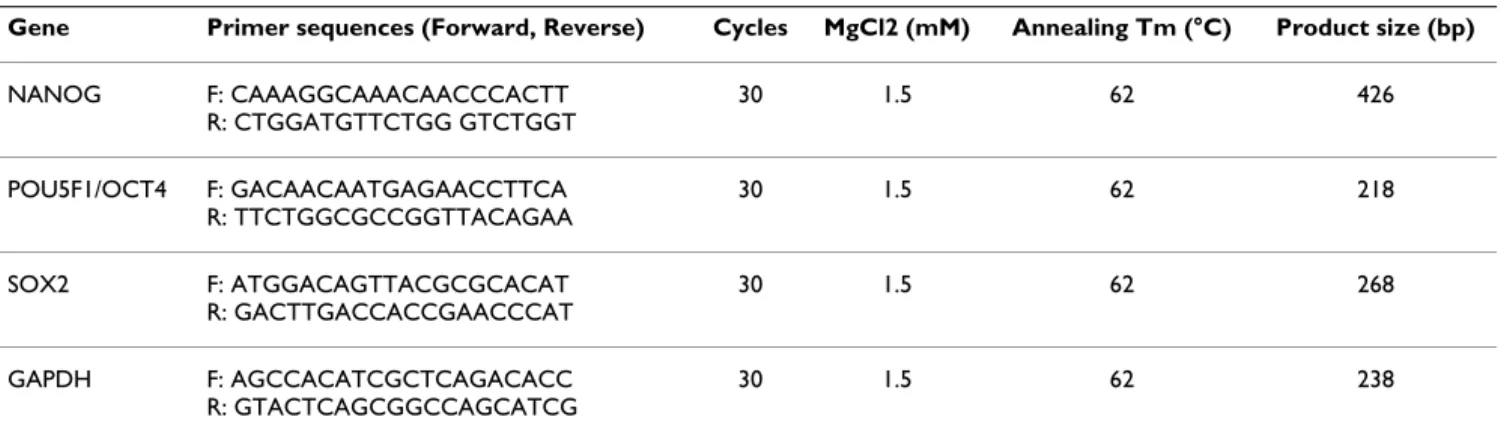
RT-PCR and quantitative PCR (QRT-PCR)
RT-PCR was carried out on total RNA isolated from hESCs grown in the absence or presence of increasing concentra-tion of MG132. PCR condiconcentra-tions and sequence for each primer are shown in Table 3. PCR products were separated on a 1% agarose gel. Expression of the housekeeping gene GAPDH was used to normalise PCR reactions. For QRT-PCR, approximately 1 μg of linear-amplified, biotin-labelled cDNA was mixed with Assays-on-Demand prim-ers and probes and TaqMan Univprim-ersal Master Mix accord-ing to the manufacturer's instructions (Applied Biosystems, Courtaboeuf, France). Real-time QRT-PCR was performed using the ABI Prism 7000 sequence detec-tion system (Applied Biosystems) and normalized to GAPDH for each sample using the following formula in which Ct is cycle threshold: 100/2ΔΔCt, where ΔΔCt = ΔCt unknown -ΔCt positive control.
Table 3: Primer sequences and conditions used for RT-PCR
Gene Primer sequences (Forward, Reverse) Cycles MgCl2 (mM) Annealing Tm (°C) Product size (bp)
NANOG F: CAAAGGCAAACAACCCACTT R: CTGGATGTTCTGG GTCTGGT 30 1.5 62 426 POU5F1/OCT4 F: GACAACAATGAGAACCTTCA R: TTCTGGCGCCGGTTACAGAA 30 1.5 62 218 SOX2 F: ATGGACAGTTACGCGCACAT R: GACTTGACCACCGAACCCAT 30 1.5 62 268 GAPDH F: AGCCACATCGCTCAGACACC R: GTACTCAGCGGCCAGCATCG 30 1.5 62 238
Immunofluorescence and cytometry flow
Cells were fixed with PBS containing 4% paraformalde-hyde for 20 minutes at room temperature and blocked with PBS containing 5% normal donkey serum for 30 minutes at room temperature. After blocking, cells were incubated with the appropriate primary antibody against POU5F1/OCT4 (sc-9081, Santa Cruz Biotechnology, Santa Cruz, CA; 1:300) or against proline 4-hybroxylase (P4H) (Dako, Trappes, France; 1:50) for 1 hour at room temperature. Cells were washed three times in PBS and incubated for 1 hour at room temperature with Alexa Fluor® 488 donkey anti-Rabbit (A-11034; Molecular
Probes; 1:1000) and Alexa Fluor® 568 goat anti-mouse
antibody (A11019, Invitrogen; 1:400) secondary antibod-ies for POU5F1/OCT4 and P4H respectively. Hoechst staining was added to first wash (Sigma, 5 μg/ml). For flow cytometry, cells were harvested by treatment with 0.05% trypsin- EDTA (Invitrogen) and were resuspended in culture media. Cell aliquots were incubated on ice with anti-CD13 MAb conjugated to phycoerythrin (PE) (A07762, Beckman-Coulter, 1:50), anti-CD44 MAb con-jugated to fluorescein isothiocyanate (FITC) (clone J-173, Immunotech; 1:50) and Tra-1-60 (90232, Chemicon) or conjugated isotypic controls. Flow cytometry was per-formed on a fluorescence-activated cell sorter (FACS Scan, Becton Dickinson), and data were analyzed with the Cel-lquest software (Becton Dickinson).
Authors' contributions
SA: conception and design, collection and assembly of data, data analysis and interpretation, manuscript writing; DC, ST, VP: collection and assembly of data; OH: provi-sion of study material; BK: design; SH: conception and design, provision of study material, manuscript writing; JDV: conception and design, provision of study material, data analysis and interpretation, manuscript writing, final approval of manuscript. All authors read and approved the final manuscript.
Additional material
Acknowledgements
We are grateful to the various labs that gave free access to their complete transcriptome data, in agreement with the MIAME recommendations [52]. We thank Isabelle Rodde-Astier and Bruno Delorme (MacoPharma) for their support, Laure Nadal for excellent technical work, Marc Piechaczyc et Isabelle Jariel for helpful discussions on the proteasome and for providing reagents and Marilyne Dijon for critical reviewing of the manuscript. Sup-ported by the University Hospital of Montpellier, the Association Française contre les Myopathies (AFM), Ferring and Organon phamaceuticals com-pagnies.
References
1. Huang JC, Lei ZL, Shi LH, Miao YL, Yang JW, Ouyang YC, Sun QY, Chen DY: Comparison of histone modifications in in vivo and
in vitro fertilization mouse embryos. Biochem Biophys Res Com-mun 2007, 354(1):77-83.
2. Santos F, Hendrich B, Reik W, Dean W: Dynamic reprogramming
of DNA methylation in the early mouse embryo. Dev Biol
2002, 241(1):172-182.
3. Campbell KH, McWhir J, Ritchie WA, Wilmut I: Sheep cloned by
nuclear transfer from a cultured cell line. Nature 1996, 380(6569):64-66.
4. Gurdon JB, Byrne JA: The first half-century of nuclear
trans-plantation. Proceedings of the National Academy of Sciences of the United States of America 2003, 100(14):8048-8052.
5. Sung LY, Gao S, Shen H, Yu H, Song Y, Smith SL, Chang CC, Inoue K, Kuo L, Lian J, et al.: Differentiated cells are more efficient than
adult stem cells for cloning by somatic cell nuclear transfer. Nature genetics 2006, 38(11):1323-1328.
6. French AJ, Adams CA, Anderson LS, Kitchen JR, Hughes MR, Wood SH: Development of human cloned blastocysts following
somatic cell nuclear transfer with adult fibroblasts. Stem cells (Dayton, Ohio) 2008, 26(2):485-493.
Additional file 1
Table S1. Detailed sample list
Click here for file
[http://www.biomedcentral.com/content/supplementary/1471-2164-10-10-S1.xls]
Additional file 2
Table S2. Transcriptome compendium. A compendium of 205
transcrip-tomes (signal and detection call).
Click here for file
[http://www.biomedcentral.com/content/supplementary/1471-2164-10-10-S2.rar]
Additional file 3
Table S3. Hierarchical clustering gene lists. Transcripts lists from gene
clusters a, b & c from Figure 1B.
Click here for file
[http://www.biomedcentral.com/content/supplementary/1471-2164-10-10-S3.zip]
Additional file 4
Table S4. Main transcript signatures. Transcripts lists from the
tures ana lyzed in the manuscript: non-cell cycle MII oocytes/hESC signa-ture (1), MII oocytes/hESC signasigna-ture (2), MII oocyte signasigna-ture (3), hESC signature (4), cell cycle MII oocytes/hESC signature (5) (Exel file). These lists can be found and datamined on our website Amazonia! http:/ /amazonia.montp.inserm.fr using the "gene list" Amazonia! tool.
Click here for file
[http://www.biomedcentral.com/content/supplementary/1471-2164-10-10-S4.zip]
Additional file 5
Figure S1. Expression bar charts for zinc finger domain containing genes. Expression bar charts for 18 transcripts overexpressed in oocytes
and hESC and containing a zinc finger domain. These charts have been generated on our online gene expression Atlas Amazonia! http://amazo nia.montp.inserm.fr. Abbreviations as in Figure 1C.
Click here for file
[http://www.biomedcentral.com/content/supplementary/1471-2164-10-10-S5.tiff]
7. Cowan CA, Atienza J, Melton DA, Eggan K: Nuclear
reprogram-ming of somatic cells after fusion with human embryonic stem cells. Science 2005, 309(5739):1369-1373.
8. Hochedlinger K, Blelloch R, Brennan C, Yamada Y, Kim M, Chin L, Jae-nisch R: Reprogramming of a melanoma genome by nuclear
transplantation. Genes & development 2004, 18(15):1875-1885.
9. Takahashi K, Yamanaka S: Induction of pluripotent stem cells
from mouse embryonic and adult fibroblast cultures by defined factors. Cell 2006, 126(4):663-676.
10. Park IH, Zhao R, West JA, Yabuuchi A, Huo H, Ince TA, Lerou PH, Lensch MW, Daley GQ: Reprogramming of human somatic
cells to pluripotency with defined factors. Nature 2008, 451(7175):141-146.
11. Takahashi K, Tanabe K, Ohnuki M, Narita M, Ichisaka T, Tomoda K, Yamanaka S: Induction of pluripotent stem cells from adult
human fibroblasts by defined factors. Cell 2007, 131(5):861-872.
12. Aoi T, Yae K, Nakagawa M, Ichisaka T, Okita K, Takahashi K, Chiba T, Yamanaka S: Generation of pluripotent stem cells from
adult mouse liver and stomach cells. Science 2008, 321(5889):699-702.
13. Boyer LA, Lee TI, Cole MF, Johnstone SE, Levine SS, Zucker JP, Guen-ther MG, Kumar RM, Murray HL, Jenner RG, et al.: Core
transcrip-tional regulatory circuitry in human embryonic stem cells. Cell 2005, 122(6):947-956.
14. Hansis C, Barreto G, Maltry N, Niehrs C: Nuclear
reprogram-ming of human somatic cells by xenopus egg extract requires BRG1. Curr Biol 2004, 14(16):1475-1480.
15. Kikyo N, Wade PA, Guschin D, Ge H, Wolffe AP: Active
remode-ling of somatic nuclei in egg cytoplasm by the nucleosomal ATPase ISWI. Science 2000, 289(5488):2360-2362.
16. Adewumi O, Aflatoonian B, Ahrlund-Richter L, Amit M, Andrews PW, Beighton G, Bello PA, Benvenisty N, Berry LS, Bevan S, et al.:
Characterization of human embryonic stem cell lines by the International Stem Cell Initiative. Nature biotechnology 2007, 25(7):803-816.
17. Irizarry RA, Warren D, Spencer F, Kim IF, Biswal S, Frank BC, Gabri-elson E, Garcia JG, Geoghegan J, Germino G, et al.:
Multiple-labora-tory comparison of microarray platforms. Nature methods
2005, 2(5):345-350.
18. Assou S, Anahory T, Pantesco V, Le Carrour T, Pellestor F, Klein B, Reyftmann L, Dechaud H, De Vos J, Hamamah S: The human
cumu-lus–oocyte complex gene-expression profile. Human reproduc-tion (Oxford, England) 2006, 21(7):1705-1719.
19. Kocabas AM, Crosby J, Ross PJ, Otu HH, Beyhan Z, Can H, Tam WL, Rosa GJ, Halgren RG, Lim B, et al.: The transcriptome of human
oocytes. Proceedings of the National Academy of Sciences of the United States of America 2006, 103(38):14027-14032.
20. Assou S, Le Carrour T, Tondeur S, Strom S, Gabelle A, Marty S, Nadal L, Pantesco V, Reme T, Hugnot JP, et al.: A meta-analysis of human
embryonic stem cells transcriptome integrated into a web-based expression atlas. Stem cells (Dayton, Ohio) 2007, 25(4):961-973.
21. Fluckiger AC, Marcy G, Marchand M, Negre D, Cosset FL, Mitalipov S, Wolf D, Savatier P, Dehay C: Cell cycle features of primate
embryonic stem cells. Stem cells (Dayton, Ohio) 2006, 24(3):547-556.
22. Dong Y, Hakimi MA, Chen X, Kumaraswamy E, Cooch NS, Godwin AK, Shiekhattar R: Regulation of BRCC, a holoenzyme
com-plex containing BRCA1 and BRCA2, by a signalosome-like subunit and its role in DNA repair. Molecular cell 2003, 12(5):1087-1099.
23. Szutorisz H, Georgiou A, Tora L, Dillon N: The proteasome
restricts permissive transcription at tissue-specific gene loci in embryonic stem cells. Cell 2006, 127(7):1375-1388.
24. Dobson AT, Raja R, Abeyta MJ, Taylor T, Shen S, Haqq C, Pera RA:
The unique transcriptome through day 3 of human preim-plantation development. Hum Mol Genet 2004, 13(14):1461-1470.
25. Adjaye J, Herwig R, Brink TC, Herrmann D, Greber B, Sudheer S, Groth D, Carnwath JW, Lehrach H, Niemann H: Conserved
molecular portraits of bovine and human blastocysts as a consequence of the transition from maternal to embryonic control of gene expression. Physiol Genomics 2007, 31(2):315-327.
26. Lemaitre JM, Danis E, Pasero P, Vassetzky Y, Mechali M: Mitotic
remodeling of the replicon and chromosome structure. Cell
2005, 123(5):787-801.
27. Meshorer E, Misteli T: Chromatin in pluripotent embryonic
stem cells and differentiation. Nature reviews 2006, 7(7):540-546.
28. Chen T, Ueda Y, Xie S, Li E: A novel Dnmt3a isoform produced
from an alternative promoter localizes to euchromatin and its expression correlates with active de novo methylation. The Journal of biological chemistry 2002, 277(41):38746-38754.
29. Roberts CW, Galusha SA, McMenamin ME, Fletcher CD, Orkin SH:
Haploinsufficiency of Snf5 (integrase interactor 1) predis-poses to malignant rhabdoid tumors in mice. Proceedings of the National Academy of Sciences of the United States of America 2000, 97(25):13796-13800.
30. Ying QL, Wray J, Nichols J, Batlle-Morera L, Doble B, Woodgett J, Cohen P, Smith A: The ground state of embryonic stem cell
self-renewal. Nature 2008, 453(7194):519-523.
31. Thornton BR, Toczyski DP: Precise destruction: an emerging
picture of the APC. Genes Dev 2006, 20(22):3069-3078.
32. Collins GA, Tansey WP: The proteasome: a utility tool for
tran-scription? Current opinion in genetics & development 2006, 16(2):197-202.
33. Yu J, Vodyanik MA, Smuga-Otto K, Antosiewicz-Bourget J, Frane JL, Tian S, Nie J, Jonsdottir GA, Ruotti V, Stewart R, et al.: Induced
pluripotent stem cell lines derived from human somatic cells. Science 2007, 318(5858):1917-1920.
34. Zhao Y, Yin X, Qin H, Zhu F, Liu H, Yang W, Zhang Q, Xiang C, Hou P, Song Z, et al.: Two supporting factors greatly improve the
efficiency of human iPSC generation. Cell Stem Cell 2008, 3(5):475-479.
35. Huangfu D, Osafune K, Maehr R, Guo W, Eijkelenboom A, Chen S, Muhlestein W, Melton DA: Induction of pluripotent stem cells
from primary human fibroblasts with only Oct4 and Sox2. Nat Biotechnol 2008, 26(11):1269-1275.
36. Okita K, Nakagawa M, Hyenjong H, Ichisaka T, Yamanaka S:
Gener-ation of mouse induced pluripotent stem cells without viral vectors. Science 2008, 322(5903):949-953.
37. Stadtfeld M, Nagaya M, Utikal J, Weir G, Hochedlinger K: Induced
pluripotent stem cells generated without viral integration. Science 2008, 322(5903):945-949.
38. Sato N, Sanjuan IM, Heke M, Uchida M, Naef F, Brivanlou AH:
Molec-ular signature of human embryonic stem cells and its com-parison with the mouse. Developmental biology 2003, 260(2):404-413.
39. Golan-Mashiach M, Dazard JE, Gerecht-Nir S, Amariglio N, Fisher T, Jacob-Hirsch J, Bielorai B, Osenberg S, Barad O, Getz G, et al.:
Design principle of gene expression used by human stem cells: implication for pluripotency. Faseb J 2005, 19(1):147-149.
40. Skottman H, Mikkola M, Lundin K, Olsson C, Stromberg AM, Tuuri T, Otonkoski T, Hovatta O, Lahesmaa R: Gene expression
signa-tures of seven individual human embryonic stem cell lines. Stem cells (Dayton, Ohio) 2005, 23(9):1343-1356.
41. Hirst CE, Ng ES, Azzola L, Voss AK, Thomas T, Stanley EG, Elefanty AG: Transcriptional profiling of mouse and human ES cells
identifies SLAIN1, a novel stem cell gene. Developmental biology
2006, 293(1):90-103.
42. Perez-Iratxeta C, Palidwor G, Porter CJ, Sanche NA, Huska MR, Suomela BP, Muro EM, Krzyzanowski PM, Hughes E, Campbell PA, et
al.: Study of stem cell function using microarray experiments. FEBS letters 2005, 579(8):1795-1801.
43. Baker DE, Harrison NJ, Maltby E, Smith K, Moore HD, Shaw PJ, Heath PR, Holden H, Andrews PW: Adaptation to culture of human
embryonic stem cells and oncogenesis in vivo. Nature biotech-nology 2007, 25(2):207-215.
44. Gasca S, Pellestor F, Assou S, Loup V, Anahory T, Dechaud H, De Vos J, Hamamah S: Identifying new human oocyte marker genes: a
microarray approach. Reproductive biomedicine online 2007, 14(2):175-183.
45. Wood JR, Dumesic DA, Abbott DH, Strauss JF 3rd: Molecular
abnormalities in oocytes from women with polycystic ovary syndrome revealed by microarray analysis. The Journal of clinical endocrinology and metabolism 2007, 92(2):705-713.
46. Ge X, Yamamoto S, Tsutsumi S, Midorikawa Y, Ihara S, Wang SM, Aburatani H: Interpreting expression profiles of cancers by
Publish with BioMed Central and every scientist can read your work free of charge "BioMed Central will be the most significant development for disseminating the results of biomedical researc h in our lifetime."
Sir Paul Nurse, Cancer Research UK Your research papers will be:
available free of charge to the entire biomedical community peer reviewed and published immediately upon acceptance cited in PubMed and archived on PubMed Central yours — you keep the copyright
Submit your manuscript here:
http://www.biomedcentral.com/info/publishing_adv.asp
BioMedcentral
genome-wide survey of breadth of expression in normal tis-sues. Genomics 2005, 86(2):127-141.
47. Su AI, Wiltshire T, Batalov S, Lapp H, Ching KA, Block D, Zhang J, Soden R, Hayakawa M, Kreiman G, et al.: A gene atlas of the
mouse and human protein-encoding transcriptomes. Proceed-ings of the National Academy of Sciences of the United States of America
2004, 101(16):6062-6067.
48. Eisen MB, Spellman PT, Brown PO, Botstein D: Cluster analysis
and display of genome-wide expression patterns. Proceedings of the National Academy of Sciences of the United States of America 1998, 95(25):14863-14868.
49. Tusher VG, Tibshirani R, Chu G: Significance analysis of
micro-arrays applied to the ionizing radiation response. Proceedings of the National Academy of Sciences of the United States of America 2001, 98(9):5116-5121.
50. Al-Shahrour F, Minguez P, Vaquerizas JM, Conde L, Dopazo J:
BABE-LOMICS: a suite of web tools for functional annotation and analysis of groups of genes in high-throughput experiments. Nucleic acids research 2005:W460-464.
51. Strom S, Inzunza J, Grinnemo KH, Holmberg K, Matilainen E, Stromb-erg AM, Blennow E, Hovatta O: Mechanical isolation of the inner
cell mass is effective in derivation of new human embryonic stem cell lines. Human reproduction (Oxford, England) 2007, 22(12):3051-3058.
52. A guide to microarray experiments–an open letter to the scientific journals. Lancet 2002, 360(9338):1019. author reply