HAL Id: hal-00475736
https://hal.archives-ouvertes.fr/hal-00475736
Submitted on 22 Apr 2010
HAL is a multi-disciplinary open access
archive for the deposit and dissemination of
sci-entific research documents, whether they are
pub-lished or not. The documents may come from
teaching and research institutions in France or
abroad, or from public or private research centers.
L’archive ouverte pluridisciplinaire HAL, est
destinée au dépôt et à la diffusion de documents
scientifiques de niveau recherche, publiés ou non,
émanant des établissements d’enseignement et de
recherche français ou étrangers, des laboratoires
publics ou privés.
and its target gene galt causing a severe ovarian defect.
Julia Halperin, Sangeeta Y Devi, Shai Elizur, Carlos Stocco, Aurora Shehu,
Diane Rebourcet, Terry G Unterman, Nancy D Leslie, Jamie Le, Nadine
Binart, et al.
To cite this version:
Julia Halperin, Sangeeta Y Devi, Shai Elizur, Carlos Stocco, Aurora Shehu, et al.. Prolactin signaling
through the short form of its receptor represses forkhead transcription factor FOXO3 and its target
gene galt causing a severe ovarian defect.. Molecular Endocrinology -Baltimore-, Endocrine Society,
2008, 22 (2), pp.513-22. �10.1210/me.2007-0399�. �hal-00475736�
Prolactin Signaling through the Short Form of Its
Receptor Represses Forkhead Transcription Factor
FOXO3 and Its Target Gene Galt Causing a Severe
Ovarian Defect
Julia Halperin, Sangeeta Y. Devi, Shai Elizur, Carlos Stocco, Aurora Shehu, Diane Rebourcet,
Terry G. Unterman, Nancy D. Leslie, Jamie Le, Nadine Binart*, and Geula Gibori*
Department of Physiology and Biophysics (J.H., S.Y.D., S.E., C.S., A.S., J.L., G.G.), College of
Medicine, and Department of Medicine (T.G.U.), University of Illinois at Chicago, and Jesse Brown
Veterans Affairs Medical Center (T.G.U.), Chicago, Illinois 60612; Institut National de la Sante´ et de la
Recherche Me´dicale U845 (D.R., N.B.), Universite´ Paris-Descartes, Faculte´ de Me´decine Rene´
Descartes, Site Necker, Unite´ Mixte de Recherche S845, Paris F-75015, France; and Division of
Human Genetics (N.D.L.), Cincinnati Children’s Hospital Medical Center and University of Cincinnati,
Cincinnati, Ohio 45229
Prolactin (PRL) is a hormone with over 300 biolog-ical activities. Although the signaling pathway downstream of the long form of its receptor (RL) has been well characterized, little is known about PRL actions upon activation of the short form (RS). Here, we show that mice expressing only RS ex-hibit an ovarian phenotype of accelerated follicular recruitment followed by massive follicular death leading to premature ovarian failure. Conse-quently, RS-expressing ovaries of young adults are depleted of functional follicles and formed mostly by interstitium. We also show that activation of RS represses the expression of the transcription fac-tor Forkhead box O3 (FOXO3) and that of the
en-zyme galactose-1-phosphate uridyltransferase
(Galt), two proteins known to be essential for nor-mal follicular development. Our finding that FOXO3
regulates the expression of Galt and enhances its transcriptional activity indicates that it is the re-pression of FOXO3 by PRL acting through RS that prevents Galt expression in the ovary and causes follicular death. Coexpression of RL with RS pre-vents PRL inhibition of Galt, and the ovarian defect is no longer seen in RS transgenic mice that coex-press RL, suggesting that RL prevents RS-induced ovarian impairment. In summary, we show that PRL signals through RS and causes, in the absence of RL, a severe ovarian pathology by repressing the expression of FOXO3 and that of its target gene Galt. We also provide evidence of a link between the premature ovarian failure seen in mice ex-pressing RS and in mice with FOXO3 gene deletion as well as in human with Galt mutation. (Molecular
Endocrinology 22: 513–522, 2008)
P
ROLACTIN (PRL) IS A polypeptide hormone thatwas originally identified by its ability to stimulate mammary development and lactation. Although PRL is involved in diverse biological processes, its actions on reproductive processes represent the largest group of functions identified for this hormone (1, 2). PRL is produced in the lactotrophic cells of the anterior pitu-itary gland as well as in other extrapitupitu-itary sites such
as the immune, decidual, mammary, epithelial, and fat cells (3–5).
PRL signals through a membrane-bound receptor (PRLR), member of the class 1 cytokine receptor su-perfamily. PRLR gene generates by alternative splicing different isoforms that are identical in their extracellu-lar ligand-binding domain but differ in the length and sequence of their intracellular domain (1). In human and rodent, long (RL) and short (RS) forms of the PRLR are differentially expressed in different tissues, sug-gesting they may activate distinct signaling pathways (6–10). PRL acting through RL activates many kinases including Janus kinase 2/signal transducer and acti-vator of transcription (11), steroid receptor coactiacti-vator kinase (12, 13), phosphatidylinositol 3-kinase/protein kinase B (14), and MAPK pathways (15). Although it has been proposed that RS could behave either as a dominant-negative isoform by inhibiting the function of the RL (16–19) or as a positive regulator in mammary gland (20), the mechanism by which PRL signals through RS remains completely unknown. One of the
First Published Online November 1, 2007
* N.B. and G.G. each made an equal contribution to this work.
Abbreviations: CL, Corpus luteum; eCG, equine chorionic gonadotropin; FOXO3, forkhead box O3; Galt, galactose-1-phosphate uridyltransferase; hCG, human chorionic gonad-otropin; IRS, insulin-responsive sequence; POF, premature ovarian failure; PRL, prolactin; PRLR, prolactin receptor; P450c17, cytochrome P450 17␣-hydroxylase; RL, prolactin receptor long; RS, prolactin receptor short.
Molecular Endocrinology is published monthly by The Endocrine Society (http://www.endo-society.org), the foremost professional society serving the endocrine community.
0888-8809/08/$15.00/0 Molecular Endocrinology 22(2):513–522 Printed in U.S.A. Copyright © 2008 by The Endocrine Society doi: 10.1210/me.2007-0399
most established functions of PRL in reproduction is its key role in maintaining the ovarian corpus luteum (CL) and progesterone production (21, 22). Indeed, one of the defects seen in PRLR null females is an early involution of the CL and infertility due to insuffi-cient levels of progesterone to support implantation and to maintain the uterus quiescent (23). Beside this defect in the CL of pregnancy, PRLR null ovaries are normal and do not present differences in either follic-ular development or ovulation rate when compared with wild types (24). Whereas PRL regulation of CL is thought, but yet never proven, to be through activation of RL (21, 22), the impact of RS activation on ovarian development is not at all known.
For the present study, we have generated PRLR⫺/⫺
females overexpressing the RS as the only isoform of the receptor, which makes this animal an ideal model to examine the putative role of the RS in vivo. Our results show, for the first time, that in absence of RL, PRL signaling through RS causes a severe follicular impairment that leads to premature ovarian failure (POF). Furthermore, activation of RS induces down-regulation of Forkhead transcription factor (FOXO3) as well as galactose-1-phosphate uridyltransferase (Galt), two molecules known to be critical for normal ovarian development (25–27). Our results on FOXO3-Galt interaction as well as their negative regulation by PRL through RS provide a mechanism that mediates the development of the acute ovarian defect displayed
by PRLR⫺/⫺RS transgenic females and also provide a
link to the similar POF displayed by the FOXO3 null mice and by women with Galt mutation.
RESULTS
Effect of RS Expression on Follicular Development
For the present study, we used females bearing a null mutation in both alleles of the PRLR gene and over-expressing a transgenic construct containing the
mouse PR-1 short isoform of the PRLR (PRLR⫺/⫺RS
mice). Because expression of this construct is driven by the elongation factor 1 promoter, RS is ubiquitously expressed in all cell types (supplemental Fig. 1, pub-lished as supplemental data on The Endocrine Soci-ety’s Journals Online web site at http://mend. endojournals.org). These mice were generated by
crossing PRLR⫹/⫺RS females with fertile PRLR⫺/⫺
males (Fig. 1). PRLR⫺/⫺RS females are outwardly
nor-mal and do not present major differences in behavior, body weight, or mortality rate as compared with
PRLR⫺/⫺ or PRLR⫹/⫹animals (not shown). Of great
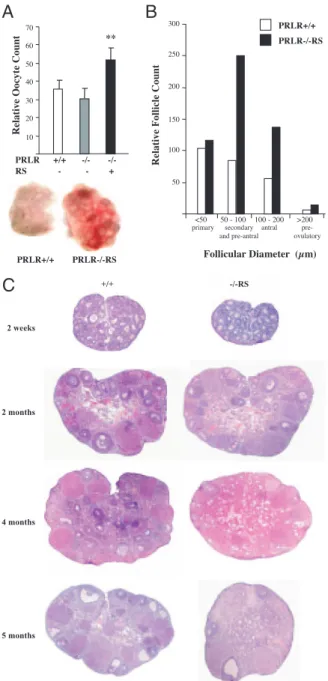
interest, however, was our finding that PRL signaling through RS profoundly impacts follicular survival. Early
during development, PRLR⫺/⫺RS ovaries are larger
than those of PRLR⫺/⫺, and when treated with human
chorionic gonadotropin (hCG), they ovulate a signifi-cantly greater number of oocytes than either the PRLR
null or the wild-type mice (Fig. 2A). At 2 months of age, the number of secondary, preantral, and antral follicles
is markedly increased in PRLR⫺/⫺RS ovaries (Fig. 2B),
indicating that premature follicular development oc-curs at an early age in these females. Interestingly, however, a severe follicular death begins from 4 wk of
age, and by the time these PRLR⫺/⫺RS females are 4
months old, the ovaries appear severely pathological (Fig. 2C). At this age, these females still cycle and accept the male, yet they cannot be superovulated (Fig. 3). Histological examination shows ovaries with severe follicular impairment and numerous holes that are the result of follicular death (Figs. 2C and 3C). In those follicles that are in the process of degeneration (Fig. 3D, upper panel), the mural granulosa cells are disorganized and the oocytes devoid of cumulus. Without the surrounding granulosa, the oocytes de-generate and lose their content. Finally, the zona pel-lucida collapses and remains surrounded by theca/
interstitial cells (Fig. 3D, upper panel). As PRLR⫺/⫺RS
females get older, (Figs. 2C and 3C), the ovaries are almost completely depleted of functional follicles and are formed mostly by theca and interstitial cells sur-rounding numerous holes containing collapsed zona pellucida that are the remnant of dead oocytes (Fig.
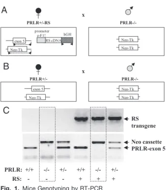
x PRLR+/-R S P RLR-/- ex on 5 Ne o- Tk p rom ot er e- F1 ! hG H RS c DNA Ne o- Tk Ne o- Tk
A
x PRLR+/ - P RLR-/- ex on 5 Ne o- Tk Ne o- Tk Ne o- TkB
RS transgene Neo cassette PRLR-exon 5 PRLR: +/+ -/- +/- +/+ -/- +/- RS: - - - + + +C
Fig. 1. Mice Genotyping by RT-PCR
A, Females expressing only the short form of the prolactin receptor (PRLR⫺/⫺RS) have been generated by mating PRLR⫹/⫺RS females with fertile PRLR⫺/⫺males. Arrows in-dicate hybridization site for the primers used for genotyping. B, PRLR⫺/⫺females were obtained by mating PRLR⫹/⫺ fe-males with PRLR⫺/⫺males. C, Homozygous, heterozygous, or null PRLR mRNA expression was detected by using prim-ers for exon 5 and for neo cassette on genomic DNA sam-ples. Expression of transgenic RS was detected by using primers for the terminal block of human GH in the eF1 ␣-PRLR-PR-1 transgenic construct.
3D, lower panel). This likely reflects widespread follic-ular initiation followed by follicfollic-ular cell death.
Although morphologically the theca/interstitial cells do not seem affected, expression of cytochrome P450
17␣-hydroxylase (P450c17), a key enzyme for
andro-gen biosynthesis, is completely absent in PRLR⫺/⫺RS
ovaries compared with PRLR⫺/⫺, indicating that
acti-vation of RS has also a negative impact on the steroi-dogenic capacity of the theca/interstitial cells (Fig. 4A, upper panel).
Early during development, PRLR⫺/⫺ as well as
PRLR⫺/⫺RS females ovulate and form CL of
preg-nancy; however, a regression of these CLs is seen within 2.5 d, and pregnancy cannot be sustained. Both genotypes show similar levels of apoptosis in the re-gressing CLs (Fig. 4A, lower panel). Subcutaneous
implantation of progesterone pellets in PRLR⫺/⫺and
PRLR⫺/⫺RS females allowed a partial rescue of
em-bryos with no significant differences between the two genotypes (Fig. 4B). Because similar expression of both isoforms were found in CL (7, 9), a role for RS on CL was suggested (9). However, the inability of the RS
to rescue the CL in pregnant PRLR⫺/⫺RS females
clearly establishes a key role for RL in the PRL main-tenance of a functionally progesterone-producing CL. The absence of RL expression and the inability to maintain the CL and pregnancy are probably the only
similarities between PRLR⫺/⫺ and PRLR⫺/⫺RS
ova-ries. In contrast to PRLR⫺/⫺RS, histological analysis of
PRLR⫺/⫺ovaries shows normal follicular development
(Fig. 3B), suggesting that the follicular defect
dis-played by PRLR⫺/⫺RS ovaries is due entirely to
acti-vation and signaling through RS. Interestingly, RS transgenic females that are wild type or heterozygous
for the PRLR gene (PRLR⫹/⫹RS or PRLR⫹/⫺RS) do
not present any sort of ovarian impairment. These Follicular Dia me ter (µ m)
PRLR+/ + PRLR-/-R S 50 100 150 200 250 300 prim ar y s ec ondary an d p re -a ntra l an tr al pre- ov ulat or y
B
C
Relative Follicle Count
Relative Oocy te Count 10 20 30 40 50 60 70 PRLR +/+ -/ - -/- **
A
RS - - + PRLR+/ + PRLR-/-RS <50 50 - 100 100 - 200 >200 2 w eek s +/ + - /-RS 2 m onth s 4 m onth s 5 m onth sFig. 2. PRL Signaling through RS Accelerates Follicular
Re-cruitment
A, Superovulation was induced with eCG and hCG in 2-month-old PRLR⫹/⫹, PRLR⫺/⫺, and PRLR⫺/⫺RS mice;
up-per panel, average number of released oocytes up-per female
(n⫽ 10 animals per group; *, P ⬍ 0.001); lower panel, gross morphology of PRLR⫹/⫹and PRLR⫺/⫺RS ovaries (note the difference in the size at 2 months of age). B, Follicular devel-opment in 2-month-old females: total primary (⬍50m), sec-ondary and preantral (50–100m), antral (100–200 m), and preovulatory (⬎200m) follicles. C, Ovarian histology at dif-ferent ages of cycling PRLR⫹/⫹(left) and PRLR⫺/⫺RS (right) ovaries. Sections were stained with hematoxylin-eosin.
PRLR+/ + PRLR-/ - PRLR-/-R S
A
theca/interstitial theca/interstitialB
C
D
Fig. 3. PRL Signaling through RS Has a Deleterious Effect
on Follicular Development
A–C, Ovarian histology of PRLR⫹/⫹(A), PRLR⫺/⫺(B), and PRLR⫺/⫺RS (C) of 4-month-old mice after superovulation with eCG and hCG. All sections are stained with hematoxylin-eosin. D, Ovarian histology of a cycling 5-month-old PRLR⫺/⫺RS female shows follicles in different stages of de-generation (upper panel). PRLR⫺/⫺RS ovaries 5 months and older are formed mostly by theca/interstitial cells surrounding holes that are remnants of dead follicles (lower panel). Halperin et al. • PRL Receptor Short and Premature Ovarian Failure Mol Endocrinol, February 2008, 22(2):513–522 515
females are fertile and have normal litters. Although they highly express RS, they display a normal follicular development (Fig. 4C), suggesting that the RL re-verses the detrimental effect of RS on follicular development.
Even though no difference in the serum PRL levels
was found between PRLR⫺/⫺ and PRLR⫺/⫺RS
fe-males (Fig. 4D), both groups presented high PRL lev-els as compared with wild-type females. These results are in agreement with data previously reported for the
high circulating PRL in PRLR⫺/⫺females (28) and
sup-port the finding that PRL through RL down-regulates its own synthesis and/or secretion at the hypothalamic and/or pituitary level (29).
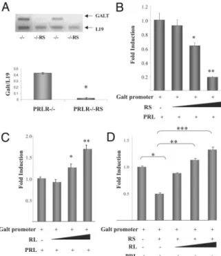
Galt Expression Is Repressed by PRL Signaling through RS
Microarray analysis performed with ovarian tissue shows that PRL through RS significantly regulates the expression of more than 80 genes that participate in different biological processes such as immune re-sponse, protein metabolism, transport, signal trans-duction, and cell communication (the entire list of
RS-regulated genes can be found in supplemental Table 1). This shows that PRL indeed signals through RS in the ovary and actively regulates the expression of several genes. Interestingly, Galt, whose mutation was shown to induce galactosemia and POF in women
(30), is down-regulated in PRLR⫺/⫺RS ovaries.
To further analyze the down-regulation of Galt by RS, we examined Galt mRNA levels in ovaries of
PRLR⫺/⫺ and PRLR⫺/⫺RS mice by semiquantitative
RT-PCR. As shown in Fig. 5A, Galt expression is
com-pletely abolished in ovaries of PRLR⫺/⫺RS females in
contrast to their PRLR null littermates (Fig. 5A). We also examined the ability of PRL to regulate Galt tran-scription in a subclone of UIII cells (31) that does not express any PRLR. Cells were transiently transfected with Galt promoter-reporter vector and RS expression vector. PRL treatment induced a marked decrease in Galt promoter activity, indicating clearly that PRL act-ing through RS represses the transcriptional activity of
PRLR+/-R S
C
PR LR +/ + P RL R- /- PR LR -/ -R S 1 100 10000 * * Seru mP RL leve l( ng/ml)D
B
* * PR LR +/ + P RL R- /- PR LR -/ -R S 0 2 4 6 8 10 numbe r o fe mbryos PRLR -/ - PRLR -/ - R S+A
PRLR -/ - PRLR -/ - R S+Fig. 4. Effect of PRL Signaling through RS on Theca, CL,
and Fetal Survival
A, Upper panel, Immunolocalization of P450c17 in 12-month-old PRLR⫺/⫺and PRLR⫺/⫺RS ovaries with immuno-reactivity shown in red and hematoxylin-counterstained nu-clei in blue; lower panel, apoptosis levels analyzed by terminal deoxynucleotide transferase-mediated nick end-la-beling in PRLR⫺/⫺and PRLR⫺/⫺RS CL. Reactivity is shown in
brown and hematoxylin-counterstained nuclei in blue B,
Number of fully developed embryos counted in uterus of PRLR⫹/⫹, PRLR⫺/⫺, and PRLR⫺/⫺RS progesterone-treated females at d 19.5 of pregnancy. Significance as compared with control (PRLR⫹/⫹) is indicated (*, P⬍ 0.05 by Dunnett’s multiple comparison post test). C, Ovarian histology of a 4-month-old PRLR⫹/⫹RS transgenic construct shows that by coexpressing RL, they display normal follicular development;
inset, detail of a preantral follicle. Sections are stained with
hematoxylin-eosin. D, Serum PRL concentration in PRLR⫹/⫹, PRLR⫺/⫺, and PRLR⫺/⫺RS virgin females. *, P⬍ 0.0001; n ⫽ 6 for each group.
Fig. 5. Activation of RS Represses Galt Expression and
Pro-moter Activity
A, Galt mRNA levels were measured by RT-PCR in PRLR⫺/⫺and PRLR⫺/⫺RS ovaries; L19 was used for loading control (top). Densitometric analysis (bottom) shows a signif-icant decrease in Galt mRNA levels in PRLR⫺/⫺RS vs. PRLR⫺/⫺ovaries: *, P ⬍ 0.001, t test. B, Full-length Galt promoter-reporter construct was transfected together with increasing concentrations of RS expression vector in UIII cells. Promoter activity was inhibited by increasing concen-trations of RS. C, Galt promoter activity is stimulated by PRL in cells expressing increasing concentrations of RL. D, Ex-pression of RL reverses the inhibitory effect of PRL through RS on Galt promoter activity in a concentration-dependent manner. Significance as compared with control is indicated: *, P⬍ 0.05; **, P ⬍ 0.01; ***, P ⬍ 0.001 by Dunnett’s multiple comparison post test.
this enzyme (Fig. 5B). Remarkably, when these cells were transfected with increasing doses of RL expres-sion vector, a clear RL dose-dependent up-regulation of the Galt promoter was observed after PRL treat-ment (Fig. 5C). Moreover, RS-mediated repression of Galt transcription is reversed by expression of RL (Fig. 5D).
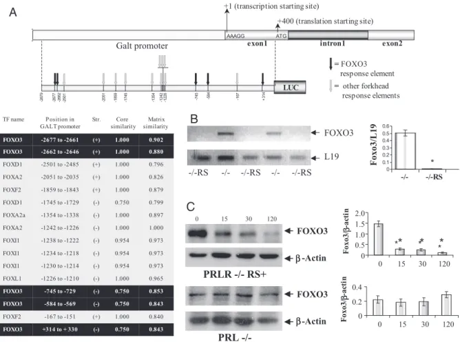
FOXO3 Expression Is Repressed by PRL Signaling through RS
The analysis of the full-length mouse Galt promoter sequence revealed 16 putative forkhead transcription factor sites, five of them being FOXO3 sites (Fig. 6A). This attracted our attention because deletion of FOXO3 gene causes an ovarian defect similar to that
seen in PRLR⫺/⫺RS mice (25, 26) as well as in women
with Galt mutation (24). This finding together with the fact that FOXO3 regulates transcriptional activity of
genes involved in glucose metabolism (32) led us to examine whether FOXO3 is repressed by PRL through RS, and whether FOXO3 regulates Galt transcription. As shown in Fig. 6B, FOXO3 is profoundly repressed
at mRNA level in the ovaries of PRLR⫺/⫺RS females as
compared with their PRLR⫺/⫺littermates.
To further examine the PRL-mediated inhibition of
FOXO3, PRLR⫺/⫺RS females were sc injected with
100 l CB-154 (1 g/l 70% ethanol) to block the
endogenously produced PRL. Six hours later, they
were injected ip with 100l PRL (60 g/100 l saline),
and ovaries were isolated at different times thereafter. Results shown in Fig. 6C (first lane) indicate that
FOXO3 is highly expressed in the PRLR⫺/⫺RS ovaries
6 h after CB-154 treatment and before PRL adminis-tration. Injection of PRL induced a drop in FOXO3 protein, and within 2 h of PRL treatment, FOXO3 ex-pression was almost completely inhibited.
0. 843 0. 75 0 (-) + 314 to + 3 30 FO XO 3 0. 840 1. 00 0 (+ ) - 167 to - 151 FO XF 2 0. 843 0. 75 0 (-) - 584 to - 569 FO XO 3 0. 853 0. 75 0 (-) - 745 to - 729 FO XO 3 0. 965 1. 00 0 (-) - 1226 to - 1210 FO XL 1 0. 973 0. 95 4 (-) - 1230 to - 1214 FO XI 1 0. 973 0. 95 4 (-) - 1234 to - 1218 FO XI 1 0. 973 0. 95 4 (-) - 1238 to - 1222 FO XI 1 1. 000 1. 00 0 (-) - 1242 to - 1226 FO XA 2 0. 897 1. 00 0 (-) - 1354 to - 1338 FO XA 2a 0. 799 0. 75 0 (-) - 1745 to - 1729 FO XD 1 0. 879 1. 00 0 (+ ) - 1859 to - 1843 FO XF 2 0. 826 1. 00 0 (+ ) - 2051 to - 2035 FO XA 2 0. 796 1. 00 0 (+ ) - 2501 to - 2485 FO XD 1 0. 880 1. 00 0 (+ ) - 2662 to - 2646 FO XO 3 0. 902 1. 00 0 (+ ) - 2677 to - 2661 FO XO 3 Ma tr ix similarity Co re simila ri t y St r. P o s itio n i n
PR L - /-
FOXO 3 β-A ctin 0 0.2 0.4 0 15 30 120 F oxo3/ -ac ti n e xon 1 AAAG G ATG in tr on 1 exon 2 +1 (t ra ns cr ip ti on st ar ti ng si te ) + 400 (t ra ns la ti on st ar ti ng si te ) Galt promoter = ot he r forkhe ad re sp ons e elemen ts = F OXO3 re sp ons e elemen t -2 677 -2 662 -7 45 -5 84 + 314 -2 879 -1 67 LU C -2 051 -1 859 -1 745 -1 354 -1 242 -1 226 -2 501A
C
12 0PRLR -/ - R S+
FOXO 3 -A ctin * * * *B
L1 9 FOXO 3 -/ - -/ - -/-R S -/-R S -/-RS 0 0. 1 0. 2 0. 3 0. 4 0. 5 0. 6 -/ - -/- RS Foxo3/L1 9 0 0.5 1.0 1.5 2.0 0 15 30 120 F oxo3/ -ac ti n*
*
*
β ββ β ββ βFig. 6. Activation of RS Represses FOXO3 Activity
A, Schematic diagram shows putative FOXO3 response elements found in the⫺2879/⫹391-bp Galt promoter sequence (black
arrows). Other forkhead response elements are indicated with gray arrows. The position of each forkhead response element as
well as their core similarity is indicated in the table. B, FOXO3a mRNA levels were measured by RT-PCR in PRLR⫺/⫺ and PRLR⫺/⫺RS ovaries; L19 was used for loading control. Densitometric analysis (right) shows a significant decrease in FOXO3 mRNA levels in PRLR⫺/⫺RS vs. PRLR⫺/⫺ovaries: *, P⬍ 0.001, t test. C, FOXO3 protein levels were analyzed by Western blot in ovaries of PRLR⫺/⫺RS and PRL⫺/⫺females injected with PRL for 0, 15, 30, and 120 min.-Actin was used as an internal loading control. Densitometric analysis shows that whereas PRL causes in vivo a significant decrease in FOXO3 expression in PRLR⫺/⫺RS ovaries (top right; *, P⬍ 0.001 by Dunnett’s multiple comparison post test), it has no inhibitory effect on FOXO3 protein levels in PRLR⫺/⫺ovaries (bottom right, P⬎ 0.5).
We examined whether PRL can repress FOXO3 in ovaries expressing both RS and RL. For this experi-ment, PRL null mice were used because they express both types of receptors but do not produce PRL. As shown in Fig. 6C, PRL has no detectable effect on FOXO3 protein levels in the ovaries of these mice. This suggests that expression of RL may prevent the down-regulation of FOXO3 induced by PRL signaling through RS.
FOXO3 Enhances Galt Transcription
To evaluate the role of FOXO3 as a regulator of Galt transcription, the full-length mouse Galt promoter was transfected into HepG2 cells in the presence or ab-sence of either wild-type or constitutively active FOXO3, also known as triple-mutant nonphosphory-latable FOXO3 (33). As shown in Fig. 7A, both FOXO3 expression vectors up-regulate Galt promoter activity.
A serial 5⬘-deletion of Galt promoter revealed that the
essential site for FOXO3 stimulation is located
be-tween ⫺613 and ⫹21 bp, a region that contains a
putative FOXO3 site at⫺584 bp (Fig. 7B). Surprisingly,
mutation of ⫺584-bp FOXO3 response element did
not prevent FOXO3-induced stimulation of the pro-moter. To examine whether this putative FOXO3 site binds to its cognate transcription factor, EMSAs were performed using oligonucleotides containing the ⫺584-bp FOXO3 response element either intact or mutated. The sequence of the FOXO3 consensus binding site-containing oligonucleotide from the IGF-binding protein IGFBP-1 gene [termed insulin-respon-sive sequence (IRS)] was used as a positive control (33). Incubation of the oligonucleotides with nuclear extract from HepG2 cells revealed the formation of two complexes (Fig. 7C). Competition with excess unla-beled wild-type oligonucleotides and supershift with anti-FOXO3 antibody indicate the specificity of FOXO3 binding to Galt promoter. Interestingly, mutation of the ⫺584-bp FOXO3 response element did not prevent the binding of this transcription factor to this piece of DNA, which suggests that FOXO3 could be stimulating Galt promoter either by binding to a novel response element not yet reported or by associating with a cofactor that binds to Galt promoter, stimulating its activity. In either case, it is clear from these results that FOXO3 enhances Galt transcriptional activity.
DISCUSSION
In the present study, we report that mice expressing only RS, specifically the PR-1 isoform of the receptor, have a severe ovarian impairment. From the three short isoforms reported in mouse (8), PR-1 has the highest homology with the rat and, more importantly, is the one that shows specific binding of PRL and mitogenic responsiveness (20). Whether the other two short isoforms have the same deleterious effect on the ovary remains to be investigated.
Our data show that PRLR⫺/⫺RS ovaries have a
pre-mature follicular development followed by massive fol-licular cell death. Although the signaling pathway downstream of RS still needs to be determined, we clearly show that PRL signaling through RS represses FOXO3 and Galt, which are important for normal fol-licular development. Normally, Galt is highly ex-pressed in ovaries and liver, which are the major sites of expression for this enzyme (34). Galt participates in the metabolism of galactose to glucose (35). Defi-ciency in Galt activity leads to accumulation of galac-tose metabolites, which causes ovarian toxicity (27, 34). This toxicity is explained by the synergism of two metabolites, galactose-1-phosphate and galactitol. Accumulation of galactose-1-phosphate is thought to inhibit enzymes involved in glucose metabolism, lead-ing to deficient glycosylation reactions and decrease in energy production in the ovarian cells. As to galac-titol, it is a galactose metabolite that cannot pass through the cellular membrane, and its accumulation into the cells causes an osmotic disequilibrium that
C
B
A
Fig. 7. FOXO3 Binds to Galt Promoter and Stimulates Its
Transcriptional Activity
A, The full-length Galt promoter-reporter construct was transfected in HepG2 cells together with either wild-type FOXO3 expression vector (WT-FOXO3) or with constitutively active vector (CA-FOXO3). Significance as compared with control is indicated: *, P⬍ 0.01 by Dunnett’s multiple com-parison post test. B, 5⬘ serial deletions of Galt promoter show that the essential region for the stimulatory effect of FOXO3 expression on the luciferase activity is located between⫺613 bp and⫹21 bp. *, P ⬍ 0.001. C, Protein-DNA complexes were analyzed by EMSA using nuclear extract from HepG2 cells and⫺596/-565-bp oligonucleotide from the Galt pro-moter containing either the intact (⫺584) or the mutated FOXO3 (m-584) binding site located at⫺584 bp. IRS oligo-nucleotide was used as a positive control for the FOXO3 binding (arrowheads) and supershift (arrow).
leads to water influx and ultimately to cell death. In women, either mutations in Galt gene or a deficiency in enzyme activity causes a disease known as galac-tosemia associated with POF (27, 30, 34). Young women with this disease are fertile early in life but become sterile in their late 20s, displaying ovaries formed by interstitial cells and devoid of follicles
sim-ilarly to those seen in PRLR⫺/⫺RS mice. Interestingly,
we found the expression of Galt to be down-regulated by PRL signaling through RS. The analysis of the con-sensus sites in the Galt promoter pinpointed FOXO3 as a possible modulator of Galt transcription. Indeed, our data show that FOXO3 markedly enhances Galt promoter activity. It has been reported that FOXO3 null mice exhibit premature follicular development at an early age followed by severe follicular death leading to ovaries formed by theca/interstitial cells surrounding collapsed zona pellucida (25, 26). In fact, the reported ovarian histology of FOXO3 null mice resembles that of RS-expressing mice at an older age (Fig. 3D). This association between FOXO3 and Galt may provide an explanation for the severe follicular death seen in the FOXO3 null mice. It has been suggested that FOXO3 plays a decisive role in controlling follicular activation and early development. Overexpression of this tran-scription factor in oocytes causes retarded oocyte growth and follicular development (36), whereas the FOXO3 null females exhibit excessive activation of primordial follicles followed by massive cell death (25, 26). In addition to regulating enzymes involved in glu-cose metabolism, FOXO3 is well known to stimulate genes involved in apoptosis (reviewed in Ref. 37). However, deletion of FOXO3 gene showed an increase in ovarian cell death rather than inhibition in the apo-ptotic process (25, 26). Because Galt is up-regulated by FOXO3, deletion of this transcription factor may have decreased Galt expression in the FOXO3 null ovary, leading to follicular death by galactose toxicity. We also show in this report that RL can prevent PRL signaling through RS and protect the ovary from the deleterious effect of such signaling. The marked inhi-bition of Galt promoter activity in cells expressing RS is clearly reversed by coexpression of RL. In addition, no down-regulation of FOXO3 and Galt is found when RL is coexpressed with RS, and the ovarian defect seen in ovaries expressing only RS is no longer ob-served in RS transgenic mice on either heterozygous or wild-type background. In fact, in wild-type animals, both isoforms are expressed in the ovaries, and their ratio varies along the estrous cycle, suggesting that the coexpression of both receptors is important for the normal physiological development of the ovarian folli-cles. Indeed, RL is expressed at much higher levels than RS in the growing follicles (9), and this may pre-vent signaling through RS.
POF is a common cause of infertility and premature aging in women, with an estimated 1% incidence; however, the vast majority of cases of POF are idio-pathic. Although it is not yet clear how PRL signals in human ovary, the possibility that this disease may be
due, in some patients, to a failure of the RL is intriguing and deserves further investigation. We have generated a mouse model for this type of POF and provide an explanation as to how PRL acting through its short cognate receptor can lead to POF.
Our findings that activation of RS by PRL represses FOXO3 and Galt and that FOXO3 stimulates Galt tran-scriptional activity provide an interesting and novel link between the POF seen in mice expressing RS and mice with FOXO3 gene deletion and in women with Galt mutation.
MATERIALS AND METHODS
Animal Model
RS transgenic females were originally generated by microin-jecting the eF1␣-PRLR-PR-1 transgenic construct encoding the mouse cDNA for RS into fertilized PRLR⫹/⫺ oocytes derived from 129 Sv pure background mice (20). This con-struct is driven by the elongation factor 1 promoter, which makes it ubiquitously expressed in the tissues along all stages of development. These PRLR⫹/⫺RS females are fer-tile, and by overexpressing the RS, they can rescue the mammary development defect displayed by PRLR⫹/⫺ fe-males (20).
For the present study, we have generated females ex-pressing only the short form of the prolactin receptor (PRLR⫺/⫺RS) by mating PRLR⫹/⫺RS females with fertile PRLR⫺/⫺males. The PRLR⫺/⫺ females were obtained by mating PRLR⫹/⫺females with PRLR⫺/⫺males.
Animals were identified by RT-PCR on genomic DNA pu-rified from tail using direct PCR lysis reagent (Viagen Biotech, Inc., Los Angeles, CA) (Fig. 1). For PRLR gene expression, the forward primers were 5⬘-GAA GAG CAA GAT CTC AAG AAC-3⬘ for the wild type and 5⬘-CCA GTC CCT TCC CGC TTC AGT-3⬘ for the mutated (Neo) strand, and the reverse primer was 5⬘-GAG AAA AAC ACC TAT GAA TGT-3⬘. For RS transgenic expression, forward 5⬘-AAG TTC GAC ACA AAC TCA CA-3⬘ and reverse 5⬘-ACT GAG TGG ACC CAA CGC AT-3⬘ primers for the human GH terminator present in the eF1␣-PRLR-PR-1 transgenic construct were used (Fig. 1). The cycling parameters for the PRLR consisted of one cycle of 94 C for 5 min and then 35 cycles of 94 C for 45 sec, 55 C for 1 min, and 72 C for 45 sec followed by a single cycle of 5 min at 72 C for extension. The molecular size for the wild-type product is 350 and 580 bp for the mutant. For the RS trans-genic construct, the cycling parameters were 94 C for 5 min and then 30 cycles of 94 C for 1 min, 64 C for 1 min, and 72 C for 1 min followed by a single cycle of 5 min at 72 C for extension, and the molecular size is 750 bp. RT-PCR prod-ucts were electrophoresed on a 1% agarose gel using 100-bp PCR markers (Invitrogen, Carlsbad, CA) as standards to determine the molecular size.
Animals were kept under conditions of controlled light (0700–1900 h) and temperature (22–24 C) with free access to standard rodent chow and water.
Experimental Animals
All experimental procedures were performed in accordance with the Guidelines of the National Institutes of Health Guide for the Care and Use of Laboratory Animals and were ap-proved by the Institutional Animal Care and Use Committee. Halperin et al. • PRL Receptor Short and Premature Ovarian Failure Mol Endocrinol, February 2008, 22(2):513–522 519
Tissue Preparation and Histology
For histological analysis, cycling females at different ages were killed at estrus. The ovaries were dissected and either frozen in liquid nitrogen for RNA and protein extraction or fixed either in Bouin or 10% formalin for histological exami-nation. Tissues were serially sectioned (5m) and stained with hematoxylin-eosin. Follicular counting was performed in all sections of each ovary. Follicles that contained oocytes with clearly visible nuclei were scored, and the total number of follicles at any particular developmental stage was calcu-lated as the sum of follicles from all sections of an ovary.
To examine whether the interstitial/thecal tissue left in the ovaries of mice expressing only RS express P450c17, ovarian sections were incubated overnight at 4 C with a primary polyclonal antibody to P450c17 and then incubated with a secondary biotinylated goat antirabbit IgG according to the manufacturer’s instructions (Vectastain ABC kit; Vector Lab-oratories, Burlingame, CA). Peroxidase activity was devel-oped with Nova Red solution (Vector)
Apoptosis levels were measured by terminal deoxynucle-otide transferase-mediated nick end-labeling using an ApopTag Peroxidase In Situ kit (Chemicon International, Te-mecula, CA) according to the manufacturer’s manual.
Superovulation Protocol and Progesterone Pellet Implantation
To induce superovulation, PRLR⫹/⫹, PRLR⫺/⫺, and PRLR⫺/⫺RS female mice were injected ip with 5 IU preg-nant mare serum [equine chorionic gonadotropin (eCG)] (Sigma Chemical Co., St. Louis, MO) followed by 5 IU hCG (Sigma) 48 h later. Ovaries were then isolated for histolog-ical examination. The difference in the number of released oocytes was determined by one-way ANOVA followed by Dunnett’s multiple comparisons post test that allows com-parisons against a control (wild type).
To determine whether PRLR⫺/⫺and PRLR⫺/⫺RS females can maintain pregnancy, they were mated with fertile PRLR⫹/⫹males, and the day that a vaginal plug was found, a progesterone pellet (25 mg; Innovative Research of Amer-ica, Sarasota, FL) was sc implanted. These females were maintained until the time of normal parturition. The difference in the number of fully developed embryos for each genotype was determined by one-way ANOVA followed by Dunnett’s multiple comparisons post test.
PRL Hormone Assay
After anesthesia, retroorbital blood samples were taken from cycling PRLR⫺/⫺, PRLR⫺/⫺RS, and PRLR⫹/⫹females, and PRL levels were measured by RIA, at the National Hormone and Pituitary Program, Harbor-UCLA Medical Center, Tor-rance, CA. The statistical differences were determined by one-way ANOVA followed by Dunnett multiple comparisons post test.
RNA Extraction and RT-PCR
RNA was extracted using TRIzol reagent (Life Technologies, Rockville, MD) following the manufacturer’s protocol. Ovarian RNA from 2-month-old females was transcribed into cDNA by superscript polymerase II. Custom oligonucleotide prim-ers were obtained from Life Technologies and used to amplify the appropriate cDNA templates by PCR. Mouse Galt, FOXO3, and L19 mRNA expression was detected using 5 ⬘-CAG TAC CCT TGG GTG ⬘-CAG AT-3⬘ (forward), 5⬘-TGG TTA GGA CCA GAC GTT CC-3⬘ (reverse); 5⬘-GTC ATG GGC CAC GAT AAG TT-3⬘ (forward), 5⬘-GGG CTG CTA ACA GTC TCT GC-3⬘ (reverse); and 5⬘-AGC GCC TCC AGG CCA AGA AGG-3⬘ (forward), 5⬘-CCA GGC CGC TAT GTA CAG ACA
CGA-3⬘ (reverse) primers, respectively. PCR product size for Galt, FOXO3, and L19 were 217, 400, and 100 bp, respectively.
Conditions for each template were optimized so that sig-nals were in the linear range of detection. The PCR products with DNA loading buffer were then separated by gel electro-phoresis on a 0.7% agarose gel. L19 concentrations were used as internal control for comparison.
Western Blot Analysis
Total ovary lysates were prepared by homogenizing the tis-sues in RIPA buffer (1⫻ PBS, 1% Nonidet, 0.5% sodium deoxycholate, 0.1% SDS) containing 1 M sodium or-thovanadate, 10g/ml phenylmethylsulfonyl fluoride, and 30 l/ml aprotinin. Proteins were resolved on 8.5% denaturing polyacrylamide. After gel electrophoresis, proteins were elec-trophoretically transferred to a polyvinylidene difluoride membrane (Millipore Corp., Billerica, MA). The blots were incubated 1 h at room temperature with 5% nonfat dry milk in Tris-buffered saline (pH 8.0) containing 0.1% Tween 20. Blots were washed and incubated overnight at 4 C with the FOXO3 polyclonal antibody (1:1000 dilution; Upstate Biotechnology, Lake Placid, NY) and then incubated with a secondary anti-body linked to horseradish peroxidase for 1 h at room tem-perature.-Actin (Abcam Inc., Cambridge, MA) was used as internal loading control. Complexes were visualized using the West Pico chemiluminescence detection kit (Pierce Biotech-nology, Inc., Rockford, IL).
Microarray Analysis
Total RNA was extracted from 2-month-old PRLR null (con-trol) and PRLR⫺/⫺RS ovaries using Atlas Glass Total RNA Isolation Kit and reverse-transcribed with PowerScript re-verse transcriptase (BD Biosciences, San Diego, CA). cDNA was labeled with [␣-33P]dATP (Amersham, Piscataway, NJ),
purified using the Atlas NucleoSpin, and hybridized overnight at 60 C with Atlas plastic mouse 5K oligo microarrays mem-branes (BD Biosciences) carrying cDNA probes for approxi-mately 5000 known mouse genes according to the manufac-turer’s instructions. Membranes were washed and exposed to a phosphorimaging screen overnight. The intensity of spots was analyzed using Atlas Image 2.7 and Atlas Naviga-tor 2.0 software. The intensity of each gene was averaged from two individual spots. A cDNA synthesis control was used as a positive control and for grid template alignment. The values were normalized using six housekeeping genes (ubiquitin, tyrosine 3-monooxygenase, ornithine decarboxyl-ase, glyceraldehyde-3-phosphate dehydrogendecarboxyl-ase, cytoplas-mic -actin, and 40S ribosomal protein S29). Genes were excluded if they were detected in only one spot or at levels near or below background. Differences over 2-fold in the intensity of the spots were considered significant.
Cell Lines and Culture
Human hepatic carcinoma cells (HepG2) were obtained from American Type Culture Collection (Manassas, VA) and cul-tured in Eagle’s MEM (with Eagle’s balanced salt solution and
L-glutamine) supplemented with 10% fetal bovine serum, 1000 U/ml penicillin G, 2.5g/ml amphotericin B, 1000 g/ml streptomycin, 1 mMsodium pyruvate, and 1⫻ nonessential amino acids. Rat uterine stromal cells (UIII) were cultured in M199 medium (with phenol red and L-glutamine) supple-mented with 10% fetal bovine serum, 1000 U/ml penicillin G, 1000g/ml streptomycin, 1 mMsodium pyruvate, and 1⫻
nonessential amino acids. Cells were incubated in a humid-ified atmosphere of 5% CO2at 37 C.
Galt Promoter Reporter Constructs
The ⫹21/⫹342-bp Galt promoter-reporter truncation was cloned by PCR from genomic DNA using primers generated with the Primer3 software (supplemental Table 2). Restriction sites for SmaI and HindIII were added to the primers. PCR was performed using 1g mouse genomic DNA as a tem-plate. PCR product was cloned into pGEMTeasy (Promega, Madison WI) according to the manufacturer’s instructions. After sequencing, it was digested with SmaI and HindIII (dual digestion in OnePhor All Buffer; Amersham), purified using the GeneClean II kit (Qbiogene Inc., Irvine, CA) according to the manufacturer’s instructions, and subcloned in pGL3 ba-sic luciferase reporter vector (Promega).
To generate the ⫺613/⫹379-bp Galt promoter-reporter construct, the full-length promoter-construct was digested with VspI, Klenow blunt-ended (Invitrogen), purified by phenol extraction, digested with NcoI, and run in a 1.5% agarose gel. The construct was purified using the GeneClean II kit (Qbio-gene) and ligated in pGL3 basic luciferase reporter vector (Promega). Mutation of the putative FOXO3 response ele-ment located at⫺584 bp of this promoter-reporter truncation was created using the Stratagene (La Jolla, CA) QuikChange II kit according to the manufacturer’s instructions and con-firmed by sequence analysis after cloning into pGEMTeasy and subcloning into pGL3 basic. Four mutations (underlined letters) were introduced into the primer with three of them in the core sequence of the FOXO3 response element (5⬘-GGT GTG CAC CAC CAC TGG CCG TTC CAT CTA CTT TTA TAT AGA TTG GGC C-3⬘).
The⫺2879/⫹391-bp mouse Galt promoter sequence was scanned with the MatInspector professional 7.4 software (available at the website http://www.genomatix.de/) using 0.75 for the core similarity and 0.8 for the matrix similarity in the matrix group for vertebrates.
Transient Transfections and Constructs
HepG2 cells were cotransfected with either full-length, trun-cated, or mutated mouse Galt promoter-reporter construct in pGL3basic and either wild-type or constitutively active FOXO3 (CA-FOXO3) in pAltermax expression vector using CaPO4as transfection method. Cells were harvested 24 h
after transfection. In all transfections, the total amount of DNA was balanced with the appropriate empty vector. Each experiment was performed at least three times in triplicate. Data are expressed as means⫾SEMand analyzed by one- or two-way ANOVA.
UIII cells were cotransfected with Galt promoter-reporter construct and with either RL or RS in pcDNA expression vectors using Lipofectamine 2000 (Invitrogen) as transfection method. Immediately after transfection, cells were treated for 24 h with PRL (1g/ml) and harvested. Each experiment was performed at least three times in triplicate. Data are ex-pressed as means⫾SEMand analyzed by two-way ANOVA.
EMSA
To prepare nuclear extracts, HepG2 and UIII cells were scraped from 75-cm2 flasks with ice-cold PBS (Ca2⫹ and
Mg2⫹free) containing 1⫻ proteases inhibitor cocktail (Sigma) and pelleted. The pellet was resuspended for 10 min in 1 ml hypotonic buffer RBS [10 mMNaCl, 3 mMMgCl2, 10 mMTris
(pH 7.4), 0.5% Nonidet NP-40, and proteases inhibitors]. The tubes were vortexed for 30 sec and centrifuged at 3000⫻ g for 5 min. The resulting pellet was resuspended in 50 l extraction buffer C [420 mMKCl, 20 mMHEPES (pH 7.9), 1.5 mM MgCl2, 0.2 mM EDTA, 20% glycerol, and proteases
inhibitors), rocked at 4 C for 20 min, and incubated on ice for 30 min. The supernatants were aliquoted and frozen at ⫺80 C.
For the EMSA, IRS-containing oligonucleotide (5⬘-ATT GCT AGC AAG CAA AAC AAA CCG CTA GCT TA-3⬘) as well as wild-type and mutated⫺584-bp FOXO3 putative binding site-containing oligonucleotides (5⬘-CAC TGG CCG TTT GGT TTA CTT TTA TAT AGA TT3-⬘ and 5⬘-CAC TGG CCG TTC
CAT CTA CTT TTA TAT AGA TT-3⬘, respectively, with the
underlined letters showing the mutation positions) were end-labeled with [␥-32P]ATP (Amersham). Nuclear extracts (1g)
were incubated for 30 min in binding buffer together with 1⫻ 105cpm labeled oligonucleotides. Six micrograms FOXO3
antibody (Upstate) were added to binding buffer for the su-pershift. Samples were electrophoresed for 2.5 h on 4% nondenaturing polyacrylamide gels. Gels were dried and ex-posed to x-ray film for 18–48 h.
Acknowledgments
We thank P. A. Kelly for helpful comments, N. D. Horseman for the PRL null mice, D. Linzer for the FOXO3 construct, B. Hales for the P450c17 polyclonal antibody, and K. Heretis for excellent technical help.
Received August 20, 2007. Accepted October 25, 2007. Address all correspondence and requests for reprints to Geula Gibori, Ph.D., 835 South Wolcott, M/C 901, Chicago, Illinois 60612. E-mail: ggibori@uic.edu.
This work has been supported by National Institutes of Health HD11119, U54 HD 40093, and HD 12356 (G.G.), In-stitut National de la Sante´ et de la Recherche Me´dicale (N.B.), and an American Physiological Society Postdoctoral Fellow-ship in Physiological Genomics (J.H.).
Disclosure Summary: The authors have nothing to disclose.
REFERENCES
1. Bole-Feysot C, GoffinV, Edery M, Binart N, Kelly PA 1998 Prolactin (PRL) and its receptor: actions signal transduc-tion pathways and phenotypes observed in PRL receptor knockout mice. Endocr Rev 19:225–268
2. Freeman ME, Kanyicska B, Lerant A, Nagy G 2000 Prolactin: structure, function, and regulation of secretion. Physiol Rev 80:1523–1631
3. Ben-Jonathan N, Mershon JL, Steinmetz RW 1996 Ex-trapituitary prolactin: distribution, regulation, functions, and clinical aspects. Endocr Rev 17:639–669
4. Prigent-Tessier A, Tessier C, Hirosawa-Takamori M, Boyer C, Ferguson Gottschall S, Gibori G 1999 Rat de-cidual prolactin. Identification, molecular cloning, and characterization. J Biol Chem 274:37982–37989 5. Hugo ER, Brandebourg TD, Comstock CE, Gersin KS,
Sussman JJ, Ben-Jonathan N 2006 LS14: a novel human adipocyte cell line that produces prolactin. Endocrinol-ogy 14:306–313
6. Davis JA, Linzer DI 1989 Expression of multiple forms of the prolactin receptor in mouse liver. Mol Endocrinol 3:674–680
7. Telleria CM, Parmer TG, Zhong L, Clarke DL, Albarracin CT, Duan WR, Linzer DIH, Gibori G 1997 The different forms of the prolactin receptor in the rat corpus luteum: developmental expression and hormonal regulation in pregnancy. Endocrinology 138:4812–4820
8. Clarke DL, Arey BJ, Linzer DIH 1993 Prolactin receptor messenger ribonucleic acid expression in the ovary dur-ing the rat estrous cycle. Endocrinology 133:2594–2603 9. Russell DL, Richards JS 1999 Differentiation-dependent prolactin responsiveness and STAT (signal transducers Halperin et al. • PRL Receptor Short and Premature Ovarian Failure Mol Endocrinol, February 2008, 22(2):513–522 521
and activators of transcription) signaling in rat ovarian cells. Mol Endocrinol 13:2049–2064
10. Buck K, Vanek M, Groner B, Ball RK 1992 Multiple forms of prolactin receptor messenger ribonucleic acid are spe-cifically expressed and regulated in murine tissues and the mammary cell line HC11. Endocrinology 130: 1108–1114
11. Frasor J, Brakai U, Zhong L, Fazleabas AT, Gibori G 2001 PRL-induced ER␣ gene expression is mediated by Janus kinase 2 (Jak2) while signal transducer and activator of transcription 5b (Stat5b) phosphorylation involves Jak2 and a second tyrosine kinase. Mol Endocrinol 15: 1941–1952
12. Berlanga JJ, Fresno Vara JA, Martin-Perez J, Garcia-Ruiz JP 1995 Prolactin receptor is associated with c-src ki-nase in rat liver. Mol Endocrinol 9:1461–1467
13. Fresno Vara JA, Caceres MA, Silva A, Martin-Perez J 2001 Src family kinases are required for prolactin induc-tion of cell proliferainduc-tion. Mol Biol Cell 7:2171–2183 14. Tessier C, Prigent-Tessier A, Ferguson-Gottschall S,
Gu Y, Gibori G 2001 PRL antiapoptotic effect in the rat decidua involves the PI3K/protein kinase B-medi-ated inhibition of caspase-3 activity. Endocrinology 9:4086–4094
15. Nemeth E, Bole-Feysot C, Tashima LS 1998 Suppression subtractive hybridization (SSH) identifies prolactin stim-ulation of p38 MAP kinase gene expression in Nb2 T lymphoma cells: molecular cloning of rat p38 MAP ki-nase. Mol Cell Endocrinol 20:151–156
16. Lesueur L, Edery M, Ali S, Paly J, Kelly PA, Djiane J 1991 Comparison of long and short forms of the prolactin receptor on prolactin-induced milk protein gene tran-scription. Proc Natl Acad Sci USA 88:824–828 17. Berlanga JJ, Garcia-Ruiz JP, Perrot-Applanatt M, Kelly
PA, Edery M 1997 The short form of the prolactin (PRL) receptor silences PRL induction of the -casein gene promoter. Mol Endocrinol 11:1449–1457
18. Perrot-Applanatt M, Gualillo O, Pezet A, Vincent V, Edery M, Kelly PA 1997 Dominant negative and cooperative effects of mutant forms of prolactin receptor. Mol Endo-crinol 11:1020–1032
19. Saunier E, Dif F, Kelly PA, Edery M 2003 Targeted ex-pression of the dominant-negative prolactin receptor in the mammary gland of transgenic mice results in im-paired lactation. Endocrinology 144:2669–2675 20. Binart N, Imbert-Bollore´ P, Baran N, Viglietta C, Kelly PA
2003 A short form of the prolactin (PRL) receptor is able to rescue mammopoiesis in heterozygous PRL receptor mice. Mol Endocrinol 17:1066–1074
21. Stocco C, Telleria C, Gibori G 2006 The molecular control of corpus luteum formation, function, and regression. Endocr Rev 28:117–149
22. Risk M, Gibori G 2001 Mechanisms of luteal cell regula-tion by prolactin. In: Horseman ND, ed. Prolactin. Boston: Kluwer; 265–295
23. Ormandy CJ, Camus A, Barra J, Damotte D, Lucas B, Buteau H, Edery M, Brousse N, Babinet C, Binart N, Kelly
PA 1997 Null mutation of the prolactin receptor gene produces multiple reproductive defects in the mouse. Genes Dev 11:167–178
24. Grosdemouge I, Bachelot A, Lucas A, Baran N, Kelly PA, Binart N 2003 Effects of deletion of the prolactin receptor on ovarian gene expression. Reprod Biol Endocrinol 1:12 25. Castrillon DH, Miao L, Kollipara R, Horner JW, DePinho RA 2003 Suppression of ovarian follicle activation in mice by the transcription factor Foxo3a. Science 301:215–218 26. Hosaka T, Biggs WH, Tieu D, Boye AD, Varki NM, Cave-nee WK, Arden KC 2004 Disruption of forkhead tran-scription factor (FOXO) family members in mice reveals their functional diversification. Proc Natl Acad Sci USA 101:2975–2980
27. Forges T, Monnier-Barbarino P, Leheup B, Jouvet P 2006 Pathophysiology of impaired ovarian function in galactosaemia. Hum Reprod Update 12:573–584 28. Binart N, Helloco C, Ormandy CJ, Barra J,
Cle´ment-Lacroix P, Baran N, Kelly PA 2000 Rescue of preimplan-tatory egg development and embryo implantation in pro-lactin receptor-deficient mice after progesterone administration. Endocrinology 141:2691–2697
29. Schuff KG, Hentges ST, Kelly MA, Binart N, Kelly PA, Iuvone PM, Asa SL, Low MJ 2002 Lack of prolactin receptor signaling in mice results in lactotroph prolifera-tion and prolactinomas by dopamine-dependent and -independent mechanisms. J Clin Invest 110:973–981 30. Kaufman FR, Kogut MD, Donnell GN, Koch R, Goebels-mann U 1979 Ovarian failure in galactosaemia. Lancet 2:737–738
31. Prigent-Tessier A, Barkai U, Tessier C, Cohen H, Gibori G 2001 Characterization of a rat uterine cell line, U(III) cells: prolactin (PRL) expression and endogenous regulation of PRL-dependent genes; estrogen receptor, ␣2
-macro-globulin, and decidual PRL involving the Jak2 and Stat5 pathway. Endocrinology 142:1242–1250
32. Onuma H, Vander Kooi BT, Boustead JN, Oeser JK, O’Brien RM 2006 Correlation between FOXO1a (FKHR) and FOXO3a (FKHRL1) binding and the inhibition of basal glucose-6-phosphatase catalytic subunit gene transcription by insulin. Mol Endocrinol 20:2831–2847 33. Brunet A, Bonni A, Zigmond MJ, Lin MZ, Juo P, Hu LS,
Anderson MJ, Arden KC, Blenis J, Greenberg ME 1999 Akt promotes cell survival by phosphorylating and inhib-iting a Forkhead transcription factor. Cell 96:857–868 34. Liu G, Hale GE, Hughes CL 2000 Galactose metabolism
and ovarian toxicity. Reprod Toxicol 14:377–384 35. Leslie ND 2003 Insights into the pathogenesis of
galac-tosemia. Annu Rev Nutr 23:59–80
36. Liu L, Rajareddy S, Reddy P, Du C, Jagarlamudi K, Shen Y, Gunnarsson D, SelstamG, Boman K, Kiu K 2007 In-fertility caused by retardation of follicular development in mice with oocyte-specific expression of Foxo3a. Devel-opment 134:199–209
37. Greer EL, Brunet A 2005 FOXO transcription factors at the interface between longevity and tumor suppression. Oncogene 24:7410–7425
Molecular Endocrinology is published monthly by The Endocrine Society (http://www.endo-society.org), the foremost professional society serving the endocrine community.