HAL Id: hal-01592721
https://hal-amu.archives-ouvertes.fr/hal-01592721
Submitted on 6 Dec 2018
HAL is a multi-disciplinary open access
archive for the deposit and dissemination of
sci-entific research documents, whether they are
pub-lished or not. The documents may come from
teaching and research institutions in France or
abroad, or from public or private research centers.
L’archive ouverte pluridisciplinaire HAL, est
destinée au dépôt et à la diffusion de documents
scientifiques de niveau recherche, publiés ou non,
émanant des établissements d’enseignement et de
recherche français ou étrangers, des laboratoires
publics ou privés.
Distributed under a Creative Commons Attribution| 4.0 International License
A three-megabase yeast artificial chromosome Contig
spanning the C57BL mouse Igh locus
Christophe Chevillard, Jennifer Ozaki, Christopher D. Herring, Roy Riblet
To cite this version:
Christophe Chevillard, Jennifer Ozaki, Christopher D. Herring, Roy Riblet. A three-megabase yeast
artificial chromosome Contig spanning the C57BL mouse Igh locus. Journal of Immunology, Publisher :
Baltimore : Williams & Wilkins, c1950-. Latest Publisher : Bethesda, MD : American Association of
Immunologists, 2002, 168 (11), pp.5659-5666. �hal-01592721�
A Three-Megabase Yeast Artificial Chromosome Contig
Spanning the C57BL Mouse Igh Locus
1
Christophe Chevillard,* Jennifer Ozaki,
†Christopher D. Herring,
‡and Roy Riblet
2§The mouse Ig H chain (Igh) complex locus is composed of >100 gene segments encoding the variable, diversity, joining, and constant portions of the Ab H chain protein. To advance the characterization of this locus and to identify all the VHgenes, we have isolated the entire region from C57BL/6 and C57BL/10 as a yeast artificial chromosome contig. The mouse Igh locus extends approximately three megabases and contains at least 134 VHgenes classified in 15 partially interspersed families. Two non-Igh pseudogenes (Odc-rs8 and Rpl32-rs14) were localized in the distal part of the locus. This physical yeast artificial chromosome map will provide important structure and guidance for the sequencing of this large, complex, and highly repetitive locus. The Journal of Immunology, 2002, 168: 5659 –5666.
T
he Ig H chain locus, termed Igh in mice, encodes the H chains of Abs. It is comprised of adjacent clusters of gene segments for VH, DH, JH, and the different isotype H chainconstant regions (CH). To produce a secreted or cell surface receptor
Ab, a B cell must assemble an active V region gene by fusing a segment from each of the V, D, and J clusters. The overall nature of the structure and functioning of these genes is well established, but much remains to be explained. If we are to thoroughly understand, and be able to predict and manipulate, the functioning of the Ab re-sponse, we must obtain a complete description of the structural loci, both coding elements and regulatory sequences of Ab H and L chains. In the human IGH locus, this goal is nearly complete (1), but for experimental manipulation these loci must be characterized in the mouse. Recently, the mouse L chain locus was extensively de-scribed by Zachau and colleagues (2). All Igk constant, joining, and variable coding elements have been identified and sequenced. For the mouse H chain locus, the constant and joining gene segments were mapped and sequenced by pioneering work of Honjo and coworkers (3), and the diversity segments have been identified, mapped, and sequenced (4 – 6). The V region gene segments, Igh-V or VH, have
posed a larger challenge; estimates of their numbers range from hun-dreds to thousands (7–10). Genetic mapping experiments indicated that the locus is a centimorgan or larger (11–13), suggesting a se-quence size of several million base pairs of DNA, consistent with a gene content of hundreds to thousands of coding elements. Detailed studies of the organization of VHgene families by Brodeur and
col-leagues (14) have provided a better understanding but much remains to be elucidated.
We have undertaken several approaches to complete the char-acterization of mouse Igh; these will ultimately lead to
determi-nation of the DNA sequence of the entire locus. In this study, we describe the assembly of a yeast artificial chromosome (YAC)3 contig, or array of overlapping clones, that spans the Igh locus in the C57BL mouse strains, and the initial characterization of its size, physical structure, and gene content. To develop this YAC contig and resultant physical map of the mouse Igh locus, we screened four YAC libraries by PCR using multiple sequence-tagged sites (STSs) within the locus and identified 36 YACs. Sev-eral additional YACs were obtained from a fifth library through reference to Internet-posted data (15). YAC insert ends were iso-lated from all clones by vector-hexamer PCR (16) to characterize YAC overlap and assemble the contig, and to detect chimeric clones. This physical YAC map will provide important structure and guidance for the sequencing of this large, complex, and highly repetitive locus.
Materials and Methods
YAC libraries
The Princeton University mouse YAC library (Princeton, NJ) was prepared from C57BL/6J female mouse genomic DNA (17, 18). It consists of 26,000 individual pYAC4 vector clones in yeast host strain AB1380. The average clone length is 250 kb and the total estimated genome coverage is 2.2 haploid genomic equivalents. This library was screened by PCR using DNA pools from Dr. S. M. Tilghman at the Howard Hughes Medical Institute, Princeton University.
The first Whitehead Institute (WI-I) mouse YAC library (Cambridge, MA) was prepared from C57BL/6J female mouse DNA (19). It consists of two groups of pYAC4 vector clones in yeast host AB1380. The first group contains 4,100 clones with an average size of 480 kb, and the second contains 15,840 clones with an average size of 640 kb. The total estimated coverage for the whole library is 4.3 haploid genomic equivalents.
The second Whitehead Institute (WI-820) mouse YAC library was made from female C57BL/6J mouse DNA (20). It contains 38,400 clones with an average size of 820 kb, providing 10-fold coverage. All clones from this library are in the pRML vector in yeast host strain J57D. Extensive char-acterization of this library and identification of YACs bearing over 600 genetic loci is published (15). The Whitehead Institute libraries can be screened by pool PCR or membrane hybridization, and clones can be ob-tained from Research Genetics (Huntsville, AL).
The Saint Mary’s Hospital Medical School (London, U.K.) RAD52 mouse YAC library was prepared from C57BL/10 female mouse DNA (21). It contains 41,568 pYAC4 vector clones in the RAD52 mutant yeast host strain 3a with an average insert size of 240 kb, providing 3.5 genome equivalents. Clones from this library are available from the Mouse Genome
*Faculty of Medicine, Immunology and Genetics of Parasitic Diseases, Institut Na-tional de la Sante´ et de la Recherche Me´dicale, Marseille, France;†Division of Ge-netic Medicine, Department of Medicine, Vanderbilt University, Nashville, TN 37232;‡Department of Genetics, University of Wisconsin, Madison, WI 53706; and §Torrey Pines Institute for Molecular Studies, San Diego, CA 92121
Received for publication November 2, 2001. Accepted for publication March 28, 2002.
The costs of publication of this article were defrayed in part by the payment of page charges. This article must therefore be hereby marked advertisement in accordance with 18 U.S.C. Section 1734 solely to indicate this fact.
1This work was supported by National Institutes of Health Grant AI23548. 2Address correspondence and reprint requests to Dr. Roy Riblet, Torrey Pines Insti-tute for Molecular Studies, 3550 General Atomics Court, San Diego, CA 92121. E-mail address: rriblet@tpims.org
3Abbreviations used in this paper: YAC, yeast artificial chromosome; STS, sequence-tagged site; PFGE, pulsed field gel electrophoresis; BLAST, basic local alignment search tool.
Table I. Sequence tagged sites in the Igh locus a Name STS Primer 1 Primer 2 PCR Product Size (bp) PCR Conditions (mM/ °C) b GenBank Accession No. Primer Source/ Reference CH region 3⬘ IgA Enhancer ctcaaggttcgagttactcattctgtgca ctcaaggttcaggatttggagcacacctacag 376 1.5/60 X96607 24 c D12Rbt6 IgA aagaagtgctggtgcgatg actggtcaccctgtttccag 159 1.5/55 J00475 This study D12Rbt7 IgG1 ctgctgcccaaactaactcc cttgtccaccttggtgctg 242 1.5/55 J00453 This study D12Rbt8 IgG3 ggggccgcctattaattaaa tcctgctgtcctttgattcc 147 1.5/55 J00451 This study D12Nds4 IgD agacttattgtaccccacatgttg tatcttccaatcctagttaggc 160 1.5/55 K02138 25 JH region D12Rbt9 J3/J4 tggtgacaatttcagggtca gtctgactagaatcacccctgg 205 1.5/55 X63166 This study DH region DH FL16.1 gagcatgttgcaggaactga tcaaaattttccccaatagg 310 1.5/60 AF018146 — c VH region D12Rbt10 VH group III gaggtgcagctggtgg gctcacagtaactttyrctcactgtg 322 1.5/55 K02890 26, this study VH 11 atggagtgggaactgagctta catacagaaatacgtggctgtgtcc 461 1.5/55 M22438 27 c D12Nds2 VH S107 acatggtaatttatgggcaa ctggatacctgcaatagtaga 190 1.5/55 X03253 25 D12Rbt11 VH Ese26.1 aagctgggtgaagcagagaa gtcctcagatgtcaggctgc 169 1.5/55 S49538 This study D12Rbt12 VH Group II caggtccaactgcagcag gatgtggttgcaacactgtg 314 1.5/55 M60252 26, this study VH 15 caggttcacctacaacag ccttgcacagtagtagat 297 1.5/55 U39293 14 c D12Rbt13 MMIGV H 28 tgcattcatttttttaaaatgc atttgcctttccacaggttg 348 1.5/55 X06862 This study D12Rbt14 MMIGD10 ttccatgtccaaacgcagt tatgggtcaacagaatattgca 147 1.5/55 X04369 This study MMIGV H 23 gtgaaagtttccatcaatatc catgtatttagagattgtgct 103 1.5/55 X06856 28 VH G8 tatcctggtagcattactactaac tcttgcacaataatagaccgc 141 1.5/45 X60424 29 c VH 186.2 tgatgcaatattctgttgaccc agagtcctcagatgtcaggct 1011 1.5/65 L26851 10 D12Nds10 MMIGHVJ2 catgtttgcaatttaccatca catggatttacagatggagct 100 and 140 2.0/55 J00507 28 YAC ends D12Rbt1 yADGC9 left arm ccatggtgagagaagtttattctt gtttcccatttgacagcatg 103 1.5/55 B07528 This study D12Rbt2 yA8D10 left arm tctactttcactcaacccagaca gcagaaagacatccatgattaca 167 1.5/55 B07577 This study D12Rbt3 yA8D10 right arm cctaatcttttcttctccctctcc agatgtaggaggaggaggaagg 156 1.5/55 B07576 This study D12Rbt4 yFCDB1 right arm agcatattcagggcatcagg tgaattccagcattatccagc 123 1.5/60 B07536 This study D12Rbt5 yFCLA12 right arm attcagaaacggactcaaaagg gctgacaggatttctaccgc 93 1.5/55 B07543 This study Mit markers D12Mit41 tgcgttaatgggtctgatagg aattccaaaacaacagcatgc 220 1.5/55 33 D12Mit134 ctatctacaacaaacttctcctggg actcagtccaaacatatacaagatgc 184 1.5/55 33 D12Mit150 cttgtcaaaatttctgttgttttaca aaaggattttgtcactaagacatgg 171 1.5/55 33 D12Mit208 tttctttctgatggaaatactttga tcaaatgaccaaaattatagtgca 134 1.5/55 33 D12Mit263 tcagatctcagcagataaatacttgg tcccctggagcatatttgac 113 1.5/55 33 Redesigned Mit markers d D12Mit134Rbt caacaaacttctcctgggttg cccactcagtccaaacatataca 180 1.5/55 This study D12Mit150Rbt gctgagaatctcattgaatgtct aacaacagaaattttgacaagaaa 117 1.5/55 This study D12Mit263Rbt gtcccaagttttggtctagcc ggttccagtgcagatggaat 114 1.5/55 This study aIndicated for each STS are the name, site, primer sequences, size of the PCR product in C57BL/6, PCR conditions ((Mg 2 ⫹)/annealing temperature), GenBank accession, and literature reference. A subset of the STSs was used to screen the YAC libraries, and all were used to characterize the content of the isolated YAC clones. bMagnesium concentrations and annealing temperatures are listed; cycling parameters were 1⫻ (94 °C, 30 s); 30 ⫻ (94 °C, 30 s; annealing temperature, 60 s; 72 °C, 60 s). cThe following colleagues graciously provided primers: R. Lieberson, enhancer ␣ ; P . Brodeur, VH 15; M. Caul fi eld, VH G8; C. Carmack, DH FL16.1 and VH 11. dThese PCR markers were redesigned from MIT sequence data (http://www-genome.wi.mit.edu/) to avoid the microsatellite CA repeat (D12Mit150 and -26 3) for use as hybridization probes or to improve performance.
Center (Harwell, U.K.; contact Dr. P. Denny: paul@har.mrc.ac.uk. See Web server at http://www.mgc.har.mrc.ac.uk/).
The Imperial Cancer Research Fund mouse YAC library (London, U.K.) was prepared from C3H male mouse DNA (22). It contains 15,000 pYAC4 vector clones in yeast host AB1380 with an average insert size of 700 kb. It covers three haploid genomic equivalents and can be screened by high-density filter colony hybridization on membranes from the Resource Cen-ter/Primary Database (Berlin, Germany) of the German Human Genome Project. (See the information server of the Resource Center/Primary Da-tabase at http://www.rzpd.de).
YAC library screening
Four YAC libraries were screened by PCR according to the protocols ac-companying the DNA pools. Screening pools for the Princeton University and Whitehead Institute (WI-I) YAC libraries were obtained from Dr. S. M. Tilghman. The pools for RAD52 and the Imperial Cancer Research Fund libraries rearrayed as the “3D” library were obtained initially from Dr. S. D. M. Brown at Saint Mary’s Hospital Medical School and later from Dr. E. Brundage at the Baylor College of Medicine (Houston, TX). Identified clones were obtained from these same sources.
A fifth library (Whitehead Institute (WI-820); Ref. 20) was produced as the vehicle for establishing a YAC physical map of the entire mouse ge-nome; it was screened with many MIT simple-sequence repeat markers on all chromosomes (this data is available at http://www-genome.wi.mit.edu; Ref. 15). Three markers used in this screening, D12Mit263, D12Mit134, and D12Mit150, are localized in the Igh locus, and the identified clones were purchased from Research Genetics.
STS content analysis of YAC clones
Clones identified in the screening were confirmed after selecting single yeast colonies. YAC DNAs were prepared as described (23). PCR primer sequences and the PCR conditions used are indicated in Table I (10, 14, 24 –29). The PCR products were separated by electrophoresis on 10% acrylamide gel and visualized by ethidium bromide staining.
Pulsed field gel electrophoresis (PFGE) and Southern blot
YAC plugs were prepared as described (23). YAC clone chromosomes were separated on 1% Seakem LE agarose gels (FMC, Rockland, Maine) using a contour-clamped homogeneous electric field apparatus (CHEF-DRIII; Bio-Rad, Richmond, California) and visualized by ethidium bro-mide staining and, when needed, blotted and probed with labeled total mouse DNA. For YAC content analysis, YAC DNAs were digested with
EcoRI (Stratagene, La Jolla, CA), electrophoresed on Seakem LE agarose
Gels (FMC), blotted onto Hybond-N⫹membranes (Amersham, Arlington Heights, IL), and probed according to Sambrook et al. (30). DNA probes were labeled by random priming with the Prime-it II kit (Stratagene). Probes included plasmid-cloned VH, DH, and CHgene segments and YAC
end PCR products.
Isolation of YAC insert ends, sequencing, and generation of STSs and probes
Both ends of each YAC were isolated by the vector-hexamer technique (16). This method consists of the PCR amplification of YAC insert ends using a nested series of vector primers in conjunction with a hexamer primer that anneals to a short arbitrary sequence randomly located in the mouse insert. Insert end PCR products were gel purified and sequenced with the ABI PRISM dye terminator cycle sequencing kit and ABI373 automated sequencer (PerkinElmer, Foster City, CA). For many ends, sev-eral products made with different hexamer primers were sequenced. Se-quences were analyzed using the basic local alignment search tool (BLAST) e-mail and Web servers (at http://www.ncbi.nlm.nih.gov/ BLAST; Ref. 31). The YAC end sequences were deposited in GenBank with accession numbers B07512 to B07602. PCR assays for STS markers were designed with PRIMER 0.5 (32). YAC insert ends were prepared for hybridization probes by reamplifying third-round gel purified ends with the innermost nested primer to reduce the content of the vector arm in the PCR product. Amplification products were electrophoresed on acrylamide gels and the correct size bands were isolated (16). Depending upon the bright-ness of the band, 1–10 microliters of gel eluate were labeled for use as probes.
Chromosome 12 synteny analysis
When YAC insert ends did not hybridize to other YACs in the contig they were tested on DNAs from somatic cell hybrid lines to determine whether they mapped to chromosome 12. Cell lines Mae28 and Mae32 were kindly
provided by Dr. P. D’Eustachio. These are Chinese hamster cell lines con-taining mouse chromosomes X and 12 and X and 16, respectively.
Results
YAC library screening
To assemble a YAC contig and establish a physical map of the mouse Igh locus with as much redundancy as possible, we screened all four available YAC libraries and isolated 36 clones. The libraries were screened by PCR for a series of STSs repre-senting C region, D region, and various V region sites: Ch␣ exon 3, DhFL16.1, VHgroup III, VH11, VHEse26.1, VHgroup II, VHG8
(Table I). Certain of the VHPCR assays (VHgroup II, VHgroup
III) were designed to detect multiple VHgenes for efficient
screen-ing; although unintended, other assays performed similarly, e.g., VHEse26.1. Two additional screenings (with yADGC9 left end
and yFCDB1 right end) were done later to close a gap in the YAC contig. More recently an additional library, prescreened for many “D_Mit_” microsatellite markers, has become available (15, 20). Two YAC clones bearing the D12Mit134 marker (y138G1 and y139H4) were added to this study.
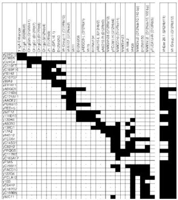
STS content
The YAC clones were analyzed with all PCR markers localized in the Igh locus. These include five microsatellites (D12Mit41, D12Mit134, D12Mit150, D12Mit208, D12Mit263) genetically mapped in the region (33). The sequences of the primers, the PCR conditions and the reference sequences for each marker are de-scribed in Table I. The results of this characterization by PCR are indicated in Fig. 1. Most YACs are positive for several loci, and the overlapping of these clones is obvious, although it is not pos-sible to construct a completely consistent marker order from this
FIGURE 1. STS content of Igh YACs. The STS content of each clone was determined by PCR with all the primer sets described in Table I. STSs are listed in approximate map order, and their presence in a YAC is indi-cated by black boxes. Although deletions in some YACs are apparent, a continuous path linking all STS markers can be traced in these YACs. Note that the 380-kb YAC D24C4 comprises a mouse Igh minilocus, bears at least 4 C region genes, all JHand DHsegments, and several VH7183 and
data. Some of the VHassays do not detect a single copy locus;
rather, they amplify several or many sites which may be widely spread throughout the locus. In addition, some YACs have internal deletions; for example, in the C region locus (the structure of which was completely determined with phage clones; Ref. 3), y36D5 contains IgA and IgG1 constant region sequences but not the intervening D12Mit41, a microsatellite in the 3⬘ flank of the
IgG2b gene.
YAC sizing and end clone characterization
To measure the length of the physical map, we determined the size of each YAC by PFGE. The sizes ranged between 100 and 800 kb. Five clones contain two different artificial chromosomes (yA8D10, yC79B11, yC110F7, yC174B4, yFEEH5), but blotting the PFGE gel and probing with an Igh region probe identified the chromo-some 12 YAC in each case.
For this study, we developed a new generalized strategy, called vector-hexamer PCR, for the isolation of insert ends from YACs (16). YAC insert ends were isolated from all 38 clones to charac-terize YAC overlap and to detect chimeric clones. Eighty-one ends were recovered from these 38 YACs. More than two ends were isolated from some of the clones which contained more than one YAC. End sequences were compared with the GenBank database with BLAST (31). A supplementary Table with GenBank acces-sions for the end sequences, BLAST results, YAC sizes, and end probe hybridization results is at http://www.tpims.org/research/ riblet-genetics.html.
Repetitive sequences were identified in 45% of ends, and most of these, 40% of the total, belonged to the LINE1, or L1, family of high-copy number repetitive sequences. Of the 81 ends, 11 were similar to known Igh sequences, usually sequences flanking coding regions. With a few exceptions, these matches were only 70 –90% identical to GenBank entries; these likely represent diverged du-plications that are not yet present in GenBank. These sequence identifications were consistent with Southern blot and PCR char-acterization of the content of these YACs, and they served to an-chor the developing YAC contig and physical map to the estab-lished genetic and deletion maps. Known genes other than Igh or repeat elements were identified by five YAC end sequences, but they appear to be the result of YAC chimerism or other artifact. For example, when yFDXB10 left (dopamine transporter), yF3A5 right (zinc finger) and left (potassium channel), and yC187G2 left (␥-aminobutyric acid receptor) ends were used as probes on panels of Igh YACs, they only hybridized to the YACs from which they were isolated, indicating chimerism. The first 129-bp portion of the yFDXB10 right end sequence is unique and hybridizes to other YACs in the contig; however, the remainder of this sequence matches the Escherichia coli mdoGH gene (accession number X64197).
Most of the PCR insert ends (53/81⫽ 65%) were suitable for hybridization probes on YAC blots, including some of the more highly diverged L1 ends. Forty ends hybridized to at least one
EcoRI fragment shared with other Igh YACs and were used to
construct the contig. The majority of these cross-hybridized to multiple fragments on Igh YACs, reflecting the duplicated nature of this locus. The other 13 probes hybridized only to the YAC from which the probe was generated, indicating that they are not present in the Igh locus. Two of these probes were from clones containing more than one YAC. Thus 11 of 38 YACs are judged chimeric.
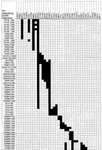
VHgene content of YACs
The content of VHgenes in each YAC was determined by
South-ern blot using probes for each of the 15 VHgene families (14). 4
Most of these families are relatively small, containing 1–10 genes and a like number of hybridizing EcoRI fragments on Southern blots. These were easily identified on Southern blots of digested YAC DNAs, and the content of small VHgene families in each
YAC is shown in Fig. 2. It is evident that there is considerable variation in size and content of the YACs, and that one YAC, ADGC9, carries nearly all of these VHgenes except the distally
mapping VH15, VH3609P, and VHJ558 families. The VHJ558
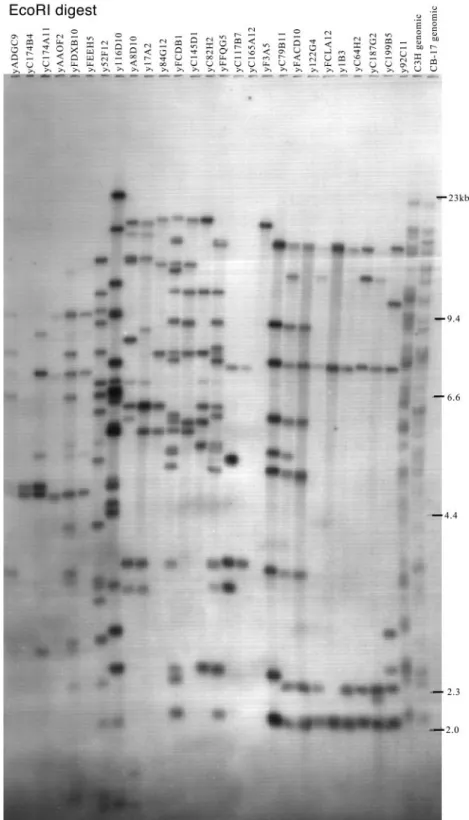
fam-ily (the largest famfam-ily) was more difficult to analyze. Seventy-seven EcoRI fragments were clearly mapped on the different
4Southern blots of YAC panels probed with 15 V
Hgene families and other probes may be seen at http://www.tpims.org/research/riblet-genetics.html.
FIGURE 2. VHgene family Southern blot analysis. YAC DNAs were
digested with EcoRI and Southern blotted. Blots were probed with repre-sentatives of each of the 15 VHgene families. The distal portion of the
locus containing some VH3609P genes and most of the complex VHJ558
family is not shown. The presence of hybridizing restriction fragments is indicated by black boxes and the absence is indicated by (-); a blank cell indicates analysis not done. Specific VHgene hybridizing fragments are
listed in only rough map order; in many cases, the data do not support unequivocal ordering. All hybridizing bands seen on C57BL/6 genomic blots are present in these YACs. It is apparent that the 765-kb YAC ADGC9 contains 40 fragments, nearly the entire proximal (3⬘) portion of the VHgene array including all families except VH15, part of VH10, and the
YACs (Fig. 3), but this is unlikely to be a complete analysis due to problems with resolving small differences in mobility, potential deletions in some or all YACs, and judging weak cross-hybridiza-tions on these YAC clone blots.
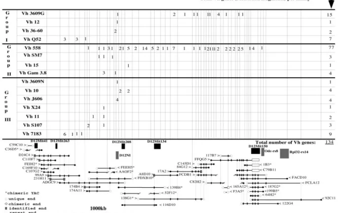
The YAC contig of Igh
The YAC clones were arranged in overlapping order using the patterns of hybridization obtained when YAC panel blots were probed with each useful insert end. This is represented in the lower
FIGURE 3. VHJ558 gene content of YACs. YAC DNAs, C3H, and C.B-17 genomic DNAs were digested with EcoRI and Southern blotted. This blot
was hybridized with the VHJ558 family probe VHA1, washed under stringent conditions (membranes were washed at 65°C for 20 min with each of: solution
1, 2⫻ SSC, SDS 0.1%; solution 2, 1⫻ SSC, SDS 0.1%; and solution 3, 0.1⫻ SSC, SDS 0.1%). Seventy-seven EcoRI bands ranging from 2 to 23 kb were detected and mapped on our YAC contig (see Fig. 4).
portion of Fig. 4. Note that the map is presented in chromosomal orientation, centromere to the left, telomere to the right, rather than the transcriptional V3D3J3C orientation usually seen (14). Note also that only three of the YACs are derived from C3H and none are crucial to the construction of the contig. The overlaps yielded a continuous path of YACs from a point midway in the constant region gene cluster at the left of the figure through all known VHgene families. All hybridizing bands visible on genomic
Southern blots of C57BL/6 DNA are present in this YAC contig indicating that we have recovered the entire Igh locus. We estimate that the overall length of the mouse Igh locus is at least 2.5 million base pairs as follows: the 3⬘ portion of the C region gene cluster containing the 3⬘ enhancer and IgA, IgE, and IgG2a CH genes
comprises⬃100 kb and is proximal (3⬘, left in Fig. 4) to YAC yD24C4, 380 kb. yD24C4 overlaps yADGC9, 765 kb, and their combined length is ⬃1100 kb. At the 5⬘ end of the locus is yFFQG5, which is 540 kb and contains the most distal VHJ558
genes, lacking only the most terminal one. Between yADGC9 and yFFQG5, but not overlapping them, is the large YAC y138G1. Its length was not determined, but it is from the Whitehead Institute (WI-820) library of large YACs, and its VHgene content and its
overlaps with other YACs indicate a length of⬃1 Mb. Thus, we conclude that Igh spans 2.5- 3 Mb.
The pattern of overlaps of YACs generated a series of sequential segments or bins along the contig ranging in size from 20 to 200 kb or more. Each PCR marker or hybridizing Southern blot band
was assigned to one of these bins producing a detailed physical map of the entire locus. The bins are indicated in the upper portion of Fig. 4 where we have summarized the blot hybridization results from Fig. 2 listing the number of hybridizing bands from each VH
gene family that reside in each bin. This physical dissection of the VHgene array provides a more accurate count of the number of
hybridizing restriction fragments and minimum estimate of the number of VHgenes (and pseudogenes). Hybridization of YAC
panel Southern blots with probes from all 15 VH gene families
reveals 134 bands. This is likely to be an underestimate of the number of VHgenes due to multiple genes on some restriction
fragments and multiple unresolved fragments appearing as single bands.
This physical map is consistent with the detailed deletion map (14); the VH7183 and VHQ52 families are nearest the DHsegments
and are thoroughly interspersed. Following the first two VH
fam-ilies are the VHS107 V1 and V3 genes and then a region of complex
interspersion of the remaining small VH families. The VHJ558
family begins in this region, as well, and extends to the distal end of the locus. The entire distal half or more of the VHgene array is
occupied by the VHJ558 gene family. Also in the distal region are
the VH3609P genes interspersed among the VHJ558 genes. The
VHJ558 family is shown extending proximally (leftward)
ap-proaching the VH7183/VHQ52 region. The identities of these most
proximal YAC bands that hybridize with a J558 probe are not known; because the effective stringency of these YAC clone blots
FIGURE 4. YAC contig of the mouse Igh locus. In the lower part of the figure, YACs are represented by horizontal bold lines: the YAC name is at either end with an arrow (⬎, ⬍) pointing to the line. The map is shown in chromosomal orientation, left is toward the centromere; this is opposite to the transcriptional orientation, 5⬘-VH3DH3JH3CH. YAC insert ends are represented as unique (no GenBank match), chimeric, identified (GenBank match),
or repeat sequences according to the key at the lower left. YACs identified as chimeric by end sequence or gene content are marked with an asterisk. Internal deletions are indicated by broken lines. The three C3H YACs, 116D10, 122G4, and 92C11, are at center bottom and bottom right of the contig; they agree with the C57BL YACs and suggest that the C3H haplotype may have the same structure. YAC overlaps are identified by hybridizations with nonrepetitive YAC end probes. These are indicated by connecting vertical lines and small boxes on the positive YACs. The positions of YAC ends divide the locus into a series of “bins”, indicated by vertical lines that are carried into the upper part of the figure where we summarize the hybridization data from Fig. 2. For each of the 15 VHgene families, the number of hybridizing restriction fragments residing in each bin is shown. Below the VHgene families, the locations
is reduced they may, in fact, be members of a related family, e.g., VHSM7. The more stringent genomic blots in the deletion
map-ping studies (14) did not suggest any VHJ558 genes in this region.
There are disagreements in gene placements between this map and the deletion map; for instance, we place a VH11 gene immediately
distal (5⬘) to a bin occupied by the VHS107 V1/V3 pair and a
VHJ558 or VHSM7 gene, while the deletion map inserts a VHX24
gene in this interval. This could reflect haplotype differences, i.e., the YAC map is of C57BL/6 and C57BL/10 (the 3 C3H YACs in our study do not contribute to the binning structure) while the deletion map is a composite of C57BL/6 and BALB/c, and in the region just discussed it is dominated by BALB/c information. Overall, this physical map confirms and provides additional detail and physical scale to the previous deletion and genetic maps of Igh.
Five D12Mit markers (33) that map in the Igh locus are clearly localized on the contig. D12Mit41 marks the IgG2b gene and is present on YACs that hybridize with CHregion probes. D12Mit263
maps in the interval between the last DHsegment, DFL16.1, and the
first VHgene, VH81X. D12Mit208 marks the region of interspersed
small VHfamilies. D12Mit134 marks the proximal (3⬘) part of the
VHJ558 region, and D12Mit150 marks the end of the VHarray. We
localized on the contig several additional genetic markers previously mapped in or near Igh. D12N1 is an anonymous DNA segment identified by an aberrant H chain rearrangement (34); it is located near D12Mit208 in the bin containing all VHJ606 and VH3609N genes.
Two pseudogenes, Odc-rs8 and Rpl32-rs14, map at the end of the VH
array just beyond D12Mit150.
Discussion
We have screened four YAC libraries representing⬃12-fold cov-erage of the mouse genome and have identified and analyzed 38 YACs bearing portions of the Igh locus. We isolated the ends of each YAC insert and used these to identify overlaps among the YACs and assemble a continuous path of YAC clones through Igh. Mouse Igh is large, 2.5–3 Mb, and contains a minimum of 134 VH
gene (or pseudogene) segments. This contrasts with the recently sequenced human IGH locus (1) which is more compact, only 1 Mb, but contains nearly as many VH segments, independently
counted as 95 (35) and 123 (1). At least half of the human VH
segments are pseudogenes. The mouse Ig L chain locus, Igk, is also large, 3.5 Mb, and contains⬃140 Vk segments of which as many as two-thirds are functional (2). The total complement of mouse VHgenes and its proportion of expressed vs pseudogenes
remain to be determined by more detailed physical characteriza-tion and sequencing. Relatively little analysis of VHgenes has
been done in the C57BL/6 or other Ighb
mouse strains relevant to this work with the notable exceptions of the VHJ558 family in
C.B-20 (9), the VHS107 family in B10.P (36), and the VH10 (37)
and VH7183 families (38) in C57BL/6. A thorough analysis of
germline and expressed VHJ558 genes identified 67 candidates
considered to be expressible germline genes (9). This estimate is surprisingly close to our band count of 77 and suggests that this gene family may have relatively few pseudogenes . All four germ-line VHS107 genes were cloned and sequenced from the closely
related B10.P strain (36); one of these, V3, was found to be a pseudogene in B10.P as it is in BALB/c. We were unable to con-sistently detect the V13 gene in our analysis and have omitted it. Langdon et al. (38) isolated 13–15 germline VH7183 genes by
PCR from C57BL/6 liver DNA; we count only nine hybridizing fragments. If the PCR experiment is correct then either some re-striction fragments carry several VH7183 genes or some
hybrid-ization bands conceal multiple similar-sized fragments.
Have we recovered the entire Igh locus in this YAC contig? Gaps remain in the representation of the constant region; presum-ably the isotype switch sequences or other repetitive elements ren-der this portion of the Igh locus especially unstable in yeast. The human IGH constant region locus was also resistant to YAC clon-ing (39). In the VHgene array, we found another potential
imped-iment to YAC stability, an extraordinary frequency of LINE1 el-ements. In our sequencing of YAC insert ends, we found that 40% belonged to the L1 family of high copy number repetitive se-quences (16); this frequency is surprisingly higher than the 5% incidence observed in another YAC contig on chromosome 11 (40), but more similar to the 25% incidence in the Igk locus (41) and a probably higher frequency in the MHC (42). Interestingly, the human IGH locus also has a 40% content of L1 sequence (1). This density of repetitive sequences makes these regions poten-tially unstable in YACs because of the high activity of the homol-ogous recombination machinery in yeast cells; this and the repet-itive nature of the VH gene families themselves likely are
responsible for the many deletions we detected in these YACs and perhaps additional deletions that we cannot detect at this level of analysis. Although we are aware of deletions in many YACs in the contig, other clones appear stable and intact, and the redundancy, or depth of coverage, that we sought in screening all available YAC libraries has resulted in apparently complete representation of all VH genes. Every hybridizing fragment seen on C57BL/6
Southern blots probed with all 15 VHfamilies is present in the
YAC contig.
Eleven of the YAC insert ends matched or were similar to known Igh sequences. The orientation of these 11 sequences rel-ative to chromosome 12 was consistent with all other known ele-ments in the H chain locus, supporting the generality that all gene segments of the Igh locus are in the same transcriptional orienta-tion and that rearrangement in Igh occurs exclusively by deleorienta-tion. The binning structure provided by the YAC overlaps has added a great deal of detail to the physical map of the VHgene array and
reveals the relatively massive scale of the VHJ558 gene family.
Acknowledgments
We thank the following for generous assistance and access to YAC librar-ies: S. Tilghman, D. Koos, and G. Guan, Princeton University; S. D. M. Brown, F. Chartier, and G. Argyropoulos, St. Mary’s Hospital Medical School; and E. Brundage, Baylor College of Medicine. We thank R. Lieberson, L. Eckhardt, P. Brodeur, M. Caulfield, and C. Carmack for primers and R. D. Miller, C. Carmack, and R. Epstein-Baak for early help and encouragement.
References
1. Matsuda, F., K. Ishii, P. Bourvagnet, K. Kuma, H. Hayashida, T. Miyata, and T. Honjo. 1998. The complete nucleotide sequence of the human immunoglob-ulin heavy chain variable region locus. J. Exp. Med. 188:2151.
2. Thiebe, R., K. F. Schable, A. Bensch, J. Brensing-Kuppers, V. Heim, T. Kirschbaum, H. Mitlohner, M. Ohnrich, S. Pourrajabi, F. Roschenthaler, et al. 1999. The variable genes and gene families of the mouse immunoglobulin locus. Eur. J. Immunol. 29:2072.
3. Shimizu, A., N. Takahashi, Y. Yaoita, and T. Honjo. 1982. Organization of the constant region gene family of the mouse immunoglobulin heavy chain. Cell
28:499.
4. Kurosawa, Y., and S. Tonegawa. 1982. Organization, structure, and assembly of immunoglobulin heavy chain diversity segments. J. Exp. Med. 155:201. 5. Wood, C., and S. Tonegawa. 1983. Diversity and joining segments of mouse
immunoglobulin heavy chain genes are closely linked and in the same orienta-tion: implications for the joining mechanism. Proc. Natl. Acad. Sci. USA 80:
3030.
6. Feeney, A. J., and R. Riblet. 1993. Dst4: a new, and probably the last, functional Dh gene in the BALB/c mouse. Immunogenetics 37:217.
7. Brodeur, P. H., and R. Riblet. 1984. The immunoglobulin heavy chain variable region (Igh-V) locus in the mouse. I. One hundred Igh-V genes comprise seven families of homologous genes. Eur. J. Immunol. 14:922.
8. Livant, D., C. Blatt, and L. Hood. 1986. One heavy chain variable region gene segment subfamily in the BALB/c mouse contains 500 –1000 or more members.
9. Gu, H., D. Tarlinton, W. Muller, K. Rajewsky, and I. Forster. 1991. Most pe-ripheral B cells in mice are ligand selected. J. Exp. Med. 173:1357. 10. Rothenfluth, H. S., A. J. Gibbs, R. V. Blanden, and E. J. Steele. 1994. Analysis
of patterns of DNA sequence variation in flanking and coding regions of murine germ-line immunoglobulin variable genes-evolutionary implications. Proc. Natl.
Acad. Sci. USA 91:12163.
11. Riblet, R., M. Weigert, and O. Makela. 1975. Genetics of mouse antibodies. II. Recombination between VHgenes and allotype. Eur. J. Immunol. 5:778. 12. Weigert, M., and R. Riblet. 1978. The genetic control of antibody variable
re-gions in the mouse. Springer Semin. Immunopathol. 1:133.
13. Riblet, R., A. Tutter, and P. Brodeur. 1986. Polymorphism and evolution of Igh-V gene families. Curr. Top. Microbiol. Immunol. 127:168.
14. Mainville, C., K. Sheehan, L. D. Klaman, C. A. Giorgetti, J. L. Press, and P. H. Brodeur. 1996. Deletional mapping of fifteen mouse VHgene families reveals a common organization for three Igh haplotypes. J. Immunol. 156:1038. 15. Nusbaum, C., D. K. Slonim, K. L. Harris, B. W. Birren, R. G. Steen, L. D. Stein, J. Miller, W. F. Dietrich, R. Nahf, V. Wang, et al. 1999. A YAC-based physical map of the mouse genome. Nat. Genet. 22:388.
16. Herring, C. D., C. Chevillard, S. L. Johnston, P. J. Wettstein, and R. Riblet. 1998. Vector-hexamer PCR isolation of all insert ends from a YAC contig of the mouse Igh locus. Genome Res. 8:673.
17. Burke, D. T., G. F. Carle, and M. V. Olson. 1987. Cloning of large segments of exogenous DNA into yeast by means of artificial chromosome vectors. Science
236:806.
18. Rossi, J. M., D. T. Burke, J. C. Leung, D. S. Koos, H. Chen, and S. M. Tilghman. 1992. Genomic analysis using yeast artificial chromosome library with mouse DNA inserts. Proc. Natl. Acad. Sci. USA 89:2456.
19. Kusumi, K., J. S. Smith, J. A. Segre, D. S. Koos, and E. S. Lander. 1993. Con-struction of a larinsert yeast artificial chromosome library of the mouse ge-nome. Mamm. Genome 4:391.
20. Haldi, M., C. Strickland, P. Lim, V. VanBerkel, X.-N. Chen, D. Noya, J. R. Korenberg, Z. Husain, J. Miller, and E. S. Lander. 1996. A comprehensive large-insert yeast artificial chromosome library for physical mapping of the mouse genome. Mamm. Genome 7:767.
21. Chartier, F. L., J. T. Keer, M. J. Sutcliffe, D. A. Henriques, P. Mileham, and S. D. M. Brown. 1992. Construction of a mouse yeast artificial chromosome library in a recombination-deficient strain of yeast. Nat. Genet. 1:132. 22. Larin, Z., A. P. Monaco, and H. Lehrach. 1991. Yeast artificial chromosome
libraries containing large inserts from mouse and human DNA. Proc. Natl. Acad.
Sci. USA 88:4123.
23. Ausubel, F. M., R. Brent, R. Kingston, D. D. Moore, J. G. Seidman, J. A. Smith, and K. Struhl. 1995. Current Protocols in Molecular Biology. John Wiley & Sons, New York.
24. Lieberson, R., S. L. Giannini, B. K. Birshtein, and L. A. Eckhardt. 1991. An enhancer at the 3⬘ end of the mouse immunoglobulin heavy chain locus. Nucleic
Acids Res. 19:933.
25. Love, J. M., A. M. Knight, M. A. McAleer, and J. A. Todd. 1990. Towards construction of a high resolution map of the mouse genome using PCR-analysed microsatellites. Nucleic Acids Res. 18:4123.
26. Schroeder, H., Jr., and J. Y. Wang. 1990. Preferential utilization of conserved immunoglobulin heavy chain variable gene segments during human fetal life.
Proc. Natl. Acad. Sci. USA 87:6146.
27. Carmack, C. E., S. A. Shinton, K. Hayakawa, and R. R. Hardy. 1990. Rearrange-ment and selection of VH11 in the Ly-1 B cell lineage. J. Exp. Med. 172:371. 28. Hearne, C. M., M. A. McAleer, J. M. Love, T. J. Aitman, R. J. Cornall, S. Ghosh,
A. M. Knight, J. B. Prins, and J. A. Todd. 1991. Additional microsatellite markers for mouse genome mapping. Mamm. Genome 1:273.
29. Caulfield, M. J., and D. Stanko. 1992. A pathogenic monoclonal antibody, G8, is characteristic of antierythrocyte autoantibodies from Coombs’-positive NZB mice. J. Immunol. 148:2068.
30. Sambrook, J., E. F. Fritsch, and T. Maniatis. 1989. Molecular Cloning: A
Lab-oratory Manual. Cold Spring Harbor LabLab-oratory Press, Cold Spring Harbor.
31. Altschul, S. F., W. Gish, W. Miller, E. W. Myers, and D. J. Lipman. 1990. Basic local alignment search tool. J. Mol. Biol. 215:403.
32. Lincoln, S. E., M. J. Daly, and E. S. Lander. 1991. PRIMER: A Computer
Pro-gram for Automatically Selecting PCR Primers. MIT Center for Genome
Re-search and Whitehead Institute for Biomedical ReRe-search, Cambridge. 33. Dietrich, W., J. Miller, R. Steen, M. A. Merchant, D. Damron-Boles, Z. Husain,
R. Dredge, M. J. Daly, K. A. Ingalls, T. J. O’Connor, et al. 1996. A comprehen-sive map of the mouse genome. Nature 380:149.
34. Bauer, S. R., L. A. D’Hoostelaere, and K. Huppi. 1988. Restriction fragment length polymorphism near the IgH locus on mouse chromosome 12. Nucleic
Acids Res. 16:8200.
35. Cook, G. P., I. M. Tomlinson, G. Walter, H. Riethman, N. P. Carter, L. Buluwela, G. Winter, and T. H. Rabbitts. 1994. A map of the human immunoglobulin VH locus completed by analysis of the telomeric region of chromosome 14q. Nat.
Genet. 7:162.
36. Perlmutter, R. M., B. Berson, J. A. Griffin, and L. Hood. 1985. Diversity in the germline antibody repertoire: molecular evolution of the T15 VHgene family.
J. Exp. Med. 162:1998.
37. Whitcomb, E. A., B. B. Haines, A. P. Parmelee, A. M. Pearlman, and P. H. Brodeur. 1999. Germline structure and differential utilization of Ighaand IghbV
H10 genes. J. Immunol. 162:1541.
38. Langdon, S. D., M. Inaioki, G. Kelsoe, and T. F. Tedder. 2000. Germline se-quences of VH7183 gene family members in C57BL/6 mice demonstrate natural selection of particular sequences during recent evolution. Immunogenetics 51:
241.
39. Mendez, M. J., H. Abderrahim, M. Noguchi, N. E. David, M. C. Hardy, L. L. Green, H. Tsuda, S. Yoast, C. E. Maynard-Currie, D. Garza, et al. 1995. Analysis of the structural integrity of YACs comprising human immunoglobulin genes in yeast and in embryonic stem cells. Genomics 26:294.
40. Nehls, M., K. Lu¨no, M. Schorpp, D. Pfeifer, S. Krause, U. Matysiak-Scholze, H. Dierbach, and T. Boehm. 1995. YAC/P1 contigs defining the location of 56 microsatellite markers and several genes across a 3.4 cM interval on mouse chro-mosome 11. Mamm. Genome 6:321.
41. Schupp, I. W., T. Schlake, T. Kirschbaum, H. G. Zachau, and T. Boehm. 1997. A yeast artificial chromosome contig spanning the mouse immunoglobulin light chain locus. Immunogenetics 45:180.
42. Jones, E. P., H. Xiao, R. A. Schultz, L. Flaherty, Z. Trachtulec, V. Vincek, Z. Larin, H. Lehrach, and K. F. Lindahl. 1995. MHC class I gene organization in