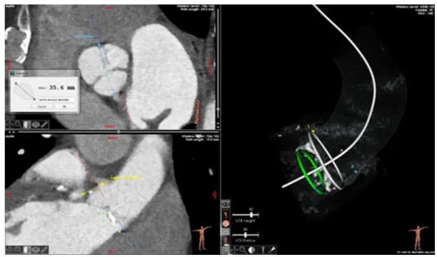
Automatic aortic root segmentation and anatomical landmarks detection for TAVI procedure planning
18
0
0
Texte intégral
Figure
+2
Documents relatifs
