Review
Local adipose tissue depots as cardiovascular risk factors
Sébastien Thalmann
b, Christoph A. Meier
a,b,⁎
a
Department of Internal Medicine, Triemli Hospital, Birmensdorferstrasse 497, CH-8063 Zürich, Switzerland
b
University of Geneva Medical School, rue Michel-Servet 1, CH-1211 Geneva 1, Switzerland Received 28 December 2006; received in revised form 20 February 2007; accepted 8 March 2007
Time for primary review 25 days Available online 14 March 2007
Abstract
Obesity is associated with increased cardiovascular morbidity and mortality. Although obesity-associated hypertension, dyslipidemia and insulin resistance account in part for this association, it becomes increasingly apparent that a systemic and local pro-inflammatory response of adipose tissue might also be a contributing factor.
White adipose tissue (WAT) is a highly active organ secreting various peptides such as cytokines, chemokines and hormone-like proteins. Besides the visceral and subcutaneous depots, WAT is also found in the close vicinity of blood vessels (perivascular adipose tissue), where it secretes cytokines such as interleukin-1, tumor necrosis factorα, pro-atherogenic chemokines, and pro-angiogenic peptides. These factors appear to contribute directly to alterations of the function and structure of the vascular wall, including chronic inflammation, alterations of vascular tone, proliferation of smooth muscle cells, neo-angiogenesis and hence to the development of atherosclerosis and cardiovascular complications. © 2007 European Society of Cardiology. Published by Elsevier B.V. All rights reserved.
Keywords: Atherosclerosis; White adipose tissue; Cytokines; Chemokines; Obesity; Cardiovascular risk; Angiogenesis
1. Introduction
In the developed world, obesity and the associated metabolic and cardiovascular complication is a major public health problem and one of the major contributors to premature death [1]. In Europe, the prevalence of obesity has tripled since the 1980s, and it has been estimated that over 4 million people become obese every year[2]. Recent data from the Centers for Disease Control and Prevention predict that the prevalence of diabetes in the USA will double by 2050 and will affect over 48 million people (or 12% of the population), which is mostly a direct consequence of the increase in overweight and obesity [3]. Moreover, the obesity pandemic is now reaching hitherto spared regions of the world, such as Southeast Asia, the Indian subcontinent and even Africa[4].
Obesity reflects the accumulation of triglycerides in fat deposits, leading to adipocyte hypertrophy and hyperplasia.
In evolutionary terms, fat deposits serve as caloric depots in case of food deprivation. However, once an advantage in the times when we were hunters and gatherers, the machinery leading to the efficient accumulation of fat stores has become deleterious. Excess white adipose tissue (WAT), leading to overweight (BMI 25–30) and obesity (BMI N30), has an impact on virtually all organ systems, including cardiovas-cular (endothelial dysfunction, coronary heart disease, hypertension, congestive heart failure), respiratory (obstruc-tive sleep apnoea, hypoventilation), gastro-intestinal (gall-bladder stones, obesity-associated gastroesophageal reflux disease), metabolic (insulin resistance, development of type 2 diabetes, hypercholesterolemia), urinary (hyperfiltration, microalbuminurie), oncologic (increased incidence of breast, endometrial and colon cancer) as well as the osteoarticular system (osteoarthritis)[5–9].
Since the discovery over a decade ago that WAT secretes various metabolically active molecules, such as tumor necrosis factorα (TNFα) and leptin, our view of adipose tissue being an inert fat storage organ has changed to that of a tissue secreting dozens, if not hundreds, of biologically active compounds,
⁎ Corresponding author. Tel.: +41 44 466 2101; fax: +41 44 466 2602. E-mail address:christoph.meier@triemli.stzh.ch(C.A. Meier).
0008-6363/$ - see front matter © 2007 European Society of Cardiology. Published by Elsevier B.V. All rights reserved. doi:10.1016/j.cardiores.2007.03.008
which are implicated in the regulation of food intake, inflammation and angiogenesis, either through systemic or local paracrine effects, ultimately resulting in a chronic low grade inflammatory state[10,11]. Hence, obesity per se is now considered as a cardiovascular risk factor contributing in the development of atherosclerosis independently of hyperten-sion, dyslipidemia and diabetes (Fig. 1)[12].
Atherosclerosis represents a heterogeneous disease of multiple different causes. Since it can be considered as a chronic inflammatory state characterized by the accumula-tion of leukocytes, the proliferaaccumula-tion of smooth muscle cells and neo-angiogenesis within the vascular wall, it is appealing
to speculate that specific localised adipose tissue depots, such as perivascular WAT (pWAT), contribute to obesity-associ-ated atherosclerosis[13]. The present review focuses on our current knowledge of the distribution, regulation and secretory activity of pWAT.
2. WAT as a secretory organ
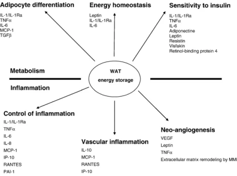
WAT secretes cytokines, chemokines and hormone-like proteins. These peptides can be subdivided according to their principal effects into molecules acting principally on metabolic processes (such as the regulation of food intake and body weight, and insulin sensitivity), and in factors modulating inflammation [14]. The WAT-derived factors can also be categorized according to their range of action, which can be local through the creation of concentration gradients (para-crine), or systemic (endocrine) (Fig. 2).
2.1. Cytokines
Cytokines are peptides produced in response to certain stimulation, which are bioactive at very low levels and play an integral role in the regulation inflammation, immunity, cell growth and maturation. Over 100 cytokines have been described and they have been classified as interleukins, interferons, chemokines, haematopoietic factors and growth factors. They are produced by cells of the haematopoietic lineage, including macrophages, T-cells, B-cells and natural killer cells, but also by non-haematopoietic cells, such as hepatocytes, epithelial cells and fibroblasts [15]. Several cytokines, such interleukin-1 (IL-1), interleukin-1 receptor antagonist (IL-1Ra), interleukin-6 (IL-6), transforming
Fig. 1. Relation between obesity and the development of atherosclerosis. Obesity has been associated with hypertension, dyslipidemia, insulin resistance, endothelial dysfunction and inflammation, which all have been linked to atherosclerosis.
Fig. 2. White adipose tissue is a highly active organ secreting various peptides implicated in inflammatory and metabolic processes, either by paracrine effect or endocrine/systemic effect.
growth factorβ (TGFβ), TNFα and certain chemokines are also produced by WAT as will be discussed in detail below. 2.2. Chemokines
Chemokines are small secreted basic proteins, classified into four subclasses according to the position of the cysteine residue (i.e. CXC, C, CX3C and CC) [16]. They are implicated in the chemoattraction of inflammatory cells through generation of local concentration gradients, acting essentially through paracrine effects [10]. Therefore, the action of these factors is often related to the proximity of the secreting tissue (e.g. adipose tissue) to the target tissue, such as leukocytes or the vascular wall. Through their capacity to recruit and activate cells, chemokines are involved in multiple systemic inflammatory disorders, such as rheuma-toid arthritis, glomerulonephritis and atherosclerosis [17]. The production of chemokines is induced by various stimuli, such as vascular injury, oxidized lipids, growth factors and cytokines [10,16]. In the often diabetic obese patient, glycated and oxidized lipids (advanced glycation end products) also contribute to augmented cytokines production
[18], therefore further accelerating atherosclerosis. 2.3. Other proteins and peptides
One of the major proteins derived from WAT is adiponectin (or also called AdipoQ, Acrp30, ApM1 or GPB28) which is the most abundant adipose-derived protein in the circulation (5–30 μg/ml), accounting for 0.01% of total plasma protein [19–21]. Adiponectin plays a role in the maintenance of insulin sensibility, and its decline observed in obesity has been linked to type 2 diabetes[22]. Moreover, the overexpression of adiponectine in leptin-deficient ob/ob mice improves insulin sensitivity[23]. Besides regulating insulin sensitivity, adiponectin has also anti-atherosclerotic effects by inhibiting proliferation of smooth muscle cells, one of the hallmarks of atherosclerosis [24], as well as by increasing inhibitors of metalloproteinases, which are necessary for the degradation of extracellular matrix zincluding the fibrous cap on atherosclerotic plaques) and neo-angiogenesis[25].
In contrast, resistin, which is another peptide secreted by monocytes and WAT, is increased in type 2 diabetes and some studies have shown a correlation between plasma resistin levels, body mass index and adiposity [26]. However, the secretion of resistin by human WAT is a matter of some debate [27]. The administration of resistin induces insulin resistance in euglycemic, hyperinsulinemic conditions in rodents, although these findings have also been controversial [28,29]. Finally, resistin could also have inflammatory properties, as it induces the expression of adhesion molecules on endothelial cells[30].
A more recently discovered peptide is visfatin, which is derived from visceral adipose tissue[31]. Visfatin decreases plasma glucose levels, and its actions have therefore been described as insulin mimetic. Like insulin, visfatin induces
phosphorylation of signal proteins downstream the insulin receptor, improving insulin sensitivity and representing a potential defence mechanism against obesity-induced insulin resistance[32]. Recently, retinol-binding protein-4 (RBP4), a new adipokine which acts as transport molecule for retinol (vitamine A), was reported to be linked to impaired glucose tolerance and type 2 diabetes, and was found to be correlated with body mass index[33,34]. Moreover, in a murine model, reduction of plasma RBP4 improved insulin resistance[35]. Leptin has been much investigated over the past decade as an appetite-regulating factor. It is a circulating protein produced by adipose tissue with central and peripheral actions on metabolic and inflammatory processes. Typically, leptin controls food intake and energy expenditure through hypothalamic pathways, thereby contributing to glucose homeostasis. However, it seems that leptin also has several immunomodulatory actions, as it regulates the secretion of chemokines and cytokines in monocytes[36].
The pro-atherogenic plasminogen activator inhibitor-1 (PAI-1) inhibits the breakdown of fibrin clots, thereby enhancing the formation of thrombi upon rupture of plaques; moreover, it contributes to vascular wall remodelling by altering the fibrinolytic balance [37,38]. Elevated plasma levels of PAI-1 are found in obesity and it has hence been suspected to represent one of the mediators of the obesity-associated excess cardiovascular risk[39].
3. Localized WAT compartments
In humans, the development of obesity leads not only to an increase in fat depots in classical locations, such as in the subcutaneous and intra-abdominal (or visceral) compart-ment, but also around specific organs, such as the heart (epicardial), blood vessels (perivascular) and the kidney (renal), which have all been described as locations for ectopic fat storage[40](Table 1). It has been demonstrated in a rabbit model of obesity that fat depots occur in various locations, such as in the neck, retroperitoneal space, mesenterium, mediastinum, renal sinus and around blood vessels[41]. Organ function may be altered by surrounding fat deposits, as they may compress the adjacent organ, thereby mechanically impairing its function. On the other hand, local fat depots could also impact on the surrounding structures through secreted factors, such as chemokines and cytokines.
3.1. Visceral WAT
Visceral WAT consists of fat deposits around anatomic structures located in the retroperitoneal, omental and mesenteric spaces. Many studies support a detrimental effect of intra-abdominal fat on metabolism and cardiovascular risk
[42], although the mechanisms of action remain largely speculative. Increased visceral WAT is a risk factor for dyslipidemia, insulin resistance, diabetes, and hypertension. Visceral adiposity is estimated by the measurement of waist
circumference, which has been included in the definition of the metabolic syndrome[43]. However, these measurements are quite imprecise and may not reflect the actual amount of visceral fat [44]. The best tools to estimate the amount of visceral fat include imaging with computer tomography (CT) or magnetic resonance imaging (MRI). Recently, it has been shown that a good correlation exists between visceral adipose tissue and epicardial fat measured by echography, therefore representing an easier tool for the estimation of visceral fat mass for clinical studies[45].
3.2. Ectopic fat storage in muscle
Ectopic lipid storage in muscle has been first described in 1967[46]. In Indian non-diabetic Pima men, an ethnic group with a pronounced tendency to obesity and type 2 diabetes, increased muscle triglyceride content was associated with insulin resistance and type 2 diabetes [47]. However, increased lipid contents of muscle were found to be predominately intracellular. Consistent with this, Goodpaster et al. found that the muscular triglycerides in obese type 2 diabetics was located within the myocytes [48]. New data suggest that extracellular muscular fat also contributes to muscle insulin resistance, as fat deposits located beneath the fascia lata in the thigh are associated with insulin resistance in obesity [49]. Mechanistically, it is plausible that adipocytes located around muscle cells produce high local concentrations of cytokines, such as TNFα or IL-6, which are all known to alter insulin sensitivity [50,51].
Alterna-tively, free fatty acids from adipose tissue may interact negatively with normal insulin sensitivity[52].
3.3. Ectopic fat storage in kidney
In animal models of diet-induced obesity, intrarenal fat deposits can directly or indirectly alter renal structure[40]. For instance, diet-induced obesity in dogs and rabbits is associated with larger kidneys and increased fat deposits in the renal sinus[53]. Consequently, renal veins and lymphatic outflow are constricted, resulting in augmentation of renal interstitial fluid pressure and prolongation of sodium transit time, leading to a significant resorption. This has been proposed to contribute to the obesity-induced hypertension observed in animals and humans [54]. However, other mechanisms have been implicated as well, such as increased renal sympathetic activity, activation of the renin-angioten-sin-aldosterone system and a direct sodium retention effect by hyperinsulinemia[55].
3.4. Epicardial adipose tissue
Obesity predisposes to the accumulation of excess epicardial fat [40], which shares with intra-abdominal fat the capacity to secrete cytokines [56]. Its quantity, as measured by echography, has been well correlated with the mass of visceral adipose tissue measured by magnetic resonance imaging [45]. Consistent with this hypothesis, a recent study showed that epicardial adipose tissue represents a novel indicator of cardiovascular risk [57]. Human epicardial adipose tissue has considerable secretory activity; for instance, epicardial adipose tissue from patients under-going elective coronary aortic by-pass grafting contained significantly more mRNA and protein for IL-1β, IL-6, monocyte chemoattractant protein 1 (MCP-1) and TNFα compared to subcutaneous adipose tissue [58]. In keeping with this finding, the cytokine concentrations in epicardial fat was correlated with an accumulation of inflammatory cells, such as T-lymphocytes, macrophages and mast cells, in the vicinity of epicardial adipose tissue. In another study, epicardial mRNA levels for CD45, a marker of macrophage infiltration, was significantly increased compared to abdom-inal fat [56]. Consistent with this, a recent study demon-strated that the expression of different pro-inflammatory cytokines, such as resistin and TNFα, augmented within 4 hours after coronary by-pass surgery in epicardial adipose tissue[59].
Adiponectin might also play a role in epicardial adipose tissue, as patients with advanced coronary heart disease have lower level of epicardial adiponectin, which potentially contributes to the increased cardiovascular risk[60]. 4. Perivascular adipose tissue and atherosclerosis
Perivascular WAT (pWAT) is an appealing candidate for mediating at least in part the obesity-associated cardiovascular
Table 1
Besides its localization in subcutaneous tissue, specific organ-associated WAT depots exist (see text for details)
WAT localisation Local factors Putative consequences Kidney Na+reabsorption Increased intravascular volume hypertension Muscle Secretion of TNFα Insulin resistance
IL-6 Free fatty acids
Visceral Secretion of IL-8 Local and systemic inflammation MCP-1
IP-10 RANTES
Epicardial Secretion of IL-6 Local inflammation and chemotaxis IL-1β
MCP-1 TNFα
Perivascular Secretion of MCP-1 Atherosclerosis systolic hypertension IL-1/IL-1Ra IL-6 IL-8 IP-10 RANTES TNFα Increased vascular tonus
risk. It is defined as the accumulation of adipocytes around vascular structures, and it can be found in the proximity of virtually all blood vessels, especially around the coronaries and the aorta, where its distance to the adventitia measures less than 0.1 mm. Conventionally, pWAT was considered to act largely as a structural support for blood vessels. However, recent data from epicardial adipose tissue and ex-vivo experiments with aorta and mesenteric arteries suggest that periadventitial fat can modulate the vascular responsiveness to vasoactive agents [61–63]. In addition, we have recently shown that pWAT secretes a variety of cytokines and chemokines[64]. Consistent with this concept, feeding of a high-fat diet to rats augmented the amount of periaortic fat by 4 to 10 times in the abdominal aorta and iliac arteries, respectively[64]. Hence, an increase in pWAT and its local secretory activity could contribute to the pathogenesis and/or progression of obesity-induced hypertension and atheroscle-rosis (Fig. 4and discussion below). Although the cytokines described below were all shown to be secreted by pWAT, only circumstantial evidence currently exists with regard to their direct pro-atherogenic action on the adjacent vasculature. 4.1. Production of chemokines and cytokines by pWAT
As the distance between pWAT and the vascular wall has been estimated to be less than 100 μm, it is easily conceivable that locally produced factors could interact with the vasculature. Consistent with this idea, we were able to demonstrate that human perivascular adipocytes are a source of cytokines, such as TNFα, IL-1, IL-1Ra, IL-6, as well as chemokines like MCP-1, interleukin 8 (IL-8), interferon-(IFN)-γ-inducible protein (IP-10), and “regulated on activated normal T-cell expressed and secreted” (RANTES), all of which have been implicated in the progression of (or protection from) atherosclerosis[64]. 4.1.1. TNFα
TNFα is one of the most studied proinflammatory cytokines. Originally described to induce the necrosis of tumors after acute bacterial infections, it is now known to be involved in inflammatory diseases, tumorgenesis, metastasis, viral replication, septic shock and fever. Endotoxins, viruses, drugs and other cytokines (e.g. IL-1) can induce its production, whereas other cytokines (IFNα, IFNβ or IL-6) are inhibitory[10]. TNFα is produced by a variety of different
cell types ranging from cells of the immune system to tumor cells, fibroblast, smooth muscle cells, as well as epicardial and perivascular adipocytes[65,66]. TNFα has been linked
to obesity in a murine model of obesity due to leptin-resistance (db/db mice), where epididymal adipose tissue was found to express TNFα at significant higher levels[67].
TNFα appears to play a certain role in insulin resistance, as it has the capacity to downregulate Glucose Transport Type 4 (GLUT4) in adipose tissue. Obese mice deficient in TNFα have lower glycemia and insulin levels, and they are more glucose tolerant than wild-type animals [50]. These
differences might be accounted for, at least partially, by a decrease in circulating free fatty acids, an increase of GLUT4 in muscle tissue, and an increase in the auto-phosphorylation of the insulin receptor in both adipose and muscle tissue.
TNFα secreted by perivascular adipocytes, could hence interact with the local vasculature by different mechanisms. First, TNFα could inhibit endothelial nitric oxide synthase (eNOS), therefore leading to the decreased production of nitric oxide (NO). Since NO is a potent vasodilator mediating e.g. the vasodilatory actions of insulin, a decrease in NO would result in an impairment in insulin-mediated vasodilatation, a reduction in muscle blood flow, thereby ultimately contributing to insulin resistance by limiting the subtract amount to the muscle[68].
4.1.2. TGFβ
TGFβ is another cytokine found in WAT and produced by adipocytes. TGFβ restricts cell growth, differentiation and cell death and it has therefore been implicated as a regulator of tumor growth[69]. Similar to TNFα, TGFβ also plays a role in adipose cell differentiation[70]. In addition, TNFα stimulates TGFβ synthesis, which in turn stimulates PAI-1 synthesis [71]. However, the precise role of TGFβ in
atherosclerosis and its secretion by pWAT remains to be elucidated.
4.1.3. IL-1 and IL-1Ra
IL-1 is a family of three related cytokines (IL-1α, IL-1β and IL-1Ra), which are produced by distinct genes. IL-1α and IL-1β have undistinguishable pro-inflammatory actions on most cell types. IL-1 is mostly produced by stimulated monocytes and tissue macrophages, and it induces fever, the acute phase liver response, the proliferation of different cell types (fibroblasts, smooth muscle and mesangial cells) and the secretion of other cytokines. IL-1 has also pro-angiogenic actions and direct effects on adipocytes, where it increases lipolysis and impairs adipocyte differentiation through the inhibition of peroxisome proliferators-activatedγ (PPARγ), a nuclear receptor playing a major role in adipocyte differentiation[72].
The production of IL-1 by pWAT may therefore play a role in development of atherosclerotic lesions by promoting neoangiogenesis and smooth muscle cell proliferation, both of which are hallmarks of atherogenesis.
In contrast, IL-1Ra is unique by the fact that it is a specific natural antagonist of IL-1 and counteracts its pro-inflamma-tory actions by binding competitively to the IL-1 receptor without inducing a signal [73]. IL-1Ra is produced by monocytes, neutrophils, fibroblasts, and epithelial cells. Like IL-1, IL-1Ra is an acute-phase protein produced by liver cells in response to inflammatory stimuli; its secretion is modulated by other cytokines, such as IL-1, interleukin 3 (IL-3), interleukin 4 (IL-4), IL-10, TNFα and interferon β (IFN-β). It is now well known that IL-1Ra levels increase markedly in response to fever and inflammation acting as a host-defence mechanism [74]. Intriguingly, leptin, which is produced by
adipocytes and principally acts on energy homeostasis, induces the production of IL-1Ra by human monocytes, suggesting a putative anti-inflammatory action[75].
Although adipocytes produce IL-1Ra as well as IL-1, the levels of IL-1Ra are selectively and markedly elevated over 7-fold in the serum of obese patients [76]. While this up-regulation appears at first sight to be anti-inflammatory, and hence beneficial, recent data in rodents suggest that excess amounts of IL-1Ra lead to insulin resistance in skeletal muscle[77].
Since perivascular adipocytes secrete IL-1 and IL-1Ra
[64], it can be hypothesized that these two cytokines play a modulating role in the development of atherosclerotic plaques, whereby IL-1Ra promotes the differentiation of adipocytes, as well as the proliferation of fibroblasts and smooth muscle cells.
4.1.4. IL-6
Together with IL-1 and TNFα, IL-6 provokes an acute phase reaction, such as fever and the hepatic production of acute phase proteins. IL-6, like IL-1Ra, also has some modulatory effects on inflammation: it decreases cytokines like TNFα and IFNγ and increases IL1-Ra[10]. Obesity is associated with increased IL-6 levels, whereas body weight loss results in a decrease[78]. IL-6 was shown to lead to a decrease in the insulin-dependent glucose uptake through the down-regulation of the expression of the insulin receptor substrate 1 (IRS-1) and GLUT4[51]. In addition, IL-6 has central effects resulting in increased energy expenditure and in lower fat pad weight[79]. With regard to atherosclerosis, IL-6 could contribute to the local balance between pro- and anti-inflammatory cytokines in pWAT as well as on the proliferation of smooth muscle cells, and could also impair local insulin sensitivity by inhibiting IRS-1.
4.1.5. MCP-1
MCP-1, also called CCL2, belongs to the family of chemokines. The basal expression of MCP-1 is very low, but can be induced by many different stimuli, including cytokines (IL-1, TNFα) and endotoxins. Even more, MCP-1 mRNA is increased in obese mice, where the plasma levels correlate with body weight[80]. In humans, MCP-1 is produced by WAT and it was shown to impair insulin signaling in skeletal muscle cells at doses which correspond to the physiological plasma concentrations (200 pg/ml). Moreover, MCP-1 controls adipogenesis and triglycerides content of adipocytes, and it plays a role in adipocyte dedifferentiation through autocrine and paracrine mechanisms[81–83].
MCP-1 is a key mediator of monocyte trafficking and promotes macrophage recruitment, an essential step in athe-rogenesis, as its receptor (CCR2) is expressed on circulating monocytes, which in turn are important contributors to the progression of atherosclerosis[84].
MCP-1 is also produced by macrophages, endothelial cells and smooth muscle cells and is also secreted by epicardial WAT, as well as by pWAT; this is correlated with
the degree of infiltration of these WAT depots by inflam-matory cells[58,64]. Immunohistochemical analysis clearly confirmed that adipocytes, rather than stromal cells, produce MCP-1[64].
It has been shown that the local perfusion of MCP-1 after unilateral ligation of the femoral artery in apoE-deficient mice induced collateral blood flow in the hindlimb with an increase in perivascular monocytes and accelerated aortic plaque formation [85]. The atherogenic role of MCP-1 is further substantiated by the observation that mice lacking MCP-1 or its receptor (CCR2) exhibit a less severe pattern of atherosclerosis, with a reduction of plaque size and macrophage content[86,87].
In humans, the expression of CCR2 has been identified on smooth muscle cells and recent findings suggest that MCP-1 could also contribute to smooth muscle cell proliferation
[88]. Moreover, MCP-1 has also systemic and paracrine actions on WAT, as it stimulates the secretion of leptin[83]. Leptin and MCP-1 are both angiogenic factors and contribute to formation of neo-vessels, which is an important component of atherosclerosis, where neo-vessels provide the necessary oxygen and nutrients for leukocyte accumulation. Taken together, these data strongly suggest that MCP-1 contributes to chemotaxis, smooth muscle cells proliferation and neo-vessel formation; hence, the local production of this chemokine in the immediate vicinity of the vascular wall is likely to have an impact on the biology of atheromatous lesions. 4.1.6. IL-8
IL-8 (or CXCL-8) is another chemokine involved in atherogenesis. IL-8 is derived from many different cells, including monocytes, lymphocytes, granulocytes, fibro-blasts, endothelial cells, bronchial epithelial cells, keratino-cytes, hepatokeratino-cytes, mesangial cells and chondrocytes. IL-1β and TNFα increase IL-8 release, whereas dexamethasone and IFNβ are inhibitory [89]. IL-8 is not only chemotactic for monocytes, but also for numerous others cell types, including neutrophils, T-cells, basophils and smooth muscle cells.
Different studies have confirmed that IL-8 plays a role in mediating the arrest of monocytes on endothelial cells, a key step in the mechanism of the infiltration of the vascular wall by leukocytes [90,91]. The IL-8 receptor (CXCR2) is strongly expressed in macrophages within atherosclerotic lesions. Consistent with these findings, mice lacking CXCR2 showed a significant reduction in atherosclerotic lesions and leukocyte accumulation [92]. In humans, IL-8 could con-tribute to obesity related low-grade inflammatory state, as IL-8 is increased in obesity[93]. However, the action of IL-8 is not limited to acute inflammation, as it stimulates the proliferation of endothelial cells and has potent angiogenic actions[94].
Since we have demonstrated that adipocytes from pWAT secrete significant amounts of IL-8[64], it is quite plausible that this factor might locally contribute to atherosclerosis. However, direct mechanistic evidence linking pWAT to atherogenesis (e.g. by examining the impact of a
pWAT-selective gene ablation of IL-1 or MCP-1 in an atheroscle-rosis prone animal model) is currently lacking.
4.1.7. IP-10
IP-10 (or CXCL-10) is produced by many different cell types after stimulation by IFN or lipopolysaccharides, including endothelial cells, keratinocytes, monocytes, respi-ratory and intestinal epithelial cells and smooth muscle cells. In WAT, it is regulated ex vivo by IFN-β, and in vivo by inflammatory conditions such as obesity, atherosclerosis and diabetes (E. Henrichot, C.A. Meier, unpublished data). Furthermore, IP-10 is also secreted by pWAT and it contributes to the chemotaxis of white blood cells, smooth muscle cells migration and neo-angiogenesis. IP-10 is chemoattractive for lymphocytes and monocytes, and it induces the proliferation and migration of smooth muscle cells from the media to the intima[95].
4.1.8. RANTES
The“regulated on activated normal T-cell expressed and secreted” chemokine (RANTES) or CCL5 is another potential candidate implicated in atherosclerosis. RANTES is chemotactic to T-lymphocytes and macrophages and decreases the amount of collagen production. It is secreted by numerous cells including endothelial cells, smooth muscle cells, activated T-cells, macrophages and platelets, and has been implicated in cardiovascular inflammation after organ transplantation or arterial injury. In patients with endstage renal disease and type 2 diabetes, polymorphisms of the RANTES gene were associated with increased mortality [96]. Veillard et al. showed that antagonism of RANTES receptor reduced atherosclerotic plaque formation in mice[97].
Human subcutaneous WAT and pWAT both produce RANTES, and this production is upregulated by systemic inflammatory cytokines such as IFN-β and IL-1 (E. Henrichot, C.A. Meier, unpublished data).
4.2. Role of extracellular matrix and metalloproteinases One of the first steps in angiogenesis is the breakdown of the subendothelial basement membrane and the surrounding extracellular matrix (ECM). This is followed by endothelial cell migration and proliferation. Metalloproteinases (MMPs) are a large family of zinc-dependent proteinases, which digest various components of the ECM, thereby stimulating the migration and attachment of endothelial cells [98]. Besides this function, MMPs also play a role in the differentiation of adipocytes [99]. The action of MMPs is inhibited by endogenous inhibitors, such as the tissue inhibitor of metalloproteinases (TIMPs). Regional distur-bance in the balance of MMPs and TIMPs could contribute to the vulnerability of the plaque and/or vascular remodel-ling. Indeed, it has been shown that oxidized lipids upregulate MMP-9 and downregulate TIMP-1 in human macrophages[100]. Moreover, IL-8 downregulates TIMP-1
expression in human atherosclerotic lesions, thereby possi-bly contributing to the development of atherosclerotic lesions[101].
Recent studies have shown that human adipocytes produce MMP-2 and MMP-9 with a diminution of TIMP-1
[102]. These findings suggest that adipose tissue might promote the development of new vessels. Leptin, which is also secreted by adipocytes, could also act to promote neovascularisation by promoting the expression of MMPs as shown in a rat corneal model [103]. Consistent with this, leptin is capable of inducing the proliferation of human umbilical endothelial cells and increases MMPs in a dose-dependent manner, contributing to the development of neo-vessels [103]. Taken together, WAT secretes several key components required for neoangiogenesis, which is in turn a proatherogenic process.
4.3. Role of angiogenesis
Angiogenesis plays an important role in tumor growth, but also in atherogenesis. As mentioned above, blood vessels
Fig. 3. Perivascular adipose tissue is characterized by increased angiogenesis as compared to subcutaneous WAT. Immunohistological staining of endothelial cells (CD34+) in human WAT: subcutaneous WAT (panel A) and pWAT (panel B). Courtesy of C. Juge-Aubry and C.A. Meier.
are necessary for plaque formation, as they provide the necessary oxygen and nutrients, but also a vascular network for inflammatory cells[104]. Intriguingly, the fat deposits in obesity are characterized by an expansion of the capillary bed (E. Henrichot, C.A. Meier, unpublished data). Further-more, human WAT is a source of many angiogenic factors, such as IL-6 and IL-8, MCP-1, leptin, fibroblast growth factor and vascular endothelial growth factor. As illustrated inFig. 3, neoangiogenesis was more pronounced in pWAT as compared to subcutaneous WAT.
Specifically, we have shown that pWAT produces significant amounts of angiogenin, which belongs to the family of pro-angiogenic factors. Angiogenin promotes the invasiveness and proliferation of cultured endothelial cells, and also serves as an adhesion molecule for endothelial cells. Indeed, primary adipocytes from pWAT induced the formation of capillary-like structures when co-cultured with aortic bovine endothelial cells; the addition of RNAsin, an inhibitor of angiogenin, entirely abolished the formation of capillary tubes (E. Henrichot, C.A. Meier, unpublished data). Hence, the secretion of angiogenic factors by pWAT
appears to be sufficient to induce angiogenesis in adjacent structures.
4.4. Role of adipose tissue derived oxidative stress
Obesity is characterized by a systemic increase in oxidative stress [105]. Subcutaneous adipose tissue from obese subjects produces more reactive oxygen species, contributing to the augmented oxidative stress in obesity
[106]. The augmented oxidative stress plays a role in vascular tone, since a recent study has shown that pWAT enhances the arterial contractile response to perivascular nerve stimulation cells through the production of superoxide[107].
4.5. Increased arterial wall stiffness and vascular reactivity Soltis and Cassis demonstrated in 1991 that pWAT significantly modifies the vascular response of aortic rings to norepinephrine in vitro [62]. More recently, another factor derived from pWAT, named adventitium derivated relaxing factor (ADRF), which is NO-independent, but calcium
Fig. 4. Hypotheses linking pWAT to atherogenesis. Arrow 1: chemoattraction of leukocyte by MCP-1, IP-10, RANTES and IL-8 (produced by perivascular adipocytes) and adhesion to endothelial cells. Arrow 2: production of pro- and anti-inflammatory cytokines such as TNFα, IL-8, IL-6, IL-1 and IL-1Ra by adipocytes. These factors contribute to a local inflammatory burst with tissue remodelling, macrophage activation and enhanced chemoattraction of leukocytes. Arrow 3: MCP-1, IP-10 and IL-6 stimulate the migration and proliferation of smooth muscle cells. Arrow 4: Extracellular matrix is digested by MMPs secreted by adipocytes, which is the first step in the formation of neo-vessels. Arrow 5: Angiogenin, leptin and MCP-1 stimulate neo-angiogenesis. This allows oxygen and nutrition delivery and apperas to contribute to plaque development. Arrow 6: Reactive oxygen species, TNFα and possibly angiotensin II interact with NO bioavaibility, thereby leading to vasoconstriction. Arrow 7: Adiponectine, visfatin, resistin, leptin, TNFα and other cytokines also enter the systemic circulation and act centrally on food intake and body weight.
dependent, was described, but its precise nature remains elusive[61].
It is well known that angiotensinogen and angiotensin-converting enzymes alter vascular tone and the circulating levels of angiotensinogen and angiotensin II are increased in human obesity [108]. Angiotensin II leads not only to vasoconstriction, but also induces the endothelial production of MCP-1, intracellular adhesion molecule-1 and vascular adhesion molecule-1 [109]. Angiotensin II also interacts with NO metabolism by enhancing the formation of oxygen free radicals, which may damage vascular tissue. NO is not only a potent vasodilatator, but also protects the wall by inhibiting the adhesion of monocytes and by decreasing vascular smooth muscle cell proliferation[11].
pWAT may also have a direct mechanical impact on the vasculature. Young obese subjects have an increased vascular stiffness[110,111]which can be reduced by weight loss, as measured by Doppler ultrasound [112]. Arterial stiffness could well be related to excess vascular rigidity caused by accumulation of periadventitial fat deposits, but also to sympathetic overactivity found in obesity[113]. In stiffer vessels the Windkessel effect is ineffective and therefore, reflection waves travel more quickly to the heart leading to an increase in blood pressure during systole.
Obesity-induced hypertension is frequent, as up to 50% of obese individuals have concomitant hypertension; the pWAT-related mechanisms detailed above could all poten-tially contribute to the pathogenesis of obesity-induced hypertension.
5. Conclusion
As obesity develops, WAT mass increases together with its secretory activity. Adipose tissue can be found in many different localisations in human body and its effects are local or systemic. Therefore, it is a very appealing concept that pWAT, which produces key molecules in inflammation and chemotaxis, contributes to obesity-associated atherosclerosis and hypertension (Fig. 4).
As obesity develops, there is hypertrophy and hyperplasia of perivascular adipocytes. TNFα, TGFβ, IL-8, IL-6, IL-1 and IL-1Ra and chemokines like IP-10, MCP-1 or RANTES are all secreted by pWAT. These peptides contribute to the chemotaxis of leukocytes to the vascular endothelium and their migration into the vascular wall. The consequence is a local inflammatory burst with production of additional cytokines and chemokines by chemo-attracted leukocytes and adipocytes. The local production of MMPs, as well as of various angiogenic factors by WAT increases neo-vascular-isation, which in turn supplies the oxygen and nutrients necessary for the maintenance and development of local inflammation and plaque formation. Some pWAT-derived chemokines, such as MCP-1 and IP-10, stimulate the migration and proliferation of smooth muscle cells.
Finally, angiotensin II and TNFα, together with the local overproduction of reactive oxygen species, contribute to a
lower bioavaibility of NO, putatively leading to vasocon-striction and impaired insulin-mediated vasodilatation, thereby ultimately enhancing insulin resistance.
Taken together, pWAT has all the characteristics of a local promoter of atherosclerosis. However, this appealing concept requires confirmation by further mechanistic research and intervention trials. Although the direct causal role of pWAT in the process of atherogenesis will be challenging to es-tablish, it might potentially provide a novel target for the prevention and treatment of obesity-associated cardiovascu-lar complications.
Acknowledgments
We thank Christina E. Juge-Aubry for providing the figures and helpful scientific advice and Dr. O. Héritier for the careful reading of the manuscript.
The work described in this paper was in part supported by grants from the Swiss National Science Foundation (to CAM).
References
[1] Adams KF, Schatzkin A, Harris TB, Kipnis V, Mouw T, Ballard-Barbash R, et al. Overweight, obesity, and mortality in a large prospective cohort of persons 50 to 71 years old. N Engl J Med 2006;355: 763–78.
[2] World Health Organisation. The challenge of obesity in the WHO European Region. Copenhagen, Bucharest; 12 September 2005. [3] Narayan KM, Boyle JP, Geiss LS, Saaddine JB, Thompson TJ.
Impact of recent increase in incidence on future diabetes burden: U.S., 2005–2050. Diabetes Care 2006;29:2114–6.
[4] IDF. Prevalence of obesity.http://wwweatlasidforg/Obesity_and_ type_2_diabetes/Prevalence_of_obesity/; 2006.
[5] Sharma AM. Obesity and cardiovascular risk. Growth Horm IGF Res 2003;13(Suppl A):S10–7.
[6] Kopelman PG. Obesity as a medical problem. Nature 2000;404:635–43. [7] Bray GA. Medical consequences of obesity. J Clin Endocrinol Metab
2004;89:2583–9.
[8] National Heart Lung and Blood institute, National Institutes of Health. Clinical guidelines on the identification, evaluation and treatment of overweight and obesity in adults. The Evidence Report; 1998. [9] Jacobson BC, Somers SC, Fuchs CS, Kelly CP, Camargo Jr CA.
Body-mass index and symptoms of gastroesophageal reflux in women. N Engl J Med 2006;354:2340–8.
[10] Juge-Aubry CE, Henrichot E, Meier CA. Adipose tissue: a regulator of inflammation. Best Pract Res Clin Endocrinol Metab 2005;19:547–66. [11] Lyon CJ, Law RE, Hsueh WA. Minireview: adiposity, inflammation,
and atherogenesis. Endocrinology 2003;144:2195–200.
[12] Brook RD. Obesity, weight loss, and vascular function. Endocrine 2006;29:21–5.
[13] Crandall DL, Hausman GJ, Kral JG. A review of the microcirculation of adipose tissue: anatomic, metabolic, and angiogenic perspectives. Microcirculation 1997;4:211–32.
[14] Schaffler A, Muller-Ladner U, Scholmerich J, Buchler C. Role of adipose tissue as an inflammatory organ in human diseases. Endocr Rev 2006;27:449–67.
[15] Feldmann M, Oppenheim JJ. Introduction to the role of cytokines in innate host defense and adaptive immunity; 2001. p. 3–20. [16] Gerszten RE, Mach F, Sauty A, Rosenzweig A, Luster AD.
Chemokines, leukocytes, and atherosclerosis. J Lab Clin Med 2000;136:87–92.
[17] Luster AD. Chemokines-chemotactic cytokines that mediate inflam-mation. N Engl J Med 1998;338:436–45.
[18] Yamagishi S, Imaizumi T. Diabetic vascular complications: patho-physiology, biochemical basis and potential therapeutic strategy. Curr Pharm Des 2005;11:2279–99.
[19] Lafontan M, Viguerie N. Role of adipokines in the control of energy metabolism: focus on adiponectin. Curr Opin Pharmacol 2006;6: 580–5.
[20] Gil-Campos M, Canete RR, Gil A. Adiponectin, the missing link in insulin resistance and obesity. Clin Nutr 2004;23:963–74. [21] Miczke A, Bryl W, Szulinska M, Pupek-Musialik D. The role of
adiponectin in pathogenesis of insulin resistance and metabolic syndrome. Pol Merkur Lekarski 2005;19:723–6.
[22] Kadowaki T, Yamauchi T, Kubota N, Hara K, Ueki K, Tobe K. Adiponectin and adiponectin receptors in insulin resistance, diabetes, and the metabolic syndrome. J Clin Invest 2006;116:1784–92. [23] Yamauchi T, Kamon J, Waki H, Imai Y, Shimozawa N, Hioki K, et al.
Globular adiponectin protected ob/ob mice from diabetes and ApoE-deficient mice from atherosclerosis. J Biol Chem 2003;278:2461–8. [24] Chandran M, Phillips SA, Ciaraldi T, Henry RR. Adiponectin: more than just another fat cell hormone? Diabetes Care 2003;26:2442–50. [25] Kumada M, Kihara S, Ouchi N, Kobayashi H, Okamoto Y, Ohashi K, et al. Adiponectin specifically increased tissue inhibitor of metallo-proteinase-1 through interleukin-10 expression in human macro-phages. Circulation 2004;109:2046–9.
[26] Azuma K, Katsukawa F, Oguchi S, Murata M, Yamazaki H, Shimada A, et al. Correlation between serum resistin level and adiposity in obese individuals. Obes Res 2003;11:997–1001.
[27] Fantuzzi G. Adipose tissue, adipokines, and inflammation. J Allergy Clin Immunol 2005;115:911–9 [quiz 20].
[28] Rajala MW, Obici S, Scherer PE, Rossetti L. Adipose-derived resistin and gut-derived resistin-like molecule-beta selectively impair insulin action on glucose production. J Clin Invest 2003;111:225–30. [29] Banerjee RR, Lazar MA. Resistin: molecular history and prognosis.
J Mol Med 2003;81:218–26.
[30] Skilton MR, Nakhla S, Sieveking DP, Caterson ID, Celermajer DS. Pathophysiological levels of the obesity related peptides resistin and ghrelin increase adhesion molecule expression on human vascular endothelial cells. Clin Exp Pharmacol Physiol 2005;32:839–44. [31] Fukuhara A, Matsuda M, Nishizawa M, Segawa K, Tanaka M,
Kishimoto K, et al. Visfatin: a protein secreted by visceral fat that mimics the effects of insulin. Science 2005;307:426–30.
[32] Hug C, Lodish HF. Medicine. Visfatin: a new adipokine. Science 2005;307:366–7.
[33] Cho YM, Youn BS, Lee H, Lee N, Min SS, Kwak SH, et al. Plasma retinol-binding protein-4 concentrations are elevated in human subjects with impaired glucose tolerance and type 2 diabetes. Diabetes Care 2006;29:2457–61.
[34] Graham TE, Yang Q, Bluher M, Hammarstedt A, Ciaraldi TP, Henry RR, et al. Retinol-binding protein 4 and insulin resistance in lean, obese, and diabetic subjects. N Engl J Med 2006;354:2552–63. [35] Yang Q, Graham TE, Mody N, Preitner F, Peroni OD, Zabolotny JM,
et al. Serum retinol binding protein 4 contributes to insulin resistance in obesity and type 2 diabetes. Nature 2005;436:356–62.
[36] Juge-Aubry CE, Meier CA. Immunomodulatory actions of leptin. Mol Cell Endocrinol 2002;194:1–7.
[37] Eitzman DT, Westrick RJ, Xu Z, Tyson J, Ginsburg D. Plasminogen activator inhibitor-1 deficiency protects against atherosclerosis progression in the mouse carotid artery. Blood 2000;96:4212–5. [38] Zaman AK, Fujii S, Sawa H, Goto D, Ishimori N, Watano K, et al.
Angiotensin-converting enzyme inhibition attenuates hypofibrinoly-sis and reduces cardiac perivascular fibrohypofibrinoly-sis in genetically obese diabetic mice. Circulation 2001;103:3123–8.
[39] Thogersen AM, Jansson JH, Boman K, Nilsson TK, Weinehall L, Huhtasaari F, et al. High plasminogen activator inhibitor and tissue plasminogen activator levels in plasma precede a first acute myocardial infarction in both men and women: evidence for the
fibrinolytic system as an independent primary risk factor. Circulation 1998;98:2241–7.
[40] Montani JP, Carroll JF, Dwyer TM, Antic V, Yang Z, Dulloo AG. Ectopic fat storage in heart, blood vessels and kidneys in the pathogenesis of cardiovascular diseases. Int J Obes Relat Metab Disord 2004;28(Suppl 4):S58–65.
[41] Carroll JF, Dwyer TM, Grady AW, Reinhart GA, Montani JP, Cockrell K, et al. Hypertension, cardiac hypertrophy, and neurohu-moral activity in a new animal model of obesity. Am J Physiol 1996;271:H373–8.
[42] Kannel WB, Cupples LA, Ramaswami R, Stokes III J, Kreger BE, Higgins M. Regional obesity and risk of cardiovascular disease; the Framingham Study. J Clin Epidemiol 1991;44:183–90.
[43] Grundy SM, Brewer Jr HB, Cleeman JI, Smith Jr SC, Lenfant C. Definition of metabolic syndrome: Report of the National Heart, Lung, and Blood Institute/American Heart Association conference on scientific issues related to definition. Circulation 2004;109:433–8. [44] Bonora E, Micciolo R, Ghiatas AA, Lancaster JL, Alyassin A,
Muggeo M, et al. Is it possible to derive a reliable estimate of human visceral and subcutaneous abdominal adipose tissue from simple anthropometric measurements? Metabolism 1995;44:1617–25. [45] Iacobellis G, Assael F, Ribaudo MC, Zappaterreno A, Alessi G,
Di Mario U, et al. Epicardial fat from echocardiography: a new method for visceral adipose tissue prediction. Obes Res 2003;11:304–10. [46] Denton RM, Randle PJ. Concentrations of glycerides and
phospho-lipids in rat heart and gastrocnemius muscles. Effects of alloxan-diabetes and perfusion. Biochem J 1967;104:416–22.
[47] Pan DA, Lillioja S, Kriketos AD, Milner MR, Baur LA, Bogardus C, et al. Skeletal muscle triglyceride levels are inversely related to insulin action. Diabetes 1997;46:983–8.
[48] Goodpaster BH, Theriault R, Watkins SC, Kelley DE. Intramuscular lipid content is increased in obesity and decreased by weight loss. Metabolism 2000;49:467–72.
[49] Goodpaster BH, Thaete FL, Kelley DE. Thigh adipose tissue distribution is associated with insulin resistance in obesity and in type 2 diabetes mellitus. Am J Clin Nutr 2000;71:885–92. [50] Ventre J, Doebber T, Wu M, MacNaul K, Stevens K, Pasparakis M,
et al. Targeted disruption of the tumor necrosis factor-alpha gene: metabolic consequences in obese and nonobese mice. Diabetes 1997;46:1526–31.
[51] Rotter V, Nagaev I, Smith U. Interleukin-6 (IL-6) induces insulin resistance in 3T3-L1 adipocytes and is, like IL-8 and tumor necrosis factor-alpha, overexpressed in human fat cells from insulin-resistant subjects. J Biol Chem 2003;278:45777–84.
[52] Boden G, Chen X, Ruiz J, White JV, Rossetti L. Mechanisms of fatty acid-induced inhibition of glucose uptake. J Clin Invest 1994;93: 2438–46.
[53] Dwyer TM, Mizelle HL, Cockrell K, Buhner P. Renal sinus lipomatosis and body composition in hypertensive, obese rabbits. Int J Obes Relat Metab Disord 1995;19:869–74.
[54] Hall JE. Mechanisms of abnormal renal sodium handling in obesity hypertension. Am J Hypertens 1997;10:49S–55S.
[55] Montani JP, Antic V, Yang Z, Dulloo A. Pathways from obesity to hypertension: from the perspective of a vicious triangle. Int J Obes Relat Metab Disord 2002;26(Suppl 2):S28–38.
[56] Baker AR, Silva NF, Quinn DW, Harte AL, Pagano D, Bonser RS, et al. Human epicardial adipose tissue expresses a pathogenic profile of adipocytokines in patients with cardiovascular disease. Cardiovasc Diabetol 2006;5:1.
[57] Iacobellis G, Ribaudo MC, Assael F, Vecci E, Tiberti C, Zappaterreno A, et al. Echocardiographic epicardial adipose tissue is related to anthropometric and clinical parameters of metabolic syndrome: a new indicator of cardiovascular risk. J Clin Endocrinol Metab 2003;88: 5163–8.
[58] Mazurek T, Zhang L, Zalewski A, Mannion JD, Diehl JT, Arafat H, et al. Human epicardial adipose tissue is a source of inflammatory mediators. Circulation 2003;108:2460–6.
[59] Kremen J, Dolinkova M, Krajickova J, Blaha J, Anderlova K, Lacinova Z, et al. Increased subcutaneous and epicardial adipose tissue production of proinflammatory cytokines in cardiac surgery patients: possible role in postoperative insulin resistance. J Clin Endocrinol Metab 2006;91:4620–7.
[60] Hug C, Lodish HF. The role of the adipocyte hormone adiponectin in cardiovascular disease. Curr Opin Pharmacol 2005;5:129–34. [61] Lohn M, Dubrovska G, Lauterbach B, Luft FC, Gollasch M, Sharma
AM. Periadventitial fat releases a vascular relaxing factor. FASEB J 2002;16:1057–63.
[62] Soltis EE, Cassis LA. Influence of perivascular adipose tissue on rat aortic smooth muscle responsiveness. Clin Exp Hypertens A 1991;13: 277–96.
[63] Verlohren S, Dubrovska G, Tsang SY, Essin K, Luft FC, Huang Y, et al. Visceral periadventitial adipose tissue regulates arterial tone of mesenteric arteries. Hypertension 2004;44:271–6.
[64] Henrichot E, Juge-Aubry CE, Pernin A, Pache JC, Velebit V, Dayer JM, et al. Production of chemokines by perivascular adipose tissue: a role in the pathogenesis of atherosclerosis? Arterioscler Thromb Vasc Biol 2005;25:2594–9.
[65] Aggarwal B, Samanta A, Feldmann M. TNFα. In: Oppenheim JJ, Feldmann M, editors. Cytokine reference. London: New York; 2000. p. 413–34.
[66] Yudkin JS, Eringa E, Stehouwer CD.“Vasocrine” signalling from perivascular fat: a mechanism linking insulin resistance to vascular disease. Lancet 2005;365:1817–20.
[67] Hotamisligil GS, Shargill NS, Spiegelman BM. Adipose expression of tumor necrosis factor-alpha: direct role in obesity-linked insulin resistance. Science 1993;259:87–91.
[68] Scherrer U, Sartori C. Defective nitric oxide synthesis: a link between metabolic insulin resistance, sympathetic overactivity and cardiovas-cular morbidity. Eur J Endocrinol 2000;142:315–23.
[69] Saunier EF, Akhurst RJ. TGF beta inhibition for cancer therapy. Curr Cancer Drug Targets 2006;6:565–78.
[70] Hausman DB, DiGirolamo M, Bartness TJ, Hausman GJ, Martin RJ. The biology of white adipocyte proliferation. Obes Rev 2001;2: 239–54. [71] Trayhurn P, Beattie JH. Physiological role of adipose tissue: white
adipose tissue as an endocrine and secretory organ. Proc Nutr Soc 2001;60:329–39.
[72] Suzawa M, Takada I, Yanagisawa J, Ohtake F, Ogawa S, Yamauchi T, et al. Cytokines suppress adipogenesis and PPAR-gamma function through the TAK1/TAB1/NIK cascade. Nat Cell Biol 2003;5:224–30. [73] Arend WP. Interleukin-1 receptor antagonist: discovery, structure and
properties. Prog Growth Factor Res 1990;2:193–205.
[74] Burger D, Dayer J. In: Oppenheim JJ, Feldmann M, editors. Cytokine reference. London: New York; 2000. p. 319–36.
[75] Gabay C, Dreyer M, Pellegrinelli N, Chicheportiche R, Meier CA. Leptin directly induces the secretion of interleukin 1 receptor antagonist in human monocytes. J Clin Endocrinol Metab 2001;86: 783–91.
[76] Meier CA, Bobbioni E, Gabay C, Assimacopoulos-Jeannet F, Golay A, Dayer JM. IL-1 receptor antagonist serum levels are increased in human obesity: a possible link to the resistance to leptin? J Clin Endocrinol Metab 2002;87:1184–8.
[77] Somm E, Henrichot E, Pernin A, Juge-Aubry CE, Muzzin P, Dayer JM, et al. Decreased fat mass in interleukin-1 receptor antagonist-deficient mice: impact on adipogenesis, food intake, and energy expenditure. Diabetes 2005;54:3503–9.
[78] Ronti T, Lupattelli G, Mannarino E. The endocrine function of adipose tissue: an update. Clin Endocrinol (Oxf) 2006;64:355–65. [79] Wallenius V, Wallenius K, Ahren B, Rudling M, Carlsten H, Dickson
SL, et al. Interleukin-6-deficient mice develop mature-onset obesity. Nat Med 2002;8:75–9.
[80] Takahashi K, Mizuarai S, Araki H, Mashiko S, Ishihara A, Kanatani A, et al. Adiposity elevates plasma MCP-1 levels leading to the increased CD11b-positive monocytes in mice. J Biol Chem 2003;278: 46654–60.
[81] Sell H, Dietze-Schroeder D, Kaiser U, Eckel J. Monocyte chemotactic protein-1 is a potential player in the negative cross-talk between adipose tissue and skeletal muscle. Endocrinology 2006;147: 2458–67.
[82] Sartipy P, Loskutoff DJ. Monocyte chemoattractant protein 1 in obesity and insulin resistance. Proc Natl Acad Sci U S A 2003;100: 7265–70.
[83] Gerhardt CC, Romero IA, Cancello R, Camoin L, Strosberg AD. Chemokines control fat accumulation and leptin secretion by cultured human adipocytes. Mol Cell Endocrinol 2001;175:81–92. [84] Boisvert WA. Modulation of atherogenesis by chemokines. Trends
Cardiovasc Med 2004;14:161–5.
[85] van Royen N, Hoefer I, Buschmann I, Kostin S, Voskuil M, Bode C, et al. Effects of local MCP-1 protein therapy on the development of the collateral circulation and atherosclerosis in Watanabe hyperlipi-demic rabbits. Cardiovasc Res 2003;57:178–85.
[86] Gosling J, Slaymaker S, Gu L, Tseng S, Zlot CH, Young SG, et al. MCP-1 deficiency reduces susceptibility to atherosclerosis in mice that overexpress human apolipoprotein B. J Clin Invest 1999;103: 773–8. [87] Boring L, Gosling J, Cleary M, Charo IF. Decreased lesion formation
in CCR2−/− mice reveals a role for chemokines in the initiation of atherosclerosis. Nature 1998;394:894–7.
[88] Hayes IM, Jordan NJ, Towers S, Smith G, Paterson JR, Earnshaw JJ, et al. Human vascular smooth muscle cells express receptors for CC chemokines. Arterioscler Thromb Vasc Biol 1998;18:397–403. [89] Bruun JM, Pedersen SB, Richelsen B. Regulation of interleukin
8 production and gene expression in human adipose tissue in vitro. J Clin Endocrinol Metab 2001;86:1267–73.
[90] Huo Y, Weber C, Forlow SB, Sperandio M, Thatte J, Mack M, et al. The chemokine KC, but not monocyte chemoattractant protein-1, triggers monocyte arrest on early atherosclerotic endothelium. J Clin Invest 2001;108:1307–14.
[91] Gerszten RE, Garcia-Zepeda EA, Lim YC, Yoshida M, Ding HA, Gimbrone Jr MA, et al. MCP-1 and IL-8 trigger firm adhesion of monocytes to vascular endothelium under flow conditions. Nature 1999;398:718–23.
[92] Boisvert WA, Santiago R, Curtiss LK, Terkeltaub RA. A leukocyte homologue of the IL-8 receptor CXCR-2 mediates the accumulation of macrophages in atherosclerotic lesions of LDL receptor-deficient mice. J Clin Invest 1998;101:353–63.
[93] Straczkowski M, Dzienis-Straczkowska S, Stepien A, Kowalska I, Szelachowska M, Kinalska I. Plasma interleukin-8 concentrations are increased in obese subjects and related to fat mass and tumor necrosis factor-alpha system. J Clin Endocrinol Metab 2002;87:4602–6. [94] Lizasa H, Matsushima K. In: Oppenheim JJ, Feldmann M, editors.
Cytokine reference. London: New York; 2000. p. 1061–7. [95] Luster A. In: Oppenheim JJ, Feldmann M, editors. Cytokine
reference. London: New York; 2000. p. 1103–9.
[96] Boger CA, Fischereder M, Deinzer M, Aslanidis C, Schmitz G, Stubanus M, et al. RANTES gene polymorphisms predict all-cause and cardiac mortality in type 2 diabetes mellitus hemodialysis patients. Atherosclerosis 2005;183:121–9.
[97] Veillard NR, Kwak B, Pelli G, Mulhaupt F, James RW, Proudfoot AE, et al. Antagonism of RANTES receptors reduces atherosclerotic plaque formation in mice. Circ Res 2004;94:253–61.
[98] Visse R, Nagase H. Matrix metalloproteinases and tissue inhibitors of metalloproteinases: structure, function, and biochemistry. Circ Res 2003;92:827–39.
[99] Maquoi E, Munaut C, Colige A, Collen D, Lijnen HR. Modulation of adipose tissue expression of murine matrix metalloproteinases and their tissue inhibitors with obesity. Diabetes 2002;51:1093–101. [100] Xu XP, Meisel SR, Ong JM, Kaul S, Cercek B, Rajavashisth TB, et al.
Oxidized low-density lipoprotein regulates matrix metalloproteinase-9 and its tissue inhibitor in human monocyte-derived macrophages. Circulation 1999;99:993–8.
[101] Moreau M, Brocheriou I, Petit L, Ninio E, Chapman MJ, Rouis M. Interleukin-8 mediates downregulation of tissue inhibitor of
metalloproteinase-1 expression in cholesterol-loaded human macro-phages: relevance to stability of atherosclerotic plaque. Circulation 1999;99:420–6.
[102] Bouloumie A, Sengenes C, Portolan G, Galitzky J, Lafontan M. Adipocyte produces matrix metalloproteinases 2 and 9: involvement in adipose differentiation. Diabetes 2001;50:2080–6.
[103] Park HY, Kwon HM, Lim HJ, Hong BK, Lee JY, Park BE, et al. Potential role of leptin in angiogenesis: leptin induces endothelial cell proliferation and expression of matrix metalloproteinases in vivo and in vitro. Exp Mol Med 2001;33:95–102.
[104] Herrmann J, Lerman LO, Mukhopadhyay D, Napoli C, Lerman A. Angiogenesis in atherogenesis. Arterioscler Thromb Vasc Biol 2006;26: 1948–57.
[105] Vincent HK, Taylor AG. Biomarkers and potential mechanisms of obesity-induced oxidant stress in humans. Int J Obes (Lond) 2006;30: 400–18.
[106] Wang HD, Pagano PJ, Du Y, Cayatte AJ, Quinn MT, Brecher P, et al. Superoxide anion from the adventitia of the rat thoracic aorta inactivates nitric oxide. Circ Res 1998;82:810–8.
[107] Gao YJ, Takemori K, Su LY, An WS, Lu C, Sharma AM, et al. Perivascular adipose tissue promotes vasoconstriction: the role of superoxide anion. Cardiovasc Res 2006;71:363–73.
[108] Berg AH, Scherer PE. Adipose tissue, inflammation, and cardiovas-cular disease. Circ Res 2005;96:939–49.
[109] Tham DM, Martin-McNulty B, Wang YX, Wilson DW, Vergona R, Sullivan ME, et al. Angiotensin II is associated with activation of NF-kappaB-mediated genes and downregulation of PPARs. Physiol Genomics 2002;11:21–30.
[110] Tounian P, Aggoun Y, Dubern B, Varille V, Guy-Grand B, Sidi D, et al. Presence of increased stiffness of the common carotid artery and endothelial dysfunction in severely obese children: a prospective study. Lancet 2001;358:1400–4.
[111] Wildman RP, Mackey RH, Bostom A, Thompson T, Sutton-Tyrrell K. Measures of obesity are associated with vascular stiffness in young and older adults. Hypertension 2003;42:468–73.
[112] Wildman RP, Farhat GN, Patel AS, Mackey RH, Brockwell S, Thompson T, et al. Weight change is associated with change in arterial stiffness among healthy young adults. Hypertension 2005;45:187–92. [113] Grassi G, Cattaneo BM, Seravalle G, Colombo M, Cavagnini F, Mancia G. Obesity and the sympathetic nervous system. Blood Press Suppl 1996;1:43–6.