COX-2 mRNA Expression is Significantly Increased
in Acid-exposed Compared to Nonexposed Squamous
Epithelium in Gastroesophageal Reflux Disease
Georg Lurje&Daniel Vallbohmer&Peter H. Collet&
Huan Xi&Stephan E. Baldus&Jan Brabender&
Ralf Metzger&Michaela Heitmann&Susanne Neiss&
Ute Drebber&Arnulf H. Holscher&Paul M. Schneider
Received: 1 June 2007 / Accepted: 1 June 2007 / Published online: 10 July 2007 # 2007 The Society for Surgery of the Alimentary Tract
Abstract
Background Little is known about the role of cyclooxygenase (COX)-2 in gastroesophageal reflux disease (GERD) and the development of Barrett’s metaplasia. The objectives of this study were to further analyze COX-2 mRNA expression in patients with GERD compared to Barrett’s esophagus (BE) and Barrett’s cancer (BC).
Methods Tissue samples from 110 patients with GERD (n=43), BE (n=20), and BC (n=47) were obtained in routine upper GI endoscopy. Expression levels of COX-2 were measured by quantitative real-time reverse trancriptase polymerase chain reaction (RT-PCR). Also, 24-h pH monitoring was performed in all patients of the GERD study group and the DeMeester composite score was used to match COX-2 mRNA expression with the severity of acid exposure in the lower esophagus. Results COX-2 mRNA is progressively upregulated within the metaplasia–dysplasia–adenocarcinoma (MDA) sequence (p=0.001). COX-2 levels of the squamous epithelium in the distal esophagus from patients with GERD and a pathologic mean DeMeester score (>14.72) were significantly higher than in patients with normal DeMeester scores (p=0.01). Conclusion In summary our findings suggest that alterations in COX-2 mRNA expression occur independently of endoscopic or histologic signs of GERD in the acid-exposed squamous epithelium of the distal esophagus. However, this early COX-2 increase in GERD is further upregulated within the MDA sequence for yet unknown reasons.
Keywords GERD . COX-2 . Esophageal cancer . Chemoprevention
G. Lurje
:
D. Vallbohmer:
P. H. Collet:
H. Xi:
J. Brabender:
R. Metzger
:
M. Heitmann:
S. Neiss:
A. H. Holscher:
P. M. Schneider
Department of Visceral and Vascular Surgery, University of Cologne,
Cologne, Germany
S. E. Baldus
:
U. DrebberInstitute of Pathology, University of Cologne, Cologne, Germany
S. E. Baldus
Institute of Pathology, Heinrich Heine University Duesseldorf, Duesseldorf, Germany
P. M. Schneider (*)
Department of Visceral and Transplantation Surgery, University Hospital Zurich,
Raemistrasse 100, 8091 Zurich, Switzerland e-mail: [email protected]
Present address: P. H. Collet
Section of Endoscopy and Sonography, Department of Surgery,
University of Mannheim, Mannheim, Germany Present address: H. Xi
Department of General Surgery, Beijing Hospital,
Introduction
Gastroesophageal reflux disease (GERD) is a common disease that affects up to 30% of the Western population.1 It is associated with esophageal adenocarcinoma, a rapidly in-creasing cancer in the Western world.2–4Cancer development is a multistep process that starts with the mucosal injury of the squamous epithelium of the distal esophagus by GERD and progresses through intestinal metaplasia and dysplasia to invasive adenocarcinoma.3Molecular events associated with the pathogenesis of esophageal adenocarcinoma have recently been identified.5Whereas most efforts have been directed at the metaplasia–dysplasia–adenocarcinoma (MDA) sequence, little is known about the molecular changes that occur in the early progression of disease, i.e., the transformation of squamous epithelium in the distal esophagus to metaplastic Barrett’s epithelium.
Cyclooxygenase (COX) is the rate-limiting enzyme in the conversion of arachidonic acid to prostaglandins. The isoform COX-1 is thought to be constitutively expressed in a variety of tissues, whereas COX-2 is induced by cytokines, growth factors, mitogens, and oncoproteins. COX-2 is involved in the regulation of a broad range of cellular processes, including angiogenesis, apoptosis, and cell proliferation. Recently, overexpression of COX-2 has been reported in various types of tumors, including esophageal adenocarcinoma.6–8 Several studies revealed an increased COX-2 expression in the MDA sequence, suggesting COX-2 to be involved Barrett’s cancer (BC) development.9–11
Less is known about the role of COX-2 in the initial phase, the conversion of squamous epithelium to Barrett’s metaplasia. Whereas studies dealing with severe reflux in rodents confirmed that inhibition of COX-2 with selective inhibitors resulted in a reduced incidence of intestinal metaplasia and cancer development, further insights in the process of COX-2 upregulation at the earliest stages of esophageal carcinogenesis might lead to new therapeutic strategies for patients with GERD.
To further elucidate the role of COX-2 in GERD and Barrett’s development, we analyzed the mRNA expression in biopsy specimens of GERD patients with and without the presence of Barrett’s metaplasia.
Material and Methods
Patients
Tissue samples of 110 consecutive patients with GERD, Barrett’s esophagus (BE), and BC were obtained at upper GI endoscopy between June 1997 and November 2002. For normal tissue controls, for each study group paired biopsies
from the proximal esophagus were obtained. Biopsy speci-mens were immediately bisected and snap-frozen in liquid nitrogen and stored at−70°C until further processing. One biopsy half was routinely fixed in 4% buffered formalin and paraffin-embedded overnight. Representative sections (be-ginning, middle, and end of sectioning) were stained with hematoxylin and eosin by a standard method and were examined by two experienced staff pathologists. For total RNA extraction and reverse trancriptase polymerase chain reaction (RT-PCR), fresh frozen biopsy halves were used without performing laser-captured microdissection.
Detailed clinicopathologic data of the GERD, BE, and BC group are shown in Tables 1,3, and4.
(1) GERD group: Patients were considered to have gastroesophageal reflux based on the presence of typical reflux symptoms, which included heartburn, regurgitation, and epigastric pain. None of the GERD study patients showed atypical symptoms of GERD, such as new-onset bronchial asthma, chronic cough, and symptomatology from ear, nose, and throat regions. Tissue samples from 43 patients of squamous epithe-lium from the distal and proximal esophagus were taken. Twenty (47.5%) patients had positive 24-h pH studies, 35 (81.4%) had evidence of histologic esoph-agitis, and 33 (76.8%) had endoscopic signs of esophagitis (Tables1and2).
(2) Barrett’s esophagus group: Samples were from 20 patients with histologically confirmed BE. Squamous epithelium from the proximal esophagus was collected as paired control tissue. Fifteen (75%) patients had no dysplasia, 4 (20%) had low grade dysplasia, and 1
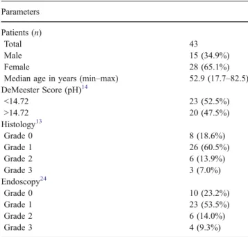
Table 1 Clinicopathologic Parameters of GERD Patients Parameters
Patients (n)
Total 43
Male 15 (34.9%)
Female 28 (65.1%)
Median age in years (min–max) 52.9 (17.7–82.5)
DeMeester Score (pH)14 <14.72 23 (52.5%) >14.72 20 (47.5%) Histology13 Grade 0 8 (18.6%) Grade 1 26 (60.5%) Grade 2 6 (13.9%) Grade 3 3 (7.0%) Endoscopy24 Grade 0 10 (23.2%) Grade 1 23 (53.5%) Grade 2 6 (14.0%) Grade 3 4 (9.3%)
(5%) patient had high-grade dysplasia. Patients with evidence of dysplasia were not included in the statistical analysis because of low patient numbers (Table3).
(3) Barrett’s cancer group: Samples were from 47 patients showing esophageal adenocarcinoma in BE. Normal squamous epithelium was taken from the proximal esophagus as paired control tissue (Table4).
Informed consent was obtained from each patient in accordance to the requirements of our institution’s board of ethics.
Definition of Reflux Esophagitis by Endoscopy and Histopathology
The criteria by Savary and Miller12 were used to define endoscopic GERD into grades I–IV. Morphologic criteria reported by Elster13 were applied for histopathologic classi-fication of reflux esophagitis into grades 0–3 (Tables1and2). All tissue specimens were evaluated by two experienced staff pathologists (S.E.B. and U.D.).
PH Monitoring
Twenty-four-hour pH monitoring was performed by posi-tioning a glass pH electrode (Medtronic Inc., Minneapolis,
MN, USA) 5 cm above the manometrically measured upper border of the lower esophageal sphincter. The electrode was connected to a digital recording device (Medtronic Inc./ Synectics Medical, EsopHogram Reflux Analysis, version 2.01, Minneapolis, MN, USA) and the pH was continuous-ly monitored for 24 h. The following parameters were measured: total percentage of time with pH less than 4, percentage of time the pH was less than 4 when subject was upright, percentage of time the pH was less than 4 when subject was supine, total number of GERD episodes longer than 5 min, time of the longest GERD episode, and composite score based on these parameters.14
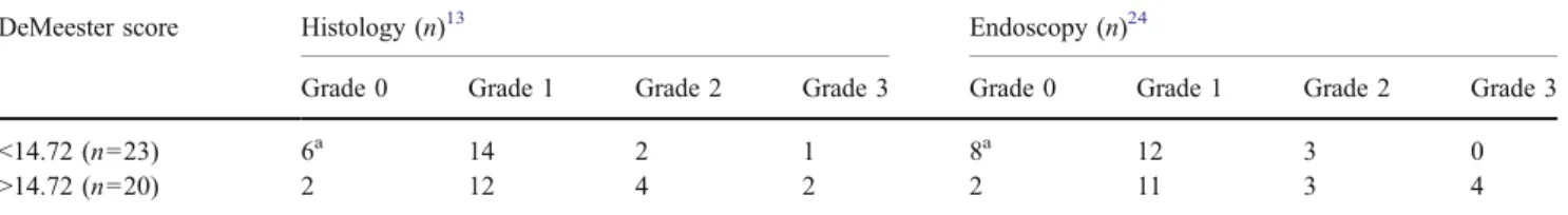
Table 2 Distribution of the DeMeester Score with Histologic and Endoscopic Signs of Reflux
DeMeester score Histology (n)13 Endoscopy (n)24
Grade 0 Grade 1 Grade 2 Grade 3 Grade 0 Grade 1 Grade 2 Grade 3
<14.72 (n=23) 6a 14 2 1 8a 12 3 0
>14.72 (n=20) 2 12 4 2 2 11 3 4
a
No patient was negative for histology and endoscopy at the same time.
Table 3 Clinicopathologic Parameters of Barrett’s Patients
Parameters Patients (n)
Total 20
Male 17 (85%)
Female 3 (15%)
Median Age (min–max) 58.9 (20.6–81.3)
Barrett’s length (n) <1 cm (ultrashort) 5 (25%) 1–3 cm (short) 7 (35%) >3 cm (long) 8 (40%) Dysplasia (n) No dysplasia 15 (75%) Low-grade dysplasia 4 (20%)
High grade dysplasia 1 (5%)
Table 4 Clinicopathologic Parameters of BC Patients Parameters
Patients (n)
Total 47 (85.1%)
Male 45 (95.7%)
Female 2 (4.3%)
Median Age (min./max.) 60.9 (41.4–81.2)
Residual tumor category
R0 40 (85.1%) R1 0 (0%) R2 1 (2.1%) not resected 6 (12.8%) c/pT category T1 20 (42.6%) T2 12 (25.5%) T3 14 (29.8%) T4 1 (2.1%) c/pN category N0 29 (61.7%) N1 18 (38.3%) c/pM category M0 38 (80.9%) M1a 5 (10.6%) M1b 4 (8.5%) Grading G1 3 (6.4%) G2 33 (70.2%) G3 11 (23.4%)
Tumor–Node–Metastasis (pTNM) Pathological Classification: c/pT =
primary tumor, c/pN = regional lymph node metastasis, c/pM = distant metastasis, G = grade of differentiation, R = residual tumor category
RNA Isolation and cDNA Synthesis
Biopsy specimens were bisected and snap-frozen in liquid nitrogen. Representative sections (beginning, middle, and end of sectioning) were stained with hematoxylin and eosin by a standard method and examined by two experienced staff pathologists.
Total RNA was isolated from fresh frozen biopsy halves using the Trizol-Kit (Life Technologies/GIBCO, Grand Island, NY, USA) according to the manufacturer’s instruc-tions. After the generation of cDNA using oligo (dT)18 primers and Moloney murine leukemia virus reverse transcriptase (Clontech Advantage™ Kit, Clontech Lab. Inc., Palo Alto, CA, USA), direct quantitative real-time RT-PCR (TaqMan™, ABI PRISM 7900HT Sequence Detec-tion System Applied Biosystems, Darmstadt, Germany) assays were performed in triplicates to determine COX-2 mRNA expression levels.
Quantitative Real-time RT-PCR
The primers and probes for COX-2 used in the study were previously reported.15 Thermal cycling conditions for COX-2 were 120 s at 50°C and 10 min at 95°C for initial denaturation followed by 40 cycles at 95°C for 15 s and 60°C for 60 s. We used serial dilutions of standard cDNA synthesized from human placenta total cellular RNA (Clontech Lab. Inc.). Triplicates of the tissue samples were assayed in each run. COX-2 levels were standardized with β-actin (ratio COX-2/β-actin) to account for loading differences. Gene expression levels (mRNA) were reported using the median as point estimator and the range of values.
Statistical Analysis
COX-2 mRNA levels and endoscopic and histopathological data were analyzed by nonparametric testing (Wilcoxon rank test, Mann–Whitney test, Kruskal–Wallis test, and Friedmann test). The level of significance was set to p< 0.05 and p values are given for two-sided testing. All
statistical tests were performed using the software package SPSS for Windows, version 11.0, Chicago, IL, USA.
Results
COX-2 Expression in Different Study Groups
COX-2 mRNA expression was detectable by quantitative real-time RT-PCR in all 110 tissue samples. According to the histopathologic group, median COX-2 mRNA expression was lowest in normal squamous epithelium of the distal esophagus (median 0.35, range 0.08–7.8), intermediate in BE (median 0.86, range 0.08–9.61), and highest in esophageal adenocar-cinoma (median 1.62, range 0.001–99.21) (p=0.001). The median value and range of expression levels of COX-2 mRNA in the three study groups are listed in Table5.
In patients with BE without dysplasia, COX-2 expres-sion was significantly higher in metaplastic tissue compared to paired normal squamous epithelium (p=0.03).
Esophageal cancer patients had higher COX-2 mRNA expression levels in cancer tissues compared to paired normal squamous epithelium and BE (p=0.001).
The mean COX-2 mRNA expression of squamous epithe-lium in all three study groups did not show any significant difference (p=0.10). Furthermore, COX-2 mRNA expression in biopsy specimens obtained from histologically and endoscopically classified GERD did not show a significant difference in distal acid-exposed tissues and paired squamous epithelium control tissues (p=0.63). No significant difference in COX-2 mRNA expression of metaplastic Barrett’s epithelium in patients with BE and patients with BC was detected (p=0.29).
COX-2 Expression and Clinicopathological Factors of Patients with GERD
Biopsy specimens obtained from patients with a mean DeMeester score >14.72 showed significantly upregulated median COX-2 mRNA levels in the distal acid-exposed (p = 0.01) esophagus compared with patients having a
Table 5 COX-2 mRNA Expression in Study Groups
Median Min Max p value
GERDa(n=43) Proximal (n=39) 0.3835 0.1058 5.9145 0.63
Distal (n=43) 0.3562 0.0853 7.8081
BE (n=15) Squamous epithelium (n=10) 0.4412 0.0754 2.0350 0.03
Intestinal metaplasia (n=15) 0.8600 0.0838 9.6151
Barrett’s adenocarcinoma (n=47) Squamous epithelium (n=38) 0.2824 0.0001 3.0755 0.001
Intestinal metaplasia (n=15) 1.2295 0.2689 8.8384
Barrett’s carcinoma (n=45) 1.6210 0.0001 99.218
a
negative DeMeester score (Fig.1). No significant correla-tion was detected between COX-2 expression and endo-scopic or histologic findings (p=0.63) (Table5).
COX-2 Expression and Clinicopathological Factors of Patients with Barrett’s Adenocarcinoma
Overexpression of COX-2 mRNA in patients with Barrett’s adenocarcinoma was not associated with grading (p=0.58), T category (p=0.95), N category (p=1.0), or patients’ survival (log-rank test, p=0.70).
Discussion
We present a study on mRNA expression of COX-2 in the reflux MDA sequence. We could reconfirm that progression of BE to esophageal adenocarcinoma is accompanied by an increase in COX-2 expression as reported by other groups.10,11As previously described by Hamoui et al., we could demonstrate that COX-2 expression was significantly correlated with exposure of the distal esophagus to acid reflux, suggesting alteration of COX-2 expression to be one of the earliest specific changes in the reflux MDA sequence.
Epidemiologic studies revealed that the use of COX-2 inhibitors was associated with a decreased risk for esophageal cancer. Much interest was focused on the potential role of COX-2 in esophageal carcinogenesis.7,8 Previous studies analyzed the expression pattern of COX-2 in the MDA sequence. Our group recently demonstrated that COX-2 protein expression by immunohistochemistry was progressively increased in metaplastic, dysplastic, and cancer tissue with the most significant differences between
squamous epithelium and metaplasia and from low-grade to high-grade dysplasia.16 Kuramochi et al.9 measured the gene expression of COX-2 by real-time quantitative polymerase chain reaction in the pathogenesis of Barrett’s adenocarcinoma and also showed a stepwise increase of COX-2 mRNA expression at the different stages. Our results are in agreement with these findings, showing that median COX-2 mRNA expression is stepwise upregulated in Barrett’s metaplasia and adenocarcinoma.
The development of esophageal adenocarcinoma is a multistep process that starts with the mucosal injury of the squamous epithelium of the distal esophagus by GERD and progresses through intestinal metaplasia, dysplasia, to cancer.2,3Whereas several molecular events associated with the progression from metaplastic to cancer tissue have been identified in recent years, little is known about the molecular changes that occur in the beginning of disease.5 This first step, conversion of squamous mucosa to columnar mucosa, is perhaps the most critical because adenocarcinoma cannot develop within squamous mucosa.3 Therefore, we additionally examined COX-2 mRNA expression in esophageal biopsies from patients with GERD. We were able to show that COX-2 expression in biopsies obtained from patients with a positive DeMeester score >14.72 was significantly upregulated compared to patients with a negative DeMeester score. These findings are in agreement with a recent study by Hamoui et al.17In their study, expression levels of several known genes were compared with the degree of acid exposure in the lower esophagus found on 24-h esophageal pH monitoring of 61 patients with GERD. They demonstrated that the expression levels of COX-2 correlated positively with the 24-h pH score, whereas there was no correlation between the expression of other tested genes and esophageal acid Figure 1 COX-2 mRNA in
GERD associated to DeMeester score.
exposure. Therefore, acid reflux disease alters gene expres-sion in esophageal mucosa, and leads to overexpresexpres-sion of COX-2, representing one of the earliest changes associated with gastroesophageal reflux, because in our study the increase in COX-2 expression was independent of the endoscopic or histologic findings in the squamous mucosa. To examine the specificity of this observation, we addi-tionally examined COX-2 mRNA expression in paired specimens derived from proximal esophageal tissue sam-ples, which appeared“normal” on endoscopy and histopa-thology, although cervical 24-h pH monitoring was not performed. Our GERD study patients showed no clinical symptoms of cervical or extra esophageal reflux disease, suggesting that the proximal esophageal epithelium was not exposed to acid reflux. Although dual channel 24-h pH monitoring was not performed, our data suggest that COX-2 mRNA expression was significantly upregulated only in the acid-exposed squamous epithelium of the distal esophagus. A field effect as shown for other genes18could not be detected in our study, thus indicating that COX-2 upregulation is probably an immediate response to acid exposure in the distal esophagus rather than a genetic variation of the entire esophagus.
Chemoprevention strategies might therefore be applied earlier in the neoplastic process because the use of selective COX-2 inhibitors might prevent progression of disease at an early stage.19–21 In fact, studies about severe reflux in rodents confirmed that inhibition of COX-2 with selective inhibitors resulted in a reduced rate of intestinal metaplasia and cancer development.22,23
Large prospective trials with the inclusion of cervical 24-h pH monitoring are needed to validate these preliminary findings.
Conclusion
In summary our findings suggest that alterations in COX-2 mRNA expression occur independently of endoscopic or histologic signs of GERD in the acid-exposed squamous epithelium of the distal esophagus. However, this early COX-2 increase in GERD is further upregulated in Barrett’s metaplasia and BC development for yet unknown reasons.
Acknowledgement This study was supported by the Deutsche
Krebshilfe/Dr. Mildred Scheel Stiftung (German Barrett Cancer Project: 70-2789-Si3, Teilprojekt G1b).
References
1. Sandler RS, Everhart JE, Donowitz M, Adams E, Cronin K, Goodman C, Gemmen E, Shah S, Avdic A, Rubin R. The burden
of selected digestive diseases in the United States.
Gastroenterol-ogy 2002;122:1500–1511.
2. Lagergren J, Bergstrom R, Lindgren A, Nyren O. Symptomatic gastroesophageal reflux as a risk factor for esophageal adenocar-cinoma. N Engl J Med 1999;340:825–831.
3. Peters JH, Hagen JA, DeMeester SR. Barrett’s esophagus. J Gastrointest Surg 2004;8:1–17.
4. Pohl H, Welch HG. The role of overdiagnosis and reclassification in the marked increase of esophageal adenocarcinoma incidence. J
Natl Cancer Inst 2005;97:142–146.
5. Wijnhoven BP, Tilanus HW, Dinjens WN. Molecular biology of
Barrett’s adenocarcinoma. Ann Surg 2001;233:322–337.
6. Buskens CJ, Ristimaki A, Offerhaus GJ, Richel DJ, van Lanschot JJ. Role of cyclooxygenase-2 in the development and treatment of oesophageal adenocarcinoma. Scand J Gastroenterol Suppl
2003:87–93.
7. Funkhouser EM, Sharp GB. Aspirin and reduced risk of
esophageal carcinoma. Cancer 1995;76:1116–1119.
8. van Rees BP, Ristimaki A. Cyclooxygenase-2 in carcinogenesis of
the gastrointestinal tract. Scand J Gastroenterol 2001;36:897–903.
9. Kuramochi H, Vallbohmer D, Uchida K, Schneider S, Hamoui N, Shimizu D, Chandrasoma PT, DeMeester TR, Danenberg KD, Danenberg PV, Peters JH. Quantitative, tissue-specific analysis of cyclooxygenase gene expression in the pathogenesis of Barrett’s adenocarcinoma. J Gastrointest Surg 2004;8:1007–1016. 10. Shirvani VN, Ouatu-Lascar R, Kaur BS, Omary MB, Triadafilopoulos
G. Cyclooxygenase 2 expression in Barrett’s esophagus and
adeno-carcinoma: Ex vivo induction by bile salts and acid exposure.
Gastroenterology 2000;118:487–496.
11. Wilson KT, Fu S, Ramanujam KS, Meltzer SJ. Increased expression
of inducible nitric oxide synthase and cyclooxygenase-2 in Barrett’s
esophagus and associated adenocarcinomas. Cancer Res
1998;58:2929–2934.
12. Savary M, Miller G. L’oesophage. Manuel et atlas d’endoscopie.
1977.
13. Elster K. [Reflux esophagitis-morphology (author’s transl)].
Langenbecks Arch Chir 1978;347:267–270.
14. Johnson LF, DeMeester TR. Twenty-four-hour pH monitoring of the distal esophagus. A quantitative measure of gastroesophageal reflux. Am J Gastroenterol 1974;62:325–332.
15. Xi H, Baldus SE, Warnecke-Eberz U, Brabender J, Neiss S, Metzger R, Ling FC, Dienes HP, Bollschweiler E, Moenig S, Mueller RP, Hoelscher AH, Schneider PM. High cyclooxygenase-2 expression following neoadjuvant radiochemotherapy is associ-ated with minor histopathologic response and poor prognosis in
esophageal cancer. Clin Cancer Res 2005;11:8341–8347.
16. Ling FC, Baldus SE, Khochfar J, Xi H, Neiss S, Brabender J, Metzger R, Drebber U, Dienes HP, Bollschweiler E, Hoelscher AH, Schneider PM. Association of COX-2 expression with corresponding active and chronic inflammatory reactions in
Barrett’s metaplasia and progression to cancer. Histopathology
2007;50:203–209.
17. Hamoui N, Peters JH, Schneider S, Uchida K, Yang D, Vallbohmer D, Hagen JA, DeMeester SR, DeMeester TR, Danenberg K, Danenberg P. Increased acid exposure in patients with gastroesophageal reflux disease influences cyclooxygenase-2 gene expression in the squamous epithelium of the lower
esophagus. Arch Surg 2004;139:712–716.
18. Brabender J, Lord RV, Metzger R, Park J, Salonga D, Danenberg KD, Danenberg PV, Holscher AH, Schneider PM. Differential
SPARC mRNA expression in Barrett’s oesophagus. Br J Cancer
2003;89:1508–1512.
19. Altorki N. COX-2: A target for prevention and treatment of esophageal cancer. J Surg Res 2004;117:114–120.
20. Dannenberg AJ, Altorki NK, Boyle JO, Dang C, Howe LR, Weksler BB, Subbaramaiah K. Cyclo-oxygenase 2: a
pharmaco-logical target for the prevention of cancer. Lancet Oncol
2001;2:544–551.
21. Taketo MM. Cyclooxygenase-2 inhibitors in tumorigenesis (part I).
J Natl Cancer Inst 1998;90:1529–1536.
22. Buttar NS, Wang KK, Leontovich O, Westcott JY, Pacifico RJ, Anderson MA, Krishnadath KK, Lutzke LS, Burgart LJ. Chemo-prevention of esophageal adenocarcinoma by COX-2 inhibitors in
an animal model of Barrett’s esophagus. Gastroenterology
2002;122:1101–1112.
23. Oyama K, Fujimura T, Ninomiya I, Miyashita T, Kinami S, Fushida S, Ohta T, Koichi M. A COX-2 inhibitor prevents the esophageal inflammation–metaplasia–adenocarcinoma sequence in rats. Carcinogenesis 2005;26:565–570.