Journal of Dairy Research (1980), 47, 185-191 Printed in Great Britain
Localization of glycosylated x-casein in bovine casein micelles by
lectin-labelled gold granules
BY MARC HORISBERGER AND MONIQUE VONLANTHEN
Nestle Research Department, CH-1814 La Tour-de-Peilz, Switzerland (Received 22 August 1979)
SUMMARY. The localization of glycosylated /c-casein in isolated bovine casein micelles
was investigated at the ultrastructural level with gold granules labelled with the
Ricinus communis lectin specific for galactose. No evidence was obtained for the
presence of glycosylated /c-casein on the surface of glutaraldehyde-fixed micelles whether or not they had been treated with neuraminidase. Glycosylated /c-casein was mainly located in the bridging network interconnecting the micelles and appeared to be loosely associated with the micelles. When thin sections of micelles were marked, no clear-cut evidence was observed for the presence of intramicellar glycosylated /c-casein.
The major components of casein in bovine milk are the as l, /?- and K-caseins which
exist primarily in the form of micelles 50-200 nm diam. (Carroll, Thompson & Melnychyn, 1971). /c-Casein is the only carbohydrate-containing casein. The carbo-hydrates exist as trisaccharide moieties (NeuNAc <x2 -» 3 Gal/?1 -» 3 GalNAc) attached to the C-terminal portion of /c-casein through /?-0-glycosidic linkages between residues of threonine and GalNAc (Jolles & Fiat, 1979). Recently a tetrasaccharide was isolated containing one additional sialic acid residue (Fournet et al. 1979). Part of/c-casein is free of carbohydrate (Woychik, Kalan & Noelken, 1966; Swaisgood, 1975). As K-casein is the principal casein affected by chymosin (E.C.3.4.23.4) during the primary phase of the milk-clotting process (Alais et al. 1953; Waugh & Von Hippel, 1956; Jolles & Alais, 1959), it plays a major role in the stabilization of the casein micelle (Waugh, 1958).
Several conflicting models have been proposed for the casein micelle. In many models the micelle has a core of asl- and /?-casein and a coat of /c-casein (Waugh &
Noble, 1965; Payens, 1966; Slattery & Evard, 1973) whereas in others K-casein is distributed throughout the interior of the micelle (Parry & Carroll, 1969; Rose, 1969; Gamier & Ribadeau Dumas, 1970; Ashoor et al. 1971). Recent results support a surface position for the carbohydrate (Slattery, 1978; Yoshikawa et al. 1978).
In view of the variety of models proposed, it was considered important to determine whether or not glycosylated /c-casein is located on the surface of the micelle by use of specific cytochemical methods at the ultrastructural level. Recently lectin-labelled gold granules have been found useful in locating carbohydrates either on cell surfaces or on thin sections, as non-specific adsorption of these granules is usually very low (Horisberger & Rosset, 1977; Horisberger, Farr & Volanthen, 1978). Due to their opacity to electrons, gold granules are easily detected by transmission electron
microscopy. The method was therefore applied to the localization of /c-casein using gold granules labelled with the Ricinus communis lectin specific for galactose.
MATERIALS AND METHODS
Materials. Human ceruloplasmin (Type III) and Ricinus communis lectin Type II,
RCAj according to the nomenclature of Nicolson, Blaustein & Etzler (1974), were obtained from Sigma, neuraminidase (500 U/ml, B grade, Vibrio cholerae) from Calbiochem (Hoechst Pharma AG, Zurich, Switzerland), purified bovine serum albumin (BSA) from Behringwerke AG, Marburg, G.F.R., Carbowax 20-M (poly-ethylene glycol compound) from Union Carbide Chemicals Co., and chloroauric acid, HAuCl4 aq. (50 % Au, purum) from Fluka AG, Buchs, Switzerland.
Buffer A was 0-15M-NaCl/0-02M-tris, pH 7-4, buffer B was buffer A containing in addition 0-5 mg/ml Carbowax 20-M and Na azide (0-02 %, w/v). Buffer C was buffer A made 20 mM in CaCl2 and buffer D was 10 mM-imidazole buffer, pH 7-1 containing
20 mM-CaCl2, 70 mM-KCl and 0-02% (w/v) NaN3 (Yoshikawa et al. 1978).
Preparation of casein micelles. Casein micelles were prepared from skim-milk (20 ml)
obtained from pooled milk of Holstein cows as described by Yoshikawa et al. (1978) with the exception that the casein micelle pellet was washed twice in buffer D. The micelle preparation was resuspended in buffer A and 25 % (w/v) glutaraldehyde was added to a final concentration of 0 2 5 % (w/v). After 15 h at 20 °C, the suspension was centrifuged at 100 000 £ for 60 min. The pellet was suspended in buffer D with a few strokes in a Potter-Elvehjem homogenizer (Berdes and Hovwin, Riedstrasse 15a, CH-8042 Zurich, Switzerland) operated by hand. The homogenate was recen-trifuged, homogenized and centrifuged at 600 £ for 1 min to remove the large aggregates. The turbid supernatant (20 ml, A600 =14) was used as
glutaraldehyde-fixed casein micelle preparation in all experiments. All procedures were performed above 20 °C to avoid the possible alterations of micelle structure observed at lower temperatures (Rose, 1968; Downey & Murphy, 1970).
The micelle preparation (6 ml) was incubated for 5 h with V. cholerae neuraminidase (0*6 ml). The suspension was centrifuged twice at 100000 g for 15 min and the pellet was redispersed in the same volume of buffer C.
Preparation of the gold markers. Gold granules Au5 (the code refers to the average
diameter in nm) were prepared by reducing chloroauric acid with white phosphorus (Horisberger & Rosset, 1977). Au5 granules (100 ml) were labelled either with
ceruloplasmin (1-2 mg; C-Au5) or with RCAt (2 mg; RCArAu5) as described previously
(Horisberger et al. 1978). The labelled colloid was recovered by centrifugation and resuspended in buffer B to an absorption A520 = 1 0 .
Marking of casein micelles with RCArAub. The micelle suspension (02 ml) was incubated for 2 h at 20 °C with an excess of RCArAu5 (0.3 ml). The micelles were
centrifuged for 10 min at 2500 £ and washed twice in buffer C. The control was performed in the presence of lactose (60 mg/ml) in the incubating medium. The micelle suspension was either deposited on grids or embedded, and examined by transmission electron microscopy.
Marking of thin sections of micelles. Thin sections were marked either by the one-step
method (Horisberger & Rosset, 1977) or the 2-step method (Horisberger et al. 1978). For the 2-step method, thin sections were successively floated on buffer B containing:
(a) 10 mg/ml BSA (10 min); (b) 1 mg/ml BSA and RCAj (30 min); (c) 1 mg/ml
BSA (5 s); (d) C-Au5 and 1 mg/ml BSA (A520 = 6-3, 30 min). For the one step-method,
Localization of glycosylated K-casein 187
The grids were finally washed 10 times with buffer B (5 s), rinsed once with distilled water and examined by transmission electron microscopy. Control experiments were performed by addition of lactose (50 mg/ml) at steps b and d.
Embedding of casein micelles. Casein micelles were embedded in small agar
micro-capsules (Salyaev, 1968). The micro-capsules were fixed in buffer B containing 2 % (w/v) glutaraldehyde for 1-5 h and stained for 15 h with 0-5% (w/v) uranyl acetate. The capsules were dehydrated and embedded according to Spurr (1969).
Electron microscopy. All preparations were examined without further staining in a
Philips EM 300 electron microscope (Philips, Eindhoven, The Netherlands).
RESULTS
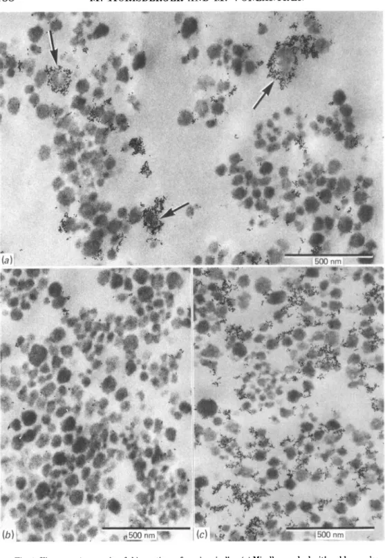
Fig. 1 (a) is an electron micrograph of casein micelles marked with RCArAu5 and
thin-sectioned. The marker indicating the presence of terminal galactose on glycosylated /c-casein was found localized mainly at interconnexions between the micelles. Approximately 70 % of the micelles were associated with one or more gold granules. Only occasionally were micelles surrounded completely by gold granules (Fig. la, arrow). The interior of the micelles was generally free of granules. A control experiment in the presence of lactose, a potent inhibitor of RCAj (Van Wauwe, Loontiens & De Bruyne, 1973), indicated that non-specific adsorption of the marker was low (Fig. 16).
When micelles were treated with neuraminidase and marked with RCAJ-AUJ
essentially the same observations were made with the exception that the proportion of marked micelles increased to 90% (Fig. lc). The control indicated again that non-specific adsorption was very low.
Neuraminidase-treated micelles were also examined without sectioning after deposition on a grid. No evidence of a surface location of glycosylated /c-casein could be obtained. Most of the gold granules were found at interconnexions between micelles (Fig. 2a) although in some cases marking was far removed from the micelle surface (Fig. 2a, arrow).
In an attempt to demonstrate an intracellular presence of glycosylated /c-casein, thin sections of micelles non-treated or treated with neuraminidase were marked. In both cases, marking was low by the one-step method. However, a higher density of marking was achieved by the two-step method (Fig. 26, c). Marking was denser on neuraminidase-treated micelles and was present mainly in the interconnexions between the micelles (Fig. 2c, arrow). No clear-cut evidence for the presence of intracellular glycosylated /c-casein could be obtained. Marking present within the micelle could as well be interpreted as a surface marking of micelle cut tangentially to the surface. Some micelles appeared to be devoid of glycosylated /c-casein (Fig. 26) even when treated with neuraminidase (Fig. 2c).
DISCUSSION
The lectin RCAX is specific for terminal galactose residues (Nicolson et al. 1974;
Surolia, Ahmad & Bachhawat, 1975) although an interaction with sialylated oligo-saccharides having a penultimate galactose residue cannot be excluded (Debray et al. 1979). While the carbohydrate of Ac-casein exists mainly as trisaccharide moieties (NeuNAc a2 -* 3 Gal/?1 -• 3 GalNAc) and tetrasaccharide moieties containing one additional sialic acid residue (Jolles & Fiat, 1979), an excess of galactose has been observed (Fiat, Alais & Jolles, 1972; Fiat et al. 1973). This heterogeneity may explain
188
Fig. 1. Electron micrographs of thin sections of casein micelles, (a) Micelles marked with gold granules labelled with the Ricinus communis lectin RCA,. The arrows indicate the presence of a few micelles completely surrounded by the marker. (6) A control experiment in the presence of lactose, (c) Casein micelles treated with neuraminidase and marked as under (a).
Localization of glycosylated K-casein
189
Fig. 2. (a) Electron micrograph of casein micelles treated with neuraminidase and marked with RCApAuj. The arrow indicates the presence of granules far removed from the micelle surface. (6) Thin sections of micelles marked by the 2-step method, (c) Thin sections of micelles treated with neuraminidase and marked as under (6).
the binding of RCAI-Au5 to the micelle preparation which was not treated with
neuraminidase (Figs l a , 26).
The present study did not give evidence for the presence of glycosylated /c-casein on the micelle surface (Figs 1 a, 6, 2a). Instead glycosylated /c-casein appeared located in the bridging network interconnecting the micelles and seemed to be loosely associated with the micelles. Similar observations have been reported with casein micelles marked with anti-/c-casein antibodies conjugated to ferritin (Parry & Carroll, 1969). However, in this case artifacts could not be ruled out since skim-milk fixed in glutaraldehyde was used instead of isolated casein micelles.
Although part of/c-casein is free of carbohydrate (Woychik et al. 1966; Swaisgood, 1975), 90% of neuraminidase-treated micelles were marked to some extent with
RCAJ-AUJ. This indicated that glycosylated /c-casein is associated with the majority of the micelles. The density of marking did not appear to be related to the micelle size. This is not necessarily at variance with the recent report that the largest micelles contain /c-casein with the largest amount of associated hexose (Slattery, 1978) since marking depends not only on the density but also on the accessibility of receptors to gold granules (Horisberger, 1979).
/c-Casein, the only major casein which contains sulphydryl groups, can form large polymers (Talbot & Waugh, 1970). These polymers are indicated by the presence of large aggregates of RCAj-Au5 (Figs l a , c, 2a).
Since all casein sub-units in a micelle are accessible to high molecular weight reagents such as carboxypeptidase A (Ribadeau Dumas & Gamier, 1970) and papain cross-linked with glutaraldehyde (Ashoor et al. 1971), intramicellar glycosylated /c-casein should also be accessible to neuraminidase whose molecular weight is approximately 90000 (Laver, Pye &.Ada, 1964). However, the apparent marking of intracellular glycosylated /c-casein (Fig. 26, c) may represent a surface or close to the surface marking of a micelle cut tangentially to the surface. Previous attempts to localize intramicellar /c-casein* with a ferritin-antibody conjugate have failed due to non-specific adsorption of the marker on thin sections (Parry & Carroll, 1969).
Recently, the presence of carbohydrate in thin sections of casein micelles was examined by the periodic acid-silver methenamine staining method (Kudo, Iwata & Mada, 1979). The micelles appeared uniformly stained. When a shorter procedure was applied staining was stronger in the outer portions of the large micelles. This was interpreted as indicating that glycosylated /c-casein is distributed throughout the micelle with the more highly glycosylated molecules being located in the outer portions of the particle. However, a slow penetration of the reagent through the micelles would produce the same results if glycosylated /c-casein is located only near the surface of the particle.
Work is therefore proceeding to re-examine the presence of intra-micellar /c-casein by marking thin sections with gold granules labelled with anti-/c-casein antibodies.
The authors thank Mrs M. Weber for the photographic work.
REFERENCES
ALAIS, C , MOCQUOT, G., NITSCHMANN, H. & ZAHLER, P. (1953). Helvetica Chiviica Ada 36, 1955-1968. ASHOOR, S. H., SAIR,, R. A., OLSON, N. F. & RICHARDSON, T. (1971). Biochimicaet Biophysica Acta229,423-430. CARROLL, R. J., THOMPSON, M. P . & MELNYCHYN, P . (1971). Journal of Dairy Science 54, 1245-1252. DEBRAY, H., DECOUT, D., STRECKER, G., MONTREUIL, J . & MONSIGNY, M. (1979). Biologie cellulaire 36, 3a. DOWNEY, W. K. & MURPHY, R. F . (1970). Journal of Dairy Research 37, 361-372.
FIAT, A.-M., ALAIS, C. & JOLLES, P. (1972). European Journal of Biochemistry 27, 408-412. FIAT, A.-M., GOUSSAULT, Y., FONT, J . & JOLLES, P . (1973). Immunochemistry 10, 355-357.
Localization of glycosylated K-casein 191
GARNIER, J . & RIBADEAU DUMAS, B. (1970). Journal of Dairy Research 37, 493-504. HORISBEROER, M. (1979). Biologie cellulaire 36 (in the Press).
HORISBEROER, M., FARR, D. R. & VONLANTHEN, M. (1978). Biochimica et Biophysica Acta 542, 308-314. HORISBEROER, M. & ROSSET, J. (1977). Journal of Histochemistry and Cytochemistry 25, 295-305. JOLLES, P. & ALAIS, C. (1959). Biochimica el Biophysica Acta 34, 565-567.
JOLLES, P . & FIAT, A.-M. (1979). Journal of Dairy Research 46, 187-191. KUDO, S., IWATA, S. & MADA, M. (1979). Journal of Dairy Science 62, 916-920. LAVER, W. G., P Y E , J . & ADA, G. L. (1964). Biochimica et Biophysica Acta 81, 177-180. NICOLSON, G. L., BLAUSTEIN, J. & ETZLER, M. E. (1974). Biochemistry 13, 196-204. PARRY, R. M. J R & CARROLL, R. J . (1969). Biochimica et Biophysica Acta 194, 138-150. PAYEKS, T. A. J. (1966). Journal of Dairy Science 49, 1317-1324.
RIBADEAU DUMAS, B. & GARNIER, J . (1970). Journal of Dairy Research 37, 269-278. ROSE, D. (1968). Journal of Dairy Science 51, 1897-1902.
ROSE, D. (1969). Dairy Science Abstracts 31, 171-175.
SALYAEV, R. K. (1968). Fourth European Regional Conference on Electron Microscopy, Rome 2, 37-38. SLATTERY, C. W. (1978). Biochemistry 17, 1100-1104.
SLATTERY, C. W. & EVARD, R. (1973). Biochimica et Biophysica Acta 317, 529-538. SPURR, A. R. (1969). Journal of Ultrastructural Research 26, 31-43.
SUROLIA, A., AHMAD, A. &, BACHHAWAT, B. K. (1975). Biochimica et Biophysica Acta 404, 83-92. SWAISOOOD, H. (1975). Journal of Dairy Science 58, 583-592.
TALBOT, B. & WAUOH, D. F. (1970). Biochemistry 9, 2807-2813.
VAN WAUWE, J. P., LOONTIENS, F. G. & D E BRUYNE, C. K. (1973). Biochimica et Biophysica Acta 313, 99-105. WAUGH, D. F. (1958). Discussions of the Faraday Society, no. 25, 186-192.
WAUOH, D. F. & NOBLE, R. W. J r (1965). Journal of the American Chemical Society 87, 2246-2257. WAUOH, D. F. & VON HIPPEL, P . H. (1956). Journal of the American Chemical Society 78, 4576-4582. WOYOHIK, J. H., KALAN, E. B. & NOELKEN, M. E. (1966). Biochemistry 5, 2276-2282.
YOSHIKAWA, M., TAKAHATA, K., SASAKI, R. & CHIBA, H. (1978). Agricultural and Biological Chemistry 42, 1923-1926.