Molecular cloning and characterization of NcROP2Fam-1,
a member of the ROP2 family of rhoptry proteins in
Neospora caninum that is targeted by antibodies
neutralizing host cell invasion in vitro
FERIAL ALAEDDINE, ANDREW HEMPHILL*, KARIM DEBACHE and CHRISTOPHE GUIONAUD
Institute of Parasitology, Vetsuisse Faculty, University of Bern, Länggass-Strasse 122, CH-3012 Bern, Switzerland
(Received 12 December 2012; revised 12 February and 16 March 2013; accepted 17 March 2013)
S U M M A R Y
Recent publications demonstrated that a fragment of a Neospora caninum ROP2 family member antigen represents a promising vaccine candidate. We here report on the cloning of the cDNA encoding this protein, N. caninum ROP2 family member 1 (NcROP2Fam-1), its molecular characterization and localization. The protein possesses the hallmarks of ROP2 family members and is apparently devoid of catalytic activity. NcROP2Fam-1 is synthesized as a pre-pro-protein that is matured to 2 proteins of 49 and 55 kDa that localize to rhoptry bulbs. Upon invasion the protein is associated with the nascent parasitophorous vacuole membrane (PVM), evacuoles surrounding the host cell nucleus and, in some instances, the surface of intracellular parasites. Staining was also observed within the cyst wall of‘cysts’ produced in vitro. Interestingly, NcROP2Fam-1 was also detected on the surface of extracellular parasites entering the host cells and antibodies directed against NcROP2Fam-1-specific peptides partially neutralized invasion in vitro. We conclude that, in spite of the general belief that ROP2 family proteins are intracellular antigens, NcROP2Fam-1 can also be considered as an extracellular antigen, a property that should be taken into account in further experiments employing ROP2 family proteins as vaccines. Key words: Neospora caninum, rhoptries, ROP2 family, invasion.
I N T R O D U C T I O N
Neospora caninum, a member of the protozoan
phylum Apicomplexa, is closely related to
Toxoplasma gondii, with whom it shares a number of features, one of which is the intracellular lifestyle. To ensure host cell entry and intracellular survival and replication, most apicomplexan parasites possess specialized secretory organelles, namely micronemes (MIC), rhoptries and dense granules (DG). These organelles play a crucial role in invasion and modul-ation of the host cell (Dubremetz et al. 1993; Carruthers and Sibley, 1997; Ngo et al. 2000; Joiner and Roos, 2002). The contents of these 3 secretory organelles are sequentially released in order to establish invasion and to ensure the maintenance of the parasite–host cell interaction (Fourmaux et al.
1996; Carruthers et al. 1999; Huynh et al. 2003; Carruthers and Tomley, 2008). Initially, MIC proteins are secreted either prior to, or immediately upon, contact with the host cell membrane and they mediate adhesion and attachment to the host cell surface. The secretion of both rhoptry neck (RON)
and MIC proteins contributes to the biogenesis of the moving junction complex (Mordue et al. 1999; Besteiro et al.2009), a transient feature that associates with the host cell plasma membrane and propels the parasite into the developing parasitophorous vacuole (PV) (Alexander et al.2005; Lebrun et al.2005). The PV membrane (PVM) is formed from the host plasma membrane together with proteins secreted from the rhoptries (Saffer et al.1992; Ngo et al.2004; Lebrun et al.2005; Besteiro et al.2009). The DG proteins are exocytosed into the PV both during and after inva-sion and they either remain soluble in the lumen of the PV or become associated with the PVM or the tubulovesicular network (TVN), a membranous structure within the PV. DG proteins modify the environment within the PV, thereby performing essential functions related to intracellular survival and replication (Mercier et al.2005).
Rhoptry proteins have been largely investigated in T. gondii. Subcellular fractionation and proteomic analysis allowed the identification of about 34 rhoptry proteins (Bradley et al. 2005), which were classified into 3 groups. The first group includes rhoptry bulb (ROP) proteins not exhibiting any homology in Plasmodium falciparum and homologous proteins were found only in genera closely related to Toxoplasma including Neospora and Sarcocystis, but
* Corresponding author. Institute of Parasitology, Vetsuisse Faculty, University of Bern, Länggass-Strasse 122, CH-3012 Bern, Switzerland. E-mail: andrew. [email protected]
not in Eimeria. The second group includes proteins restricted to the RON, which are conserved through-out the phylum Apicomplexa, including Plasmodium spp. The third group is formed of proteins encoding functions that are common to most eukaryotic cells, such as kinases, phosphatases and proteases (Bradley et al.2005).
Proteins of the ROP2 family are among the best-studied rhoptry proteins in Toxoplasma and have been implicated in vaccine development (Dlugonska,
2008). This family possesses at least 12 members that share a number of features. All precursors of the ROP2 family have a signal peptide and most of them harbour a pro-region that is cleaved during matu-ration. The eventual maturation site is followed by an arginine-rich (R-rich) domain of 100–150 residues. Their C-termini consist of a kinase domain (El Hajj et al.2006; Reese and Boothroyd,2009), but the neces-sary catalytic residues for ATP-dependent phos-phorylation are found only in some proteins of the family (such as ROP11, 17 and 18) but not in others (ROP2, 4, 5, 7 and 8). Rhoptry proteins have also been identified in N. caninum (Marugán-Hernández et al.2011; Regidor-Cerrillo et al. 2011).
The function of most ROP2 family proteins is still largely unknown. Most members appear to play a role in PV biogenesis and in the modulation of the func-tional properties of the PVM. Anti-sense RNA-mediated depletion of TgROP2 resulted in several harmful effects, such as an impairment of rhoptry biogenesis and cytokinesis, a reduced capacity to invade and replicate in vitro, as well as an attenuation of virulence in mice (Nakaar et al. 2003). It was initially suggested that T. gondii ROP2 forms a tight interaction between the PVM and the host cell mitochondria and endoplasmic reticulum (Sinai and Joiner, 2001; Nakaar et al. 2003; Boothroyd and Dubremetz, 2008); however, recent work by Pernas and Boothroyd (2010) showed this was not the case.
Several studies demonstrated essential roles for TgROP18 in parasite virulence (Saeij et al. 2006; Taylor et al.2006; Melo et al.2011; Niedelman et al.
2012). In vivo, overexpression of TgROP18 increased intracellular tachyzoite multiplication (El Hajj et al.
2007b) and TgROP18 was shown to phosphorylate other ROPs (ROP2, 4 and 8) on the PVM. In ad-dition, TgROP18 binds to and phosphorylates immunity-related GTPases, preventing the clearance of intracellular parasites within inflammatory mono-cytes and IFN-γ-activated macrophages, thus con-ferring parasite survival in vivo and promoting virulence (Fentress et al. 2010; Steinfeldt et al.
2010). In contrast to T. gondii, ROP18 in N. caninum is a pseudogene and the parasite is unable to phos-phorylate the immunity-related GTPase 6 (Reid et al.2012).
To date, no rhoptry protein has been described and characterized in N. caninum and only comparative
genomics of T. gondii and N. caninum revealed that although both the genome and gene expression are remarkably conserved between species, some rhoptry genes are highly divergent (Reid et al. 2012). However, several studies from our group demon-strated that a recombinant, bacterially expressed C-terminal moiety of NcROP2Fam-1 (formerly de-signated recNcROP2) induced highly effective im-munity against challenge infection with N. caninum tachyzoites in both cerebral and fetal infection mouse models (Debache et al.2008,2009,2010).
In the present work, we report on the cloning of the cDNA encoding a full-length NcROP2Fam-1, the molecular characterization of the protein, its localization and its translocation during invasion of target cells by tachyzoites. Moreover, we show that NcROP2Fam-1 is expressed in both tachyzoite and bradyzoite stages. Lastly, using antisera produced against NcROP2Fam-1 antigenic peptides, potential immunoprotective regions of the protein have been identified.
M A T E R I A L S A N D M E T H O D S Cell and parasite cultures
Vero cells and human foreskin fibroblasts (HFF)
were cultured at 37 °C under 5% CO2 in RPMI
1640 medium containing 10% fetal calf serum (Gibco-Invitrogen, Carlsbad, CA, USA), 2 mM L-glutamine, 50 IU mL− 1penicillin and 50μg mL− 1 streptomycin. Neospora caninum (Nc-Liverpool iso-late) tachyzoites were grown in Vero cells in the same conditions but replacing fetal calf serum by 10%
horse serum (Hemphill and Gottstein, 1996).
Cultures of N. caninum-infected keratinocytes were prepared as described previously (Vonlaufen et al.
2002).
Free parasites were obtained by repeatedly passing infected cells through a 25G, 5/8 needle, washed in ice-cold medium and filtered through PD-10 col-umns (GE Healthcare, Piscataway, NJ, USA), as previously described (Hemphill and Gottstein,
1996).
Tachyzoite to bradyzoite in vitro stage conversion was performed using a published protocol (Guionaud et al. 2010), except that sodium nitroprusside (SNP; Sigma, St Louis, MO, USA) was used at 50μMfor 6 days instead of 25μMfor 9 days. BAG1, a classical bradyzoite-specific marker (McAllister et al. 1996; Vonlaufen et al. 2002, 2004), was used to monitor the conversion efficiency. For this purpose, parasites were purified from SNP-treated cells and the expression levels of BAG1 and BAG1 mRNA were compared with those of tachyzoites by immunofluorescence (IF) and quantitative RT-PCR (qRT-RT-PCR), respectively (Fig. S1– in Online version only).
Oligonucleotides
Neospora caninum-specific primers were designed based on nucleotide sequences in the ToxoDB database ((Kissinger et al. 2003; Gajria et al.2008);
http://toxodb.org/toxo/) and on the formerly avail-able ApiDots database ((Li et al. 2003);http://www. cbil.upenn.edu/apidots/). All primers were pur-chased from MWG (Ebersberg, Germany).
RNA isolation andfirst strand cDNA synthesis Total RNA was isolated from N. caninum Liverpool tachyzoites and first-strand cDNA synthesis re-actions were performed as described previously (Matz et al. 1999; Guionaud et al. 2010). Primers NcROP2Fam-1-R (Table 1) and T-primer 3 (5
′-AAGCAGTGGTATCAACGCAGAGTAC(T)30
VN-3′, where V = A, C or G and N = any nucleo-tide) were used to prime the first-strand cDNA synthesis reaction for the 5′ RACE or conventional RT-PCR and 3′ RACE, respectively. A manual hot-start PCR was used in order to reduce background amplifications in conventional PCR and 3′-RACE reactions.
The cDNA corresponding to the 5′-end of
NcROP2Fam-1 was amplified by RT-PCR in a
100μL reaction volume containing 5 μL (1/10th) of
the NcROP2Fam-1-R-primed first strand cDNA,
20 pmol of each NcROP2Fam-1-5′F
(5′-TTCAGA-AACATGCCCCGCTAAC-3′) and
NcROP2Fam-1-5′R (5′-TTGGAGAGGAAGAAGGCGGT-3′)
primers, 0·2 mMof each dNTP and 2 U of Phusion High-Fidelity DNA polymerase (Finnzymes) in the supplied HF reaction buffer. Thirty-five PCR cycles with an annealing temperature of 58 °C were per-formed.
The remainder of the NcROP2Fam-1 cDNA was obtained by 3′-RACE. The 3′-RACE was carried out in a 100μL reaction mixture containing 2 μL
(1/10) of the first-strand cDNA prepared with
T-primer 3, 4 pmol of Heel-carrier primer (5 ′-
CTAATACGACTCACTATAGGGCAAGCAGT-GGTATCAACGCAGAGT-3′), 20 pmol of each
NcROP2Fam-1-3′F
(5′-GAGTAGTATCTTCAT-ACAGAAGAACGG-3′) and Heel-specific
(5′-CTAATACGACTCACTATAGGGC-3′) primers,
0·2 mM of each dNTP and 2·5 U of Phusion High-Fidelity DNA polymerase in the supplied reaction buffer. The PCR reaction comprised 10 cycles with an annealing temperature (Ta) of 62 °C and 30 further cycles with a Ta of 55 °C.
Sequencing and sequence analyses
NcROP2Fam-1 5′-end and 3′-RACE amplicons were cloned into pCR Blunt II TOPO (Invitrogen) and sequenced on both strands using a primer walking approach. Five independent clones were sequenced
T able 1. Primers used for quantita tiv e R T-PCR (The slope and corr ela tion coe ffi cient (R 2 ) a re indica ted for ea ch qRT-PCR.) Primer Sequence (5 ′ to 3′ ) Ta (°C) a T arget cDNA T oxoDB ID (NCLIV_) Loca tion b qRT-PCR Slope R 2 NcR OP2F am-1-F TT CTT CCT CT CC AAGCGA C A 55 NcR OP2F am-1 001970 e1 − 3·51 0·998 NcR OP2F am-1-R TTGAGT CGTT CCCGAAGTTG e1 NcR OP2F am-2-F GC AA TGGA TGCTT GGAGT CTG 55 NcR OP2F am-2 001950 e1 − 3·42 0·999 NcR OP2F am-2-R AGTTGCGGTT C A CCTGGTTG e1 NcTUBA-F GGT AA CGCCTGCTGGGAG 58 NcTUBA (alpha tubulin ) 058890 e1 – e2 − 3·32 0·999 NcTUBA-R GCT CC AAA T C C AAGAAGA CGC A e3 NcBAG1-F CCT CCCT CCGCT GCT A T G 5 5 NcBAG1 (br adyzoite antigen 1) 027470 e2 − 3·33 0·998 NcBAG1-R TT AGCGGCCTT CT C A C CTTTG e3 – e4 a Annealing temper a tur e. b Primers located within a single exon or spanning one exon – exon boundary ar e sho wn as e and e – e, respectiv ely, with e xon number(s) indica ted.
for each PCR product. Sequencing reactions were
carried out using BigDye v3.1 fluorescent dye
terminators and run on an ABI PRISM 3100 Genetic Analyzer (Applied Biosystems, Foster City, CA, USA). Raw sequencing data were assembled and edited with the Staden package (http://staden. sourceforge.net/).
Homology searches were done using BLAST (http://www.ncbi.nlm.nih.gov/blast/) and the Con-served Domain Database CD-Search (http://www. ncbi.nlm.nih.gov/Structure/cdd/wrpsb.cgi) with de-fault settings. Proteins were aligned on the Muscle server (http://www.drive5.com/muscle/), minimally edited and formatted with GeneDoc (http://www. nrbsc.org/gfx/genedoc/). Residue grouping and shading was according to the structurally derived matrix (SDM) 12 reduced alphabet (Prlic et al.2000; Solis and Rackovsky, 2000; Peterson et al. 2009). Potential signal peptide cleavage sites were identified with SignalP 3.0 (http://www.cbs.dtu.dk/services/ SignalP/).
Potential alpha helices in the RAH domain were searched using Jpred3 (http://www.compbio. dundee.ac.uk/www-jpred/), PSIPRED v3.0 (http:// bioinf.cs.ucl.ac.uk/psipred/) and PSSpred (http:// zhanglab.ccmb.med.umich.edu/PSSpred/). The pro-perties of the helices (mean hydrophobicity [kHl] and mean hydrophobic moment [kμHl]) were analysed on the HeliQuest server ((Gautier et al. 2008); http:// heliquest.ipmc.cnrs.fr) and helical wheel projections were plotted using a Java applet (http://rzlab.ucr. edu/scripts/wheel/wheel.cgi).
For Neospora (Liverpool) vs Toxoplasma (ME49) genomic sequence comparisons, 50 kb segments of genomic DNA centred on the loci of interest were extracted from the ToxoDB database and low complexity regions were identified with Re-peat Masker (http://www.repeatmasker.org/cgi-bin/ WEBRepeatMasker). Genomic dot plots were gen-erated using Advanced PipMaker ((Schwartz et al.
2000);http://pipmaker.bx.psu.edu/cgi-bin/pipmaker? advanced).
Quantitative reverse transcription-PCR
RNA transcript steady-state levels were determined by two-step qRT-PCR. Total RNA from free N. caninum tachyzoites or from free parasites cul-tured in bradyzoite conditions (up to 6 days in the presence of 50μM SNP, as indicated) was prepared using Trizol reagent (Invitrogen) and RNA concen-trations were adjusted to 200 ngμL− 1. Since some of the transcripts analysed derive from intron-less genes, RNA was digested with RNase-free DNaseI to degrade contaminating genomic DNA. Briefly, 5μg RNA was incubated for 10 min at room temper-ature with*7 Kunitz units of DNaseI in 100 μL of the 1 × supplied buffer (Qiagen). DNaseI was heat-inactivated for 5 min at 95 °C and RNA cleaned-up
using RNeasy total RNA purification kit columns (Qiagen). RNA was eluted from the columns with RNase-free water and its concentration measured with a spectrophotometer (NanoDrop ND-1000; Thermo Scientific). First-strand cDNA synthesis
was carried out in 40μL, using 3 μg RNA and
Omniscript reverse transcriptase (RT) following the manufacturer’s instructions (Qiagen), except that
1μg random hexamers (Promega) were used to
prime RT reactions. We performed control reactions without RT to exclude the presence of remaining genomic DNA in the cDNA preparations. RT was inactivated by heating for 5 min at 95 °C.
Quantitative PCR was carried out using the
QuantiTect SYBR Green PCR Kit (Qiagen). The reaction mix contained 1 × SYBR Green Mix, 0·5μM each primer (Table 1), cDNA (*40 ng) and water to afinal volume of 20 μL. Quantitative PCR was per-formed on the Rotor-Gene 6000 (Corbett Research, Sydney, Australia). Gene expressions were deter-mined (each sample run in triplicate) using the second derivative maximum method and a standard curve based on 4-fold dilutions (ranging from 1 : 4 up to 1 : 256, each in triplicate) of a positive cDNA sample. Gene expression values were normalized to the housekeeping Neospora alpha tubulin transcripts. For the procedure and associated calculations, we followed the ‘relative standard curve method (Singleplex) using an independent sample for a standard curve’ guidelines, as described in the Ap-plied Biosystems online guide to real-time quantitat-ive PCR (http://www3.appliedbiosystems.com/cms/ groups/mcb_support/documents/generaldocuments/ cms_042380.pdf).
After each run, a high-resolution melting analysis
was performed to confirm the identity of the
amplified products.
Anti (α-) NcROP2Fam-1 polyclonal antibodies (Ab)
Regions of NcROP2Fam-1 towards which a ffinity-purified Ab are directed are presented in Fig. S2A and B– in Online version only.
A fragment of NcROP2Fam-1 (aa 238–594), en-compassing the whole kinase-like domain and re-ferred to here as recNcROP2Fam-1, was expressed in Escherichia coli as a recombinant (His)6-tagged fusion protein. Details on the construction of the recom-binant protein, on the production and a ffinity-purification of rabbit α-NcROP2Fam-1 polyclonal Abs and use thereof to neutralize invasion in vitro can be found elsewhere (Debache et al. 2008). The re-combinant protein, previously dubbed recNcROP2, was used in a number of vaccination trials in mice (Debache et al.2008,2009, 2010).
In addition to α-recNcROP2Fam-1
anti-peptide antisera were produced. During the first round of peptide selection we only retained pep-tides with a high potential antigenicity, as predicted by the consensus epitope search algorithm of the BEPITOPE software (Odorico and Pellequer,2003). Peptides with significant identity to mammalian cell antigens or to other ROP2 family members were eliminated.
The 2 selected NcROP2Fam-1 peptides, P1
(aa 459–472; CH3
CO-CSRQGRSYDPRKHKR-CONH2) and P2 (aa 581–594; CH3
CO-CQGDDGVDETEDTRL-CO2H), with an
N-terminal cysteine residue added for coupling (underlined), were synthesized and used to immu-nize rabbits (Thermo Scientific). Anti-peptide
poly-clonal α-P1 and α-P2 Abs were affinitypurified
using the Thio-Link Gel Kit (Severn Biotech, Kidderminster, UK).
Protein extracts, SDS-polyacrylamide gel electrophoresis (SDS-PAGE) and Western blots
Total protein extracts from free parasites or from infected or uninfected Vero cells were prepared as previously described (Peixoto et al.2010). We used 10μL mL− 1Halt protease inhibitor cocktail (Pierce-Thermo Scientific) in the lysis buffer. Extracts were clarified by centrifugation and the protein concen-tration in the supernatant was measured using the DC Protein Assay Kit (Bio-Rad, Hercules, CA, USA).
Protein samples were mixed with reducing, dena-turing sample buffer, boiled for 5 min and resolved by SDS-PAGE. Proteins were transferred onto nitrocellulose or PVDF membranes (Bio-Rad) for 1 h at 100 V using a wet transfer apparatus (Bio-Rad). Membranes were blocked by incubation in TBST (20 mMTris, 150 mM NaCl, 0·1% (v/v) Tween 20) containing 5% non-fat dry milk. Blots were then incubated for 2 h at room temperature with the diluted primary Abs. We used affinity-purified α-recNcROP2Fam-1,α-P1 and α-P2 Abs at a dilution of 1 : 1500. Detection was performed with alkaline phosphatase-conjugated secondary Abs (Pierce-Thermo Scientific) at a dilution of 1 : 10 000 and NBT/BCIP (nitroblue tetrazolium/bromochloroin-dolyl phosphate; Sigma) for colorimetric detection or Lumi-Phos WB substrate (Pierce-Thermo Scientific) for detection by electrochemiluminescence on a LAS-3000 imaging system (Fujifilm).
Immunofluorescence (IF) analysis
Immunofluorescence staining was performed on
purified parasites or on N. caninum-infected Vero cells grown on poly-L lysine-coated glass coverslips. Free parasites were centrifuged (2000g, 2 min at 4 °C), re-suspended by gentle pipetting in 100μL of fixative (2·5% paraformaldehyde (PFA)/0·05%
glutaraldehyde in PBS), distributed onto poly-L-lysine coated coverslips, left for 15 min at room temperature in thefixative and subsequently kept in pre-cooled acetone/methanol (v/v) for 30 min at −20 °C for permeabilization.
Unless otherwise stated (time-course of infection in vitro, see below), cells were fixed and simul-taneously permeabilized with acetone/methanol. Briefly, coverslips in 24-well plates were rinsed once in PBS, immersed into pre-cooled (−20 °C) acetone/ methanol (v:v) and incubated at−20 °C for 2 h.
For IF staining, coverslips were subsequently washed 3 times with PBS, 5 min each, incubated in
blocking buffer (PBS/3% BSA) for 2 h and the
following primary antibodies (Abs), in PBS/0·3% BSA, were applied for 1 h: (a) affinity-purified rabbit polyclonal Abs α-P1, α-P2 (this work) or α-recNcROP2Fam-1 ((Debache et al.2008) and this work), used each at a 1 : 2000 dilution; (b) α-whole N. caninum extract rabbit antiserum (Hemphill et al.
1996), diluted 1 : 2500; (c) CC2, a rat monoclonal Ab reacting with a yet uncharacterized cyst wall antigen (Gross et al. 1995), diluted 1 : 300; (d) α-BAG1, a polyclonal rabbit Ab directed against BAG1, a classical bradyzoite-specific antigen (McAllister et al.
1996; Vonlaufen et al. 2002, 2004), diluted 1 : 300; (e)α-SAG1, a rat monoclonal Ab directed against the major immunodominant tachyzoite surface antigen (Bjorkman and Hemphill,1998), diluted 1 : 300; and (f) E7, a mouse monoclonal Ab directed against alpha-tubulin (Developmental Studies Hybridoma Bank), diluted 1 : 2000.
Incubations with primary antibodies were fol-lowed by 3 washes in PBS for 5 min each. Alexa Fluor (AF)-labelled secondary antibodies (Invitrogen) at a dilution of 1 : 3000 (AF-488, AF-568) or 1 : 300 (AF-350) in PBS/0·3% BSA were then applied. Finally, the preparations were washed 3 times in PBS for 5 min each and samples were mounted in ProLong Gold anti-fade reagent (Invitrogen). Nuclei were eventually counterstained with 4 ′-6-diamidino-2-phenylindole (DAPI; Sigma) at 1μg mL− 1. Images were acquired on an Axioskop 2fluorescence microscope (Carl Zeiss, Oberkochen, Germany). Confocal microscopy was performed on a FluoView
FV1000 laser scanning microscope (Olympus,
Tokyo, Japan). Images were processed with Image J 1.38 (http://rsb.info.nih.gov/ij/).
Immunogold-labelling and transmission electron microscopy
LR-White embedding and on-section labelling of N. caninum cultures were performed as previously described (Hemphill et al. 1997). Sections were loaded onto Formvar-carbon coated grids and non-specific binding sites were blocked for 2 h in PBS/1% BSA. They were then incubated with rabbit a ffinity-purified α-P1 or α-P2 antibodies diluted at 1:100 in
PBS/0·1% BSA for 1 h. After washing in 5 changes of PBS (2 min each), goatα-rabbit secondary Ab con-jugated to 10 nm diameter gold particles (Amersham) was applied at a dilution of 1:5 in PBS/0·1% BSA. After extensive washing in PBS, grids were air-dried and stained with lead citrate and uranyl acetate. Specimens were viewed on a Philips 400 TEM (Philips Electronics, Eindhoven, the Netherlands) operating at 80 kV.
In order to determine the subcellular localization of NcROP2Fam-1, we analysed a series of trans-mission electron microscopy (TEM) photographs labelled usingα-P2 Ab. We only took into consider-ation rhoptries/portions of rhoptries (N = 34, 23 longitudinal/oblique sections and 11 transversal sections) with (i) sufficient contrast to check for the eventual localization of gold particles within electron-dense rhoptries and (ii) for which the rhoptry surface was clearly delineated.
Immunolocalization of NcROP2Fam-1 during a time-course of infection in vitro
The fate of NcROP2Fam-1 during infection in vitro was assessed by IF after large numbers of parasites were left to adhere to cells for a short duration. NcROP2Fam-1 was then chased for up to 24 h. Briefly, Vero cells (5×10⁴) were seeded onto glass poly-L-lysine-coated coverslips in 24-well tissue cul-ture plates and culcul-tured for 24 h until a subconfluent monolayer (*105 cells) was obtained. The culture medium was aspirated from the tissue culture plates and 2 × 10⁶ freshly purified parasites in 200 μL of RPMI 1640 supplemented with horse serum (com-plete RPMI medium) were added to each well (parasites : cells ratio = 20). After 3 min at room tem-perature, free non-adherent parasites were washed away twice with PBS and once with RPMI 1640. Pre-warmed (37 °C) complete RPMI medium (1 mL) was then added to the wells and plates were returned to the 37 °C/5% CO2 incubator for further short (5–90 min) or longer (24 h) periods of incubation. Infected cultures were then washed with PBS and either (i) fixed with 4% PFA in PBS for 20 min at room temperature and processed immediately for IF analysis or (ii)fixed with 4% PFA in PBS for 20 min at room temperature and permeabilized with 0·1% saponin (Sigma) in PBS for 10 min at room tem-perature.
Invasion inhibition assays
Invasion inhibition assays were carried out in 6-well plates, as previously described (Debache et al.2008). Wells were inoculated with 2 × 10⁵HFF and grown to confluency. Briefly, freshly purified tachyzoites (1 × 10⁶) were pre-incubated for 30 min at room tem-perature in 200μL RPMI 1640 medium containing 1 : 100 dilutions of the following rabbit polyclonal
Abs: α-P1, α-P2, α-recNcROP2Fam-1 (kinase-like domain) or rabbit α-NC1 (whole N. caninum strain NC1 lysate). As controls, tachyzoites were pre-incubated in rabbit pre-immune serum (1 : 100) or in RPMI 1640 medium.
Parasite suspensions were added to HFF mono-layers in 6-well plates for 30 min at 37 °C/5% CO2. Monolayers were washed 3 times with cold RPMI 1640 and incubated with RPMI 1640 medium containing 100μM pyrrolidine dithiocarbamate (PDTC), 0·2μM CuSO2 and α-N. caninum hyper-immune serum (1 : 200) for 2 h at 37 °C/5% CO2to permeabilize adherent, non-invaded (extracellular) tachyzoites. Wells were then washed 3 times with cold RPMI 1640 and incubated for 1 h at 37 °C/5% CO2 with RPMI 1640 containing DNaseI (Roche; 1 mg mL− 1). Wells were then washed twice with cold RPMI 1640 and a third time with RPMI 1640 containing 1 mM EDTA. Finally, monolayers were trypsinized and the DNA was purified (High Pure PCR Purification kit; Roche Diagnostics). DNA concentrations were measured by Hoechst 33 258 (Sigma) fluorimetry (Ausubel et al. 1997) on a Synergy HT plate reader (Biotek Instruments, Winooski, VT, USA). The number of parasite equivalents in each DNA sample (200 ng per assay, each in triplicate) was assessed by real-time PCR on a Light Cycler instrument (Roche Diagnosis, Basel, Switzerland) using an established protocol (Müller et al.2002).
NcROP2Fam-1 cDNA accession number
NcROP2Fam-1 cDNA was submitted to the DDBJ/ EMBL/GenBank databases under accession number HM587954.
R E S U L T S
Cloning and sequence analysis of NcROP2Fam-1
A sequence homology search against N. caninum expressed sequence tags (ESTs) in the ApiDots data-base (Li et al. 2003) using T. gondii ROP2 (TgROP2) cDNA (Beckers et al.1994) as query sequence iden-tified DT.92484732 as best hit. This contig spans *2·8 kb and potentially encodes a 594 amino acid (aa) protein with an overall 47% identity to TgROP2. To confirm the DT.92484732 data, we initially planned to clone and sequence the corresponding cDNA as 2 overlapping 5′- and 3′-RACE products. First-strand cDNA synthesized from total RNA isolated from N. caninum Liverpool tachyzoites was prepared for this purpose. Several attempts to amplify the 5′-end of the transcript by 5′-RACE after tailing thefirst-strand cDNA with either (dG)n or (dA)n homopolymers were not successful and resulted in 5′-truncated products (data not shown). Finally, we amplified a nearly complete 5′-end cDNA
by conventional RT-PCR. The 3′-end of the
DT.92484732 cDNA was obtained by 3′-RACE.
We cloned the 5′-end RT-PCR and 3′-RACE pro-ducts and sequenced 5 clones of each. A composite, consensus 2278 bp cDNA sequence made out of the 5′-end RT-PCR and 3′-RACE overlapping products was submitted to the DDBJ/EMBL/GenBank data-bases under accession number HM587954.
The cDNA possesses a 1785 nucleotides open reading frame (ORF) starting with an ATG initiation codon that is part of a sequence (TTCACCATGG) matching 7 positions of the previously described T. gondii translation initiation consensus sequence
(gNCAAaATGg) (Seeber, 1997). The ORF
poten-tially encodes a 594 aa protein matching perfectly
the predicted translated product of ApiDots
DT.92484732/ToxoDB NCLIV_001970 transcripts.
In addition to the *47% identity shared with
T. gondii ROP2 and ROP8, the most closely related rhoptry proteins in Toxoplasma (Fig. S3– in Online version only), the predicted protein also shares 52% identity with the predicted protein encoded by NCLIV_001950, a tandem, expressed gene (Figs S3 and S4B– in Online version only) located on N. caninum Liverpool chromosome Ia. Both genes are located in a region of synteny shared with the region around the ROP4/7 locus on Toxoplasma
ME49 chromosome Ia (Fig. S5A – in Online
version only, left panel). Such a syntenic region does not exist on chromosome X between Neospora
NCLIV_001970/NCLIV_001950 and the
Toxoplasma ROP2/8 locus (Fig. S5A, right panel). Therefore, in spite of a higher homology of Neospora
NCLIV_001970 and NCLIV_001950 predicted
proteins with TgROP2 and TgROP8 (Fig. S3), we do not believe these gene pairs are orthologous. A recent report (Reid et al. 2012) established NCLIV_001950 and NCLIV_001970 as the syntenic orthologues of Toxoplasma ROP4 and ROP7, respectively. While this might be correct for the pair NCLIV_001950/ROP4, our genomic analysis
suggests that it is unlikely that ROP7 and
NCLIV_001970 are orthologues (Fig. S5B). For this reason, in the present work, NCLIV_001970 was named NcROP2Fam-1 (N. caninum ROP2 family
member 1) and NCLIV_001950 was named
NcROP2Fam-2.
NcROP2Fam-1 possesses all features of ROP2 family proteins
The predicted NcROP2Fam-1 precursor is 67 kDa and its deduced primary sequence exhibits all char-acteristics of ROP2 family members (Fig. S3A). The protein contains an N-terminal predicted signal peptide with a computed cleavage site between resi-dues 28 and 29. In its N-terminal domain, a sequence is found (SWLQ, aa 82–85), that is reminiscent of the SФxE consensus (where Ф is a hydrophobic residue
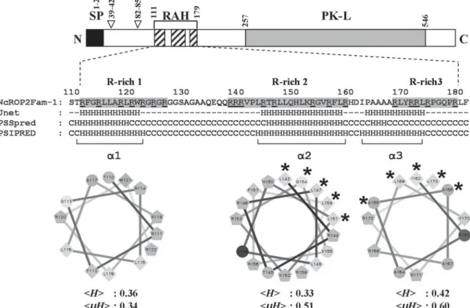
and x is any aa) proposed in T. gondii as a likely processing site for the TgSUB2 protease by Miller et al. (2003) and that was recently re-defined as SФx (E/D) by Hajagos et al. (2011). Whether this site in NcROP2Fam-1 actually represents a cleavage site for a TgSUB2-like Neospora maturase is uncertain, since there is a negatively charged (E) to neutral (Q) amino acid substitution at position P1. A second, although perfect match to the TgSUB2 consensus (SWDD, aa 39–42) was identified upstream in the NcROP2Fam-1 precursor. This site is not conserved in other ROP2 family members (Fig. S3A). Three arginine-rich sequences (R-rich 1–3), collectively referred to as the RAH (R-rich Amphipathic Helix) domain, were also found in the N-terminal domain of NcROP2Fam-1 (aa 111–126, 138–159 and 168–179; precursor num-bering; Fig. S3A and Fig. 1). This domain was recently shown to mediate the attachment of ROP2 family proteins to membranes and especially to the PVM (Reese and Boothroyd, 2009; Fentress et al.
2012). Combining secondary structure predictions andα-helix properties analysis, we inferred the pres-ence of 2 amphipathic α-helices within the portion of the RAH domain containing R-rich 2 and R-rich 3 (Fig. 1). Whereas R-rich 1 harboured a predicted α-helix (aa 110–122) it did not have a marked hydrophobic face (Fig. 1).
Additionally, a serine/threonine protein kinase-like domain was detected in the C-terminal moiety of NcROP2Fam-1 (aa 257–546;Fig. 1and Fig. S3A). However, this domain lacks 3 of the most invariant key residues (K, D and D residues of Hanks motifs II, VIb and VII, respectively) commonly found in ATP-binding and catalytic sites of active kinases (Hanks and Hunter, 1995). It is therefore unlikely that NcROP2Fam-1 is an active kinase. NcROP2Fam-1 also possesses a hydrophobic stretch (aa 473–489; Fig. S3A) encompassing Hanks domain IX that is buried within the core of other rhoptry proteins adopting a kinase fold (El Hajj et al.2006,
2007a,2007b; Labesse et al.2009; Qiu et al.2009). Lastly, a non-canonical motif (FENI; aa 546–549, Fig. S3A), reminiscent of the YxxФ motif (where x is any aa andФ is hydrophobic), as well as 7 dileucine motifs (LL; not shown) that may function as sorting signals mediating trafficking to the rhoptries (Hoppe et al. 2000; Ngo et al. 2003) were found in NcROP2Fam-1.
NcROP2Fam-1 undergoes a proteolytic maturation
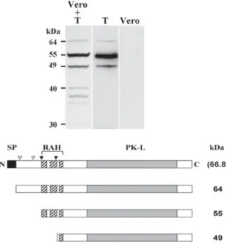
On Western blots of lysates of free N. caninum tachyzoites, α-P2 antibodies, raised against the C-terminal tail of NcROP2Fam-1, detected 2 major bands of 55 and 49 kDa, as well as a fainter band of 64 kDa, the latter being compatible with the predicted size of the pro-NcROP2Fam-1 precursor (Fig. 2). The 55 and 49 kDa bands were repeatedly observed in all lysates of tachyzoites, regardless of
which of the 3 antibodies was used (Fig. S2C– in Online version only) or if parasites were directly boiled in sample buffer or not. We therefore exclude that the major 55 kDa or the fainter 49 kDa bands may be artifactual (either resulting from protein degradation in a sample or from the cross-reactivity of one antibody with a ROP protein or any unrelated Neospora protein). The same protein pattern was also observed in lysates of parasite-infected cells, although the 64 kDa pre-pro-protein was not visible in all samples (data not shown). Additional faint bands of 30–40 kDa, not present in lysates of free tachyzoites, were detected in infected cells lysates and are likely to be due to protein degradation. There was no visible cross-reactivity of α-P2 with uninfected Vero cell lysate (Fig. 2).
The positions of the processing sites inferred from the sizes of the observed mature NcROP2Fam-1 are not compatible with a cleavage at any of the putative TgSUB2 sites mentioned above. Indeed, a cleavage of pro-NcROP2Fam-1 would give rise to a 62·5 kDa
or a 58 kDa mature protein, depending on whether the upstream or the downstream site is used, respectively. Instead, our results indicate that pro-NcROP2Fam-1 is cleaved around F 112 (pre-pro-protein numbering; major 55 kDa band), leaving the whole RAH domain intact, or around F 157 (minor 49 kDa band), leaving only R-rich 3 on mature NcROP2Fam-1 (Fig. 2).
NcROP2Fam-1 is associated with rhoptry bulbs in parasites
Affinity-purified antibodies were used to localize NcROP2Fam-1 by IF and TEM. In routine cultures of infected cells, after membrane permeabilization, NcROP2Fam-1 was detected within the apical region of intracellular parasites, at locations compatible with a rhoptry staining (Fig. 3A). In some infected cells, and only with a standard but not with a confocal microscope, we observed a punctate staining within the PV and in the host cell cytosol (not shown).
Fig. 1. Helical wheel projections of putativeα helices within the NcROP2Fam-1 RAH domain. Top: schematic representation of pre-pro-NcROP2Fam-1. Black box: signal peptide (SP); empty arrowheads: regions reminiscent of the classical SΦx(E/D) pro-ROP processing site; hatched boxes: arginine-rich (R-rich) regions 1–3 of the RAH domain; grey box: protein kinase-like (PK-L) domain. Middle: sequence of NcROP2Fam-1 RAH domain (aa 111–179, precursor protein numbering) with R-rich regions 1–3 shaded and arginine residues underlined (single letter code, numbering according to the precursor protein). Secondary structure predictions by the Jnet, PSSpred and PSIPRED servers are displayed below NcROP2Fam-1 RAH domain sequence (H: helix; C: coil; dashes: undefined). Bottom: helical wheel projections of the corresponding putative alpha helices bracketed (α1–α3). Diamonds: hydrophobic residues; pentagons: positively charged residues; circles: hydrophilic, uncharged residues. Mean hydrophobicities (kHl) and mean
hydrophobic moments (kμHl), as determined by the HeliQuest server, are indicated. Amino acids on the hydrophobic face, if any, are indicated by asterisks.
Immunogold-TEM was performed on N. caninum-infected keratinocytes usingα-P1 or α-P2 antibodies. In tachyzoites, labelling was clearly associated with rhoptry bulbs (Fig. 3B; Fig. S2D and Table S1– in Online version only).
NcROP2Fam-1 is secreted and associates with the PVM and the surface of invaded and adherent parasites
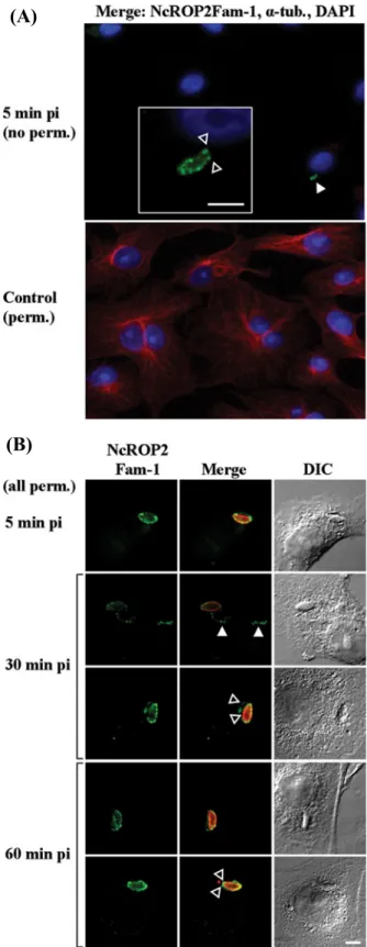
Neospora caninum tachyzoites were allowed to
interact with host cells for 3 min and non-invaded/ non-adherent parasites were washed away. This time-point was designated as time-time-point zero of infection. At 5 min post-infection (p.i.) and without permea-bilization, anti-NcROP2Fam-1 antibodies revealed a punctate staining pattern on the surface of a small amount of parasites per coverslip, while a monoclonal Ab directed against cellular alpha-tubulin applied simultaneously failed to stain the host cell cytoske-leton, confirming that under our experimental con-ditions the cellular membrane was not compromised
and therefore that only adherent, but not already invaded, parasites were accessible to antibodies (Fig. 4A). The labelling on the parasite pictured presents a truncation at the anterior end, where it already has penetrated the host cell; this indicates that the parasite membrane was also impermeant to antibodies and that NcROP2Fam-1 is indeed on its surface. The small number of parasites we detected per slide is most certainly due to the fact that invasion is known to process rapidly and therefore most parasites were already within the host cells at 5 min p.i. After 5 min p.i., tachyzoites were not detected anymore without permeabilization, indicating that they all had completely invaded the host cells (data not shown), suggesting that the parasites that were labelled with NcROP2Fam-1 antibodies at 5 min p.i. were viable and not arrested in the adhesion phase.
Fig. 2. NcROP2Fam-1 is expressed in tachyzoites and undergoes processing. Top: affinity-purified polyclonal antibodies raised against the C-terminus of
NcROP2Fam-1 (α-P2) were used to detect the protein byWestern blot in lysates of infected Vero cells containing *5×10⁵
tachyzoites (Vero + T), 5 × 10⁵purified tachyzoites (T), or 5 × 10⁴uninfected Vero cells (Vero). Apparent MWs of the bands detected are indicated. Bottom: a schematic representation of pre-pro-NcROP2Fam-1 is shown (66·8 kDa, deduced from the cDNA sequence and not visible on the blot) together with the proteins inferred from the bands detected by Western blot. Black box: signal peptide (SP); empty arrowheads: regions reminiscent of the classical SΦx(E/D) pro-ROP processing site;filled arrowheads: actual processing sites, as deduced from the Western blot; hatched boxes: R-rich regions 1–3 of the RAH domain; grey box: protein kinase-like (PK-L) domain.
(A)
(B)
Fig. 3. NcROP2Fam-1 is a rhoptry protein associated with rhoptry bulbs. (A) Confocal laser scanning microscopy. NcROP2Fam-1 was detected in a double staining indirect immunofluorescence experiment in fixed and permeabilized infected cells. Primary antibodies used wereα-P2 (NcROP2Fam-1; green) and α-Neospora whole extract (red in Merge pictures). The images show a unique plane. DIC: differential interference contrast images. Scale bars = 5μm; (B) Transmission electron microscopy. Immunogold staining of NcROP2Fam-1 in N. caninum-infected HFF cells was performed using affinity-purified α-P2 primary Ab and a colloidal gold-conjugated secondary Ab. A longitudinal section of a parasite (only the apical end shown) is presented. Average gold particles diameter = 10 nm.
In permeabilized preparations, antibodies strongly labelled the newly formed PV (Fig. 4B). In addition, NcROP2Fam-1 was also observed as punctuations associated with cytoplasmic filaments that we ident-ified as evacuoles (Hakansson et al.2001; Saeij et al.
2006; Taylor et al. 2006; Turetzky et al. 2010) (Fig. 4B,filled arrowheads). At 30–60 min p.i., most parasites appeared closely associated with the host cell perinuclear region. In addition, NcROP2Fam-1-immunoreactive evacuoles were longer, sometimes branched, and most of them surrounded the host cell nucleus (not shown). On some occasions we clearly observed that NcROP2Fam-1 was not only detected on the nascent PVM, but also on the surface of intracellular parasites (Fig. 4, empty arrowheads). NcROP2Fam-1 staining intensity decreased pro-gressively starting from 60–90 min p.i. and at 24 h p.i., the protein was exclusively distributed at the parasite apex, consistent with its localization in rhoptries (not shown).
Tachyzoites and bradyzoites express similar levels of NcROP2Fam-1
Neospora caninum bradyzoites were generated in vitro by growing parasites for 6 days in the presence of SNP added to the culture medium. Preparations of
(A)
(B)
Fig. 4. NcROP2Fam-1 is a secreted rhoptry protein that associates with the PVM and the surface of invaded and adherent parasites. Freshly purified tachyzoites were left to invade Vero cells for 3 min and non-adherent parasites were washed away. After addition of pre-heated culture medium, preparations were returned to the incubator and infection was left to proceed for times ranging from 5 min to 24 h. Preparations werefixed and processed for double-staining IF analysis without (no perm.) or with prior permeabilization (perm.). For simplification purposes the times pi indicated omit the 3 min during which parasites
were left in contact with cells. (A) NcROP2Fam-1 is present on the parasite surface of some adherent Neospora caninum tachyzoites. The following primary antibodies and appropriate Alexa Fluor-labelled secondary Abs were used: 12G10, a monoclonal Ab reacting against a broad range of alpha-tubulins (α tub., red) was used to check the integrity of the host cell membrane in the absence of permeabilization andα-P2 Ab (green) was used to stain NcROP2Fam-1. Nuclei were counterstained with DAPI. Only the merged (3-colours) pictures are presented. Top picture: no permeabilization prior to staining. Thefilled arrowhead points towards an adherent parasite coated with NcROP2Fam-1. A higher magnification view of the parasite is shown in the box. Note the truncated aspect of the extracellular NcROP2Fam-1 staining in the apical region (empty arrowheads), indicating that (i) this adherent parasite was actively invading the host cell when invasion was arrested byfixation and (ii) the parasite body was itself impermeant to antibodies. Bottom: a
permeabilized control slide was processed in parallel to check the efficiency of alpha-tubulin staining by 12G10 mAb (red). Scale bar = 5μm; (B) NcROP2Fam-1 is present on the PVM, in evacuoles and on the surface of intracellular parasites after invasion. Images were taken using a laser scanning confocal microscope showing a unique plane. NcROP2Fam-1 was detected withα-P2 Ab (green) andα-SAG1 was used to stain the parasites (red in Merge panel) after permeabilization. DIC images are presented on the left. Filled arrowheads: staining of evacuoles; empty arrowheads: staining of both the PVM and the parasite surface. Scale bars = 5μm.
tachyzoites and bradyzoites were split into 2 aliquots each. Thefirst aliquots of infected cells were used for IF experiments. Free parasites were purified from the second aliquots and were used for controlling the tachyzoite to bradyzoite stage conversion by IF, for the assessment of NcROP2Fam-1 levels by real-time RT-PCR and for Western blot experiments.
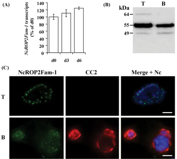
The efficiency of the stage conversion was con-trolled at day (d) 6 by checking the expression of NcBAG1 (a classical bradyzoite marker) in parasites isolated from the culture. Nearly 70% of individual d6 parasites displayed a stronger BAG1 staining than d0 parasites (tachyzoites), while BAG1 transcripts increased by 15-fold (Fig. S1).
NcROP2Fam-1 transcripts in tachyzoites and bradyzoites were quantified by real-time RT-PCR. NcROP2Fam-1 steady-state levels were very similar in tachyzoites (d0), at d3 of stage conversion and in cultures containing mainly bradyzoites (d6) (Fig. 5A), indicating that NcROP2Fam-1 is not a gene with a stage-specific pattern of transcription. Using the same approach we could show that NcROP2Fam-2 is also transcribed in both tachy-zoites and bradytachy-zoites, but appears to be 2-to3-fold more expressed in d6 bradyzoites than in d0 tachyzoites (Fig. S4B).
Western blots of lysates prepared from purified tachyzoites (d0) and from d6 bradyzoites indicated
(A)
(B)
(C)
Fig. 5. NcROP2Fam-1 is expressed by bradyzoites and accumulates in the cyst wall of‘cysts’ produced in vitro. (A) Quantitative RT-PCR. Total RNA was isolated from purified parasites at days (d) 0, 3 and 6 of a tachyzoite to bradyzoite conversion time-course. NcROP2Fam-1 steady state levels were determined by real-time RT-PCR and values were normalized to Neospora alpha-tubulin. Results were expressed as the mean percentage of the value obtained at d0 (tachyzoites) ±S.D. (assays were carried out in triplicate and each one with triplicate PCR reactions); (B) Western blot analysis. Affinity-purified α-P2 antibodies were used to probe NcROP2Fam-1 in lysates of *5×10⁵parasites purified from untreated Vero cells (T; tachyzoite culture conditions) or from SNP-treated Vero cells (B; d6 of bradyzoite culture conditions). Apparent MWs are indicated on the left; (C) IF analysis. Neospora caninum were cultured in untreated Vero cells (T; d0) or in SNP-treated cells (B; d6) and intracellular parasites were then analysed by indirect IF afterfixation and permeabilization. Primary antibodies were: α-P2 affinity-purified polyclonal Ab (NcROP2Fam-1; green), a mAb directed against a cyst-specific antigen (CC2; red) and a rabbit antiserum directed against N. caninum crude extract (Nc; blue in Merge panel). Image acquisition was performed with identical exposure time for bradyzoite and tachyzoite stages. Scale bar = 10μm.
that both stages expressed the two major forms (55 and 49 kDa) of mature NcROP2Fam-1 (Fig. 5B).
Next, we compared by IF the localization of NcROP2Fam-1 in infected cells cultured in the presence of SNP for 6 days (bradyzoite conditions) to its localization in infected cells grown in tachyzoite conditions (no SNP, 48 h p.i.). NcROP2Fam-1 stain-ing was observed in the rhoptries of all intracellular parasites, irrespective of the culture conditions. We also noticed that NcROP2Fam-1 staining was much more prominent in d6 cysts (bradyzoite culture) than in PVs (tachyzoite culture; staining not visible with the exposure settings in Fig. 5C). NcROP2Fam-1 and the bradyzoite-specific CC2-reactive cyst wall antigen are both found at the periphery of cysts (Fig. 5C).
Selected specific anti-peptide antibodies directed against NcROP2Fam-1 partially neutralize host cell invasion in vitro
In a previous study from our group (Debache et al.
2008), incubation of Neospora tachyzoites in the presence of antibodies against rec-NcROP2Fam-1
significantly reduced invasion in vitro, thus suggest-ing that NcROP2Fam-1 may have a previously un-suspected function in adhesion and/or invasion and that it may also be accessible to antibodies. Since rec-NcROP2Fam-1 likely shares common epitopes with other ROP2 family members in Neospora (Fig. S3A), the neutralization effect observed could possibly have been due to recognition of a cross-reacting antigen on the parasite. We therefore tested whether anti-peptide antibodies prepared against unique regions of NcROP2Fam-1 (α-P1 and α-P2) would also inhibit invasion in vitro.
No reduction of invasion was observed when tachyzoites were pre-incubated with normal rabbit serum (NRS) in RPMI medium. In contrast, pre-incubation of parasites with α-P1 or α-P2 reduced invasion by 46% and 58%, respectively. Likewise, invasion was also significantly reduced by 72% or 58% withα-recNcROP2Fam-1 or with α-Nc Ab, an anti-serum against N. caninum whole extract, respectively (Fig. 6). Altogether these results demonstrate that antibodies directed against specific NcROP2Fam-1-epitopes can partially neutralize invasion in vitro.
D I S C U S S I O N
In T. gondii, the ROP2 family of rhoptry proteins was initially described as a family of 16 proteins sharing a number of features with the ROP2 prototype mem-ber (El Hajj et al.2006); yet no protein of this family has so far been characterized in the related genus Neospora. Using a reverse genetics approach, we iden-tified and cloned a cDNA encoding a ROP2 family member in N. caninum. The cDNA sequence was in perfect agreement with the original NCLIV_001970 in ToxoDB v. 7.1 (Kissinger et al. 2003; Gajria et al.2008). The NCLIV_001970 deduced primary amino-acid sequence showed most significant hom-ology to NCLIV_001950, an expressed, tandemly arranged gene on Neospora chromosome Ia, and to a lesser extent to Toxoplasma rhoptry proteins (ROP2, 4, 7, 8 and 18). It was also surprising to notice that apparently Neospora does not possess a ROP18 gene, hence NcROP18 is pseudogenized, as recently demonstrated by Reid et al. (2012). In addition, within the same clade only two genes (NcROP2Fam-1 and NcROP2Fam-2) were found in Neospora, whereas Toxoplasma has a set of four to five (Tg ROP2 (a/b), 4, 7 and 8). These differences may reflect a degree of redundancy in the functions of Toxoplasma proteins or could contribute to different strategies of adaptation of the two species in their respective hosts (Reid et al. 2012). The predicted NcROP2Fam-1 protein possesses the hallmarks of ROP2 family proteins (El Hajj et al. 2006), each of which will be further discussed. In the N-terminal moiety of the predicted NcROP2Fam-1 we found a potential signal peptide, as well as an approximate match (SWLQ) and a perfect match (SWDD) to the
Fig. 6. Antibodies raised against unique peptides of NcROP2Fam-1 partially neutralize invasion in vitro. Dilutions of antibodies (1:100) in medium were tested for their ability to block parasite invasion. Freshly isolated Neospora caninum tachyzoites were incubated in the presence of medium alone (RPMI), NRS,
affinity-purified antibodies against unique, NcROP2Fam-1-derived peptides (α-P1 and α-P2; Fig. S2A and B – in Online version only) or against the whole PK-L domain (α-recNcROP2Fam-1) or antibodies against a whole N. caninum extract (Nc). Parasites were then used to infect HFF cells and the amount of invaded, intracellular parasites was determined by real-time PCR using Neospora-specific primers. Results were expressed as the mean percentage of the value obtained for RPMI alone ±S.D. (triplicate PCR reactions). These experiments were done 3 times with a virtually identical outcome, and 1 representative result is shown.
SΦx(E/D) consensus for the TgSUB2 maturase (Bradley and Boothroyd, 1999; Miller et al. 2003; Hajagos et al.2011), suggesting that NcROP2Fam-1 might be synthesized as a pre-pro-protein. The N-terminal moiety of NcROP2Fam-1 also contained an RAH domain made out of three R-rich regions and a serine/threonin protein kinase-like (PK-L) domain.
Due to the high overall homology of the
NcROP2Fam-1 PK-L domain with those of
TgROP2 and TgROP8, for which the structure has been established (Labesse et al.2009; Qiu et al.
2009), we anticipate that the PK-L domain of NcROP2Fam-1 certainly also adopts the kinase fold. Unlike TgROP18, the only ROP2 family protein with a demonstrated kinase activity (El Hajj et al.2007b) and similar to most other members of the ROP2 family in Toxoplasma (El Hajj et al. 2006), NcROP2Fam-1 lacks essential, conserved residues present within the catalytic core of classical kinases
(Hanks and Hunter, 1995). NcROP2Fam-1 can
therefore be qualified as a pseudokinase (Boudeau et al.2006). However, it would be difficult to make any assumption as to whether NcROP2Fam-1 might nevertheless have a kinase activity since a number of pseudokinases, such as WNKs (with-no-lysine (K)) or CASK (Ca2 +/calmodulin-dependent serine kinase) have evolved alternative ways of binding ATP and performing the phosphoryl transfer reaction (Xu et al. 2000; Mukherjee et al.2008).
In order to proceed with the characterization of NcROP2Fam-1, it was necessary to obtain specific antibodies that would not cross-react with e.g. other ROP2 family members. The selection process we used in choosing unique regions of NcROP2Fam-1 for the generation of specific anti-peptide antibodies is fully detailed in the Materials and Methods section. The fact that both anti-NcROP2Fam-1 anti-peptide antibodies (α-P1 and α-P2) recognized the same patterns in Western blot and immunolocalization (IF and TEM) experiments provides strong evidence that our selection strategy was successful.
Most rhoptry proteins studied are synthesized as pre-pro-proteins (Sadak et al.1988; Bradley and Boothroyd, 1999; Carey et al. 2004; El Hajj et al.
2006, 2007b; Turetzky et al. 2010; Hajagos et al.
2011). The signal peptide is cleaved as soon as the precursor enters the secretory pathway; however, the removal of the pro-domain occurs much later, within the nascent rhoptries (Soldati et al.1998).
Western blot analysis of purified tachyzoite lysates with three different antibodies raised against the C-terminal moiety of the protein revealed that at least two N-terminal processing events must occur follow-ing the synthesis of the NcROP2Fam-1 precursor. A faint band of 64 kDa was observed, the size of which was compatible with the predicted pro-NcROP2Fam-1. In addition, we observed two ad-ditional bands of 55 and 49 kDa (the 55 kDa band being prominent), indicating a further maturation of
NcROP2Fam-1. TgSUB2, a subtilisin-like pro-tease present in rhoptry bulbs, is viewed as the most likely maturase candidate for N-terminal maturation of rhoptry proteins (Miller et al. 2003). In earlier studies, it was shown that the cleavage occurs at SΦxE sites on rhoptry proteins (Bradley and Boothroyd,
1999; Miller et al.2003) and that mutations at the P1 position of the cleavage site (E in SФxE) had a dele-terious effect on the processing (Bradley et al.2002; Miller et al.2003; Turetzky et al.2010). However, a recent study by Hajagos et al. (2011) showed that the P1 site could accommodate a conservative E to D substitution, thus re-defining the TgSUB2
consen-sus as SΦx(E/D). In NcROP2Fam-1, we found a
perfect match to the cleavage consensus (SWDD (aa 39–42)) at a position not conserved in ROP2 family proteins and a poorer match (SWLQ (aa 82–85)) at a conserved position, but with a negatively charged (E) to neutral (Q) substitution at the P1 site that would likely impair cleavage. Remarkably, neither the 55 nor the 49 kDa bands that we observed in Western blots are compatible with a maturation of NcROP2Fam-1 at any of the above-mentioned sites (the expected sizes would be 62·5 and 58 kDa). We therefore conclude that the processing of pro-NcROP2Fam-1 in Neospora involves a protease with a different substrate specificity than that of TgSUB2. The bands we observed also do notfit with the yet unidentified rhoptry protein maturase acting at RAMA1-like sites (SxL) in T. gondii (Hajagos et al.
2011). We therefore cannot determine the exact locations of the two maturation events that take place in pro-NcROP2Fam-1. In lysates of infected cells, additional bands of 30–40 kDa, which were not present in lysates of cell-free, purified parasites, were detected. This indicates that NcROP2Fam-1 may be subjected to a variety of additional proteolytic activities once it has been secreted by the parasites.
In eukaryotic cells, sorting of integral proteins to subcellular organelles involves conserved mech-anisms mediated by cytoplasmic adaptor complexes (APs), which recognize short motifs, usually either tyrosine-based (YxxΦ)ordileucine-based (LL),expo-sed on the cytoplasmic tail of the cargo (Bonifacino and Lippincott-Schwartz, 2003; Robinson, 2004). Toxoplasma rhoptry proteins are also sorted through the secretory pathway (Joiner and Roos, 2002; Sheiner and Soldati-Favre, 2008) using similar mechanisms. Indeed, it was shown that targeting of TgROP2 to the rhoptries is mediated by the clathrin/ adaptor protein 1 (AP1) complex (Ngo et al.2003). Likewise, a number of rhoptry-addressing signals have been identified either in the pro-region or in mature rhoptry proteins (Hoppe et al.2000; Bradley and Boothroyd, 2001; Striepen et al. 2001). In the case of TgROP2, there is strong evidence that these rhoptry-addressing signals are tyrosine-based (YxxΦ) or dileucine-based (LL) (Hoppe et al.2000; Ngo et al.2003). Although NcROP2Fam-1 harbours
a non-canonical, most likely non-functional, tyro-sine-based-like motif, it also possesses 7 LL motifs, one of which (aa 535–536) is conserved across most ROP2 family members and was shown to be involved in the targeting of TgROP2 to the rhoptries (Ngo et al.2003). However, structural determination of PK-L domains of TgROP2 and TgROP8 has established that these domains do not contain any transmembrane helix (Labesse et al.2009; Qiu et al.
2009), thus questioning the possibility of a direct interaction of the previously identified C-terminal sorting motifs (Hoppe et al.2000; Ngo et al. 2003) with cytosolic APs. Interestingly, in P. falciparum most rhoptry proteins are not integral proteins with a cytoplasmic tail but they are nonetheless targeted to the rhoptries (Kats et al. 2006). In Plasmodium, RAP1 (Rhoptry Associated Protein 1) isfirst directed to the rhoptries via its interaction with the glycophosphatidyl inositol-anchored RAMA1 (Rhoptry Associated Membrane Antigen 1), which in turn interacts with components of the trafficking machinery, possibly via a transmembrane escorter (Richard et al. 2009). Such a complex scaffolding mechanism may also exist for members of the ROP2 family in Toxoplasma and Neospora.
We confirmed by IF and TEM that
NcROP2Fam-1 indeed belongs to the set of rhoptry proteins. NcROP2Fam-1 was found in the subapical region of free or intracellular parasites and was associated with rhoptry bulbs, consistent with the findings of other ultrastructural localization studies of ROP2 family proteins in T. gondii (Sadak et al.
1988; Saffer et al.1992; Soldati et al.1998; Lee et al.
2001; Carey et al. 2004; Taylor et al. 2006; Sloves et al.2012).
The invasion of a cell by T. gondii is accompanied by a rapid discharge of the contents of rhoptries that is concomitant with the formation of the PV. The trigger for the secretion is presently unknown but it is widely accepted that it involves an interaction between the parasite apex and one or more cellular receptor(s). During the secretion process, RONs serve as ducts that direct the contents of the rhoptries towards an opening at the apex of parasites (Nichols et al. 1983; Porchet-Hennere and Nicolas, 1983; Dubremetz et al.1993; Carruthers and Sibley,1997; Alexander et al. 2005; Boothroyd and Dubremetz,
2008). Upon secretion, ROP proteins either remain in the lumen of the PV, associate with the PVM, or are injected into the host cell cytoplasm and may then reach further locations e.g. the host cell nucleus (Hakansson et al.2001; Carey et al.2004; Hajj et al.
2006; Saeij et al. 2006; Taylor et al. 2006; El Hajj et al. 2007a; Turetzky et al. 2010). We detected NcROP2Fam-1 on the surface of intracellular para-sites, within the nascent PVM and cysts, and antibodies also stained weakly the PVM of mature vacuoles. This indicates that the protein is secreted and is able to bind a variety of biological membranes.
It was recently shown that the RAH domain of ROP2 family members is composed of R-rich, amphipathic helices that individually confer rhoptry proteins the property to associate with membranes and act in concert to bind preferentially to the PVM (Labesse et al. 2009; Reese and Boothroyd,2009). An RAH domain composed of three R-rich regions was found in NcROP2Fam-1. In NcROP2Fam-1, the amphi-pathic characteristics of the α-helices predicted within R-rich regions 1–3 were not as obvious as with Toxoplasma ROP2 family proteins. In fact, only the predicted α-helices within R-rich 2-R-rich 3 clearly appeared amphipathic. Interestingly, whereas each region can individually bind cellular mem-branes, R-rich 2 is the main determinant of the preferential association with the PVM (Reese and Boothroyd,2009).
By Western blot analysis, we observed that the processing of pro-NcROP2Fam-1 led to 2 mature proteins of 55 and 49 kDa, predicted to have retained either the whole RAH domain or only R-rich 3, respectively. It would be of interest to test which form(s) of mature NcROP2Fam-1 can actually associate with membranes and if the short, 49 kDa mature NcROP2Fam-1, having lost R-rich 2, pos-sesses a skewed membrane selectivity compared with the 55 kDa protein.
NcROP2Fam-1 was also found on intracyto-plasmic ribbon-like structures in the host cell. We identified these structures as being evacuoles since they were similar to the evacuoles observed with other rhoptry proteins in the absence of treatment by cytochalasin D (Hakansson et al. 2001; Saeij et al.2006; Taylor et al.2006; Turetzky et al.2010). The parasite migration towards the host cell nucleus followed the growth of evacuoles in the direction
of the perinuclear region, suggesting that
NcROP2Fam-1 may play a role in parasite migration or in the establishment of the PV in the vicinity of the host cell nucleus. Finally, and certainly most intri-guing, was the fact that NcROP2Fam-1 was also detected on the surface of extracellular, adherent tachyzoites that had partially invaded the host cell, as well as on the surface of intracellular parasites. To the best of our knowledge, this is thefirst time a rhoptry protein is observed on the surface of tachyzoites at the time of entry and immediately after invasion of the host cell. Rhoptry protein secretion takes place exclusively in the apical region of the parasite and is triggered by contact with a host cell (Joiner and Roos,
2002). In this context, it is likely that some of the NcROP2Fam-1 proteins exocytosed during the secretion burst that follows adhesion to the host cells bind to the parasite membrane in a fashion reminiscent of T. gondii micronemal proteins MIC2 (Carruthers et al. 1999, 2000) and MIC3 (Garcia-Reguet et al.2000), both of which are exocytosed at the parasite apex but redistribute to the whole surface of the parasite body.
Since similar levels of NcROP2Fam-1 transcripts were found in tachyzoites and bradyzoites, transcrip-tion of the NcROP2Fam-1 gene is most certainly not developmentally regulated in these stages. We also showed that tachyzoites and bradyzoites express the same forms of NcROP2Fam-1 proteins. NcROP2Fam-1 IF labelling of the nascent PVM was always strong shortly after invasion but rapidly fell at levels close to the detection threshold past 90 min p.i., thus requiring much longer exposure times. The protein could not usually be detected on the PVM of older vacuoles. The decrease of NcROP2Fam-1 staining intensity associated with the PVM that we observed over time has also been shown for T. gondii rhoptry proteins in the hours following invasion. It was usually attributed to protein turn-over and to the dilution of PVM-bound ROP proteins consequent to the expansion of the PV surface as the vacuole grows. Although staining of the PVM fell below detectable levels past 90 min p.i., NcROP2Fam-1-labelling reappeared as punctua-tions on the PVM of a few mature vacuoles contain-ing a few parasites (tachyzoite growth conditions), but was readily visible on cyst walls (bradyzoites). Taken together, these observations strongly suggest that a continuous secretion of NcROP2Fam-1 by intracellular parasites still takes place within the PV and the bradyzoite-containing cysts. The stronger staining intensity observed in cysts may be due to a higher affinity of NcROP2Fam-1 for cyst wall com-ponents and/or to a difference in protein secretion rate or turnover between these stages. We propose
that the few mature PV that showed some
NcROP2Fam-1 staining in tachyzoite culture con-ditions belonged to vacuoles containing parasites having acquired some bradyzoite features.
The region of NcROP2Fam-1 including the PK-like domain and the C-terminal tail that was bacterially expressed as the recNcROP2Fam-1 re-combinant protein appears to play an unsuspected role in the invasion process. Indeed, the recombinant protein itself ((Debache et al.2008) and this work) and antibodies directed against recNcROP2Fam-1-derived peptides (this work) partially neutralized N. caninum tachyzoite host cell invasion in vitro. This observation provides further support that the protein may be accessible on the surface of parasites during the invasion process. RecNcROP2Fam-1 is an efficient antigen against cerebral infection by N. caninum in adult mice and against a vertical transmission of the infection from the mother to the offspring (Debache et al. 2008, 2009, 2010). Classically, Th1 responses are elicited against intra-cellular antigens while Th2 responses are elicited against extracellular antigens. In all vaccination studies in mouse, recNcROP2Fam-1 induced pre-dominantly either IgG2a (Th1-biased) or IgG1 (Th2-biased) Abs, depending on the adjuvant used but, nevertheless, the type of response elicited did not
affect the protectivity of the antigen against lethal challenge. This indicates that the protective effects of recNcROP2Fam-1 can be attributed to a
combi-nation of cellular (Th1) and humoral (Th2)
responses.
We have demonstrated that highly immunogenic NcROP2Fam-1-specific peptides (P1 and P2), dis-playing no cross-reactivity with mammalian cells, could be used to produce antibodies specific to NcROP2Fam-1 that inhibit host cell invasion in vitro. This suggests that peptides P1 and P2, as well as yet-to-be-discovered similar peptides, could be included in future experimental vaccine formu-lations. In addition, NcROP2Fam-2, which is also transcribed in tachyzoites and bradyzoites, should be investigated for its potential role as a vaccine candidate, either alone or synergistically with NcROP2Fam-1.
A C K N O W L E D G E M E N T S
We are thankful to J.-F. Dubremetz for helpful discussions regarding the interpretation of our TEM data. D. Williams and A. J. Trees (University of Liverpool) are gratefully acknowledged for providing the Nc-Liverpool isolate. We are indebted to W. Bohne and U. Gross (University of Götttingen, Germany) for providing mAb CC2 and to M. McAllister (University of Adelaide, Australia) for the gift of α-BAG1 Ab. The 12G10 mAb developed by J. Frankel and E. M. Nelsen was obtained from the Developmental Studies Hybridoma Bank developed under the auspices of the NICHD and maintained by The University of Iowa, Department of Biology, Iowa City, IA 52242, USA.
F I N A N C I A L S U P P O R T
This study was supported by the Vetsuisse Faculty (University of Bern) and the Swiss National Science Foundation (A.H., grant number 31-127374).
R E F E R E N C E S
Alexander, D. L., Mital, J., Ward, G. E., Bradley, P. and Boothroyd, J. C. (2005). Identification of the moving junction complex of Toxoplasma gondii: a collaboration between distinct secretory organelles. PLoS Pathogens1, e17. doi: 10.1371/journal.ppat.0010017.
Ausubel, F. M., Brent, R., Kingston, R. E., Moore, D. D., Seidman, J. G., Smith, J. A. and Struhl, K. (1997). Current Protocols in Molecular Biology. John Wiley & Sons, Somerset, NJ, USA.
Beckers, C. J., Dubremetz, J. F., Mercereau-Puijalon, O. and Joiner, K. A. (1994). The Toxoplasma gondii rhoptry protein ROP 2 is inserted into the parasitophorous vacuole membrane, surrounding the intracellular parasite, and is exposed to the host cell cytoplasm. Journal of Cell Biology127, 947–961. doi: 10.1083/jcb.127.4.947.
Besteiro, S., Michelin, A., Poncet, J., Dubremetz, J. F. and Lebrun, M. (2009). Export of a Toxoplasma gondii rhoptry neck protein complex at the host cell membrane to form the moving junction during invasion. PLoS Pathogens5, e1000309. doi:10.1371/journal.ppat.1000309.
Bjorkman, C. and Hemphill, A. (1998). Characterization of Neospora caninum iscom antigens using monoclonal antibodies. Parasite Immunology 20, 73–80. doi: 10.1046/j.1365-3024.1998.00127.x.
Bonifacino, J. S. and Lippincott-Schwartz, J. (2003). Coat proteins: shaping membrane transport. Nature Reviews Molecular Cell Biology 4, 409–414. doi: 10.1038/nrm1099.