demonstration using a CCR6 EGFP knock-in mouse. Eur J Immunol 2002; 32: 104–112
22. McDonald KG, McDonough JS, Wang C et al. CC chemokine recep-tor 6 expression by B lymphocytes is essential for the development of isolated lymphoid follicles. Am J Pathol 2007; 170: 1229–1240 23. Neusser MA, Kraus AK, Regele H et al. The chemokine receptor
CXCR7 is expressed on lymphatic endothelial cells during renal allo-graft rejection. Kidney Int 2010; 77: 801–808
24. Cohen CD, Frach K, Schlondorff D et al. Quantitative gene expres-sion analysis in renal biopsies: a novel protocol for a high-throughput multicenter application. Kidney Int 2002; 61: 133–140
25. Segerer S, Cui Y, Hudkins KL et al. Expression of the chemokine monocyte chemoattractant protein-1 and its receptor chemokine re-ceptor 2 in human crescentic glomerulonephritis. J Am Soc Nephrol 2000; 11: 2231–2242
26. Ghadjar P, Rubie C, Aebersold DM et al. The chemokine CCL20 and its receptor CCR6 in human malignancy with focus on colorectal cancer. Int J Cancer 2009; 125: 741–745
27. Tanida S, Yoshitomi H, Nishitani K et al. CCL20 produced in the cyto-kine network of rheumatoid arthritis recruits CCR6 + mononuclear cells and enhances the production of IL-6. Cytokine 2009; 47: 112–118 28. Brand S. Crohn's disease: Th1, Th17 or both? The change of a para-digm: new immunological and genetic insights implicate Th17 cells in the pathogenesis of Crohn's disease. Gut 2009; 58: 1152–1167 29. Williams IR. CCR6 and CCL20: partners in intestinal immunity and
lymphorganogenesis. Ann NY Acad Sci 2006; 1072: 52–61 30. Lugering A, Floer M, Westphal S et al. Absence of CCR6 inhibits
CD4 + regulatory T-cell development and M-cell formation inside Peyer's patches. Am J Pathol 2005; 166: 1647–1654
31. Westphal S, Lugering A, von Wedel J et al. Resistance of chemokine receptor 6-deficient mice to Yersinia enterocolitica infection: evidence of defective M-cell formation in vivo. Am J Pathol 2008; 172: 671–680
32. Turner JE, Paust HJ, Steinmetz OM et al. CCR6 recruits regulatory T cells and Th17 cells to the kidney in glomerulonephritis. J Am Soc Nephrol 21: 974–985
33. Cohen CD, Calvaresi N, Armelloni S et al. CD20-positive infiltrates in human membranous glomerulonephritis. J Nephrol 2005; 18: 328–333 34. Segerer S, Mack M, Regele H et al. Expression of the C-C chemokine
receptor 5 in human kidney diseases. Kidney Int 1999; 56: 52–64 35. Woltman AM, de Fijter JW, van der Kooij SW et al. MIP-3alpha/
CCL20 in renal transplantation and its possible involvement as dendritic cell chemoattractant in allograft rejection. Am J Transplant 2005; 5: 2114–2125
36. Gerritsen ME, Tomlinson JE, Zlot C et al. Using gene expression profiling to identify the molecular basis of the synergistic actions of hepatocyte growth factor and vascular endothelial growth factor in human endothelial cells. Br J Pharmacol 2003; 140: 595–610 37. Hillyer P, Mordelet E, Flynn G et al. Chemokines, chemokine
recep-tors and adhesion molecules on different human endothelia: discrim-inating the tissue-specific functions that affect leucocyte migration. Clin Exp Immunol 2003; 134: 431–441
38. Punj V, Matta H, Schamus S et al. Induction of CCL20 production by Kaposi sarcoma-associated herpesvirus: role of viral FLICE inhibi-tory protein K13-induced NF-kappaB activation. Blood 2009; 113: 5660–5668
39. Li M, Ransohoff RM. The roles of chemokine CXCL12 in embryonic and brain tumor angiogenesis. Semin Cancer Biol 2009; 19: 111–115 40. Takabatake Y, Sugiyama T, Kohara H et al. The CXCL12 (SDF-1)/ CXCR4 axis is essential for the development of renal vasculature. J Am Soc Nephrol 2009; 20: 1714–1723
41. Beider K, Abraham M, Begin M et al. Interaction between CXCR4 and CCL20 pathways regulates tumor growth. PLoS ONE 2009; 4: e5125 Received for publication: 20.12.09; Accepted in revised form: 19.8.10
Nephrol Dial Transplant (2011) 26: 1220–1228 doi: 10.1093/ndt/gfq558
Advance Access publication 14 September 2010
Anti-C1q autoantibodies do not correlate with the occurrence or
severity of experimental lupus nephritis
Cornelia Bigler
1, Helmut Hopfer
2, Doris Danner
1, Monica Schaller
1, Michael J. Mihatsch
2and Marten Trendelenburg
11
Clinical Immunology, Department of Biomedicine, University Hospital Basel, Switzerland and
2Institute for Pathology, University
Hospital Basel, Switzerland
Correspondence and offprint requests to: Marten Trendelenburg; E-mail: [email protected]
Abstract
Background. In systemic lupus erythematosus patients, a
strong association between the occurrence of antibodies
against complement C1q (anti-C1q) and lupus nephritis
can be observed. However, the predictive value of
anti-C1q titres for a renal flare remains to be determined.
In-creasing titres of anti-C1q before the occurrence of clinical
apparent nephritis might not only serve as a clinical
param-eter but also indicate a direct pathogenic mechanism of
anti-C1q.
Methods. The aim of this study was to analyse the
occur-rence of anti-C1q before the onset of experimental lupus
nephritis in MRL/MpJ +/+ mice and to correlate anti-C1q
titres with the type and severity of glomerulonephritis (GN)
developing at advanced age.
Results. As judged by a number of morphological and
im-munological analyses, GN in MRL/MpJ +/+ mice
re-sembled human lupus nephritis and occurred in variable
degrees of severity. We also observed an abundant and early
presence of anti-C1q. However, anti-C1q neither correlated
© The Author 2010. Published by Oxford University Press on behalf of ERA-EDTA. All rights reserved. For Permissions, please e-mail: [email protected]
with overall survival nor with any histological marker of
severity of GN.
Conclusions. The absence of a correlation between the
presence of anti-C1q and the occurrence of experimental
lupus nephritis contradicts the hypothesis that anti-C1q
are pathogenic. However, different pathogenic mechanisms
of experimental lupus nephritis and human proliferative
lupus nephritis cannot be excluded.
Keywords: autoantibodies; complement; lupus nephritis
Introduction
In patients with systemic lupus erythematosus (SLE), a
strong correlation between the occurrence of
autoanti-bodies against C1q (anti-C1q), the first component of
the classical pathway of complement, and lupus
neph-ritis has been demonstrated. Furthermore, a rise in
anti-C1q titre was suggested to be predictive for a renal flare.
However, the predictive value of anti-C1q for severe lupus
nephritis remains controversial [1,2]. Such an analysis in
pa-tients is limited by the difficulty of close follow-up in
rela-tion to biopsy-confirmed nephritis. In contrast, the analysis
of lupus-prone mice for the presence of anti-C1q in relation
to the occurrence of severe glomerulonephritis (GN) might
allow the predictive value of anti-C1q to be determined.
Several mouse models of human SLE have been
de-scribed. Although these models differ considerably from
each other, they mostly share two phenotypic
characteris-tics: the occurrence of autoantibodies and GN [3–5].
Among other autoantibodies, anti-C1q have been described
in the three best-characterized lupus-prone strains: MRL/
MpJ-lpr/lpr, BXSB and (NZBxNZW) F1 [6–9]. The highest
levels of anti-C1q have been detected in MRL/MpJ-lpr/lpr,
and in this strain, anti-C1q were shown to be associated
with low C1q levels [7]. However, this mouse model of
SLE is not ideal to dissect the time course of the disease
because of its fast and severe course. Furthermore, all
MRL/MpJ-lpr/lpr mice had anti-C1q antibodies and all
developed a severe and very early GN. Therefore, no
clear correlation between anti-C1q and nephritis could
be established [9].
In contrast, MRL/MpJ +/+ mice, which lack the lpr
mu-tation spontaneously, develop an SLE-like autoimmune
syndrome in a more delayed and less uniform fashion. In
this strain, GN is a late event and occurs in variable degrees
of severity. Although GN is considered to resemble human
lupus nephritis, detailed histopathological analyses of this
strain are scarce [10]. Lack of C1q has been shown to
ac-celerate the disease [11,12], supporting the hypothesis that
anti-C1q play a role in the pathogenesis of GN in MRL/
MpJ +/+ mice. In addition, preliminary studies in a small
cohort of MRL/MpJ +/+ mice had shown that some but
not all of these mice developed elevated titres of anti-C1q
early in life, when compared to normal BALB/c mice.
Fur-thermore, anti-C1q could not be detected in MRL/MpJ +/+
mice being C1q-deficient (unpublished data). Thus,
anti-C1q associated with low anti-C1q could be involved in the
pathogenesis of murine GN in a disease-modifying way.
To test the predictive value of anti-C1q antibodies and
the hypothesis that these have a pathogenic role in lupus
nephritis, we followed MRL/MpJ +/+ mice and analysed
potential correlations between anti-C1q titres before the
onset of nephritis and the final severity of GN.
The aims of the presented study were (i) to give a
de-tailed morphological description of the glomerular
histo-pathology of autoimmune MRL/MpJ +/+ mice and (ii) to
analyse the occurrence of anti-C1q before the onset of
nephritis and to correlate anti-C1q titres with the type and
severity of GN.
Materials and methods
Animals and experimental protocol
Female MRL/MpJ +/+ mice were obtained from the Jackson Laboratory (Bar Harbor, ME, USA) at 4 weeks of age. Control BALB/c mice were maintained at our animal facility. All animals had free access to water and standard chow. Animal care and experimentation were performed in ac-cordance with the national guidelines (Federal Veterinary Office) for the care and use of laboratory animals. Serum and urine were first collected every month, then every second month. Mice were euthanized according to the national guidelines for the care and use of laboratory animals with a carbon dioxide chamber or with pentobarbital followed by axle bleeding and collection of kidneys for further analysis.
Three cohorts of 30 MRL/MpJ +/+ mice each were analysed. In the first cohort, the survival of MRL/MpJ +/+ mice was determined and com-pared to the survival of 12 BALB/c control mice. In the second and third cohorts, MRL/MpJ +/+ mice were euthanized at 11 and 14 months in order to analyse the degree of GN at these ages. As a control, kidneys and spleens from 6-week-old and 14-month-old BALB/c mice were used.
Morphological studies
All morphological studies were performed with histological sections of 11- and 14-month-old MRL/MpJ +/+ mice. Eleven-month-old mice had only mild nephritis which didn’t allow a distinct grading required for the correlation with anti-C1q titres. Therefore, detailed morphological ana-lyses were only performed in 14-month-old mice.
Light microscopy. The kidneys were divided lengthwise. A slice of kidney was fixed in 4% phosphate-buffered formalin and then embedded in paraf-fin. Three-micrometre-thick sections were stained with H&E, PAS, tri-chrome (chromotrope aniline blue) and methenamine silver.
Electron microscopy. Remaining tissue was used for electron microscopic studies. Fixation was done in 3% phosphate-buffered glutaraldehyde, em-bedding in epon. Ultra-thin sections were stained with osmium tetroxide and contrasted with lead citrate.
Morphometry. Unstained paraffin sections were mounted with a medium containing Hoechst blue (H33258). Photographs of 10 consecutive glom-eruli of each mouse were taken under fluorescent light using a DAPI filter as well as under visible light using phase contrast with a Zeiss microscope equipped with an Axiocam. The Hoechst blue-positive nuclei were counted manually in each glomerular cross section. The area of a glomerular cross section was determined in the phase contrast photographs using the Zeiss Axiovision AutoMeasure module.
Light microscopic evaluation. The following morphological findings were systematically assessed by light microscopy and graded 0–3+ (absent– severe): mesangial matrix expansion, mesangial hypercellularity, intraca-pillary hypercellularity, lobulation of the glomerular tuft, mesangial and peripheral protein deposits (as seen with trichrome stain) and active lesions (mesangiolysis, tuft necrosis, fibrin exudation into the capsular space, crescents, protein thrombi, vasculitis). The total score could reach a
max-imum of 18 (range 0–18). In addition, the following features were recorded as present/not present: obsolescent glomeruli, arteriolar hyalinosis and lymph follicules.
For statistical evaluation, three groups each containing seven animals with increasing scores (0–4, 5–9, 10–18) were used.
Immunohistochemistry
To reveal IgG and C3 deposition, sections were deparaffinated and hydrated according to standard protocols. Sections were digested with protease XXIV (0.03% 50μL per section) (Sigma, Missouri, USA) for 5 min (IgG) and 10 min (C3) and washed in 100% ethanol. Then they were
rehy-drated in PBS and blocked with normal goat serum (Vector Laboratories, Burlingame, CA). Sections were incubated overnight in PBS, 1% BSA with biotinylated goat anti-ms IgG 1/1000 (SouthernBiotech, Alabama USA) and rabbit anti-human C3c 1/2000 (DakoCytomation, Glostrup, Denmark), which crossreacts with mouse C3c. After washing in PBS, sections were incubated for 45 min in PBS 1% BSA with biotinylated goat anti-rabbit IgG (Vector Laboratories, Burlingame, CA). They were washed again and incubated with VECTASTAIN Elite ABC reagent (Vector Laboratories, Burlingame, CA) for 30 min, then washed again and incubated with freshly prepared DAB solution (Vector Laboratories, Burlingame, CA) for 2 min until a suitable colour had developed. Counterstaining was carried out with Mayer’s haematoxylin (J.T. Baker, Mallinckrodt Baker, Inc., Philipsburg, NJ, USA) for 2 min fol-lowed by 2 min each of rinsing in tap water, distilled water, ethanol 70%, 96%, 100% and UltraClear (J.T. Baker, Mallinckrodt Baker, Inc., Philipsburg, NJ, USA). Finally, samples were mounted with UltraKitt mounting medium (J.T. Baker, Mallinckrodt Baker, Inc., Philipsburg, NJ, USA). For staining of nuclei, sections were deparaffinated and hy-drated, followed by staining with bisbenzimide H33258 fluorochrome trihydrochloride (Calbiochem, Merck, Darmstadt, Germany) for 1 min. Then sections were washed three times and mounted with Ultrakitt mounting medium (J.T. Baker, Mallinckrodt Baker, Inc., Philipsburg, NJ, USA). The degree of IgG and C3 deposition was systematically assessed by light microscopy and graded 0–3+.
Glomerular T cells were stained using deparaffinated and hydrated paraffin sections that were blocked with normal goat serum and incubated with a polyclonal rabbit anti-mouse CD3 antibody (Dako Denmark A/S, Glostrup, Denmark). After washing, the binding of the primary antibody was revealed by a biotinylated goat anti-rabbit IgG antibody (Vector Labo-ratories, Burlingame, CA) followed by incubation with VECTASTAIN Elite ABC reagent (Vector Laboratories, Burlingame, CA) for 30 min, AEC Substrat-Chromogen (Dako Denmark A/S, Glostrup, Denmark) and Mayer’s haematoxylin counterstaining (J.T. Baker, Mallinckrodt Baker, Inc., Philipsburg, NJ, USA) as outlined before. In analogy, glomerular macrophage staining was performed using a rat anti-mouse Mac2 antibody (Cedarlane Laboratories, Hornby, Ontario, Canada) revealed by a bio-tinylated secondary goat anti-rat IgG antibody (Southern Biotechnology, Birmingham, AL, USA). For both, numbers of cells per glomerulus were expressed as the median count of 50 glomeruli.
Fig. 1. Survival analysis of MRL/MpJ +/+ and BALB/c mice. By 15 months the mortality rate in the MRL/MpJ +/+ group was 30% compared with no mortality observed in the BALB/c wild-type group. (By 22 months the mortality rate in the MRL/MpJ +/+ group was 100% compared to 8.33% in the BALB/c wild-type controls). The survival curves differed significantly (by log-rank test, P < 0.0001).
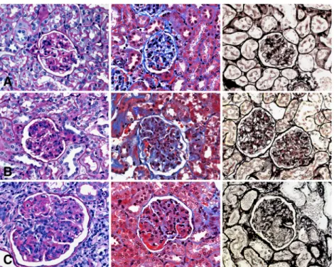
Fig. 2. Typical morphological lesions in MRL/MpJ +/+ mice of Groups I (A), II (B) and III (C) by light microscopy (LM ×600), in the PAS, trichrome and silver stain (from left to right). a. Mild mesangial expansion and hypercellularity in the mesangium. b. Moderate mesangial expansion and hypercellularity in the mesangium and few capillary loops. c. Severe lobulation of the glomeruli with prominent mesangial expansion and mesangial as well as intracapillary hypercellularity. Protein deposits and thrombi (reddish/red) in the PAS and trichrome stain. Severe mesangiolysis in the silver stain. Note increase of glomerular size from (A) to (C).
Immunofluorescence
Cryostat sections of 4μm of snap-frozen kidneys were stained for the deposition of C1q. Sections were blocked with 5% normal rat serum (Sigma-Aldrich, St. Louis, MO, USA) in PBS for 30 min, then incubated with FITC-labelled rat anti-mouse C1q (Cedarlane, Burlington, Canada) diluted 1/10 in PBS for 60 min. Sections were washed three times using PBS then mounted with UltraKitt mounting medium (J.T. Baker, Mallinckrodt Baker, Inc., Philipsburg, NJ, USA). Grading of immuno-fluorescence was as follows: 1 = focal, 2 = diffuse weak and 3 = diffuse strong. For evaluation purposes, two groups were formed according to the degree of complement deposition (score: < = 2/>2).
Stainings for complement IgG and C3 were performed in analogy using normal goat serum for blocking followed by a FITC-labelled
poly-clonal goat anti-mouse IgG (Sigma, St. Louis, Missouri, USA) and goat anti-mouse C3 (ICL, Newberg, OR, USA), respectively.
Detection of total IgG, autoantibodies against complement C1q and anti-nuclear antibodies
For the detection of murine anti-C1q, a highly significant correlation of results was obtained comparing assays in which purified human C1q or purified mouse C1q was used as the antigen [6]. Therefore, ELISA plates (Nunc, Rosklide, Denmark) for anti-C1q measurements were coated over-night at 4°C with purified human C1q (gift from Bühlmann Laboratories, Schönenbuch, Switzerland; >99% pure as judged by SDS–PAGE) at a concentration of 0.5μg/well or for determination of total IgG with goat anti-ms Ig (H+L) (SouthernBiotech, Alabama, USA) at a concentration of 2μg/mL. For the anti-C1q ELISA, serum samples were diluted 1:50 in PBS Tween (0.05%), 1% FCS containing 1 M NaCl. For the detection of IgG, serum samples were diluted 1/800 000 in PBS. After incubation with serum samples, plates were washed and bound IgG was detected using biotinylated polyclonal goat anti-mouse IgG (SouthernBiotech, Alabama, USA) and horseradish peroxidase-labelled Streptavidin (Jackson ImmunoResearch Europe, Suffolk, UK). A monoclonal mouse anti-human C1q (generated by immunization of C1qa-deficient mice, clone 23D11)
[13] was used to generate a standard curve. Anti-C1q are expressed as units per millilitre. To calculate the amount of IgG, a standard mouse IgG preparation (SouthernBiotech, Alabama, USA) was used.
Anti-nuclear antibodies (ANA) were determined as described previous-ly [14] (with the kind help of Prof. Rolink, Basel). In short, snap-frozen sections of kidneys from RAG-2−/− mice were incubated with sera diluted 1:20 and bound antibodies revealed with an FITC-labelled anti-mouse IgG antibody.
Proteinuria, haematuria and serum creatinine
Urine was regularly collected and analysed using dipsticks (Multistix® 5 SG, Bayer Diagnostics, Bridgend, UK) and a urine chemistry analyser (Clinitek 50, Bayer Diagnostics, Bridgend, UK). Serum creatinine was measured by quantitative colorimetric determination at 510 nm using a commercially available assay (Creatinine Assay Kit, Biochain, Hayward, CA, USA).
Antibody elution from kidneys
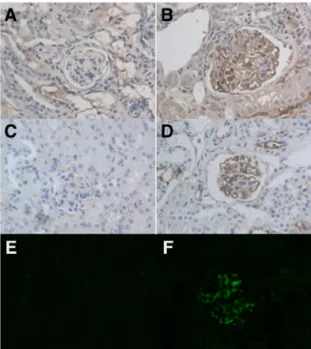
The procedure was performed as described before [9]. In short, six kidneys from six mice with high titres of anti-C1q antibodies were pooled, minced and collected in 3 mL PBS containing protease inhibitor cocktail (Roche, Mannheim, Germany). The mixture was centrifuged at 3000 rpm for 5 min and supernatants were collected. Pellets were washed and resuspended in 1.5 mL elution buffer consisting of 0.1 M glycine–HCl, 0.15 M NaCl, pH 2.5 and sonicated on ice with three bursts of 30 s and amplitude of 25. After overnight rotation at 4°C, samples were centrifuged for 10 min at 10 000 rpm and supernatants were collected and adjusted to pH 7.5. Samples Fig. 3. Deposition of C3, IgG and C1q in renal sections of MRL/MpJ +/+
mice. MRL/MpJ +/+ mice having deposition of C3, IgG and/or C1q varied in their positivity, whereas others were negative. (A) Glomerulus of a MRL/MpJ +/+ mouse, no deposition of C3. (B) Deposition of C3 in a glomerulus of another mouse. (C) Glomerulus without IgG deposition. (D) Glomerular deposition of IgG. (E) Glomerulus of a MRL/MpJ +/+ mouse, no deposition of C1q. (F) Deposition of C1q in a glomerulus of another mouse. Original magnification ×40.
Table 1. Light microscopical findings and immune deposits in three groups of animals with different degrees of morphological lesions
Parameter, median (range) Group I Group II Group III P-value
Total score 3 (2–4) 6 (5–8) 12 (9–15) 0.0001 Mesangial enlargement 1 (1–2) 2 (2–2) 2 (2–3) 0.0007 Mesangial proliferation 1 (1–2) 2 (2–2) 3 (2–3) 0.0033 Intracapillary hypercellularity 0 (0–1) 1 (0–1) 2 (1–3) 0.0010 Lobulation 0 (0) 0 (0–2) 2 (2–3) 0.0004 Protein deposits 0 (0–1) 0 (0–2) 1 (1–3) 0.0114 Glomerular area (×103μm2) 24.6 (16.2–29.5) 29.1 (23.1–32.4) 33.7 (25.6–39.4) 0.0043
Glomerular DAPI+ area (×103μm2) 9.1 (5.6–13.4) 11.0 (9.0–12.4) 12.7 (11.1–13.7) 0.0091
Glomerular DAPI+ area per glomerular area 0.37 (0.34–0.42) 0.37 (0.33–0.43) 0.39 (0.30–0.46) 0.7799
Glomerular CD3+ cells 5.4 (3.1–6.6) 6.0 (4.3–9.2) 8.1 (6.4–9.8) 0.0113
Glomerular Mac2+ cells 0.7 (0.2–2.4) 3.5 (0.6–6.5) 5.3 (4.8–6.8) 0.0008
Glomerular IgG deposits 0 (0–2) 0 (0–2) 0 (0–2) 0.9823
Glomerular C3 deposits 0 (0–1) 1 (0–3) 2 (1–3) 0.0079
were then tested by ELISA for the presence of anti-C1q and IgG as de-scribed above.
Statistics
Kaplan–Meier curves for the analysis of survival curves, column statis-tics, area under the anti-C1q curves (AUC anti-C1q), non-parametric cor-relation tests (Spearman) and Mann–Whitney U-test or Kruskal–Wallis tests for the comparison of multiple groups were performed where
appro-priate. Levels of severity of complement deposition were analysed by Chi-square tests and mortality by log-rank test. All statistics were performed using GraphPad Prism version 4 (GraphPad Software, San Diego, USA).
Results
Survival analysis
First, a survival study of 30 MRL/MpJ +/+ and 12 BALB/c
control mice was performed. The onset of death within the
MRL/MpJ +/+ group occurred after 5 months, and 50%
mortality was reached at 18 months. After 22 months all
MRL/MpJ +/+ mice were dead. At this time, mortality in
the BALB/c control mice group was 8.33% (1 out of 12).
The difference in mortality between the two mouse strains
was significant (by log-rank test, P < 0.0001) (Figure 1).
Renal pathology
Since 11-month-old mice had only mild nephritis, all the
following morphological analyses were only performed in
14-month-old mice. At this age, 95% of MRL/MpJ +/+
mice were ANA positive.
Light microscopy. At low magnification, the basic
struc-ture of the kidneys was well preserved. In the cortex,
enlarged glomeruli were apparent exhibiting variable
de-grees of hypercellularity. Occasional (< 5%) obsolescent
glomeruli were seen in practically all mice. The
tubulo-interstitial space was unremarkable, apart from variable
Fig. 4. Typical morphological lesions in MRL/MpJ +/+ mice of Groups I (A), II (B) and III (C) by electron microscopy (EM × 3500). a. Slight mesangial expansion due to matrix increase and minute mesangial electron-dense deposits (not seen at this magnification), no cell increase. Basement membranes unchanged. One polymorphonuclear leukocyte in the capillary lumen. No foot process fusion of podocytes. b. Very similar morphology as compared with (A), but large and lumpy electron-dense deposits in the mesangium (arrow). c. In addition to (B), numerous electron dense deposits of variable size on the outside of the basement membrane (arrow). Complete foot process effacement of podocytes.
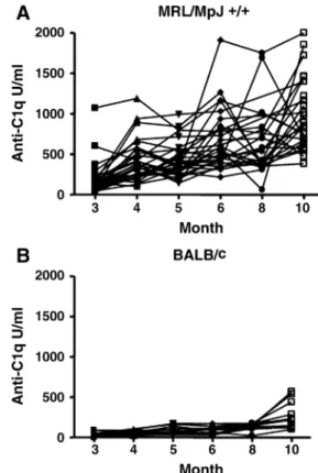
Fig. 5. Anti-C1q autoantibody titres in individual mice over time. (A) MRL/MpJ +/+ and (B) BALB/c mice. Data are expressed relative to a standard monoclonal mouse anti-C1q antibody. In MRL/MpJ +/+ mice, anti-C1q levels strongly varied over time.
numbers of cortical and medullary lymph follicles seen in
most animals. Individual mice exhibited small foci of
interstitial plasma cell aggregates.
Hyalinosis was rare in the arterioles. The arteries (with
two exceptions) and the veins were unremarkable. At higher
magnification, a variable degree of mesangial expansion
was seen, in part due to an increase of the mesangial matrix
and partly due to a highly variable hypercellularity or both.
Protein deposits were visible by trichrome stain in the
mesangium and less frequently in the periphery. This basic
picture of glomerular injury had several variants. The more
severe the mesangial hypercellularity, the more frequent was
a generally segmental intracapillary hypercellularity due to
mononuclear cells. In cases of particularly severe
hypercel-lularity, the glomeruli were often segmentally or globally
lobulated. The BALB/c control mice studied in the same
way showed no pathology at all.
In addition, occasional so-called active lesions were
found, in the form of segmental tuft necrosis, segmental
proliferative or proliferative sclerosed crescents,
mesangio-lysis, protein thrombi or vasculitis. These active lesions
were extremely rare and involved at most 1% of the
glom-eruli. The peripheral loops were occasionally thickened,
but clear cut doubling of the basement membrane with
me-sangial interposition was not observed (Figure 2).
Immunohistology. In immunohistochemical stainings,
me-sangial, and more rarely peripheral, deposits of varying
in-tensity were seen for complement C3 (62%) and IgG
(29%). Using immunofluorescence, C3 and IgG stained
positive in 94 and 100% of mice, respectively. C1q
posi-tivity was found in 71% of mice where weakly positive
findings dominated (Figure 3).
Semi-quantitative and quantitative evaluation. The
arbi-trary assignment of the animals into three groups
accord-ing to increasaccord-ing scores of injury reflects the level of
variability and the pattern of injury (Table 1).
Group 1: The group with the lowest injury score is
essentially distinguished by a slight increase in mesangial
matrix and cell number, without regular presence of
im-munoglobulin or complement deposits. The basic pattern
corresponded to minor glomerular abnormalities or mild
mesangial proliferative GN.
Group II: A marked increase in mesangial matrix and
cell number is accompanied by mild segmental
intracapil-lary hypercellularity. Cell proliferation paralleled the
in-crease in size (glomerular DAPI + area/glomerular area).
Slight C3 or C1q deposits could be seen. The lesions seen
corresponded to severe mesangio-proliferative GN.
Group III: In this group with the highest injury scores,
the mesangial changes and the segmental, intracapillary
hypercellularity were even more prominent than in group
II. Protein deposits were seen with the trichrome stain,
identified as complement deposits (C3 and/or C1q) by
im-munofluorescence. Quantitative evaluation showed, in
addition to a further increase in glomerular cell size and
number, an over proportional cell proliferation (glomerular
DAPI+ area/glomerular area). The intracapillary
hypercel-lularity was mainly due to an increase in monocytes more
than lymphocytes (see Table 1). All cases with so-called
active lesions (see above) were found in group III. The
pattern of injury corresponded to a diffuse proliferative
GN with active lesions.
The level of severity of the GN correlated with the level
of complement deposition (C3) seen by light microscopy
(P = 0.0079).
Electron microscopy. Group I and II: Both groups of mice
exhibited mesangial enlargement resulting from a modest
increase in matrix material and prominent mesangial cell
activation. The mice of group I revealed numerous small,
osmiophilic deposits along the para-mesangial basement
membrane, in the sample from group II deposits were large
and lumpy. There were no deposits in the periphery and no
fusion of the podocyte foot processes.
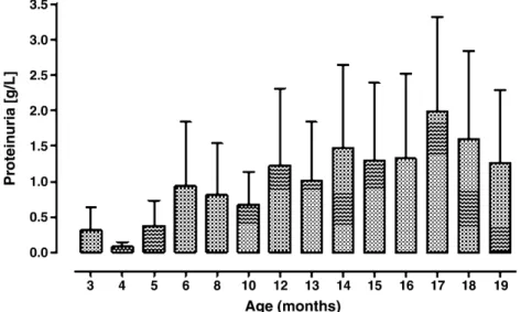
Fig. 6. Proteinuria in ageing MRL/MpJ +/+ mice. Proteinuria in MRL/MpJ +/+ mice was determined using dipstick analyses. Data are shown as mean +/− standard deviation.
Group III: These mice had massive mesangial expansion,
due to cell proliferation and cell activation, modest matrix
expansion and massive accumulation of osmiophilic
depos-its of varying size. In addition, subepithelial deposdepos-its of
varying size irregularly distributed along the peripheral
basement membrane were seen, in part flanked by spikes.
The podocytes were highly activated and exhibited
com-plete fusion of the foot processes. Subendothelial deposits
were comparatively small and rarer. In the endothelium,
tubulo-reticular or fingerprint-like structures could not be
found in any animal. The vessels and the tubulo-interstitial
space were unremarkable, in particular no osmiophilic
deposits were detected along the peripheral capillaries
(Figure 4).
Detection of anti-C1q autoantibodies in serum
Already at 3 months of age, most MRL/MpJ +/+ mice had
elevated anti-C1q when compared to BALB/c control
mice. Although all mice showed a rise in titre at later
time-points, individual mice showed a high variability in
anti-C1q titres (Figure 5). No correlation between survival
and either peak anti-C1q levels or areas under the anti-C1q
curves (AUC anti-C1q) was found.
Correlation between anti-C1q autoantibodies and renal
damage
Using dipstick analyses, we observed a progressive
in-crease in proteinuria (Figure 6) but did not detect a
signifi-cant haematuria in ageing MRL/MpJ +/+ mice.
No correlation was found between degrees of proteinuria
and serum creatinine concentrations (median 0.56 mg/dL,
range 0.39
–1.41 mg/dL in 14-month-old MRL/MpJ +/+
mice) on the one hand and any parameter of anti-C1q, i.e.
areas under the anti-C1q curves, the peak anti-C1q levels or
the anti-C1q levels at the time of euthanasia, on the other
hand.
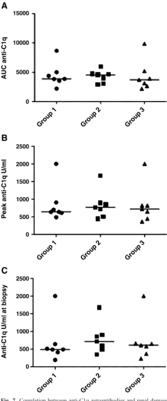
According to the renal pathology, mice were separated in
three groups (see above) and analysed for their correlation
with anti-C1q autoantibodies. No significant differences
could be found between the three groups with regard to
the areas under the anti-C1q curves (AUC anti-C1q), the
peak anti-C1q levels or the anti-C1q levels at the time of
euthanasia (Figure 7). Furthermore, no correlations were
found between AUC anti-C1q, peak anti-C1q or anti-C1q
titres at the time of euthanasia and any parameter of renal
pathology (studied by light microscopy or
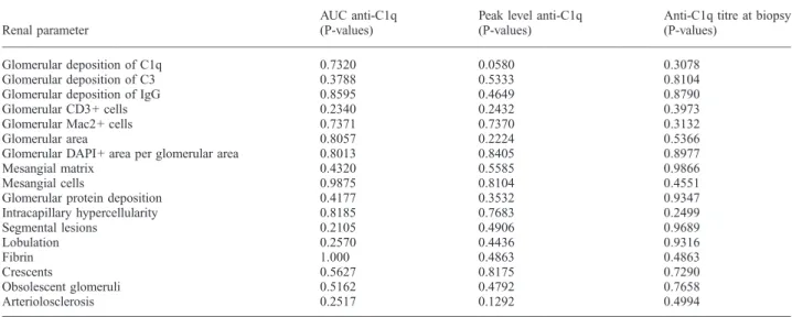
immunohisto-chemistry, Table 2).
Detection of anti-C1q autoantibodies in kidney eluate
The lack of correlation between anti-C1q autoantibodies
and renal damage was not due to the lack of anti-C1q
deposition in kidneys. Anti-C1q autoantibodies were
detected in the eluate of kidney tissue from 14-month-old
MRL/MpJ +/+ mice having high titres of anti-C1q, but not
in eluates of kidney tissue from BALB/c control mice.
Comparing anti-C1q levels in the eluate relative to eluted
total IgG with corresponding serum anti-C1q levels relative
to total serum IgG, a strong enrichment of anti-C1q in the
kidney eluate could be seen of MRL/MpJ +/+ but not in
BALB/c mice (Figure 8).
Fig. 7. Correlation between anti-C1q autoantibodies and renal damage. Mice were divided into three groups (Groups 1–3) according to renal pathology and analysed for their correlation with anti-C1q autoantibodies. No significant difference could be found between the three groups when looking at areas under the anti-C1q curves (AUC anti-C1q), peak levels of anti-C1q or titres at time of biopsy. Medians are shown as horizontal lines.
Discussion
Our study provides a detailed morphological analysis of
the lupus nephritis in MRL/MpJ +/+ mice but fails to
dem-onstrate a correlation between anti-C1q antibodies and the
severity of GN.
MRL/MpJ +/+ mice develop a highly variable picture of
GN at 14 months which reflects the picture seen in human
lupus nephritis. There is a range from minor glomerular
lesions, with only tiny osmiophilic deposits in the
mesan-gium without regular complement deposition, to
mesangio-proliferative GN with marked mesangial expansion and
lumpy mesangial deposits and to diffuse proliferative GN
which is typical for severe lupus nephritis in man. These
latter cases have, in addition to marked mesangial
expan-sion with cell proliferation, large, lumpy mesangial deposits
accompanied by minor subendothelial deposits as well as
in part massive subepithelial deposits and dominant
com-plement deposition. However, the so-called active lesions
were only observed in very few glomeruli. In most other
mouse models of lupus nephritis (NZBxW, MRL/1,
BXSB, MRL/MpJ-lpr/lpr), a lower degree in the variability
of the GN severity is seen between individual animals. At
any particular time point, and often early (3–6 months), a
severe, homogenous GN of diffuse proliferative type can be
observed [3,5,15, personal observations]. Occasionally,
these lesions are accompanied by severe exudative changes
with crescent formation and, after an observation period
exceeding 1 year, extensive glomerular obsolescence. The
latter type of changes were virtually absent in our study.
In contrast to the other SLE mouse models, we did not
observe regular IgG deposition in the MRL/MpJ +/+ model.
As described for other lupus-prone mouse strains, the
MRL/MpJ +/+ mice developed elevated levels of
anti-C1q antibodies early in life. Levels of anti-anti-C1q antibodies
varied which allowed the analysis of differences between
mice with high levels of anti-C1q and mice with low levels
of anti-C1q. Fourteen-month-old mice with high titres of
C1q antibodies in serum had an enrichment of
anti-C1q in their kidneys. This finding is in line with the finding
of anti-C1q antibodies in renal tissue of MRL/MpJ-lpr/lpr
mice [9] as well as in post-mortem material of end-stage
kidneys from patients with lupus nephritis [16]. At the
age of 14 months some MRL/MpJ +/+ mice exhibited
GN with increased glomerular cellularity as well as
glom-erular IgG, C1q and/or C3 deposition. This is in accordance
with previous studies showing glomerular deposition of C3
and IgG starting already at 7 months [11,17].
However, we found no correlation between levels of
anti-C1q and any parameter of glomerular inflammation. In
Table 2. No correlation of anti-C1q antibodies with parameters of glomerular histology in MRL/MpJ +/+ mice
Renal parameter
AUC anti-C1q (P-values)
Peak level anti-C1q (P-values)
Anti-C1q titre at biopsy (P-values)
Glomerular deposition of C1q 0.7320 0.0580 0.3078
Glomerular deposition of C3 0.3788 0.5333 0.8104
Glomerular deposition of IgG 0.8595 0.4649 0.8790
Glomerular CD3+ cells 0.2340 0.2432 0.3973
Glomerular Mac2+ cells 0.7371 0.7370 0.3132
Glomerular area 0.8057 0.2224 0.5366
Glomerular DAPI+ area per glomerular area 0.8013 0.8405 0.8977
Mesangial matrix 0.4320 0.5585 0.9866
Mesangial cells 0.9875 0.8104 0.4551
Glomerular protein deposition 0.4177 0.3532 0.9347
Intracapillary hypercellularity 0.8185 0.7683 0.2499 Segmental lesions 0.2105 0.4906 0.9689 Lobulation 0.2570 0.4436 0.9316 Fibrin 1.000 0.4863 0.4863 Crescents 0.5627 0.8175 0.7290 Obsolescent glomeruli 0.5162 0.4792 0.7658 Arteriolosclerosis 0.2517 0.1292 0.4994
AUC = area under the anti-C1q curve.
Fig. 8. Renal elution of anti-C1q antibodies. Anti-C1q reactivity per microgram IgG in eluate of kidney tissue and serum of 14-month-old MRL/MpJ +/+ mice compared to age-matched BALB/c control mice. An enrichment of anti-C1q autoantibodies is seen in the kidney eluate of MRL/MpJ +/+ but not in BALB/c mice when compared to serum.
addition, levels of anti-C1q did not correlate with the overall
survival of MRL/MpJ +/+ mice. Although we cannot
ex-clude a role for deposited antibodies, our observational data
suggest that anti-C1q are not involved in the pathogenic
mechanism of GN in lupus-prone MRL/MpJ +/+ mice
and do not support the hypothesis that anti-C1q have a
pathogenic role in SLE. The finding is also in conflict with
data from Trouw et al. showing that the injection of a
monoclonal anti-C1q in mice pretreated with
subnephrito-genic doses of C1q-fixing anti-glomerular basement
mem-brane antibodies resulted in exacerbation of the subclinical
renal disease [18]. In another study, the injection of
poly-clonal rabbit anti-mouse C1q into healthy mice resulted in
glomerular complement activation, leukocyte influx and
mild albuminuria [19]. As a consequence, we think that
the type of GN might be critical in the determination
of the predictive value and pathogenic role of anti-C1q.
Independently, it has been recognized that translation of
results obtained in experimental autoimmune diseases
into the human situation is difficult [20]. Thus, in spite
of morphological similarities, different mechanisms might
be involved between the pathogenesis of human lupus
nephritis and the GN seen in MRL/MpJ +/+ mice. Only
large studies on SLE patients that are closely followed
over long periods might provide a definitive answer to
the question of the true predictive value of anti-C1q for
renal flares.
In conclusion, we did not observe a correlation of
anti-C1q with the survival or the severity of GN in lupus-prone
mice. Therefore, our data do not support the hypothesis
that anti-C1q have a pathogenic role in SLE. However,
dif-ferent pathogenic mechanisms might be involved in the
GN of lupus-prone MRL/MpJ +/+ mice and human
prolif-erative lupus nephritis.
Acknowledgements. We thank Mrs Brigitte Schneider, Mrs Ursula Duermueller and Mrs. Hedwig Niederer for their important help in the handling of animals and in the histopathological analyses. The study was supported by a SCORE fellowship from the Swiss National Foundation (3232BO-107248/2).
Conflict of interest statement. None declared.
References
1. Moroni G, Trendelenburg M, Del Papa N et al. Anti-C1q antibodies may help in diagnosing a renal flare in lupus nephritis. Am J Kidney Dis 2001; 37: 490–498
2. Grootscholten C, Dieker JWC, McGrath FD et al. A prospective study of anti-chromatin and anti-C1q autoantibodies in patients with
proliferative lupus nephritis treated with cyclophosphamide pulses or azathioprine/methylprednisolone. Ann Rheum Dis 2007; 66: 693–696 3. Theofilopoulos AN, Dixon FJ. Murine models of systemic lupus
erythematosus. Adv Immunol 1985; 37: 269–390
4. Peutz-Kootstra CJ, de Heer E, Hoedemaeker PJ et al. Lupus nephritis: lessons from experimental animal models. J Lab Clin Med 2001; 137: 244–260
5. Andrews BS, Eisenberg RA, Theofilopoulos AN et al. Spontaneous murine lupus-like syndromes. Clinical and immunopathological manifestations in several strains. J Exp Med 1978; 148: 1198–1215 6. Hogarth MB, Norsworthy PJ, Allen PJ et al. Autoantibodies to the collagenous region of C1q occur in three strains of lupus-prone mice. Clin Exp Immunol 1996; 104: 241–246
7. Trinder PK, Maeurer MJ, Schorlemmer HU et al. Autoreactivity to mouse C1q in a murine model of SLE. Rheumatol Int 1995; 15: 117–120
8. Uwatoko S, Mannik M, Oppliger IR et al. C1q-binding immuno-globulin G in MRL/l mice consists of immune complexes containing antibodies to DNA. Clin Immunol Immunopathol 1995; 75: 140–146 9. Trouw LA, Seelen MA, Visseren R et al. Anti-C1q autoantibodies in
murine lupus nephritis. Clin Exp Immunol 2004; 135: 41–48 10. Hewicker M, Kromschröder E, Trautwein G. The pathogenesis of
glomerulonephritis in MRL mice - murine lupus erythematosus. Verh Dtsch Ges Pathol 1989; 73: 129–132
11. Mitchell DA, Pickering MC, Warren J et al. C1q deficiency and auto-immunity: the effects of genetic background on disease expression. J Immunol 2002; 168: 2538–2543
12. Trendelenburg M, Manderson AP, Fossati-Jimack L et al. Monocy-tosis and accelerated activation of lymphocytes in C1q-deficient autoimmune-prone mice. Immunology 2004; 113: 80–88
13. Bigler C, Schaller M, Perahud I et al. Autoantibodies against com-plement C1q specifically target C1q bound on early apoptotic cells. J Immunol 2009; 183: 3512–3521
14. Bénard A, Ceredig R, Rolink AG. Regulatory T cells control auto-immunity following syngeneic bone marrow transplantation. Eur J Immunol 2006; 36: 2324–2335
15. Gunn HC, Ryffel B. Glomerulonephritis in NZB/W mice: therapeutic effect of cyclosporine. Clin Nephrol 1986; 25: 1189–192
16. Mannik M, Wener M. Deposition of antibodies to the collagen-like region of C1q in renal glomeruli of patients with proliferative lupus glomerulonephritis. Arthritis Rheum 1997; 40: 1504–1511 17. Cortes-Hernandez J, Fossati-Jimack L, Petry F et al. Restoration of
C1q levels by bone marrow transplantation attenuates autoimmune disease associated with C1q deficiency in mice. Eur J Immunol 2004; 34: 3713–3722
18. Trouw LA, Groeneveld TWL, Seelen MA et al. Anti-C1q autoanti-bodies deposit in glomeruli but are only pathogenic in combination with glomerular C1q-containing immune complexes. J Clin Invest 2004; 114: 679–688
19. Trouw LA, Seelen MA, Duijs JMGJ et al. Glomerular deposition of C1q and C1q antibodies in mice following injection of anti-mouse C1q antibodies. Clin Exp Immunol 2003; 132: 32–39 20. Davis MM. A prescription for human immunology. Immunity 2008;
29: 835–838