HAL Id: hal-00330218
https://hal.archives-ouvertes.fr/hal-00330218
Submitted on 29 Sep 2004HAL is a multi-disciplinary open access
archive for the deposit and dissemination of sci-entific research documents, whether they are pub-lished or not. The documents may come from teaching and research institutions in France or abroad, or from public or private research centers.
L’archive ouverte pluridisciplinaire HAL, est destinée au dépôt et à la diffusion de documents scientifiques de niveau recherche, publiés ou non, émanant des établissements d’enseignement et de recherche français ou étrangers, des laboratoires publics ou privés.
The environment recording unit in coral skeletons:
structural and chemical evidences of a biochemically
driven stepping-growth process in coral fibres
J. P. Cuif, Y. Dauphin
To cite this version:
J. P. Cuif, Y. Dauphin. The environment recording unit in coral skeletons: structural and chemical evidences of a biochemically driven stepping-growth process in coral fibres. Biogeosciences Discussions, European Geosciences Union, 2004, 1 (1), pp.625-658. �hal-00330218�
BGD
1, 625–658, 2004
The environment recording unit in coral skeletons
J. P. Cuif and Y. Dauphin
Title Page Abstract Introduction Conclusions References Tables Figures J I J I Back Close Full Screen / Esc
Print Version Interactive Discussion © EGU 2004 Biogeosciences Discussions, 1, 625–658, 2004 www.biogeosciences.net/bgd/1/625/ SRef-ID: 1810-6285/bgd/2004-1-625 © European Geosciences Union 2004
Biogeosciences Discussions
Biogeosciences Discussions is the access reviewed discussion forum of Biogeosciences
The environment recording unit in coral
skeletons: structural and chemical
evidences of a biochemically driven
stepping-growth process in coral fibres
J. P. Cuif1and Y. Dauphin2
1
Universit ´e Paris XI-Orsay, Bat. 504, G ´eologie ,UMR IDES, F-91405 Orsay, France
2
Universit ´e Paris VI-UPMC, Micropal ´eontologie, t. 46–56 E5, 4 place Jussieu, UMR IDESF, F-75252 Paris cedex 05, France
Received: 31 August 2004 – Accepted: 21 September 2004 – Published: 29 September 2004 Correspondence to: J. P. Cuif ([email protected])
BGD
1, 625–658, 2004
The environment recording unit in coral skeletons
J. P. Cuif and Y. Dauphin
Title Page Abstract Introduction Conclusions References Tables Figures J I J I Back Close Full Screen / Esc
Print Version Interactive Discussion
© EGU 2004
Abstract
To improve our understanding of the environment recording by scleractinian corals, a detailed study of the skeleton microstructure has been carried out. A series of physico-chemical in situ characterizations was made, an approach that provides us with struc-tural and biochemical information at the micrometric and nanometric scales. Gathering
5
of these data results in a significant change in our concept of the growth of coral skele-tons. In contrast to the usual view of an aggregate of purely mineral units (the coral fibres) independently growing by a simple chemical precipitation, coral skeletons ap-pear to be biochemically controlled structures. Both structural and biochemical data reveal the micron-scaled stepping growth-mode of fibres, and its global coordination.
10
In this process, sulfated acidic proteoglycans probably play a major role, due to their ability to create polymeric frameworks. Atomic force microscopy confirms the close relationship of organic and mineral phases at the nanometric scale.
A new microstructural model of coral skeleton formation is proposed, that places coral skeletons among the typical “matrix mediated structures”. From a practical
stand-15
point, these results may contribute to develop a new high resolution approach in the study of paleoenvironments.
1. Introduction
Various chemical or isotopical proxies have placed coral skeletons among the most important sources of environmental information but, surprisingly, no agreement exists
20
about the formation of these widely used biological archives. The process by which the basal ectoderm of coral polyps produces the underlying skeleton was the matter of a first controversy by the end of the 19th century when, in contrast to the von Heider’s theory (1881), it was recognized that calcification does not occur within the ectoderm cells themselves. An extracellular calcification occurring outside the ectodermal cell
25
BGD
1, 625–658, 2004
The environment recording unit in coral skeletons
J. P. Cuif and Y. Dauphin
Title Page Abstract Introduction Conclusions References Tables Figures J I J I Back Close Full Screen / Esc
Print Version Interactive Discussion
© EGU 2004
the control of skeleton organization remains an impending question. As summarized by Le Tissier (1991), models of calcification in corals range from a pure physicochemical (i.e. Barnes, 1970) to a biologically controlled process (i.e. Johnston, 1977–1980).
Crystallization of the coral aragonite being an extracellular phenomenon, the hypoth-esis of a biological control exerted on skeleton formation should be supported first by
5
evidences concerning the place where crystallization occurs: the interface between the polyp basal ectodermal cell layer and the underlying skeleton. Noticeably, in spite of the Goreau’s pioneering investigation (1956–1959), research focussing on physico-chemical characteristics of the subectodermal space are extremely rare. The complex-ity of coral skeletons prevents an easy access to the interface compartment, in contrast
10
to Molluscs (i.e. Pelecypods), where the mineralization space between the outer side of the mantle and the shell is much more easily accessible. As a result, extraction of mineralizing fluids to study their composition and mineralizing capabilities was carried out in Molluscs as early as 1974–1976 (Wada and Fujinuki, 1974). To date, no equiva-lent research exists for coral mineralization process. The first pH measurement at the
15
interface between the basal ectoderm and the growing surface of a coral skeleton was attempted in 2003 only (Al-Horani et al., 2003).
Instead of an unapproachable direct analysis of the mineralizing sites, complete de-calcification of coral skeletons that give access to organic materials entrapped during skeletal growth has been much more used by investigators. This resulted in a series
20
of papers that have emphasized the importance of either sugars (Wainwright, 1963; Wilfert and Peters, 1969; Dauphin, 2001) or amino acids (Young, 1971, 1973; Mitterer, 1978; Constantz and Weiner, 1988; Cuif et al., 1999). Nevertheless, as pointed out by Johnston (1980), research dealing with biochemical composition of organic materials extracted from coral skeletons results in mineralization models made “in ignorance of
25
this material spatial distribution and micro architecture within the skeleton”.
Additionally, uncertainty persists with respect to the amount of these organic compo-nents. Since Wainwright (1963), the fraction of organic material embedded in the coral skeletons has been commonly estimated to 0.1%wt, a proportion recently reevaluated
BGD
1, 625–658, 2004
The environment recording unit in coral skeletons
J. P. Cuif and Y. Dauphin
Title Page Abstract Introduction Conclusions References Tables Figures J I J I Back Close Full Screen / Esc
Print Version Interactive Discussion
© EGU 2004
to about 1%wt (Cohen and McConnaughey, 2003). On the other hand, thermogravi-metric analyses (TGA) have shown that 2.5 to 3%wt of coral skeletons were lost before aragonite thermal decomposition (Cuif et al., 1997). Such a discrepancy casts doubts on the possible influence of these organic compounds on the crystallization process.
In response to the Johnston’s remark, this paper aims to gather a series of
re-5
cent data based on a different approach. Owing to the development of high resolu-tion analytical instruments, combinaresolu-tion of results from scanning electron microscopy, synchrotron X-ray fluorescence and atomic force imaging allows a fine scale in situ physico-chemical characterization of coral skeletons to be obtained. This results in a new set of information about coral skeleton units examined at the micrometric and
10
nanometric levels, allowing the calcification process to be reexamined.
2. Material and methods
Coral specimens have been collected alive, mostly by authors during the last few years. To remove tissues, polyps were decayed by a three-hour immersion in pure water that causes the cells to be destroyed. Removal of the remaining living tissues was done
15
by using water-jet, followed by an immersion in 0.1M sodium hypochlorite to clean the deeper parts of the skeletons. Then, specimens were rinsed and room-temperature air dried. In all cases, only the very upper parts of skeletons were used for imaging. The specimens were selected by SEM observation of polished and etched surfaces, allowing the fibrous microstructures to be examined. Samples invaded by endolithic
20
borers were discarded.
2.1. Materials
Corallites belonging to the following species have been used:Favia stelligera from Polynesian Archipelago (Moorea Island); Montastrea curta from Mururoa Island (J.P. Chevalier coll.); Diploastrea heliopora, Leptoria phrygia, Porites c.f. australiensis,
BGD
1, 625–658, 2004
The environment recording unit in coral skeletons
J. P. Cuif and Y. Dauphin
Title Page Abstract Introduction Conclusions References Tables Figures J I J I Back Close Full Screen / Esc
Print Version Interactive Discussion
© EGU 2004
pora digitifera, Merulina scabricula from New Caledonia lagoon; Caryophyllia ambrosia
from Atlantic Ocean (Gascogne Gulf); Cladocora caespitosa from Mediterranean Sea (Marseille coastal region);Favia fragum from Carribean Sea (Guadalupe Island).
2.2. Methods
2.2.1. Structural and chemical characterizations
5
Microstructural features of fibres have been first checked by microscope observation of ultra-thin sections in polarized light. This improvement of the usual microscopic technique requires fine polishing of the sample surface and a reduced thickness of slides. Instead of the standard 30 microns, grinding is continued up to some 4–5 micrometers thickness, that allows the primary colours of aragonite to be visible. Final
10
polishing of the upper surface ensures an accurate microstructural observation.
2.2.2. Scanning Electron Microscopy (SEM) of coral skeleton
Observations were carried out not only on simple fracture surfaces but mostly on pol-ished and etched surfaces. Etching by very light acidic solutions (one per mil formic acid including the fixative glutaraldehyde 3 to 5%) reveals that differences in
solubil-15
ity do exist within the crystal like fibres. Additionally, the global organization of coral septa is well evidenced, with emphasize on the trace of the early mineralization zone (commonly called “centres of calcification”).
2.2.3. X-ray Absorption Near Edge Spectrum (XANES) characterization and mapping of sulfated polysaccharides
20
At the European Synchrotron Radiation Facility in Grenoble (France), the ID-21 line has developed a high resolution analytical device based on detection of X-ray fluo-rescence. By carefully selecting X-ray wavelengths, characterization of bound energy in different oxidation state of sulfur is possible. After a calibration phase that allows
BGD
1, 625–658, 2004
The environment recording unit in coral skeletons
J. P. Cuif and Y. Dauphin
Title Page Abstract Introduction Conclusions References Tables Figures J I J I Back Close Full Screen / Esc
Print Version Interactive Discussion
© EGU 2004
the different XANES spectra from sulfated standard molecules to be clearly separated (Figs. 1a to 1d), in situ characterization of organic sulfate can be made. Moreover, high performance lense focalization allows this characterization to be obtained with an infra-micronic spatial resolution. A two-dimensional piezoelectric driven specimen holder makes possible a micronic step displacement of the polished sample surface,
5
resulting in a biochemical mapping of the coral skeleton in response to the selected energy.
2.2.4. Atomic Force Microscopy (AFM)
Scanning probe microscopy encompasses a family of techniques that measures sur-face topography and properties at the atomic scale. The atomic force microscope
10
simultaneously produces maps the topography of surfaces (height images or derived amplitude images) and phase images. In phase imaging, a variant of tapping mode, the phase lag of the cantilever oscillation relative to the signal sent to the cantilver’s piezo driver is used as a basis for image generation. Phase images can be generated as a consequence of variations in material properties such as composition,
viscoelas-15
ticy, adhesion. AFM observations were conducted with Digital Instruments (Veeco) Nanoscope III Dimension 3100 at room temperature and air. The probe consisted of a cantilever with integrated Si3N4tips (Digital Instruments). Micron scale images were acquired using tapping mode.
For AFM observation of biominerals, no routinely applicable procedure exists.
Vari-20
ous preparative process of the observed surface have been used, that all aim to reduce the possible changes in relationships between mineral and postulated organic compo-nents. Consistency between phase and height images is essential for interpretation. The procedures of the sample preparations are given in the figure captions.
BGD
1, 625–658, 2004
The environment recording unit in coral skeletons
J. P. Cuif and Y. Dauphin
Title Page Abstract Introduction Conclusions References Tables Figures J I J I Back Close Full Screen / Esc
Print Version Interactive Discussion
© EGU 2004
3. Results
3.1. The incremental growth mode of fibres and global microstructural patterns evi-dencing the stepping growth mode of coral skeletons
Checking of the microstructural characteristics emphasizes the common patterns of coral fibres: they appear as groups of elongated units (Fig. 2a) with global
monocrys-5
talline behaviour in polarized light (Figs. 2b, 2c). However, on a polished and etched surface, a completely different pattern appears, that reveals the incremental growth of fibres (Fig. 2d). This result is exemplified here by a microstructural sequence in a Porites c.f. australiensis colony that shows the morphology of skeletal component (Figs. 3a–3c). The fracturated skeletal rod (Fig. 3c) shows the radial organization of
10
fibres (Fig. 3d). Ultra-thin section (Fig. 3e) confirms the monocrystalline behaviour of fibres, whereas after etching the concentric growth lines transversal to fibre c-axes are well visible (Fig. 3f).
To date, no exception is known to this micron-scaled incremental growth of fibres. Attention must be drawn on the fact that visualisation of the stepping growth patterns
15
depends on orientation of fibres with respect to the observation surface. It is long known that tridimensional arrangements of fibres are complex and very different among corals from various families. With respect to growth direction (Fig. 4a, white arrows) fibres are globally oblique to the septal median plan or growth axis. Consequently, within a given surface (Fig. 4b, line S), fibres may appear under different sections
20
between two extreme conditions exemplified in Fig. 4c: from perfectly longitudinal (L) to transversal sections (T ). Figures 4d and 4e give equivalent SEM pictures, showing that incremental growth patterns is variously readable depending on orientation of fibres.
Etched surfaces also show that fibres do not grow as independent units. Between adjacent fibres, a well-marked synchronism in the high solubility zones creates
con-25
centric lines evidencing the existence of a cyclic growth process. When the global ori-entation of fibres is favourable, continuity of concentric growth lines can be observed in the whole septum (Figs. 5a–5c), showing that this stepping growth pattern is
coordi-BGD
1, 625–658, 2004
The environment recording unit in coral skeletons
J. P. Cuif and Y. Dauphin
Title Page Abstract Introduction Conclusions References Tables Figures J I J I Back Close Full Screen / Esc
Print Version Interactive Discussion
© EGU 2004
nated at a global scale. The series of few microns thick growth layers surround regions that exhibit specific microstructural patterns, in which Ca-carbonate shows a granular microstructure (Fig. 5c, EMZ). Specificity of these internal regions is also evidenced by the results of biochemical maps (see below 3.2).
3.2. Biochemical maps: layered distribution of sulfated polysaccharides within fibres,
5
high concentration of S-polysaccharides in Early Mineralization Zones
All polished surfaces exposed to the 2.4825 keV X-ray energy produce a strong and exclusive signal corresponding to the presence of organic sulfated polysaccharides (Fig. 6). In contrast, sulfated aminoacids are practically undetectable. To take the full advantage of the ID-21 mapping device, a preliminary exploration of the surface
10
is useful, as exposed in Figs. 7a–7c. Part of a polished section (wall and septa) in a
Montastrea corallite is firstly exposed to UV light (365 nm) on a reflection microscope,
allowing the EMZ zones to be localized (Fig. 7c, arrows). The infra micronic 2.4825 keV beam, is then applied to the specimen, the distance between measurement points and lines being one micron. Time exposure for each point is 0.8 second.
15
This results in the map of sulfur (Fig. 7d), i.e. the distribution of sulfated polysac-charides between EMZ and fibres on a 80×100 mm field view. Presently no precise quantitation is possible, owing to the heterogeneity of the biogenic material that does not allows any calculation concerning the absorbed/emitted radiations. However, the higher concentration of polysaccharides in EMZ is well visible, as well as the banding
20
patterns in the fibrous part of the field view (arrows).
Very comparable results are obtained on corallite from other species: Diploria
labyrinthica (Figs. 8a–8d), Porites c.f. australiensis (Figs. 9a and 9b), Acropora digi-tifera (Figs. 9c and 9d). In all samples, a correspondence is evidenced between the
microstructural pictures and the biochemical maps. The exact correlation between the
25
mineral growth layer arrangements and spatial distribution of acidic polysaccharides detected by their sulfated sulfur indicates that organic and mineral components are
BGD
1, 625–658, 2004
The environment recording unit in coral skeletons
J. P. Cuif and Y. Dauphin
Title Page Abstract Introduction Conclusions References Tables Figures J I J I Back Close Full Screen / Esc
Print Version Interactive Discussion
© EGU 2004
interrelated within the growth layer itself, i.e. at an infra-micronic scale.
3.3. Spatial relationships between organic and mineral phases at the nanometric scale
AFM examination confirms the infra-micronic spatial relationships between organic and mineral components within skeleton growth layers. Pictures of Merulina scabricula
5
are particularly significant. Densely packed nanograins are well visible (Figs. 10a and 10b). Looking more closely (Fig. 10c and 10d), the grains appear coated by a thin layer made of a material that exhibits strongly different physico-chemical properties, as shown by the phase image (Fig. 10d). Due to effectiveness of AFM phase imaging to detect properties (i.e. elasticity) linked to chemical compositions, we can assess that
10
the thin layers that appear as low relief at the grain surfaces (Fig. 10c, arrows) are very different in chemical composition from the grains themselves (Fig. 10d). Remarkably, similar pictures have been obtained on coral skeletons of every studied species, and at any place in the fibrous structures of each specimen (Figs. 11a–11b: Favia stelligera; Fig. 11c–11d: Cladocora caespitosa; Fig. 11e–11f: Caryophyllia ambrosia).
15
This simultaneously shows that: (1) a nanometric granular structure is the basic organization of the fibre calcareous growth layers and (2) the few tenths to hundred nanometre grains are included in a material that exhibits different physico-chemical properties, suggesting an organic composition.
4. Discussion
20
When Johnston published his transmission electron microscope pictures (1977, Fig. 1; 1980, Fig. 18), the organic reticulum visible within the upper growth layer of a coral fibre was the first ever seen evidence of a sub micronic relationship between organic and mineral material within coral skeletons. Of course, no mineral was visible within the reticulum spaces, due to the TEM preparative process.
BGD
1, 625–658, 2004
The environment recording unit in coral skeletons
J. P. Cuif and Y. Dauphin
Title Page Abstract Introduction Conclusions References Tables Figures J I J I Back Close Full Screen / Esc
Print Version Interactive Discussion
© EGU 2004
4.1. Consistency of AFM data with Johnston results
Dimensions of the reticulum units in Johnston pictures remarkably correspond to those of the nanograins evidenced by AFM (Figs. 9–10). This correspondence fully supports the interpretation that the grains visible on the AFM pictures are made of aragonite. Accordingly, the material that separates the mineral grains, with its strongly interactive
5
properties (in phase images, black means “attractive”), can be supposed to be made of organic material.
Owing to the preparative process, Johnston was able to characterize the organic reticulum at the growing tips of fibres only. He suggested that the reticulum might “disappear” in deeper parts of the skeleton. In contrast, we have noted that the XANES
10
signal of sulfated polysaccharides is obtained on the whole surface of the sections, whatever the distance to the growing surface. Although the accurately focussed X-ray beam is less than 1 micron in diameter, it is much wider than the nanograin mean diameter. Therefore the polysaccharide signal is always observed, whatever the place at which the X-ray beam is applied. Thus, the sulfated polysaccharide signal persists in
15
coral skeletons far deeper than Johnston thought. However, there is no doubt that some change may occur within a coral colony lifetime, specifically in the global composition of organic compounds (Cuif et al., 1997).
Recent data show that the organic component in coral skeletons structurally includes water (Cuif et al., 2004), a result that also contributes to create a consistent view of this
20
material. From a quantitative stand point, the hydrated matrix of coral skeletons has been shown to represent up to 2.5 - 3% of the skeleton total weight, depending upon species. More than a half of this weight is made by water, a result that is close to the proportion suggested by Gaffey (1988) from infrared observations. Thus, biochemical compounds (properly speaking) represent about 1 per cent of the skeletal weight, a
25
result that supports the estimate proposed by Cohen and McConnaughey (2003). This hydrated organization of the organic material must be compared with analyti-cal results (chromatography of electrophoresis) that have more precisely established
BGD
1, 625–658, 2004
The environment recording unit in coral skeletons
J. P. Cuif and Y. Dauphin
Title Page Abstract Introduction Conclusions References Tables Figures J I J I Back Close Full Screen / Esc
Print Version Interactive Discussion
© EGU 2004
the proteoglycan composition of the skeletal organic phases. 2D electrophoresis is specifically relevant (Dauphin, 2001) because Alcian blue staining allows acidic sul-fated glucidic compounds to be evidenced (Fig. 12a) whereas associated proteins are silver stained (Fig. 12b). By their biochemical composition, mineralizing matrices of fibre growth layers belong to this category of glycoconjuguates (proteoglycans close
5
to aggrecan, versican, etc.), organic compounds well known for their ability to include water in their complex molecular assemblages. They usually include a core protein (MW 200 to 500 kD) with carbohydrate lateral chains, resulting in MW that may reach several megaDaltons. These data are consistent with the results concerning global characterization of organic compounds extracted from coral fibres.
10
A remark must be made that a 2.5–3% ratio “in weight” for hydrated organic com-pounds suggests that the hydrated-organic phase in coral fibres may be in a ratio about three times higher when considering their “volumes” (aragonite density: 2.99). Even assuming a high density for this hydrated organic phase (i.e. 1.5), this may result in a global proportion of about 5–7% of the skeleton volume occupied by the non-carbonate
15
component, a result consistent with the proportion of nanograins and organic phases on the AFM pictures.
Profiles of thermal decompositions observed on the TGA experiments show that sig-nals corresponding to the decomposition of organic compounds are observed up to 550◦C (Cuif et al., 2004). This unexpected resistence of organic material to heating
20
is explainable by the nanometric interfingering of mineral and organic components. From a practical standpoint, this raises questions about isotope ratio measurements. The heating phase included in the preparative process, before acidic decomposition of calcareous materials, is usually limited to 350/400◦C, a temperature that is unable to completely remove the intra crystalline organic phases: the main part of the organic
25
component is decomposed above this temperature. Consequently, isotopical peculiari-ties of the remaining organic compounds may be suspected to influence the measured ratio. Such a possibility is still more probable when using SIMS methods that do not include a thermal degradation step.
BGD
1, 625–658, 2004
The environment recording unit in coral skeletons
J. P. Cuif and Y. Dauphin
Title Page Abstract Introduction Conclusions References Tables Figures J I J I Back Close Full Screen / Esc
Print Version Interactive Discussion
© EGU 2004
4.2. “The hypothesized organic matrix mediation of crystal growth (Johnston, 1977; Jackson and Gladfelter, 1985) is difficult to support”
This statement (Constantz, 1986, p. 155) illustrates the Le Tissier’s comment (1991) about the diversity of biomineralization models for coral fibres, due to the difficulty to characterize organic compounds within calcareous skeletons. During decades, we
5
were unable to connect microstructural evidences of the layered organization of fi-brous or prismatic microstructural units (Cuif et al., 1981, 1997, 1998) to chemical of biochemical data obtained at a corresponding scale. For instance, information about sulfur in biominerals has long been collected (Cuif et al., 1986), but the chemical status of this element was not established, no more than its close relationship with the growth
10
steps of biominerals.
Data reported here favours the interpretation of coral fibre growth as a biologically controlled process.
4.3. A polycyclic model for the growth of the coral fibres
The reported structural and chemical data can be summarized in a simple model of the
15
growth layer formation (Fig. 13).
At the starting point of a biomineralization cycle (Fig. 13a), the basal ectoderm of the polyp is in close contact with the upper surface of fibres (F1 to F3), each of them with its c-axis in global conformity with fibre length (note that orientations of the two other axes differ from fibre to fibre, see Fig. 2).
20
4.3.1. The matrix secretion step
Although physiology of the matrix secretion is thoroughly studied (see Tambutt ´e, 1996; Allemand, 2004), molecular organization outside the ectodermal layer is still poorly understood. Chromatographic and electrophoretic characterizations have emphasized the presence of acidic high molecular weight compounds in the organic part of coral
BGD
1, 625–658, 2004
The environment recording unit in coral skeletons
J. P. Cuif and Y. Dauphin
Title Page Abstract Introduction Conclusions References Tables Figures J I J I Back Close Full Screen / Esc
Print Version Interactive Discussion
© EGU 2004
skeletons (Dauphin, 2001; Dauphin and Cuif, 1997), but recent results (Allemand et al., priv. com) have also evidenced an important proportion of very low weight molecules (peptides). Owing to the importance of polysaccharides in skeletal matrices, this con-trast suggests that a self-assembly process may occur within the mineralizing compart-ment, resulting in a polymeric organic framework. The historical observation by Goreau
5
(1956) of a plucidic layer on the external side of the basal ectoderm of the polyp may correspond to this secretory step.
This self-assembly step may be also the explanation for the crystallographic conti-nuity between the successive growth layers of fibres. At the beginning of the biomin-eralization cycle, the organic compounds involved in the hypothesized self-assembly
10
process are in contact with the upper surface of the aragonite fibres, that are facing the basal ectoderm. Each fibre provides a template that allows the organic framework resulting from the self-assembly process to be oriented in conformity with the charac-teristics of each of the underlying fibres (Fig. 13b).
4.3.2. The growth layer crystallization step
15
To create Ca-carbonate nanograins, calcium is transported to the subectodermal space (Tambutt ´e et al., 1995; Marshall, 1996). Many analyses have shown that both glucidic and protein matrix components have compositional peculiarities that favour interaction with mineral ions. Glucids are sulfated and very acidic in composition (Dauphin, 2001), as well as aspartic/glutamic rich proteins (Mitterer, 1978; Constantz and Weiner, 1988).
20
Taking into account the very low pI of matrix components, the free mineral ions certainly found numerous sites to be fixed on the organic framework, starting the phase 2 of the biomineralization cycle, the crystallization process properly speaking.
In this mineralization step, the hypothesized polymeric pre-oriented organic frame-work may play a major role, being responsible for the position of the initial
crystal-25
lization sites. Thus, after having been oriented by the underlying mineral surface, the organic framework itself may ensure the crystallographic coherence of the nanograins, an essential condition to maintain the global crystallinity of fibres. It must be noticed,
BGD
1, 625–658, 2004
The environment recording unit in coral skeletons
J. P. Cuif and Y. Dauphin
Title Page Abstract Introduction Conclusions References Tables Figures J I J I Back Close Full Screen / Esc
Print Version Interactive Discussion
© EGU 2004
however, that a close SEM observation of fibres (Fig. 14a) shows that they are not so perfectly monocrystalline than believed by Bryan and Hill (1941) based on optical observations.
Within the mineralizing layer, growth of nanograins progressively reduces the space available for the initial hydrated organic medium secreted in phase 1 of the
biomineral-5
ization cycle. At the end of this crystallization step (Fig. 13c), the organic phase simply appears as a cortex surrounding nanograins (see Figs. 10a and 10b).
4.3.3. The coral fibre: a matrix mediated biocrystal
In contrast to the still widely admitted monocrystalline concept, coral fibres belong to this category of matrix mediated biominerals built by repeatedly produced micron-thick
10
growth layers. A comparison can be made with the most classical of them, the calcitic prism of Pinna nobilis, a Pteriomorphid Pelecypod. The following points emphasize the similarity of the growth process between mollusc prisms and coral fibres (Fig. 14).
– The micronic stepping growth mode (now evidenced also in fossil corals, Stolarski,
2003) is common to both microstructures, with quite comparable patterns. Mean
15
thickness and coordination pattern of growth units show a global scale growth control.
– In both cases growth layers are built by nanograins.
– From a biochemical stand point, hydrated glycoconjugate components are
present at the nanometric level in both structures and exhibit the same spatial
20
relationships (for XANES pictures of Pinna prisms see Dauphin et al., 2003). Not only the polycyclic mineralization process and growth mode of coral fibre agree with the reported results, but they could help to interpret some recent chemical or isotopical data that begin to raise question about the common views about environment recording by corals.
BGD
1, 625–658, 2004
The environment recording unit in coral skeletons
J. P. Cuif and Y. Dauphin
Title Page Abstract Introduction Conclusions References Tables Figures J I J I Back Close Full Screen / Esc
Print Version Interactive Discussion
© EGU 2004
4.4. Relevance of the polycyclic model of coral fibre to recent results of fine scale chemical and isotopical measurements
During recent years, numerous evidences have shown that proxies deriving from min-eralized skeletons do not give us a perfectly reliable information. Not only various dis-crepancies have been reported between environmental conditions and the values
mea-5
sured on biocarbonates (Allison et al., 2001; Finch et al., 2003), but when measure-ments are made using high resolution devices, chemical variability within a given spec-imen appears unexpectedly high, strongly overstepping what the influence of some physiological process (i.e. photosynthesis in reef corals) is able to explain (Rollion et al., 2003). Moreover, these unexpected variations also occur within deep sea corals
(Bla-10
mart, 2002; Adkins et al., 2003). Clearly, application of simple thermodynamical laws cannot provide us with an accurate interpretation of environmental signals recorded in biominerals (Juillet-Leclerc, 2004)), and a new strategy (Lough, 2003) is now required. Data reported above concerning the fine structure and the biomineralization patterns in coral skeletons may help to explain the present difficulties.
15
To illustrate the rapid compositional changes that may occur in successive growth layers of polycyclic biominerals, comparizon with the Pteriomorphid Pelecypod can be continued. In the Pinna prisms, microprobe maps of Mg and S (Figs. 13f and 13g) ex-emplify the rapid compositional changes in superimposed growth layers. Obviously, the chemical layering strictly corresponds to microstructural pattern and variability along
20
the growth process is clearly different for the two mapped elements: S varies indepen-dently of Mg.
Such a compositional diversity shows that the biomineralization layer is the basic environment recording unit and demonstrates that biological regulation does not pre-vent various influences to be recorded in the composition of growth layers. Although no
25
equivalent document exists for coral skeletons, experiments have shown that mineraliz-ing activity is very sensible to physico-chemical conditions such as pH values (Marubini et al., 2002).
BGD
1, 625–658, 2004
The environment recording unit in coral skeletons
J. P. Cuif and Y. Dauphin
Title Page Abstract Introduction Conclusions References Tables Figures J I J I Back Close Full Screen / Esc
Print Version Interactive Discussion
© EGU 2004
The rapid changes in fractionation ratios evidenced by high resolution SIMS mea-surements in corals (Meibom, 2003; Rollion et al., 2003) are probably related to this high sensitivity, that causes surprizingly rapid variations in environmental signals. As measurements made by most of sampling methods (even by computer driven hole driller, laser or ICPMS) involve several growth layers (i.e. environment recording units)
5
variations within individual growth layers are probably higher than shown by recent papers. Only NanoSIMS method approaches the relevant measurement level.
Interestingly, the first mapping experiment applying NanoSIMS method to coral fi-brous tissues (Meibom, 20041) has produced a result that gives the final touch to the hypothesis of a biological control on the crystallization process in corals. A clear Mg
10
signal has been observed at the top of each fibre growth layer, the role of which could be to repress the crystallization at the end of the biomineralization cycle (Fig. 13c). There is a striking contrast between the meaningless low concentration of Mg in coral fibrous aragonite measured by low resolution method and the biologically significant interpretation that can be proposed when Mg localized distribution is evidenced at the
15
relevant level.
Clearly, to develop the “new strategy” that must be now elaborated to improve the use of coral proxies (Lough, 2003), a detailed understanding of biomineralization process is a prerequisite, an approach that should be based on the notion of “environment record-ing unit”. Precise relationships between environmental conditions and the composition
20
of a given biomineral layer have to be established and reciprocally, signals created by the recording process within each skeleton growth layer have to be measured at the relevant scale.
1
Meibom, A., Cuif, J. P., Hillion, F.; Constantz, B. R., Juillet-Leclerc, A., Dauphin, Y., Watan-abe, T., and Dunbar, R. B.: Distribution of magnesium in coral skeleton, Geophys. Res. Lett., submitted, 2004
BGD
1, 625–658, 2004
The environment recording unit in coral skeletons
J. P. Cuif and Y. Dauphin
Title Page Abstract Introduction Conclusions References Tables Figures J I J I Back Close Full Screen / Esc
Print Version Interactive Discussion
© EGU 2004
5. Conclusions
1. Coral fibres are built by superimposition of few micron-thick growth layers.
2. Organic compounds are distributed within each growth layer at a sub-micronic scale.
3. Growth layers are made of mineral nanograins densely packed within an organic
5
phase.
4. Concentric growth patterns within a given corallite indicate that the growth process is coordinated by polyp physiology.
5. Consequently, each growth layer is the basic Environment Recording Unit.
References
10
Adkins, J. F., Boyle E. A., Curry, W. B., and Lutringer A.: Stable isotopes in deep-sea corals and a new mechanism for “vital effects, Ceochimica et Cosmochimica Acta, 67, 6, 1129–1143, 2003.
Al-Horani, F. A., Al-Moghrabi, S. M., and de Beer, D.: The mechanism of calcification and its relation to photosynthesis and respiration in the scleractinian coral, Galaxea fascicularis,
15
Marine Biology, 142, 419–426, 2003.
Allemand, D., Ferrier-Pages, C., Furla, P., Houlb `eque F., Puverel S., Reynaud S., Tambutt ´e E., Tambutt ´e S., and Zoccola, D.: Biomineralization in reef-building corals: from molecular mechanisms to environmental control. C.R. Acad. Sc; Paris, in press, 2004.
Barnes, D. J.: Coral skeletons: An explanation of their growth and structure. Science, 170,
20
1305–1308, 1970.
Blamart, D., Cuif, J. P., and Juillet-Leclerc, A.: O-stable isotopes distribution in deep-sea corals from SIMS measurements, 27th Gen. Ass. European Geophysical Soc., Geophys. Res. Ab-str., 4, EGS02-A-01713, Nice 21–26 April, 2002.
Bryan, W. H. and Hill, D.: Spherulitic crystallization as a mechanism of skeletal growth in the
25
BGD
1, 625–658, 2004
The environment recording unit in coral skeletons
J. P. Cuif and Y. Dauphin
Title Page Abstract Introduction Conclusions References Tables Figures J I J I Back Close Full Screen / Esc
Print Version Interactive Discussion
© EGU 2004
Cohen, A. L. and McConnaughey, T. A.: Geochemical perspective on coral mineralization, Rev. Mineral. Geochem., 54, 151–187, 2003.
Constantz, B.: Coral Skeleton Construction: a Physiochemically dominated Process, Palaios, 1, 152–157, 1986.
Constantz, B. and Weiner, S.: Acidic macromolecules associated with the mineral phase of
5
scleractinian coral skeletons, J. Exp. Zool., 248, 253–58,1988.
Cuif, J. P., Denis, A., and Gaspard, D.: Recherche d’une m ´ethode d’analyse ultrastructurale des tests carbonat ´es d’Invert ´ebr ´es, Bull. Soc. g ´eol. Fr., s ´er., 9, 28/5, 525–534,1981.
Cuif, J. P., Dauphin, Y., Flamand, D., Frerotte, B., and Gautret, P.: La mesure localis ´ee du taux de soufre comme indicateur de l’origine et de l’ ´etat diag ´en ´etique des biocristaux carbonat ´es
10
C. R. Acad. Sc. Paris, 303, II, 3, 251–256, 1986.
Cuif, J. P., Dauphin, Y., Denis, A., Gautret, P., Kiyashko, S., and Massault M.: Facteurs de la di-agen `ese pr ´ecoce des biomin ´eraux: exemple d’un polypier de Porites de Nouvelle Cal ´edonie, Geobios, MS 20, 171–179, 1997.
Cuif, J. P., Dauphin, Y., and Gautret, P.: Biomineralization features in scleractinian coral
skele-15
tons: source of new taxonomic criteria, Bol. Real. Soc. Hist; Nat. (Geol.), 92, 1–4, 129–141, 1997.
Cuif, J. P. and Dauphin, Y.: Microstructural and physico-chemical characterization of “centres of calcification” in septa of some recent Scleractinian corals, Pal. Zeit., 72, 3/4, 257–270, 1998. Cuif, J. P., Dauphin, Y., and Gautret, P.: Compositional diversity of soluble mineralizing matrices
20
in some recent coral skeletons compared to fine-scale growth structures of fibres. Discussion of consequences for biomineralization and diagenesis, Int. Journ. Earth Sciences, 88, 582– 592, 1999.
Cuif, J. P., Dauphin, Y., Berthet, P., and Jegoudez, J.: Associated water and organic compounds in coral skeletons: quantitative thermogravimetry coupled to infrared absorption
spectrome-25
try, Geochemistry, Geophysics, Geosystems, in press, 2004.
Dauphin, Y.: Comparative studies of skeletal soluble matrices from some Scleractinian corals and Molluscs, Int. Journ. of Biol. Macromol., 28, 293–304, 2001.
Dauphin, Y. and Cuif, J. P.: Isoelectric properties of the soluble matrices in relation to the chem-ical composition of some Scleractinian skeletons, Electrophoresis, 18, 1180–1183, 1997.
30
Dauphin, Y., Cuif, J. P., Doucet, J., Salom ´e, M., Susini, J., and Williams, C. T.: In situ mapping of growth lines in the calcitic prismatic layers of molluscs shells using X-ray absorption near edge structure (XANES) spectroscopy at the sulphur k-edge, Marine Biology, 142, 299–304,
BGD
1, 625–658, 2004
The environment recording unit in coral skeletons
J. P. Cuif and Y. Dauphin
Title Page Abstract Introduction Conclusions References Tables Figures J I J I Back Close Full Screen / Esc
Print Version Interactive Discussion
© EGU 2004
2003.
Gaffey, S.: Water in skeletal carbonates, J. Sedimentary Petrology, 58/3, 397–414, 1988. Gautret, P., Cuif, J. P., and Freiwald, A.: Composition of soluble mineralizing matrices in
Zoox-anthellate and Non-ZooxZoox-anthellate Scleractinian corals: biochemical assessment of photo-synthetic metabolism through the study of skeletal features, Facies, 36, 189–194, 1997.
5
Goreau, T.: Histochemistry of mucopolysaccharide – like substances and alkaline phosphatase in Madreporaria, Nature, 177, 1029–1030, 1956.
Goreau, T. F.: The physiology of skeleton formation in corals. I. A method for measuring the rate of calcium deposition by corals under different conditions, Biol. Bull., 116, 59–75, 1959. Heider, A. von: Die Gattung Cladocora Ehrenb. Sitzungber. d. K. Akad d. Wiss. Wien, 44,
10
707–735, 1881.
Hidaka, M.: Surface structure of skeletons of the coral Galaxea fascicularis formed under dif-ferent light conditions, Proc. 6th Int Coral reef Symp., 3, 95–100, 1988.
Hidaka, M.: Deposition of fusiform crystals without apparent diurnal rhythm at the growing edge of septa of the coral Galaxea fascicularis Coral Reefs, 10, 41–45, 1991.
15
Johnston, I. S.: Aspect of a skeletal organic matrix and the process of skeletogenesis in the reef coral Pocillopora damicornis, Proc. third Int. Coral Reef Cong., Brisban, 2, 301–320, 1977.
Johnston, I. S.: The ultrastructure of skeletogenesis in hermatypic corals. Internat, Review of Cytology, 67, 171–213, 1980.
20
Koch, G. von: Ueber die Entwicklung des Kalkskeletes von Astroides calycularis u. dessen Morphologische Bedeutung, Mitth. Zool. St. Neapel, 3, 284–292, 1882.
Le Tissier, M. D’A. A.: The nature of the skeleton and skeletogenetic tissues in the Cnidaria, Hydrobiogena, 216/217, 397–402, 1991.
Lough, J. M.: A strategy to improve the contribution of coral data to high-resolution
paleoclima-25
tology, Paleoceanography, Paleoclimatology, Paleontology, 204, 115–143, 2003.
Marshall, A. T.: Calcification in hermatypic and ahermatypic corals, Sciences, 271, 637–639, 1966.
Marubini, F., Ferrier-Pages, C., and Cuif, J. P.: Suppression of skeletal growth in scleractinian corals by decreasing ambient carbonate-ion concentration: a cross-family comparizon, Proc.
30
R. Soc. Lond., B 270, 179–184, 2002.
Mitterer, R. M.: Aminoacid composition and metal binding capability of the skeletal protein of corals, Bull. Mar. Sci., 28/1, 173–180, 1978.
BGD
1, 625–658, 2004
The environment recording unit in coral skeletons
J. P. Cuif and Y. Dauphin
Title Page Abstract Introduction Conclusions References Tables Figures J I J I Back Close Full Screen / Esc
Print Version Interactive Discussion
© EGU 2004
Stolarski, J.: Three-dimensional micro- and nanostructural characteristics of the scleractinian coral skeleton:A biocalcification proxy, Acta. Paleont. Pol., 48, 4, 497–530, 2003.
Tambutt ´e, E., Allemand, D., Bourge, I., and Jaubert J.: An improved Ca45 protocol for investi-gating physiological mechanisms in coral calcification, Mar. Biol., 122, 453–459, 1995. Tambutt ´e, E.: Processus de calcification d’un Scl ´eractiniaire hermatypique, Stylophora pistillata
5
(Esper, 1797), Th `ese Univ. Nice, 247 p., 1996.
Wada, K. and Fujinuki, T.: Physiological regulation of shell formation in Molluscs, I. Chemical composition of extrapallial fluids, Bull. Natl. Pearl Res. Lab., 18, 2085–2110, 1974.
Wada, K. and Fujinuki, T.: Biomineralization in bivalve molluscs with emphasis on the chemical composition of the extrapallial fluid, The Mechanisms of Mineralization in the Invertebrates
10
and Plants, edited by Watabe, N. and Wilbur, K. M., The Belle W. Baruch library in Marine Science, 5, 175–190, 1976.
Wainwright, S. A.: Skeletal organization in the coral Pocillopora damicornis, Quart. J. micr. Sci., 104/2, 169–183, 1963.
Wilfert, M. and Peters, W.: Vorkommen von Chitin bei Coelenteraten, Z. Morph. Tiere, 64,
15
77–84, 1969.
Young, S. D.: Organic materials from scleractinian coral skeletons, I Variation in composition between several species, Comp. Biochem. Physiol., 40B, 113–120, 1971.
BGD
1, 625–658, 2004
The environment recording unit in coral skeletons
J. P. Cuif and Y. Dauphin
Title Page Abstract Introduction Conclusions References Tables Figures J I J I Back Close Full Screen / Esc
Print Version Interactive Discussion © EGU 2004 2.47 2.45 2.49 2.51 2.53 cystine C-S-S-C 2.47 2.45 2.49 2.51 2.53 C-S-04 chondroitin sulfate 2.4825 energy (keV) energy (keV) 2.47 2.45 2.49 2.51 2.53 2.4825 2.47 2.45 2.49 2.51 2.53 methionineC-S-C 2.473 H-S-C cysteine Ca-S-04 mineral sulfate
a
b
c
d
BGD 0035 fig. 1
Fig. 1. Differences between XANES spectra from four sulfur organic bounds and sulfur in a
calcium sulfate. Not only is the edge energy significant, but also the “near edge” oscillation: in the SO4case, it allows to clearly separate the sulfur linked to an organic carbon from the sulfur in the mineral species.
BGD
1, 625–658, 2004
The environment recording unit in coral skeletons
J. P. Cuif and Y. Dauphin
Title Page Abstract Introduction Conclusions References Tables Figures J I J I Back Close Full Screen / Esc
Print Version Interactive Discussion
© EGU 2004
BGD 0035 Fig. 2
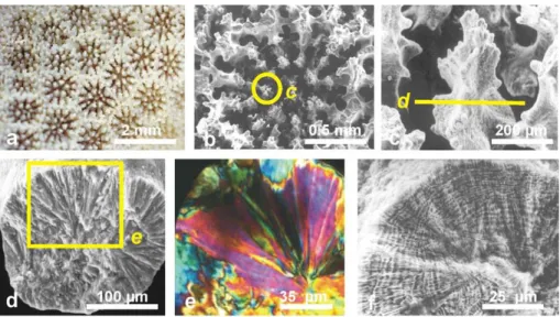
Fig. 2. Three different aspects of the same group of coral fibres. (a): Morphology of fibres
on a fracture surface of a corallite (Favia stelligera). (b)–(c): Ultra-thin section observed in
polarized light. Fibres are groups of subunits with globally similar behaviour with respect to polarization directions (orientation of the yellow arrow).(d) Aspect of the same skeletal sector
after polishing and enzymatic etching. Global organization of fibres is still recognizable, but growth layers have been made visible by synchronous differences in sensitivity to etching. This results in coordinated changes in thickness and spacement of growth layers, allowing a precise description of fibre growth to be made.
BGD
1, 625–658, 2004
The environment recording unit in coral skeletons
J. P. Cuif and Y. Dauphin
Title Page Abstract Introduction Conclusions References Tables Figures J I J I Back Close Full Screen / Esc
Print Version Interactive Discussion
© EGU 2004
BGD 0035 Fig. 3
Fig. 3. From morphology to elemental growth layer in a Porites skeleton. (a)–(b): Morphology of
Porites c.f. australiensis corallite.(c) Longitudinal view of a vertical unit. (d) Radial disposition
of fibres in a vertical unit. (e) Ultra-thin slide in a vertical unit: radial disposition of fibre fans is
well visible, a typical example of what led numerous authors to emphasize the similarity of coral fibres with abiotic crystallisations. (f) Etching of the fractured surface in the Porites corallite,
BGD
1, 625–658, 2004
The environment recording unit in coral skeletons
J. P. Cuif and Y. Dauphin
Title Page Abstract Introduction Conclusions References Tables Figures J I J I Back Close Full Screen / Esc
Print Version Interactive Discussion
© EGU 2004
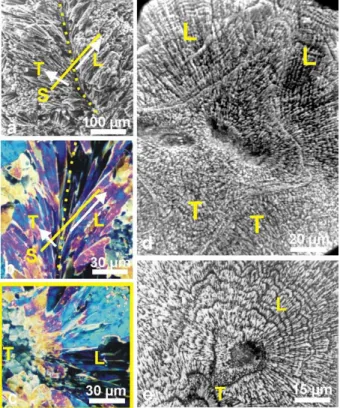
BGD 0035 Fig. 4
Fig. 4. Diversity of fibre directions on a corallite section, and consequences on the observation
of etching results. (a) Symmetrically diverging fibres on both sides of a septum median plan
(yellow dotted lline).(b) Diverging fibres observed in thin slides, polarized light. S is the surface
transverse to the view. Depending on their orientation, fibres may appear transversely (T ) or longitudinally (L) cut. (c) This yellow framed view corresponds to the b transverse section. On
the left side, fibres appear as polygonal units because they are transversely cut (T ). On the opposite they are longitudinally cut (L). (d)–(e) When looking at etched surfaces, fibre growth
layers are well visible in fibre longitudinal sections only: d: Leptoria phrygia; e: Diploastrea
BGD
1, 625–658, 2004
The environment recording unit in coral skeletons
J. P. Cuif and Y. Dauphin
Title Page Abstract Introduction Conclusions References Tables Figures J I J I Back Close Full Screen / Esc
Print Version Interactive Discussion
© EGU 2004
BGD 0035 Fig. 5
Fig. 5. Synchronism of growth layers in a septum of Favia stelligera. Concentric growth layering
indicates that mineralizing activity of the ectodermal cell layer is controlled at a global level, in contrast to the usual concept of “crystal growth competition”.
BGD
1, 625–658, 2004
The environment recording unit in coral skeletons
J. P. Cuif and Y. Dauphin
Title Page Abstract Introduction Conclusions References Tables Figures J I J I Back Close Full Screen / Esc
Print Version Interactive Discussion © EGU 2004 2.45 2.47 2.49 2.51 2.53 in te n s it y S aminoacids 2.473 a b 2.45 2.47 2.49 2.51 2.53 in te n s it y energy (keV) energy (keV) Diploria Acropora Early Min. Zone Fibres Porites sulfate 2.482 sulfate 2.482 organic sulfate organic sulfate S aminoacids 2.473 BGD 0035 Fig. 6
Fig. 6. Responses of polished surfaces in corallites of Diploria labyrinthica, Acropora
digiti-formis and Porites c.f. australiensis to a 2.4825 keV X-ray beam. In all cases a strong signal
corresponding to a sulfur bond in organic sulfates is detected, whatever the location of the X-ray beam on the corallite surface, including the Early Mineralization Zones (the “centres of calcification”). In contrast, no response is obtained for S-bond in amino acids.
BGD
1, 625–658, 2004
The environment recording unit in coral skeletons
J. P. Cuif and Y. Dauphin
Title Page Abstract Introduction Conclusions References Tables Figures J I J I Back Close Full Screen / Esc
Print Version Interactive Discussion
© EGU 2004
BGD 0035 Fig. 7
Fig. 7. Mapping of sulfated polysaccharides in Montastrea curta. (a) Morphology of the
coral-lites.(b) Specimen submitted to analyse, in the sample holder. (c) Selection of the region to be
mapped by UV fluorescence: the Early Mineralization zones show a strong response (red ar-rows).(d) Biochemical map of the selected zone. The Early Mineralization Zones exhibit a high
sulfated polysaccharide concentration. In fibres, the well visible banding pattern in exact con-formity with fibre growth layers (arrows) shows that mineral phase and sulfated polysaccharides are associated at a submicronic scale within fibres.
BGD
1, 625–658, 2004
The environment recording unit in coral skeletons
J. P. Cuif and Y. Dauphin
Title Page Abstract Introduction Conclusions References Tables Figures J I J I Back Close Full Screen / Esc
Print Version Interactive Discussion
© EGU 2004
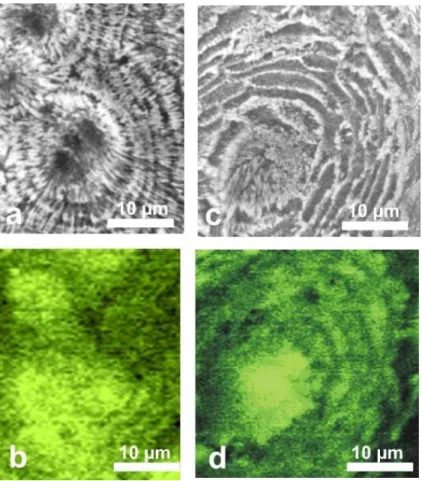
BGD 0038 Fig. 8
Fig. 8. Growth layers and sulfated polysaccharide repartition in Diploastrea labyrinthica. (a)
Morphology of growing edges of septa built by a simple series of conical units.(b) At the tip of
conical units, the Early Mineralization Zone (EMZ) surrounded by the beginning of fibrous zone
(c) Etched section, showing the concentric growth layers surrounding the EMZ. (d) XANES
BGD
1, 625–658, 2004
The environment recording unit in coral skeletons
J. P. Cuif and Y. Dauphin
Title Page Abstract Introduction Conclusions References Tables Figures J I J I Back Close Full Screen / Esc
Print Version Interactive Discussion
© EGU 2004
BGD 0035 Fig. 9
Fig. 9. Microstructural patterns and corresponding XANES mapping of organic sulfur bond on
BGD
1, 625–658, 2004
The environment recording unit in coral skeletons
J. P. Cuif and Y. Dauphin
Title Page Abstract Introduction Conclusions References Tables Figures J I J I Back Close Full Screen / Esc
Print Version Interactive Discussion
© EGU 2004
BGD 0035 Fig. 10
Fig. 10. AFM images of skeletal nanograins in Merulina scabricula. Amplitude images (a) and (c) and phase images (b) and (d) show the dual composition of skeletal grains. Phase imaging
reveals the importance of the very weak relief that can be seen on a and c pictures of the grain surfaces (arrows). The very high contrast produced by these structures demonstrates that they are basically different, from a chemical standpoint, from the nanograins themselves. The XANES in situ characterization of sulfated polysaccharides at a submicronic scale fully supports the interpretation of this high phase-contrast material as an organic coating of skeletal grains.
BGD
1, 625–658, 2004
The environment recording unit in coral skeletons
J. P. Cuif and Y. Dauphin
Title Page Abstract Introduction Conclusions References Tables Figures J I J I Back Close Full Screen / Esc
Print Version Interactive Discussion
© EGU 2004 BGD 0035 Fig. 11
Fig. 11. Equivalent AFM amplitude and phase pictures from corallite belonging to three species: (a)–(b): Favia stelligera; (c)–(d): Cladocora caespitosa; (e)–(f): Caryophyllia
am-brosia. Note that surface topography remains rather unprecise on height images (a, c, e), but
phase imaging (b, d, f) provides clear information about the basic granular structure, that has been found in all observed coral skeletons.
BGD
1, 625–658, 2004
The environment recording unit in coral skeletons
J. P. Cuif and Y. Dauphin
Title Page Abstract Introduction Conclusions References Tables Figures J I J I Back Close Full Screen / Esc
Print Version Interactive Discussion
© EGU 2004
BGD 0035 Fig. 12
Fig. 12. 2D electrophoretic characterization of sulfated polysaccharides (a) and protein
com-pounds(b) from coral skeletons. Alcian blue staining of acidic sulfated polysaccharides shows
that, after isoelectric focussing that establish their low pI, most of them remain included at the top of the 2D gel (arrows), showing that their molecular weight is higher than 300 kD, that is the upper MW accepted by the mass characterization gel.
BGD
1, 625–658, 2004
The environment recording unit in coral skeletons
J. P. Cuif and Y. Dauphin
Title Page Abstract Introduction Conclusions References Tables Figures J I J I Back Close Full Screen / Esc
Print Version Interactive Discussion
© EGU 2004
BGD 0035 Fig. 13
Fig. 13. Scheme of a growth layer formation in a coral skeleton, summarizing the structural,
BGD
1, 625–658, 2004
The environment recording unit in coral skeletons
J. P. Cuif and Y. Dauphin
Title Page Abstract Introduction Conclusions References Tables Figures J I J I Back Close Full Screen / Esc
Print Version Interactive Discussion
© EGU 2004
BGD 0035 Fig. 14
Fig. 14. Similarity between the fine structure of coral fibrous tissue and a typical “matrix
me-diated” mollusc microstructure: the prismatic layer of Pinna nobilis. (a) Close view of coral
fibres: each fibre is not single crystal but a cluster of elongated subunits with globally similar polarizing behaviour (see Fig. 2).(b) Polished and etched surface in the fibrous zone of a coral
skeleton: fibre subunits are still visible but the dominant pattern is the stepping growth mode.
(c)–(e) Morphology, ultra-thin slide (polarized light) and etching surface in the Pinna prismatic
shell layer. Calcitic prism morphology (c) is more precisely defined than coral fibres, due to their strong organic envelope, and monocrystalline behaviour (d) of the prisms is also better marked. However, an obvious similarity between the fine structures of coral fibres appears after etching (e). (f)–(g) Microprobe maps (Mg and S) on a longitudinal section of the Pinna prismatic shell
layer. Note the fine layering of minor element repartition, the high contrast between successive growth layers. Also remarkable is the independence in concentration changes between the two elements (Microprobe mappings by C.T. Williams, NHM London).