Biology and Potential Biogeochemical Impacts of Novel Predatory Flavobacteria By
Erin C. Banning
B.S., Syracuse University, 1999
B.S., University of South Florida, 2004
Submitted in partial fulfillment of the requirements for the degree of Doctor of Philosophy
at the MASS
MASSACHUSETTS INSTITUTE OF TECHNOLOGY and the
WOODS HOLE OCEANOGRAPHIC INSTITUTION
June 2010
© 2010 Erin C. Banning All rights reserved
The author hereby grants to MIT and WHOI permission to reproduce and to distribute publicly paper and electronic copies of this thesis document in whole or in part in any
medium now known or hereafter created.
ARCHIVES
ACHUSE TS INSTITUTE u)FTECHNOLYUTUN 0
2
2010
IBRARIES
Signature of AuthorJoint Program in Oceanography/Applied Ocean Science and Engineering Massachusetts Institute of Technology and Woods Hole Oceanographic Institution May 21, 2010
Certified by
I/ /2l
Karen Casciotti, Associate Scientist, WHOI Thesis Co-Supervisor
S--- Elizabeth Kujawinski, Associate Scientist, WHOI Thesis Co-Supervisor
Simon Thorrold, Senior Scientist, WHOI Chair, Joint Committee for Biological Oceanography
Biology and Potential Biogeochemical Impacts of Novel Predatory Flavobacteria By
Erin C. Banning
Submitted to the Department of Biology on May 21, 2010 in Partial Fulfillment of the Requirements for the Degree of Doctor of Philosophy in Biological Oceanography
ABSTRACT
Predatory bacteria are ubiquitous in aquatic environments and may be important players in the ecology and biogeochemistry of microbial communities. Three novel strains belonging to two genera of marine flavobacteria, Olleya and Tenacibaculum, were cultured from coastal sediments and found to be predatory on other bacteria on surfaces. Two published species of the genus Tenacibaculum were also observed to grow by lysis of prey bacteria, raising the possibility that predation may be a widespread lifestyle amongst marine flavobacteria, which are diverse and abundant in a variety of marine environments. The marine flavobacterial clade is known to include species capable of photoheterotrophy, scavenging of polymeric organic substances, pathogenesis on animals, the degradation and lysis of phytoplankton blooms and, now, predation on bacterial communities. Strains from the two genera were found to exhibit divergent prey specificities and growth yields when growing predatorily. Olleya sp. predatory cells accumulated to an order of magnitude greater cell densities than Tenacibaculum sp. cells on equivalent prey cell densities. Experiments were conducted to constrain the potential of the novel isolates to affect prey communities under more environmentally relevant conditions. An investigation of the minimum number of predatory cells needed to generate clearings of prey cells found that the inoculation of individual predatory flavobacteria cells can ultimately result in dense lytic swarms. In some cases, the
susceptibility of particular prey species to lysis by a flavobacterial predator was found to vary based on the growth state of the prey cells or the presence of their spent growth media. A novel methodology for the experimental study of biofilms was used to assess the impact of exposure to predatory marine flavobacteria on the release of macronutrients from prey biofilms. The Olleya sp. predator had a stimulative effect on macronutrient release while the Tenacibaculum sp. did not, further suggesting the two groups of predators are adapted to different ecological niches.
Thesis supervisor: Karen Casciotti Title: Associate Scientist
Thesis supervisor: Elizabeth Kujawinski Title: Associate Scientist
ACKNOWLEDGMENTS
During my graduate career, I have been supported by the National Science Foundation (NSF) Division of Molecular and Cellular Biosciences Grant
(MCB-0348425), the MIT Student Assistance Fund, the Woods Hole Oceanographic Institution (WHOI) Academic Programs Office and the WHOI Coastal Ocean Institute (COI). The work described in this thesis has been supported by a WHOI Ocean Venture Fund grant, COI and the WHOI Ocean Life Institute.
I am grateful to my advisers, Liz Kujawinski and Karen Casciotti, for taking me on as a student when I needed to change labs and develop a new thesis project. I am indebted to them for their support through the many obstacles and difficulties that arose during the execution of this project. In addition, I would like to thank my committee members for their forbearance and advice during this process: Martin Polz of MIT, John Waterbury of WHOI, Ed Leadbetter of the University of Connecticut and Mark Martin of the University of Puget Sound. I am also grateful to the chairs of my thesis proposal and thesis defenses, Stefan Sievert and Sonya Dyhrman respectively, for their support and advice at the beginning and end of my thesis research.
I would also like to thank the students, technicians and postdocs who have worked alongside me here at WHOI for their field assistance, helpful discussions and laboratory support. They include Matt McIlvin, Dan Rogers, Carly Buchwald, James Saenz, Alyson Santoro, Misty Miller, Krista Longnecker, Maya Bhatia and Cornelia Wuchter, amongst others. I am indebted to Jeff Donnelly and Maya Gomes of WHOI for their loan of a vibracoring rig and core barrels which facilitated the collection of the samples upon which this project was based; to Louie Kerr of the Marine Biological Laboratory (MBL) for his technical assistance with confocal microscopy; to Kevin Kroeger, Sandy Baldwin and Laura Erban of the United States Geological Survey for field support and assistance; to Tracy Mincer for the use of his dissecting microscope and camera; and to Stefan Sievert of WHOI for the use of his lab for fluorescent in-situ hybridization. I am also very grateful to Vicke Starczak for her guidance and assistance with statistical analyses.
Table of Contents
A BSTRA CT ... 3
A CK N O W LED G M EN TS... 5
CH A PTER 1: IN TR O D UC TIO N ... 9
A B ST R A C T ... 9
PREDATION IN MICROBIAL COMMUNITIES... ... ... 9
PREDATORY BACTERIA ... 10
TOWARDS AN ASSESSMENT OF THE IMPORTANCE OF SURFACE-ASSOCIATED PREDATORY BACTERIA... 12
TABLE 1: LIST OF DESCRIBED PREDATORY BACTERIA... ... ... ... 16
CHAPTER 2: NOVEL STRAINS ISOLATED FROM A COASTAL AQUIFER SUGGEST A PREDATORY ROLE FOR FLAVOBACTERIA ... 17
A B ST R A C T ... 17
INTRODUCTION... 17
M ATERIALS & M ETHODS ... 20
R E S U L T S ... 2 8 D ISC U S S IO N ... 3 3 F IG U R E 1 ... 3 9 F IG U R E 2 ... 4 0 F IG U R E 3 ... 4 2 F IG U R E 4 ... 4 3 TABLE 1: PREY SPECIFICITY OF PREDATORY STRAINS... 44
TABLE 2: PHYSIOLOGICAL CHARACTERISTICS OF PREDATORY STRAINS AND CLOSE RELATIVES... 45
CHAPTER 3: BIOLOGY OF PREDATORY MARINE FLAVOBACTERIA ... 47
ABSTRACT ... 47
INTRODUCTION ... 47
M ATERIALS AND M ETHODS... 51
R E SU L T S ... 5 6 DISCUSSION... 64 F IG U R E 1 ... 7 4 F IG U R E 2 ... 7 5 F IG U R E 3 ... 7 7 F IG U R E 4 ... 7 9 TABLE 1: V ARYING CONDITIONS EXPERIMENT TREATMENTS... 80
TABLE 2: CALCULATED GENERATION TIMES (HOURS) ACROSS TEMPERATURE AND SALINITY GRADIENTS 80 TABLE 3: PREDATION ACROSS TEMPERATURE AND SALINITY GRADIENTS . ... ... 81
TABLE 4: ENZYME ACTIVITY RESULTS AFTER GROWTH IN MARINE BROTH ... ... 82
TABLE 5: ENZYME ACTIVITY RESULTS AFTER DIFFERENT GROWTH CONDITIONS... 83
TABLE 6: ESTIMATED PREDATOR CELL DENSITIES ON FILTERS AFTER ADDITION OF -101 PREDATOR CELLS ... ... 8 5 TABLE 7: G ROW TH FEATURES W ITH B. SUBTILIS PREY ... 85
TABLE 8: GROW TH FEATURES W ITH E. COLI PREY ... 86
TABLE 9: G ROW TH FEATURES W ITH H . HALODURANS PREY ... ... ... 87
TABLE 10: GROW TH FEATURES W ITH K .KRISTINAE PREY ... 88
TABLE 11: G ROW TH FEATURES W ITH P. CORRUGATA PREY ... 89
TABLE 12: GROW TH FEATURES W ITH S. ONEIDENSIS PREY ... 90
CHAPTER 4: BIOGEOCHEMICAL EFFECTS OF FLAVOBACTERIAL PREDATION... 91
IN T R O D U C T IO N ... 9 1I
M A TER IA LS & M ETH O D S ... 95
R ESULTS ...1:... 102
D ISC U S S IO N ... 1 0 6 TABLE 1: EXPERIMENTAL DESIGN... 115
TABLE 2: RESULTS OF 2-WAY ANOVA OF FLUID CHEMISTRY DATA... 115
F IG U R E I... 1 16 F IG U R E 2... 117 F IG U RE 3 ... 1 18 F IG U R E 4... 119 F IG U R E 5... 120 F IG U R E 6... 12 1 FIGURE 7 ...VOBAT ... 124 F IG U R E 8 ... 12 6 FIG U R E 9 ... 127
CHAPTER 5: IMPLICATIONS OF BACTERIOLYTIC PREDATION AS A LIFESTYLE OF MARINE FLAVOBACTERIA ... ... 129
ABSTRACT TH. RY... L... ... 1292.... INTRODUCTION S RA -S CA DPA...TORY... 129
ECOLOGY OF MARINE FLAVOBACTERIA... 130
CONSTRAINING THE PREDATORY LIFESTYLE... 134
EVOLUTION OF SURFACE-ASSOCIATED PREDATORY BACTERIA... 137
APPENDIX ... ... .. ... 139
MEDIA RECIPES ... 139
CHAPTER 2 SUPPLEMENTARY MATERIAL: ... 141
F IG U R E S I ... 14 2 F IG U R E S2 ... ... . ... ... . . ... 14 3 F IG U R E S3 ... ... . ... 144
F IG U R E U ... ... .. ... ... .. ... ... ... ... ... . ... ... 14 5 REFERENCES ... 147
CHAPTER 1: INTRODUCTION
Abstract
Predation as a general ecological phenomenon has been frequently found to be important in structuring and regulating microbial communities. Although
phylogenetically and physiologically diverse predatory bacteria have been described and studied, our knowledge of their ecological roles in microbial communities remains small. Their great variety of predatory strategies suggests that they may have novel effects on microbial communities, relative to those already known for protozoan grazers and viruses.
Predation in microbial communities
Predators have been found to have diverse impacts on communities on a variety of scales, from macroscopic to microscopic. These can include the maintenance of community diversity through selective predation on otherwise competitively dominant species [1,2,3,4,5], the stimulation of primary production [6,7] and direct selection for predation-resistant physiologies and behaviors [8]. Top-down, or mortality-driven,
control of microbial communities can be exerted by organisms as diverse as metazoa [9,10], protozoa [11,12], bacteria [13,14] and viruses [15].
Predation has been associated with alteration in the rates of microbially mediated biogeochemical processes, including nitrogen fixation [16], nitrification [17], carbon fixation [6], the production of dissolved organic carbon (DOC) [18,19] and jet fuel degradation [20]. In some cases in which predators are grazing directly on the microorganisms responsible for the process in question, evidence of lower prey populations and higher community productivity suggests increases in the prey per-cell metabolic activity [16,17]. However, it is unclear if this is a direct response to predation on the part of individual prey cells or an indirect result of lower prey population densities because of grazing pressure. Another major consequence of predation is the conversion and release of grazed prey biomass as inorganic nutrients and incompletely broken-down
organic matter [7,21,22,23,24], which could sustain additional prey production and make nutrients available to other parts of the microbial community. These processes are
integral to the operation of the microbial loop in aquatic ecosystems, which leads to more efficient cycling of organic matter and other nutrients [25].
The effect of a specific predator in a particular ecosystem is dependent on its predation mechanism and other adaptations to a predatory lifestyle. Since most protozoan grazers ingest their prey by phagocytosis into food vacuoles, they can only engulf prey cells within a particular size range, an example of limitations imposed by the predation mechanism [11]. As a result, prey communities often respond to protozoan grazing pressure with increases and/or decreases in cell size distribution or the formation of aggregates too large for protozoan grazers to engulf [26,27]. Other adaptations besides the predatory mechanism itself can also influence the components of a microbial community that are susceptible to a particular predator as well. For example, different heterotrophic microflagellates have divergent motility adaptations that appear to control whether they are specialized to grazing on attached or suspended prey populations [28]. Two microflagellate species with robust swimming ability were found to reduce
suspended populations of prey bacteria without any decreases in attached bacterial
populations. On the other hand, two microflagellates with weak swimming capability, but rapid movement over surfaces as a result of adaptations of their flagella, mainly fed on
attached bacteria [28].
Predatory bacteria
Although prokaryotic microorganisms are often assumed to occupy basal roles in microbial food webs as consumers of dissolved organic carbon (DOC), diverse bacteria
are known to grow by the lysis of a variety of prey microorganisms [29,30,31,32,33]. Their trophic role in microbial ecosystems could be more similar to that played by protozoan grazers and viruses than that of heterotrophic, non-predatory bacteria.
Cultured predatory bacteria belong to a wide variety of phylogenetic groups (Table 1). The best- and longest-studied groups both belong to the 6-Proteobacteria: the
surface-associated myxobacteria and the genera Bdellovibrio, Bacteriovorax and
Peredibacter. In addition, predatory bacteria have been isolated that belong to the a-,
p-and y-Proteobacteria p-and the phyla Bacteroidetes, Chloroflexi p-and Actinobacteria. In some cases, such as the myxobacteria and the Bdellovibrio-like genera, all cultured members of these groups are predatory [34,35] while most of the other known predatory bacteria are closely related to apparently non-predatory species. For example, Ensifer
adhaerens, an a-Proteobacterium isolated from soil that predates on the gram-positive Micrococcus luteus [30], is closely related to plant-associated rhizobia [36], none of
which have been reported to possess predatory capabilities.
In addition to their broad phylogenetic diversity, predatory bacteria also utilize a variety of lytic mechanisms to attack prey cells. A distinctive periplasmic invasion mechanism is utilized by the 6-Proteobacterial genera Bdellovibrio, Bacteriovorax and
Peredibacter in which individual predatory cells attach to and penetrate the outer
membranes of susceptible Gram-negative bacteria [29,35]. The predatory cell then degrades the cytoplasm and inner membrane of the prey, grows and divides before bursting the outer membrane and repeating the cycle. Some predatory bacteria belonging to the genera Micavibrio and Ensifer have been reported to attach to and degrade prey organisms' from a position on their outer surfaces, a strategy termed epibiotic predation [30,37,38,39,40,41]. In most other documented cases, some degree of cell-to-cell contact short of the tight association described as epibiotic has been observed
[42,43,44,45,46,47]. Although these lytic mechanisms have not been well-characterized, they presumably involve either the release of lytic factors into the local cellular
environment or the action of outer-membrane associated hydrolytic enzymes.
In addition to the diversity in the mechanisms of cell lysis, there is also diversity amongst predatory bacteria in their hunting strategies. The periplasmic predators and at least one of the described epibiotic predators, Micavibrio, possess flagella and swim at high speeds [37,48]. In addition to their solitary hunting habit, these swimming predators have all been found to be obligate predators, incapable of growth in the absence of live prey cells although host-independent mutants of Bdellovibrio sp. have been developed in
culture. This obligate predatory metabolism stands in contrast to nearly all other described predatory bacteria, which can be grown non-predatorily as well as with live prey as a sole carbon source.
Amongst the non-obligate predatory bacteria, many groups lack flagella entirely and are motile by gliding on solid surfaces, including myxobacteria, Saprospira sp., Lysobacter sp. and Herpetosiphon sp. [33,34,44,45,49]. All of these gliding, non-obligate predatory bacteria, collectively referred to hereafter as surface-associated predatory bacteria, have been observed to lyse prey on solid surfaces and swarm cooperatively in a habit often termed wolfpack predation [13,50]. Despite their lack of motility in
suspension, at least one species of myxobacteria has been observed to form their own particulate surfaces composed of floating microcolonies, which can trap and lyse cyanobacteria in suspension [46,51]. At least for the myxobacteria and Saprospira sp., the wolfpack growth habit does not appear to be required for predation to occur since both groups have been observed to singly lyse individual prey cells [43,45]. In fact,
Saprospira grandis has been described as utilizing an additional strategy termed
ixotrophy, or the feeding on prey caught on a sticky surface [45]. In suspension, S.
grandis was observed to catch flagellated Vibrio cells by the ends of their flagella and
'reel' them in up and down the predator cell's length.
Most cultured predatory bacteria have been isolated from soil and aquatic sediments, as shown in Table 1, although some cultures have also been isolated from seawater and freshwater lakes and ponds. A cursory search of the Genbank sequence database will reveal that culture-independent surveys of microbial 16S rRNA genes have detected sequences closely related to known predatory bacteria in nearly all aquatic environments [14]. Although predatory bacteria are clearly ubiquitous, the question of their ecological and biogeochemical importance has rarely been investigated.
Towards an assessment of the importance of surface-associated predatory bacteria In light of present knowledge of predatory bacteria, they seem likely to affect microbial communities in ways distinct from those of protozoan grazers and viruses. As a
result of their high intra-guild diversity, different groups of predatory bacteria may also diverge from each other with respect to their effects on prey communities. Similar to the coevolutionary interactions between prey and protozoa, many predatory bacteria seem likely to select for certain cell wall and membrane characteristics as suggested by already described prey specificities.
Although predatory bacteria have been relatively less studied than both larger and smaller predators, some have been shown to affect prey communities. Much of this work has focused on the Bdellovibrio-type predatory bacteria, which are capable of drastically lowering the viability and density of Gram-negative bacterial biofilms [52,53] and provoking increases in cell size [54]. Coevolutionary interactions between Bdellovibrio-type predators and their prey have been inferred from the fact that predators isolated from the Great Salt Lake have prey specificities apparently better adapted to the indigenous heterotrophic bacteria relative to reference strains [55].
In a study of the removal of Escherichia coli cells from estuarine water, predation
by Bdellovibrio-type predator was detected but judged to be less important than
protozoan grazing, since the presence of protozoan grazers was associated with greater decreases in E. coli cells [56]. However, the study was carried out using estuarine surface water communities, which may not have contained a predatory bacterial community adapted to efficiently utilize E. coli. In addition, surface-associated predatory bacteria were unlikely to have been present in the samples used. Particularly with respect to surface-associated predatory bacteria, little is known regarding their impacts on the surrounding microbial communities.
The non-obligate predatory metabolism possessed by all known surface-associated predatory bacteria presents a significant challenge to interpreting the consequences of their presence in the environment. For instance, myxobacteria can be difficult to separate from solid substrates, rendering traditional approaches used for
Bdellovibrio-type predators of little use [34]. Even though modem molecular techniques
have made it possible to quantify the numbers of known non-obligate predators in an environment, it is currently impossible to tell with certainty whether non-obligate
predators detected in the environment are growing predatorily or non-predatorily. In addition, since most non-obligate predators are closely related to non-predatory species
[36,49,57,58], a priori knowledge of which species are predatory and which are not is
necessary to interpret the results of culture-independent diversity surveys.
For example, even when a molecular technique can be targeted to a particular ecological category, the molecular identification by itself is insufficient to identify novel predators. A stable-isotope probing study using 13C-labeled E. coli prey added to soil microcosms detected an interesting diversity of microorganisms within the labeled RNA fraction at various time points in the experiment [59]. Sequences closely related to those of known non-obligate predatory bacteria, including myxobacteria and Lysobacter sp., were detected in the labeled RNA fractions. It is important note, however, that labeled sequences were detected at the same time points from a variety of other phylogenetic groups, which may or may not represent predatory bacteria. Since non-obligate predatory bacteria lyse their prey extracellularly, it is possible that some of the labeled biomass would be consumed by predation-resistant scavenging heterotrophs in the same vicinity. The fact of assimilation of labeled biomass, by itself, is of limited usefulness in
identifying novel predatory bacteria.
Therefore, the most viable approach currently available for determining the potential ecological and biogeochemical importance of surface-associated predatory bacteria is to conduct laboratory experiments with controlled cocultures of predator and prey. Such an approach has been widely used in exploring the effects of protozoan grazing on prey communities [22,23,24,27,60,61,62].
The following thesis chapters describe the results of the enrichment and isolation of surface-associated predatory bacteria and experiments conducted to elucidate their predatory biology and potential biogeochemical effects on a simple prey community. I addressed the following questions:
* What surface-associated predatory bacteria can be cultured from coastal sediments?
* How are the isolates' predatory activities influenced by factors such as the size and distribution of predatory inocula and the composition and activity of prey communities?
* How is macronutrient release from a simple prey community affected by exposure to the predatory isolates?
Table 1: List of described predatory bacteria
Group Predatory Known habitats Known prey References
strategy a-Proteobacteria
Alcaligenes n.d. Eutrophic freshwater cyanobacteria [57]
denitrificans pond
Ensifer adhaerens Epibiotic Soil Gram(+) bacteria [30]
Micavibrio sp. Epibiotic Soil, sewage Gram(-) bacteria [37,63,64,65]
Candidatus Midichloria Periplasmic Tick ovary mitochondria [66,67]
mitochondrii
|3-Proteobacteria
Cupriavidus necator n.d. Soil Gram(-)/(+) bacteria [68]
Aristabacter necator n.d. Soil Gram(-)/(+) bacteria, yeast [69]
and other fungi y-Proteobacteria
Lysobacter sp. Wolfpack, Soil, freshwater Cyanobacteria, Gram(-)/(+) [49,70,71,72]
cell contact bacteria
Stenotrophomonas n.d. Stratified lake, soil Chlorobi, Gram(-)/(+) [58]
maltophila bacteria
5-Proteobacteria
Bdellovibrio sp., Periplasmic Soil, freshwater, Gram(-) bacteria [29,35,64]
Bacteriovorax sp., estuaries, sewage,
Peredibacter sp. marine sediments
Myxobacteria Wolfpack, Soil, dung, bark, Bacteria, fungi, protozoa, [31,34,43,73]
cell contact sediments nematodes
Chloroflexi
Herpetosiphon sp. Wolfpack, Freshwater lakes Un-encapsulated bacteria [33,74]
cell contact
Bacteroidetes
Saprospira sp. Ixotrophy, Coastal sediment, sea Bacteria, cyanobacteria, [32,44,45,75]
wolfpack, water diatoms
cell contact
Actinobacteria
Agromyces ramosus n.d. Soil Gram(-) bacteria, yeast [42,76]
CHAPTER 2: NOVEL STRAINS ISOLATED FROM A COASTAL AQUIFER SUGGEST A PREDATORY ROLE FOR FLAVOBACTERIA
A ccepted for publication by and included with permission of FEMS Microbiology Ecology
Abstract
Three newly isolated strains of flavobacteria from coastal aquifer sediments have been found to be predatory, lysing a range of live and pasteurized microbial prey. The three strains have been classified on the basis of 16S rRNA gene phylogeny as belonging to the recently described Olleya (strains VCSA23 and VCSM12) and Tenacibaculum (strain VCSA14A) genera. Two of the closest cultured relatives to the strain VCSA14A,
T. discolor and T. gallaicum, were also found to be bacteriolytic. These five predatory
strains exhibit gliding motility and have been observed to lyse prey cells after surrounding them with social swarms, similar to known predatory bacteria such as myxobacteria and members of the genus Lysobacter. Flavobacteria are often numerically significant in a wide variety of freshwater and marine environments, particularly in association with particles, and are thought to be involved in the degradation of
biopolymeric substances. If predatory capability is widespread among flavobacteria, they may be a previously unrecognized source of 'top-down' bacterial mortality with
influence on the composition and activity of surrounding microbial communities.
Introduction
Microbial communities can be structured by top-down mortality resulting from the activity of predators, which include protists, viruses and bacteria. The action of microbial predators also causes the release of dissolved organic matter (DOM), much of which is accessible to heterotrophic microorganisms and can support secondary
production in the microbial loop [25]. Many experimental studies have shown that protistan grazing can strongly influence the morphological, physiological and
metabolic activity [16,78,79] of prey communities. Due to high host specificity and rapid production rates, viruses are hypothesized to affect the most abundant populations in a community, promoting high overall community diversity [15]. The impacts of protistan-and viral-mediated mortality on microbial communities' composition protistan-and function have garnered significant study recently. On the other hand, the activities of predatory bacteria are virtually unconstrained. Indeed, many studies have assumed that bacteria can be approximated as a single trophic level with respect to predation pressure [15,25]. However, this assumption ignores the trophic diversity known to exist within the bacterial domain.
Predatory bacteria, capable of growing with live bacterial prey as a sole substrate, have been detected in and cultured from a wide variety of environments and phylogenetic groups [13,14]. The longest-studied predatory bacteria, the myxobacteria and the genus
Bdellovibrio and its close relatives, belong to the 6-Proteobacteria [35,80], but predatory
species have also been characterized from the a-,
P-
and y-Proteobacteria [30,37,65,68,70,72] and the phyla Chloroflexi [33], Bacteroidetes [45] andActinobacteria [42]. These well-characterized predatory species have been cultured from
soils, estuaries, rivers, lakes, bogs and marine and freshwater sediments, and have been detected using PCR-based techniques in an even wider range of environments, including groundwater, human skin and hydrothermal vents. In addition to these relatively well-studied predatory bacteria, a growing number of less well-characterized lytic bacteria have been reported in culture, microscopy and stable-isotope probing studies
[57,58,59,66,81,82,83]. Many of these potentially predatory bacteria belong to genera such as Cytophaga, Pseudoalteromonas, and Alcaligenes which, while actively studied,
had not been previously thought to include predatory species [57,82,83].
Predatory bacteria employ a wide array of predatory mechanisms, ranging from the invasion of Gram-negative periplasms carried out by Bdellovibrio and its relatives
[29] to the production of lytic exoenzymes by myxobacteria [84,85] and some members of the genus Lysobacter [70]. Other predatory mechanisms include attachment to prey cells coupled with production of a diffusible lytic factor [30,77] and the capture of prey
cells by their flagella [45]. Predatory bacteria can be considered obligate or non-obligate predators, depending on whether they can assimilate exogenous DOM as a growth substrate in addition utilizing live prey. Most described predatory bacteria are non-obligate predators, the exceptions being members of the genera Bdellovibrio, Bacteriovorax and Peridibacter in the 6-Proteobacteria and Micavibrio in the a-Proteobacteria, which require live prey as their growth substrate.
The great diversity in the phylogeny and physiology of known predatory bacteria makes them difficult to study them as a group using culture-independent techniques. For example, some predatory bacteria, such as Ensifer adhaerens, are very closely related to non-predatory bacteria [86], which renders 16S-rDNA-based phylogenetic probes incapable of distinguishing predatory from non-predatory organisms. In addition, the culture-independent study of non-obligate predatory bacteria is complicated by the current inability to assess whether an organism was actively lysing prey or merely
assimilating exogenous DOM at the time of its detection. One recent study [59], designed to detect predatory bacteria using 13C-labeled prey bacteria, successfully detected known non-obligate predatory bacteria such as myxobacteria and a member of the genus
Lysobacter, as well as many gene sequences belonging to groups of bacteria containing
no well-characterized predatory species. However, this approach is complicated by the fact that predatory bacteria are unlikely to be the only assimilators of lysed prey biomass, resulting in the possibility of uptake of 13C-labeled biomolecules by non-predatory heterotrophs.
In order to assess the efficacy of culture-independent approaches for the detection of predatory bacteria and to examine the environmental and ecological significance of the functional guild as a whole, a wider range of predatory bacteria needs to be isolated and characterized. With this goal in mind, we conducted a study to identify the presence and diversity of culturable predatory bacteria from a coastal aquifer on Cape Cod, MA. This site contains a variety of chemical niches [87,88,89] in a surface-rich environment which could provide substrate for microbial communities supporting a variety of predatory bacteria. Aquifers have been found to harbor significant protist communities, as reviewed
by Novarino et al. [90], but other than the detection of Bdellovibrio in a groundwater-fed cave [91], other sources of top-down bacterial mortality have not been extensively studied in groundwater systems.
Materials & Methods
Field collection: A vibrocorer was used to sample aquifer sediments beneath the intertidal zone at the Waquoit Bay National Estuarine Research Reserve, Cape Cod, MA, in November 2007. The Cape Cod aquifer is primarily composed of fine to coarse sand and gravel [92]. A mixing zone between fresh groundwater and sea water intruding into the aquifer from the head of the bay results in a steep, tidally and seasonally influenced salinity gradient [87,89,93] providing a variety of chemical environments. Before coring, a piezometer profile was taken, sampling every six inches, to locate the salinity gradient. A vibrocoring rig and four-inch-diameter aluminum barrels, rinsed with sea water from the intertidal zone, were used for coring within the intertidal zone at the head of Waquoit Bay. The core barrel was split lengthwise with an electric saw and sediment collected at four-inch depth intervals from the center of the core using flame-sterilized spatulas. The sediment samples were transported to the laboratory in autoclaved glass jars on ice and kept refrigerated for less than 24 hours before being sampled for culturing.
Reference and prey bacterial cultures: Tenacibaculum discolor DSM 18842, T. gallaicum DSM 18841, Kocuria kristinae DSM 20032, Pseudomonas putida DSM 50906
and Planctomyces maris DSM 8797 were obtained from the German Collection of
Microorganisms and Cell Cultures (DSMZ). Bacillus subtilis PY79 was kindly provided by Tonja Bosak, Saccharomyces cerevisiae by Lynn Miller, Flavobacterium johnsoniae
by Mark McBride, Shewanella oneidensis MR- 1 by Dianne Newman, P. corrugata by
Edouard Jurkevitch and Escherichia coli JM 109 by Daniel Rogers. Olleya marilimosa
strain CAM03OT was kindly provided by Carol Mancuso Nichols. Nitrosomonas sp. C-11 3a (Red Sea isolate) is maintained in the lab of Karen Casciotti. Halomonas
halodurans is maintained in the lab of Elizabeth Kujawinski (originally isolated by G
from Thermo Fisher Scientific, Inc., Waltham, Massachusetts, USA. The details of the culture media used are described in the supplemental material.
Predatory culture isolation and purification: We prepared bacterial prey for initial enrichment and isolation from cultures of Serratia marcescens, S. oneidensis MR-I
and Nitrosomonas sp. C-113a, grown as described in the supplemental material. Cultures
of S. oneidensis and S. marcescens were harvested by centrifugation and washed three times in HEPES buffer at a salinity of 20. The final cell suspension represented a twenty-fold concentration of the original culture. As a result of relatively low cell densities,
Nitrosomonas sp. C-113a was handled differently - 300 to 500 mL of a stationary phase culture was filtered onto a 0.2 ptm filter and resuspended in its own sterile medium, with a final concentration factor of at least a hundred-fold. The bacterial prey smears were prepared by spreading 30 ptL of the concentrated prey suspension over an area about one centimeter wide and several centimeters in length on no-nutrient water agar containing cycloheximide (WCX agar) [34]. Cycloheximide was included in the initial enrichment agar to prevent fungal growth, but was excluded after the first two to three transfers. The smears were allowed to dry before inoculation with sediment aliquots.
Small (approximately thimble-sized) aliquots of sediment, taken from different depths in the sediment core, were placed directly at one end of bacterial prey smears. After inoculation, plates were observed every one to three days using a dissecting microscope at magnifications of 6X and 12X. Swarms associated with unambiguous clearing of the prey smear were transferred to fresh prey smears by cutting out a small block of agar from the leading edge of the swarm with a sterile syringe needle.
Purification was accomplished by transfer of the freshest, leading edges of expanding swarms to new prey smears at least ten times and then to dilute nutrient agar (DNa; see recipe in supplemental material), from which isolated colony morphologies were repeatedly transferred. Purity was assessed by phase-contrast microscopy using a 1OOX oil-immersion objective lens of wet-mounted slides from agar chunks of spreading swarms, direct sequencing of 16S rRNA gene PCR products with the general primer
Cultures were maintained non-predatorily on DNa of the appropriate salinity and predatorily on water agar without cycloheximide (WAT) once fungal contaminants had been eliminated. Two salinities were used for the initial enrichments to represent the
salinity range of the sampling site: one using deionized water, and the other substituting artificial sea water (ASW, recipe in the supplemental materials) for 71.5% of the final volume to reach a final salinity of about 25.
Culture experiments: The production of lytic exoenzymes by VCSA23 was tested by resuspending a live S. oneidensis cell concentrate in the cell-free supernatant
from a heterotrophically grown VCSA23 stationary-phase culture, grown in Marine broth. The cell-free supernatant was obtained by pelleting the VCSA23 cells by
centrifugation and then filtering the collected fluid through a 0.2-ptm syringe filter. The S.
oneidensis culture was split into two equal volumes and harvested by centrifugation. One
of the resulting pellets was resuspended in spent Marine broth from VCSA23 (10 mL) and the other was resuspended in the spent Marine broth from the S. oneidensis culture (10 mL). The absorbance of the two suspensions was monitored by spectrophotometry over the course of two weeks relative to sterile Marine broth.
Unless specified otherwise, DN broth with a salinity of 25 was used as a basal medium for non-predatory growth tests, and WAT with a salinity of 25 (WAT25) was used as a basal medium for predatory growth tests with S. oneidensis MR-I as the live prey organism. Pasteurized prey smears were also used as a predatory growth substrate, prepared by exposing the S. oneidensis prey to 70'C for at least 15 minutes followed by normal washing procedures as described above.
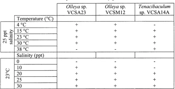
Both predatory and non-predatory growth of VCSA23, VCSM12 and VCSA14A were tested at five different temperatures (4'C, 15'C, 23'C, 30'C and 37C) and
salinities (0, 10, 20, 25 and 30). The response of the three strains to increasing
concentrations of organic matter was also assessed by growing them on WAT25 amended with 0.01, 0.1 or 1 gram of yeast extract per liter with pasteurized S. oneidensis MR-I prey washed and concentrated to about 10 times the stationary culture cell density (final concentration about 1010 cells/mL). Degradation of casein and starch by the three isolates
and 0. marilimosa, T discolor and T. gallaicum were tested by inoculation onto FMM agar [94,95], prepared in the same seawater matrix as WAT25 and amended with 0.4% w/v casein or starch. Requirement for sea salts was tested by growth on DN agar at 25 ppt salinity with only sodium chloride as a salt.
Prey specificity was tested by inoculating each test strain onto live smears of each prey organism, prepared by repeated centrifugation and resuspension as described above, except for Nitrosomonas sp. C-113a and P. maris, which were concentrated by filtration.
Two different prey densities were tested for many of the other prey organisms - about 10
times the prey's stationary phase cell density and about one tenth the prey's stationary phase cell density. Clearing presence and progress was monitored every two to three days via dissecting microscope. A strain was scored as predatory on a particular prey organism if macroscopically visible clearing expanded progressively in either of the replicate smears for the test prey.
To facilitate observation of the predatory behavior and spatial relationships between predators and prey, cultures were grown on autoclaved polycarbonate membrane filters (Millipore, Billerica, MA; 0.2 ptm pore size) placed onto WAT25 and analyzed by fluorescent in-situ hybridization (FISH) and confocal microscopy. 180 piL of S.
oneidensis live cell suspension was spread evenly onto the filters using a pipet tip. In
parallel, prey was applied in the same manner directly onto the agar to facilitate the monitoring of clearing progress during the experiment. Excess fluid from the suspensions was allowed to absorb overnight. For each predatory strain tested (strains VCSA23,
VCSM12, VCSA14 and T. gallaicum A37. 1T), a small agar block covered with the strain
in question was inoculated onto the center of the prey-covered membrane filters, as well as onto prey spots on the agar. Each experiment included three replicate plates per predatory strain, with each plate containing two prey-covered filters inoculated with predator, one filter inoculated with predator only and two filter-less prey spots, one inoculated with predator and one not. In addition, a separate plate contained prey-covered filters which were not inoculated with predator. The experiment was monitored daily by checking the filter-less predator + prey spots using a dissecting microscope.
Once clearing zones of approximately 1-cm diameter had developed
(approximately three days after predator inoculation), one predator + prey filter from each plate and three prey-only filters were fixed and frozen. When clearing zones in the filter-less predator + prey spots came close to the edges of the prey lawns, the remaining filters were fixed and frozen (after five days for strains VCSA23, VCSM12 and
VCSA14A and after 12 days for T. gallaicum). Filters were fixed in Petri dishes using 4% paraformaldehyde in phosphate-buffered saline (PBS; for recipe see supplemental material) for one to two hours at 4'C. Filters were washed by successive transfer through three Petri dishes containing PBS, with full immersion in each dish for at least five minutes at room temperature. After washing, each filter was dipped into a 1:1 mixture of PBS and ethanol and dried in a Petri dish before freezing at -20' C.
16S rRNA gene sequencing: Cells from strains VCSA23, VCSM12 and VCSA14A were suspended in HEPES buffer at a salinity of 20 and frozen at -20' C. Thawed suspensions (1 pL per reaction) were used as the template for polymerase-chain-reaction (PCR) amplification. General bacterial 16S rRNA primers (10 pM; 27F: 5'-AGA GTT TGA TCC TGG CTC AG -3', 1492R) and 2X GoTaq Green Master Mix (Promega, Madison, WI) were used in the reaction mixes. Thermal cycling was
performed under the following conditions: denaturation at 94'C for two minutes followed by 30 cycles of denaturation at 94'C for 30 seconds, annealing at 470C for 90 seconds and extension at 72'C for three minutes followed by a final extension of 10 minutes at 72'C. PCR products were checked for single, coherent bands of the appropriate size (about 1500 base pairs) by agarose gel electrophoresis and ethidium bromide staining.
Full-length 16S rRNA gene sequences (about 1500 base pairs) were cloned from VCSA23, VCSM12 and VCSA14A using the pGEM@-T Easy cloning kit (Promega Corp., Madison, WI) according to the manufacturer's instructions. For each strain, 16 colonies were picked and grown up overnight in LB at 37*C with shaking at 180 rpm. No colonies were recoverable from the VCSM12 clone library, which was not further
pursued due to the high similarity between the partial 16S rRNA gene sequences of VCSM12 and VCSA23. Cells were collected by centrifugation and plasmids were
extracted and purified by a Beckman-Coulter BiomekFX alkaline-lysis plasmid
preparation machine and used as template in two parallel sequencing reactions with the M13F (5'- GTA AAA CGA CGG CCA G -3') and M13R primers (5'- CAG GAA ACA GCT ATG AC -3'), respectively. Partial sequences (600-800 base pairs) were acquired
for strain VCSM 12 by using the full-length PCR product as the template for sequencing with the 1492R primer after purification with the Wizard SV PCR Cleanup Kit (Promega, Madison, WI). Sequencing reactions were carried out in 6-ptL volumes using Applied Biosystems BigDye 3.1 chemistry. All sequencing was performed using an Applied Biosystems 3730XL capillary sequencer at the Josephine Bay Paul Center at the Marine Biological Laboratory in Woods Hole, MA.
Phylogenetic analysis: Bases were called and vector sequences were trimmed from the 16S rRNA gene clone sequences using the Ribosomal Database Project pipeline
[96,97]. The trimmed sequences were imported into ARB [98, version December 2007] and aligned to closely related sequences in the Greengenes ARB database [99] using the ARB aligner. The ARB editor was used to construct consensus sequences from cloned
16S rRNA gene sequences for each half of the full 16S rRNA gene, which were then exported to FASTA files and manually merged into a single consensus full-length contig and imported into the Silva Reference database [100, SSURef 97 release]. The full-length consensus sequences from VCSA14A and VCSA23 and a partial sequence from
VCSM12 have been deposited in GenBank under the accession numbers G0996383, G0996384 and G0996385, respectively. The percent similarity between the full-length consensus 16S rRNA gene sequences of VCSA23 and VCSA14A, the approximately 750-base-pair partial sequence of VCSM12 and their closest relatives was determined using the "Align two sequences" function of the Basic Local Alignment Search Tool (BLAST) [101].
The full-length consensus sequences for strains VCSA23 and VCSA14A were aligned with the ARB aligner and manually checked against the 16S rRNA sequences of 204 species of marine flavobacteria in the Living Tree Project database release 100 [102]. The 16S rRNA sequence of 0. marilimosa CAM030T was obtained from Genbank and
aligned in the Living Tree Project database as well. A positional conservancy filter was calculated using the filter by base frequency method in ARB at 30% minimal similarity, then manually checked against the 207 aligned sequences and ultimately included 1,266 positions present and aligned in all sequences. Trees were constructed and compared in ARB using the neighbor-joining (ARB), maximum likelihood (RAxML v7.04 [103]), and maximum parsimony (DNAPars vl.8 [104]) methods. A smaller set of 76 species was exported using the conservancy filter for tree-building in PHYLIP. Trees were built using the neighbor-joining, maximum likelihood and maximum parsimony methods in PHYLIP [104]. The maximum-likelihood tree was converted to an extended post script file using the PHY- F online tree drawing tool and manually formatted in Adobe Illustrator. Bootstrap values were obtained for 100 replicates for the maximum likelihood tree in PHYLIP.
Probe design and optimization: Oligonucleotide probes were designed using the Probe Design tool in ARB [98] in the Greengenes database [99] and their specificity checked using BLAST [101]. The VCSA23 probe (5'- GTC ATC TCT CAC CGT AAC CT -3') is also an exact match for VCSM12, but not to any other sequences in GenBank. The VCSA14A (5'- ACC GAT CTC TCA GTC TGT CAC TCT AC -3') probe matches
T. discolor and T. litoreum exactly, but no other sequences in GenBank. 0. marilimosa CAM03OT and T. gallaicum A37. 1T have single mismatches to the VCSA23 and
VCSA14A probes, respectively, and were used as negative controls during probe optimization experiments. Both probes were determined to be specific to the single mismatch level at a formamide concentration of 35%, although the VCSA14A probe did exhibit weak hybridization to T. gallaicum cells at formamide concentrations up to 50% (data not shown). This was not a concern for the two-strain predator/prey tests described here, but could be significant if the VCSA14A probe is used on environmental samples. Finally, neither VCSA23 nor VCSA14A probes hybridized to fixed suspensions of S.
oneidensis with 35% formamide in the hybridization buffer. The GAM42a probe [105],
as a prey-specific probe since it hybridized reliably to S. oneidensis cells but not to VCSA23 or VCSA14A cells at 35% formamide.
In all cases, FISH was carried out on whole membrane filters by placing them on a microscope slide and pipetting 300 ptL of hybridization buffer (35% formamide) and 9 [tL of each probe's working stock (GAM42A conjugated with fluorescein and a predator probe conjugated with Cy3 on each filter as appropriate; 100 ng/pL) onto each filter. Hybridization and wash buffers and probe stock solutions were prepared according to previously published protocols [106]. The slides were hybridized in 50-mL centrifuge tubes prewarmed to 46'C with hybridization-buffer-soaked Kim-wipes inside.
Hybridization was conducted at 46'C for two to three hours before removing the filters from the humidification chambers and gently placing them in Petri dishes filled with wash buffer and treating them as described elsewhere [106]. To hybridize filters inoculated with T. gallaicum, which has a single mismatch with the VCSA14A probe, hybridization buffer containing 0% formamide was used. After hybridization, each filter was mounted with a small drop of DAPI mountant mix [107] on a precleaned microscope slide with a large coverslip (24 by 50 mm) and kept at 4'C in the dark until imaging.
Confocal microscopy: While control slides were examined using a Zeiss Axioplan 2 microscope, experimental filters were imaged on a Zeiss LSM 510 META NLO confocal microscope using 488-nm Argon gas and 543-nm Helium-Neon gas lasers
and a Zeiss Plan-Apochromat 63X oil-immersion objective with a numerical aperture of 1.4. In most cases, the filter was initially explored in fluorescence mode using a mercury lamp to visualize DAPI, Cy3 and FITC labels and to mark locations of interest in the LSM software for later imaging. Detector gain and amplifier offset settings were manually optimized on each filter in response to varying signal-to-noise ratios between filters. Cy3 and FITC image z-stacks were collected in frame scanning mode using the averaging method over four scans at either 1024-by-1024 or 2048-by-2048 pixel resolution. For each stack, slices were captured at 0.5 ptm intervals and pinhole settings were optimized for one airy unit. Images were collected primarily in transects crossing the predatory-prey interaction zones.
Image analysis: Z-stacks were exported as tiff files using the Zeiss LSM Image Browser version 4.2.0.121 and imported into the daime digital image analysis tool, version 1.2 [108]. The histogram stretch and noise reduction tools of daime were used to improve the contrast between cells and filter backgrounds. The same settings were used for all images collected in a given channel from a particular filter. Two-dimensional image projections were then calculated using the maximum intensity projection tool of daime and exported to the tiff file format for display. Scale bars were added manually using Adobe Photoshop.
For labeled cell volume measurements, sub-transects of 128-by-128 pixel stacks were cropped from selected full image stacks (1024-by-1024 pixels) in transects using Adobe Photoshop and individually processed in daime using the same settings for histogram stretch and noise reduction as for the larger image stacks. The cropped image stacks were segmented using the 3D segmentation tool in daime with the edge detection algorithm, ignoring any putative objects of 5 voxels or smaller. Volumes for each channel in each cropped image stack were measured using daime's measurement tool.
Results
Predatory activity: Live bacterial prey were used as a sole carbon source to isolate three strains of predatory bacteria, designated VCSA23, VCSM12 and VCSA14A, from a Cape Cod aquifer. These isolates generated macroscopic clearing zones in a variety of live and pasteurized bacterial prey smears and swarmed beyond the prey smears on water agar. In addition, two already-described species closely related to
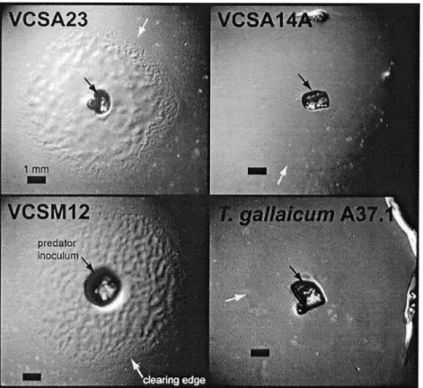
strain VCSA14A, T. discolor and T gallaicum, also cleared prey smears. Figure 1 contains images of typical macroscopic clearings caused by T. gallaicum and strains VCSA23, VCSM12 and VCSA14A on live prey lawns of S. oneidensis. 0. marilimosa
strain CAM03OT, which is closely related to strains VCSA23 and VCSM12, did not generate any clearings on the same prey bacteria. We cannot currently determine if 0.
marilimosa CAM03OT was never predatory or if it has lost its predatory capability in
The predatory specificity of the five strains was tested on 11 prey organisms selected on the basis of cell wall structure and taxonomic diversity (Table 1). Strains
VCSA23 and VCSM12 had the broadest prey specificity of the strains tested, showing
unambiguous and expanding clearing on smears of all but three of the prey organisms onto which they were inoculated. With the Gram-positive K. kristinae and the yeast S.
cerevisiae, clearing zones developed around the inoculum site and expanded to varying
degrees before ceasing growth well short of full prey consumption. Neither strain cleared
H. halodurans. The three Tenacibaculum strains (VCSA14A, T. discolor LLO4 I1.,.1I
and T gallaicum A37. IT) displayed narrower specificity than strains VCSA23 and
VCSM 12. Strain VCSA14A had the narrowest prey specificity tested, unambiguously
clearing only S. oneidensis and F. johnsoniae. T discolor and T. gallaicum each cleared both of those prey as well as B. subtilis, . coli and, in the case of T gallaicum,
Nitrosomonas sp. C-i 13a. None of the three Tenacibaculum strains visibly cleared K. kristinae, P. maris, S. cerevisiae or any of the pseudomonads tested. Like the Olleya
strains, none of the Tenacibaculum strains affected H. halodurans. All five strains were also found to be non-obligate predators, capable of growing heterotrophically on complex organic media.
Since predatory bacteria are known to employ a variety of lytic mechanisms [13,14], the predatory flavobacteria were further investigated to constrain the mechanism of prey lysis. We investigated the possibility that lytic exoenzymes are released into culture fluid by the predator, as has been observed for a Lysobacter species [70]. In our study, live S. oneidensis cells were resuspended into cell-free culture supernatant collected from a stationary-phase broth culture of VCSA23. No decrease in cell density was observed relative to a control culture resuspended in spent media from S. oneidensis (data not shown), suggesting that the enzymes responsible for cell lysis either (1) are not released extracellularly by VCSA23 or (2) are not expressed during non-predatory growth of VCSA23.
We also explored the possibility that direct cell-to-cell contact was required for prey lysis. Live cell microscopy was used in an attempt to visualize lytic events caused
by cell contact between individual cells of all three isolated strains and the S. oneidensis prey. The predators frequently exhibited gliding motility on slides in wet mount
preparations, but no direct lysis of prey cells by single predator cells was observed. However, it is possible that the wet mount environment is not conducive to predatory activity (or its observation) due to a number of factors, such as low predator cell density or disturbance associated with preparation of a wet mount slide.
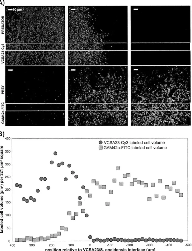
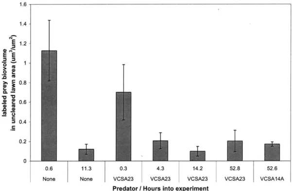
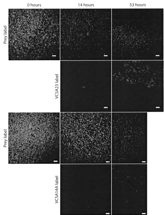
In order to facilitate imaging of predator-prey spatial relationships with a minimum of physical disturbance, membrane filters were used as a growth surface for three- to five-day incubations and visualized using FISH. The two sets of predatory flavobacteria (Olleya sp. VCSA23 and VCSM12 and Tenacibaculum sp. VCSA14A and
T. gallaicum A37. 1T) exhibited different patterns of prey clearing when visualized at high
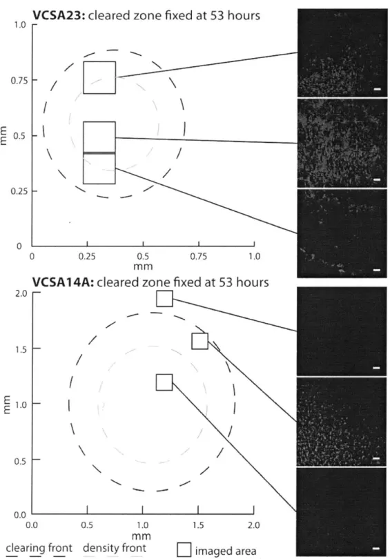
magnification. On prey-coated filters inoculated with strain VCSA23, a decrease in prey abundance was observed coincident with the position of a dense, expanding front of VCSA23 cells (Figure 2). Measurements of labeled prey and predator biovolume show that S. oneidensis cell volume decreased by approximately two orders of magnitude within about 150 pim crossing into the predator swarm (Figure 2). At least one millimeter
inward from the main interaction zone, both VCSA23 and VCSM12 formed very dense round aggregates with small central hollows. These aggregates appeared to expand over time to become a dense mass of predator cells (Figures Sl and S2). However, despite
extremely high densities of predator cell in the aggregates, a small number of prey cells were still present. Strain VCSM12 showed a broadly similar pattern, with predator cells eventually clearing most, but not all of the prey cells.
On filters inoculated with strain VCSA14A, prey cell density dropped quickly to very low levels coincident with the predator cell front (Figure 3), with most of the drop in
S. oneidensis volume complete within 50 pim. Strain VCSA14A's cell front had a visibly
lower density than that of strain VCSA23 (approximately six times less labeled cell volume), and no dense aggregates of VCSA14A cells could be found in the cleared areas of the filter. Instead, low densities of small spherical cells labeled with the VCSA14A-specific probe were observed (Figure S3). Such shortened, often spherical cells have been
frequently reported from older, late exponential or stationary phase cultures in several
Tenacibaculum species [94,109,110,111], and may represent a dormant life stage. T gallaicum A37. I was observed to have a similar pattern of predation on S. oneidensis cells (Figure S4).
For all strains tested, at least a few cells hybridizing with the prey-specific probe could be found in cleared areas, suggesting that not all susceptible prey cells are lysed. In addition, small numbers of scattered predatory cells were observed well in advance of the main density fronts for all four strains. Despite this, the areas ahead of the predator density fronts appeared essentially identical to prey lawns on control filters that never received predatory bacteria. A general tendency of higher prey cell volumes in close proximity to predatory cell fronts was observed on most filters. We attribute this to the regrowth of prey cells on DOM released from neighboring prey cell lysis at and behind the predatory cell front.
Since VCSA23, VCSM12 and VCSA14A are non-obligate predators, we examined whether their lytic behavior was affected by availability of exogenous DOM using yeast extract as a complex DOM source. All three strains cleared the pasteurized prey smears at all of the DOM concentrations tested. However, the growth habits of strains VCSA23 and VCSM12 changed from a swarming growth habit with filamentous margins to a thick, slimy growth habit with smooth-edged, entire margins at the highest DOM concentration tested (0.1% w/v yeast extract). In contrast, strain VCSA14A retained a swarming growth habit with filamentous margins at 0.1% yeast extract. Strain VCSA14A did, however, switch to a thicker growth habit with initially entire edges when grown on marine agar, which has a concentration of complex DOM of about 0.6%. These results showed that although all three of the tested strains continued to track and lyse pasteurized prey cells in the presence of high concentrations of exogenous DOM, they changed their social and motility behavior in response to increasing DOM concentrations.
Phylogenetic analysis: The 16S ribosomal RNA (16S rRNA) gene sequences of VCSA23 (1483 base pairs) and VCSM12 (three partial sequences between 708 and 756 base pairs) are nearly identical, with 99% identity between the overlapping regions of the
full-length consensus sequence for VCSA23 and the partial sequences for VCSM12, suggesting that they may be two strains belonging to a single species. Phylogenetic analysis based on 1,299 well-aligned base pairs of the 16S rRNA gene sequences indicates that VCSA23 belongs to the genus Olleya (Figure 4). The 16S rRNA gene sequence of VCSA23 is 97% identical to 0. marilimosa CAM03OT, which is the only described species in the genus [112]. These levels of 16S rRNA gene sequence identity suggest that VCSA23 and VCSM12 could represent a second species in the Olleya genus, given the often-used threshold of 97% identity in this marker gene for species-level differentiation. The next closest cultured relative on the basis of 16S rRNA gene sequence is Lacinutrix algicola, which is 95% identical to VCSA23.
The 16S rRNA sequence (1486 base pairs) of the third isolate, VCSA14A, is 89 to 91% identical to those of VCSA23 (full length) and VCSM 12 (partial), respectively. It branches tightly within the genus Tenacibaculum, and is most closely related to the
recently described species T. discolor, T. gallaicum [95], and T. litoreum [113], which are
99%, 97% and 99% identical to VCSA14A in their 16S rRNA gene sequences, respectively (Figure 4).
Physiology: Physiological tests used to compare the three newly isolated strains with other closely related cultured strains are shown in Table 2. With respect to the physiological characteristics tested, strains VCSA23 and VCSM 12 are both very similar to their closest cultured relative, 0. marilimosa strain CAM03OT. These three strains have
gliding motility, are mesophiles incapable of growth at 37'C and require at least some salt for growth. In fact, the only major characteristics assessed in which the two predatory strains differ from strain CAM03OT is the ability to grow by lysing prey bacteria and a requirement for sea salts. Their next closest cultured relative, L. algicola strain AKS293T, cannot glide, is incapable of growing at 30 C and does not require salt [114]. Strain VCSA14A was found to be very similar to its closest relatives, T discolor strain LLO4
11.1.1 , T. litoreum strain CL-TF 13T and T gallaicum strain A37. 1 .All four strains possess gliding motility, are capable of growth at 37'C and are incapable of utilizing either (+)-D-glucose or citrate as sole carbon sources with ammonia and nitrate as
nitrogen sources. All six of the strains tested in this study were unable to degrade starch with ammonia and nitrate as nitrogen sources but were able to degrade casein, exhibiting clearing halos indicative of diffusible proteases on casein FMM agar plates.
Discussion
Surface-associated predatory flavobacteria such as those described in this study could influence the biogeochemistry of a wide variety of environments. In a manner similar to that already known for predatory protozoa [11,12], they could increase the mobility and cycling of organic matter and suppress or stimulate particular members of microbial communities. The strength and persistence of such an influence is dependent on a variety of factors, including the accessibility of susceptible prey and details of the predatory biology of particular predatory flavobacteria present, such as the breadth of their prey specificity and environmental cues that either stimulate or discourage predatory activity. The isolates described in this study are undergoing further characterization with respect to their predatory biology. In addition, the discovery that two type strains of the
genus Tenacibaculum are predatory raises the possibility that existing culture collections
may contain clues as to the phylogenetic distribution of additional predatory bacteria. This study provides the first definitive evidence for predatory activity within the
class Flavobacteria. Flavobacteria are found in a wide variety of habitats, including
seawater [115], lakes [116,117], marine sediments [118,119], polar sea ice [120] and hydrothermal settings [121]. This group has been reported as being important in the degradation of polymeric substances in aquatic environments [122,123,124,125]. They are also frequently associated with particulate matter in the oceans [126,127], as well as with phytoplankton and bacterial blooms [117,128]. If some fraction of the flavobacteria in these habitats is accessing living biomass as a growth substrate, our observations have broad implications for the role of flavobacteria in microbial ecology and
biogeochemistry.
Our results suggest that the predatory flavobacteria described in this study might employ a lytic mechanism requiring at least close proximity, if not direct contact, to prey