colchicine-binding site at the interface between - and -tubulin
Sébastien Fortin a,b,c,*, Lianhu Wei d, Emmanuel Moreau c, Philippe Labrie a,b, Éric Petitclerc a, Lakshmi P. Kotra d,e,f, René C.-Gaudreault a,*
a Unité des Biotechnologies et de Bioingénierie, Centre de recherche, C.H.U.Q., Hôpital Saint-François d’Assise, Université Laval, Québec, Canada G1L 3L5
b Faculté de Pharmacie, Université Laval, Pavillon Vandry, Québec, Québec, Canada G1V 0A6
c Université Clermont 1, UFR Pharmacie, Laboratoire de Chimie Organique, Clermont-Ferrand, F-63001, France
d Center for Molecular Design and Preformulations, Toronto General Research Institute, University Health Network, Toronto, Ontario, Canada M5G 1L7
e Departments of Pharmaceutical Sciences and Chemistry, University of Toronto, Toronto, Ontario, Canada M5S 2S2
f Department of Chemistry and Biochemistry, The University of North Carolina at Greensboro, Greensboro, NC 27402, USA
* Corresponding authors. Address: Unité des Biotechnologies et de Bioingénierie, Centre de recherche, C.H.U.Q., Hôpital Saint-François d’Assise, Université Laval, Québec, Canada GIL 3L5. Fax: +1 418 525 4372. E-mail addresses: sebastien.fortin.1@ulaval.ca (S.
Abstract
Computational tools such as CoMSIA and CoMFA models reported in a recent study revealed the structure-activity relationships ruling the interactions occurring between hydrophobic N-phenyl-N’-(2-chloroethyl)ureas (CEU) and the colchicine-binding site (C-BS) on bII-tubulin. Here, we describe the mechanisms involved in the covalent binding of three subsets of CEU derivatives to the C-BS. The Flexi-Dock experiments confirmed that the interaction of non-covalent portions of the CEU auxophore moiety of CEU is involved in the binding of the drug to the C-BS facilitate the nucleophilic attack of Glu-198 rather than Cys-239. In addition, these studies suggest that Cys-239 together with Asn-99, Ser-176, Thr-177, Leu-246, Asn-247, Ala-248, Lys-252 and Asn-256 are implicated in the stabilization of a C-BS-CEU complex prior to the acylation of Glu-198 by CEU. Our molecular models propose the formation of a stabilized C-BS-CEU complex before the completion of the Glu-198 acylation; acylation triggering conformational changes of -tubulin, microtubule depolymerization and anoikis. The computational models presented here might be useful to the design of selective and more potent C-BS inhibitors. Of interest, in vivo acylation of acidic amino acid residues by xenobiotics is an unusual reaction and may open new approaches for the design of irreversible protein inhibitors such as tubulin.
Keywords. Molecular modeling; Docking; Phenylchloroethylurea; CEU; Antimicrotubule agents; Antitubulin agents; Anticancer drugs; Colchicine-binding site ligands; FlexiDock
1 Introduction
N-Phenyl-N’-(2-chloroethyl)urea compounds (CEU) are antiproliferative agents derived initially from the hybridization of aromatic nitrogen mustards and aliphatic nitrosoureas.1, 2 CEU exhibit antiproliferative activity on a large number of tumor cell lines and they are significantly more active than chlorambucil and carmustine.2–7 In addition, CEU efficiently blocked angiogenesis and tumor growth in three distinct animal models.8 On the basis of the apparent innocuousness of CEU and on their specific biodistribution to organs of the gastrointestinal tract, CEU represent a promising new class of anticancer drugs, notably against cancers of the gastrointestinal tract.8,9 Mechanistic studies indicate that CEU are acting as antimicrotubule agents and that they exhibit a mechanism of action similar to colchicine and combretastatin A-4 (Figure 1) by binding to the colchicine-binding site (C-BS) on -tubulin.10, 11 The binding of CEU to -tubulin triggers a cascade of events involving inhibition of tubulin dynamics, microtubule depolymerization, cytoskeleton disruption and cell death by anoikis.8, 12 Of interest, the pharmacophoric moiety of CEU has been recently shown useful to the design of potent inhibitors of proteins such as the mitochondrial voltage-dependent anion channel,13 thioredoxin-114, 15 and prohibitin-1.16 O O O O O N H O O O O O OH NH NH O Cl R4 R5 R3 Auxophore moiety
Pharmacophore moiety binding to Cys-239
Pharmacophore moiety acylating Glu-198
Colchicine Combretastatin A-4 Chloroethylureas (CEU)
CEU 1st class: R
3, R4 and R5= lower alkyl or branched alkyl (C1-C6) and halogens CEU 2nd class: R
3 = -(CH2)4-7-R6 and R6= OH, -OCH3, -CH3, -C(O)CH3
CEU 3rd class: R3 or R4= or
Figure 1 Molecular structure of colchicine, combretastatin A-4 and three different classes of CEU studied so far. Dashed rectangle and the gray area delimited the pharmacophore
and the auxophore groups of colchicine, combretastatin A-4 and CEU. Circle line delimited the pharmacophore moiety acylating Glu-198.
Molecular pharmacology and mass spectrometry experiments have recently showed that antimicrotubule CEU bind covalently (acylation) to II-tubulin on the glutamic acid residue in position 198 (Glu-198), which is embedded into a small hydrophobic pocket located in strand S6 adjacent to the C-BS and behind the cysteine residue in position 239 (Cys-239). That mechanism of action is different of the mechanisms of action exhibited by colchicinoids, combretastatins, podophyllotoxins and several other C-BS inhibitors.17 The latter molecules are characterized notably by the presence in their structure of a trimethoxylated phenyl ring that is essential to their anchoring in close vicinity of Cys-239 and to important conformational modifications of the protein.7, 10, 11, 18–20
The acylation of Glu-198 by CEU gives rise to the formation of a -tubulin adduct that is evidenced using SDS-PAGE and a monoclonal anti--tubulin antibody, as an immunoreacting -tubulin band exhibiting an apparent lower molecular weight. Such a fast-moving tubulin band has been observed also by Wiesen, after the mutation of Glu-198 into Gly-Glu-198 in K20T cells.21 That Glu-198-Gly mutation of -tubulin may exert its effect by causing a conformational change of -tubulin and the -, -tubulin heterodimer leading to the hypoacetylation of Lys-40 on -tubulin. The acylation of Glu-198 by CEU results also in hypoacetylation of Lys-40 (unpublished results).
The model proposed initially for the alkylation of the C-BS on -tubulin by CEU10 was based on the three-dimensional structure of -tubulin reported by Nogales et al.22 The latter suggested that the hydrophobic cavity (Figure 2), behind Cys-239 and adjacent to the C-BS, could bind lower alkyl substituted CEU through hydrophobic bonds therein leaving the electrophilic 2-chloroethyl urea moiety near the nucleophilic Cys-239. Afterwards, the CEU-tubulin complex was hypothesized to be stabilized through a hydrogen bond formed between the urea moiety of CEU and Arg-241 near the adjacent pocket. Finally, the nucleophilic reaction involving the 2-chloroethyl urea moiety and the thiol group on Cys-239 was taking place.10 That mechanism leading to the alkylation of
Cys-239 has been reported also for the covalent binding of several other antimicrotubule agents such as chloroacetylcolchicine,23,24 2,4-dichlorobenzyl thiocyanate25 and 3-(haloacetamido)-benzoylureas.26–31
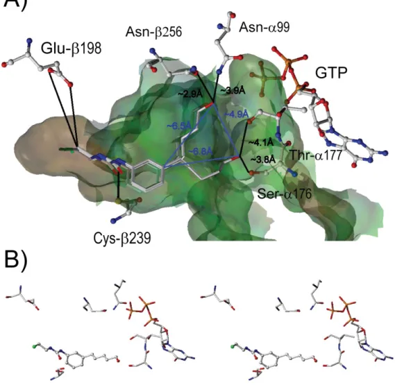
Figure 2 (A) Cartoon representation of - and -tubulin heterodimer (-helices are shown as red ribbons, -sheets in blue, and the loops in yellow). Residues important for the binding of the ligand, GDP, GTP, and colchicine are shown in capped-stick representation (C: white, O: red, N: blue, S: yellow). A translucent Connolly water accessible surface
spans the colchicine-binding site. (B) Rectangle delimits the zoomed region. (C) Orientation of HPCEU docked in the C-BS in regard of the position of colchicine.
The formation of a tubulin adduct upon reaction with CEU that migrates faster than -tubulin on SDS-PAGE unseen with other C-BS alkylating agents and the recent publication by Wiesen that the Glu-198-Gly mutation results in the formation of a similar tubulin by-product on SDS-PAGE21 prompted us to revisit the mechanism of action of CEU on -tubulin. As aforementioned, mass spectrometry studies were conducted using 4-ICEU (Figure 1) that showed the nucleophilic reaction between CEU and -tubulin was occurring on Glu-198 instead of Cys-239 (Figure 2).11 Interestingly, the in vivo acylation of either glutamic or aspartic acid residues by xenobiotics is poorly documented and is a rather unusual, if not unique, mechanism of action.
Prior to docking studies of CEU into the C-BS, we conducted Comparative Molecular Field Analysis (CoMFA) and Comparative Molecular Similarity Indices Analysis (CoMSIA) studies to confirm that the molecular structure of CEU was compatible with the tridimensional model of -, -tubulin heterodimer described by Ravelli.20,32 At first, we have conducted docking experiments using CEU substituted by different alkyl chains on position 4 of the phenyl ring (data not shown) that exhibit a more favorable binding mode that facilitate the nucleophilic attack of Glu-198 rather than Cys-239.20 Afterward, we have extended our studies to two subsequent classes of CEU substituted either on position 3 or 4 of the phenyl ring; substitutions that increase the antiproliferative activity by five to 10-fold on tumor. The first group of CEU derivatives is divided into two subsets: (i) CEU substituted by alkyl chains bearing 4 to 6 carbon atoms and (ii) CEU substituted by alkyl chains comprising 4 to 6 carbon atoms and bearing methoxy, -hydroxy or terminal methylketyl groups.18, 19, 33 The second CEU group is bearing either a meta-Z- or meta-hydrogenated arylethenyl, or a para-Z-arylethenyl moiety (Figure 1).19 The second CEU group were synthesized to assess the hypothesis that the trimethoxyphenyl moiety of combretastatin A-4 could be replaced by the aryl-3-(2-chloroethyl)urea moiety of the CEU without affecting the mechanism of action of the drug on tubulin, and its antiproliferative activity.19
Based on the hypothesis brought about by the X-ray structure published by Ravelli32 showing that the tubulin-colchicine complex is compatible with the acylation of Glu-198 by CEU, we have conducted molecular modeling experiments using FlexiDockTM on CEU structure. At first, the experiments were designed to evaluate the role of key amino acids in the stabilization of the CEU-tubulin complex. Second, we wanted to anticipate which amino acids would be targeted by CEU of the second and the third classes. Finally, we have assessed the effect of: (i) the chain length substituting the phenyl group, (ii) the nature of the group substituting in position of the alkyl chain and (iii) the effect of the rigidification of a arylethenyl group substituting the aromatic ring of our stilbenelike CEU (e.g., compounds # 7 and 8). These studies were intended to develop novel CEU that would be more active and more selective toward the Glu-198 residue imbedded in the colchicine-binding site.
2 Methodology
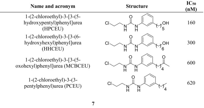
A series of eight CEU derivatives were selected for this study (Table 1). The biological activity of each compound is expressed in terms of IC50, which is the inhibitory concentration of compounds required to inhibit 50% of HT-29 cell growth.18, 19, 34
Table 1 Structure of the CEU derivatives and their antiproliferative activity on HT-29 tumor cells docked with FlexiDockTM into the colchicine-binding site18, 19
# Name and acronym Structure IC50
(nM) 1 1-(2-chloroethyl)-3-[3-(5-hydroxypentyl)phenyl]urea (HPCEU) NH NH O Cl OH 5 ( ) 160 2 1-(2-chloroethyl)-3-[3-(6-hydroxyhexyl)phenyl]urea (HHCEU) NH NH O Cl OH 6 ( ) 300
3 oxohexyl)phenyl]urea (MCBCEU)
1-(2-chloroethyl)-3-[3-(5-N H N H O Cl ( ) 4 O 600
4 pentylphenyl)urea (PCEU)
1-(2-chloroethyl)-3-(3-N H N H O Cl ( ) 4 620
5 hydroxybutyl)phenyl]urea (HBCEU) 1-(2-chloroethyl)-3-[3-(4-N H N H O Cl ( )OH 4 710 6 1-(2-chloroethyl)-3-[3-(5-methoxypentyl)phenyl]urea (MPCEU) NH NH O Cl O 5 ( ) 840 7 1-[4-(3-hydroxy-4- methoxystyryl)phenyl]-3-(2-chloroethyl)urea (4ZCombCEU) N H N H O Cl O OH 2400 8 1-[3-(3-hydroxy-4- methoxyphenethyl)phenyl]-3-(2-chloroethyl)urea (3HCombCEU) N H N H O Cl O OH 9600
Three-dimensional structure building and all modeling were performed using the SYBYL 7.0 (Tripos Inc. 2004) program package.35 The crystal structure of the complex of tubulin-colchicine: stathmin-like domain was recovered from the Brookhaven Protein Data Bank (http://www.rcsb.org/pdb/ with the entry code 1SA0). The potential of the 3-D structure of tubulin was assigned according to the Tripos Force Field36 with Kollman-all-atom37 charges subprograms in SYBYL 7.0. All atoms of GTP and GDP embedded within the protein structure were modified according to the biological structure. All partial atomic charges were calculated using Gasteiger-Hückel method.38 Initial structures of CEU were generated with sketch molecules, which is the fastest way to build a molecular model. A clean-up procedure was made to perform a torsional hunt or rough dynamics of the molecules studied. Energy minimization was performed using the Tripos Force Field with a distance-dependent dielectric and the Powell conjugate gradient algorithm that had a convergence criterion of 0.05 kcal x mol-1 x Å-1. The C, D and E chains of protein were deleted to simplify the molecular structure of the protein. To confirm that the acylation of Glu-198 is a plausible mechanism of action of CEU compatible with the Ravelli’s models and our results using MALDI-TOF mass spectrometry, colchicine was extracted from the model and CEU were merged into the colchicine-binding site in such a way that the pharmacophoric aryl-3-(2-chloroethyl)urea moiety was at the center of the pocket adjacent to the C-BS. Amino acids were shown 11 Å around the docked CEU to see as clearly as
possible the interactions with the key amino acids constituting the C-BS. The docked ligands were then subjected to flexible docking calculation employing the option ‘FlexiDock’ in SYBYL 7.0. During our flexible docking calculation, all the single bonds of the ligands and the side chains of the amino acid residues Cys-239 and Glu-198 were taken as flexible within the interaction region. Thus we obtained the initial structure of the ligand-receptor complexes. For sake of simplicity, all amino acids located farther than 5 Å away from the CEU were deleted. However, GTP and Glu-198 were kept to locate the C-BS and to show the distance between the acylating group and its nucleophilic target. The illustration of the 3-D complex was made to illustrate the interactions between the HPCEU and the complex as clearly as possible. Moreover, to simplify the models, hydrogen atoms were not displayed. In this study, we searched for the heteroatomic interactions occurring between CEU and tubulin. Amino acids that were not displaying hydrogen bonding or dipole-dipole interactions were discarded to simplify the models. The interatomic distances between the heteroatoms of HPCEU and the heteroatoms of neighboring amino acids were displayed with blackened lines. When several conformers were superimposed and expressed in the same space, the mean distances of each conformer were used. The blue line between the two different modes of stabilization of HPCEU (Figure 3a) represents the distances between the phenyl ring and their respective hydroxyl groups. The surface around the C-BS was created using the Molcad Surface tool (with unaltered protein) and it was defined in the absence of any ligand. The surface of the pocket is shown as Connolly-channel and is color-coded according to hydrophobicity (polar to hydrophobic from blue to brown via green). To a better view of the HPCEU 3-D model, a stereo view of the best score of HPCEU was shown in Figure 3b.
Figure 3 (A) 3-D Structure of the two modes of stabilization of HPCEU complex (4 conformers) with key amino acids of tubulin. Black lines represent the distance between the heteroatoms of key amino acids and heteroatoms present on CEU. When several conformers are occupying the same space, the mean distances between the heteroatoms on CEU and the heteroatoms of amino acids involved were assessed as the mean distances. Blue line in panel B represents the distance of the two modes of stabilization of HPCEU between the phenyl ring and the hydroxyl group. (B) Stereoview of the best score of HPCEU.
3 Results and discussion
‘Clean-up tool’ was used to perform a torsional hunt or rough dynamics of the molecules studied and the minimization energy conformation of CEU structure was used as starting docking material because the 3-D models of crystallized CEU are unavailable. The minimum energy conformation was used to perform flexible docking calculation with
FlexiDock with the CEU identified in Table 1. Figure 3 shows the best way of stabilization of the lead compound HPCEU (1) as a template listed in Table 1 prior to its acylation by Glu-198.
CEU are composed of a pharmacophoric phenyl-3-(2-chloroethyl)urea moiety and an auxophore moiety identified as the -alkyl chain substituting the aromatic ring or the aryl ethenyl group. In regards to the interaction described by Bai et al.,17,23,32 the trimethoxy phenyl moiety of colchicine interact with Cys-239 and that interaction is the key interaction to anchor the drug in the C-BS. On one hand, as illustrated in Figure 3, the 2-chloroethylurea moiety of CEU is also anchoring the drug to Cys-239. On the other hand, the auxophore moiety of CEU contributes to stabilize the molecule between the - and -tubulin heterodimer. That result prompted us to hypothesize that the CEU pharmacophore could replace the trimethoxy phenyl group of colchicine and many other C-BS antagonists. The biological data obtained with the different CEU tested confirm that the interactions occurring between Cys-239 and the pharmacophore moiety of CEU are essential. To that end, the mutation Cys-239-Ser found in III-tubulin abrogates the covalent binding of CEU to that -tubulin isoform.10 The interactions between the auxophore moiety of CEU and -tubulin are also important for the antiproliferative activity (Table 1) as confirmed by the biological inactivity of the unsubstituted CEU derivative (data not shown).
In this study, our analyses were not based on the energy stabilization of the complex ligand-receptor notably because the antiproliferative activity of CEU is not based only on the stabilization of the protein by the ligand. The stabilization of the protein plays an important role in the mechanism of action but the position of the ligand and the nature of the interactions occurring into the pocket are still more important for the acylation of Glu-198. However, the docking scores show that the non-covalent binding portions of the CEU support a superior or more favorable binding mode to trigger the nucleophilic attack by Glu-198 as compared to Cys-239. In addition, all models are showing that the 2-chloroethylurea moiety of CEU is protruding inside the adjacent pocket toward Glu-198 in such a way that the acylation is favored. It is also important to note that all models of the CEU-tubulin complexes are showing interactions occurring between the urea moieties of
CEU and Cys-239 with distances ranging from 2.4 to 3.5 Å. Cys-239 is probably one of the most important amino acid residue involved in the stabilization of the CEU-tubulin complex prior to the acylation of Glu-198.
The interactions occurring between the SH group of Cys-239 and the urea group of CEU probably lock the 2-chloroethylurea moiety into the pocket containing Glu-198. The distance between the urea moiety of CEU and the SH group of the binding site is an important parameter favoring Glu-198 acylation. We hypothesize that the shorter the distance between the electrophilic group of CEU and the -COOH of Glu-198 is, the higher the rate of acylation.
Moreover, other important interactions occur between the heteroatom positioned at the -position of the substituent on the aromatic ring of CEU or the aryl ethenyl group and heteroatoms on Asn-99, Ser-176, Thr-177, Leu-246, Asn-247, Ala-248, Lys-252 or Asn-256 residues. A 3-D model was built to correlate the activity and the structure to design new and more active CEU. The 3-D model takes into account the interactions occurring between CEU and the hydrophobic pocket adjacent to C-BS according to the following parameters: (i) the number and the strength of heteroatom interactions (correlated with the distance); (ii) the nature and the position of the amino acids involved in the heteroatomic interactions; and (iii) the global position of CEU into the colchicine-binding site. To confirm the importance of the aforementioned parameters on the stabilization of the CEU-protein complex before the acylation reaction, we have listed the interactions obtained between eight potent CEU and the C-BS (Table 2). The mean distances between the carbon atom bearing the chlorine atom of CEU (electrophile) and the carboxyl of Glu-198 (nucleophile) were measured to evaluate the minimal distance required to initiate the nucleophilic reaction. As illustrated in Table 2, when several conformers were occupying the same space, the mean distances between the heteroatoms of CEU and the amino acids of -tubulin were calculated and were taken as a mode of stabilization and the lowest energy scores of the docking of the CEU complex were used to describe the molecular stabilization of the CEU-C-BS complexes.
Table 2 Docking scores of FlexiDock and distances (Å) between heteroatoms of the key amino acids and the CEU in eight different CEU models.
CEU Acronym/ lower energy score* CEU interacting group Amino acid involved Amino acid interacting group Interatomic distance (Ǻ)*
HPCEU OH (-group) Asn-256 C=O 2.9
OH (-group) Asn-99 NH2 3.9
-7.37 OH (-group) Ser-176 OH 3.8
OH (-group) Thr-177 C=O 4.1
C=O (urea) Cys-239 SH 2.4
C-Cl Glu-198 O-C=O 6.0
HHCEU OH (-group) Asn-256 C=O 4.2
OH (-group) Asn-99 NH2 3.6
-6.59 OH (-group) Thr-177 C=O 3.8
C=O (urea) Cys-239 SH 3.8
C-Cl Glu-198 O-C=O 7.0
MCBCEU C=O (-group) Asn-99 NH2 4.3
NH (urea) Cys-239 SH 3.8
-19.58 C-Cl Glu-198 O-C=O 5.8
PCEU C=O (urea) Cys-239 SH 3.5
1.48 C-Cl Glu-198 O-C=O 5.1
HBCEU OH (-group) Asn-256 C=O 3.7
OH (-group) Thr-177 C=O 4.0
-13.27 C=O 3.8
C=O (urea) Cys-239 SH 3.7
C-Cl Glu-198 O-C=O 5.7
MPCEU OH (-group) Asn-99 NH2 4.5
C=O (urea) Cys-239 SH 3.3
-15.80 NH (urea) Cys-239 SH 4.3
C-Cl Glu-198 O-C=O 5.8
C-Cl Glu-198 O-C=O 5.0
4ZCombCEU OH (aryl ethenyl) Leu-246 C=O 4.2
O-Me (aryl ethenyl) C=O 4.5
-16.70 OH (aryl ethenyl) NH 3.8
C=O (urea) Cys-239 SH 4.3
C-Cl Glu-198 O-C=O 7.1
3HCombCEU OH (aryl ethenyl) Lys-252 NH2 3.2
O-Me (aryl ethenyl) NH2 3.5
O-Me (aryl ethenyl) C=O 3.2
OH (aryl ethenyl) Ala-248 NH 3.2
C=O (urea) Cys-239 SH 3.2
C-Cl Glu-198 O-C=O 8.5
*Most docking scores have generated several conformers (limited to 10). When several conformers were in the same area, the mean distances between the heteroatoms on CEU and the targeted amino acids neighboring the C-BS were calculated and they were taken as a mode of stabilization. When many modes of stabilization were given, the lowest energy score are shown.
We have classified the CEU derivatives in three groups based on their antiproliferative activities: strongly active (e.g., HPCEU (1) and HHCEU (2)), fairly active (e.g., HBCEU (3), MPCEU (4), MCBCEU (5) and PCEU (6)) and weakly active (e.g., 4ZCombCEU (7) and 3HCombCEU (8)). In this study, all 3-D models studied gave a position of the electrophilic moiety of CEU into the pocket adjacent to the C-BS that allows the acylation of Glu-198. Experimentally, HPCEU is the most active CEU studied so far. It exhibits a IC50 of 160 nM on human HT-29 colon carcinoma cells. The computational model obtained using HPCEU has generated two different mode of stabilization. In each case, there is formation of hydrogen bonds between the thiol group of Cys-239 and the urea moiety of CEU (distance of 2.4 Å). The first mode of stabilization of compound 1 generates a second hydrogen bond between the carbonyl group of Asn-256 and the hydroxyl group of the alkyl chain substituting the phenyl moiety of CEU (distance of 2.9 Å). It forms also a weak dipole-dipole interaction between the amidic group of Asn-99 and the -OH on the alkyl chain of HPCEU (distance of 3.9 Å). The second mode of stabilization of compound 1 exhibits two weak interactions between the hydroxyl group of the alkyl chain of CEU and the hydroxyl group of Ser-176 as well as with the carbonyl group of Thr-177 (distance of 3.8 Å and 4.1 Å, respectively). HHCEU is the second most potent molecule (IC50 = 300 nM) and has generated one mode of stabilization. Compound 2 form three weak interactions occurring between the -hydroxyl of the alkyl chain of CEU and the amide of Asn-99, the carbonyl groups of Thr-177 and Asn-256 (distance of 3.6 Å, 3.8 Å and 4.2 Å, respectively). The carbonyl group of the urea moiety of 2 interacts also with the thiolate of Cys-239 (distance of 3.8 Å).
MCBCEU (3), PCEU (4), HBCEU (5) and MPCEU (6) are fairly active CEU exhibiting IC50 of 600, 620, 710 and 840 nM, respectively. The model using MCBCEU generates one mode of stabilization. Compound 3 interacts with the C=O of the -group and the NH2 of Asn-99 with a distance of 4.3 Å. The distance between the NH group of the urea moiety from compound 3 neighboring the phenyl group and the SH of Cys-239 is 3.5 Å. The model of PCEU presents a single weak interaction between C=O of the urea and the SH of Cys-239. In addition, the position of the aliphatic chain is at the center of the C-BS. The van-der-Waals interactions between the auxophore of CEU and C-BS are stabilizing and orienting the position of the CEU into the C-BS. The importance of these interactions in the mechanism of action of CEU is exemplified by the inactivity of the unsubstituted phenyl CEU derivative. The molecular model based on compound 5 exhibits two modes of stabilization. First, two weak interactions are occurring between the -OH group of the alkyl chain substituting the phenyl moiety and the carbonyl of Thr-177 as well as the amide of Asn-256 (distance of 4.0 Å and 3.7 Å, respectively). Second, compound 5 presents a weak interaction between the -OH of CEU and the carbonyl of Thr-177 (distance of 3.8 Å). The carbonyl group of the urea moiety from the two conformer groups forms a weak interaction with the SH group of Cys-239 with a distance of 3.7 Å. The MPCEU model presents two different modes of stabilization. The difference between these two modes of stabilization is that the 3-phenyl-(2-chloroethyl) urea moiety is occupying two different spatial configurations when the position of the -methoxy group is maintained in the same position. The distance between the O-Me of compound 6 and the NH2 of the Asn-99 is 4.5 Å. The distance between the carbonyl of the urea moiety from the first mode of stabilization of compound 6 and the SH of C-BS is 3.3 Å while the distance between NH of the second mode of stabilization of compound 6 and the SH of C-BS is 4.3 Å. The energy of the second mode of stabilization is higher but it occupies a deeper position in the pocket that may favor the acylation of Glu-198.
4ZCombCEU and the 3HCombCEU were prepared based on their potential higher affinity for the C-BS and were expected to exhibited improved antiproliferative activities. However, the addition of the aryl ethenyl moiety to CEU leads to low IC50 of 2400 and
9600 nM, respectively. Our computational models show that 4ZCombCEU interacts with the carbonyl and the NH groups of Leu-246. The molecule forms two weak interactions between the carbonyl of Leu-246, and the OH and the O-Me of 4ZCombCEU that are separated by a distance of 4.2 Å and 4.5 Å, respectively. The hydroxyl group of 4ZCombCEU also interacts with the NH group of Leu-246 with a distance of 3.8 Å. The distance between the C=O of the urea group of 4ZCombCEU and the SH group of the Cys-239 is 4.3 Å. The 3HCombCEU generates two weak dipole interactions with Lys-252 and Asn-247 and forms a hydrogen bond with the C=O of Asn-247. Interestingly, the OH group of 3HCombCEU weakly interacts with the NH2 of Lys-252 with a distance of 3.2 Å while the other O-Me group on 3HCombCEU exhibits the same weak interaction with the NH2 of Lys-252 with a distance of 3.5 Å. The O-Me group of 3HCombCEU interacts also with the C=O group of Asn-247 with a distance of 3.2 Å while the OH group of 3HCombCEU generates a hydrogen bond with the C=O of Asn-247 with a distance of 2.3 Å. The distance between C=O of the urea of 3HCombCEU and the SH of the Cys-239 is 3.2 Å. The energy stabilization is favor in the 4ZCombCEU complex and highly disfavour in the 3HCombCEU complex. In our models, the stabilization of tubulin does not correlate well with the antiproliferative activity of CEU. That phenomenon might be explained as follows. First, the position of the 4ZCombCEU and 3HComb-CEU in the pocket results in higher distances between the carbon atom bearing the chlorine atom of CEU (electrophile) and the carboxyl of Glu-198 (nucleophile) than the ones measured in the models of strongly and fairly active CEU. This long distance between the electrophilic and the nucleophilic entities seem to unfavor the efficient acylation of Glu-198. Moreover, many unfavorable steric interactions and conformational constraints between the drug and the C-BS decrease the drug-tubulin complex stability, and partly explain the lowering of antiproliferative activity of 3HCombCEU. Furthermore, are the nature and the position of the amino acids involved in the stabilization of the drug-C-BS complexes playing a role in the decrease of energy during the acylation of Glu-198? To assess this, we compared the different subsets of CEU studied versus the nature and position of the amino acid residues involved in the drug-C-BS interactions. We observed that CEU exhibiting IC50 below 1 M
4ZCombCEU and 3HCombCEU interact with: Leu-246, Asn-247, Ala-248 and Lys-252 while CEU that are strongly and fairly active interact with: Asn-99, Ser-176, Thr-177 and Asn-256. The latter amino acid residue, except Lys-252 and Asn-256 (helix 8), are located between the rigid backbone structure of C-BS that is constituted of the beta-sheets (B3 and B5) and the alpha helices (H3, H5, H7 and H8). Asn-99, Ser-176, Thr-177, Leu-246, Asn-247 and Ala-248 are interfacing B3 and H3, B5 and H5, B5 and H5, H7 and H8, H7 and H8, H7 and H8, respectively.22 We believe that the intermolecular interactions between these amino acids and CEU might decrease the energy of activation of the acylation reaction by destabilizing one part of the protein that synergistically induces favorable conformational changes.
Finally, the antiproliferative activity does not correlate well with the interatomic distances between the carboxyl group of Glu-198 and the electrophilic carbon atom of CEU. However, it is postulated that the distance and the ligand position are also important parameters involved in the initiation or not of the acylation of Glu-198. We have dissected the mechanism underlying the acylation of Glu-198 by CEU in four main steps that are illustrated in Figure 4 by the acylation of HPCEU. First, there is stabilization of the CEU-tubulin complex via the auxophore group of CEU that forms dipole-dipole or hydrogen bond interactions and van-der-Waals interactions with amino acids (Asn-99, Ser-176, Thr-177 and Asn-256, respectively). Concomitantly, the urea moiety anchors the drug to Cys-239 through a hydrogen bond locking the CEU in a spatial conformation favoring the nucleophilic addition. The latter hydrogen bond favors also the electronic induction of the 2-chloroethylamino group by decreasing the energy requirements for the acylation. Third, the acylation occurs between the carboxyl group of Glu-198 and the carbon atom (electrophilic) bearing the chlorine atom of CEU. Finally the acylation of Glu-198 leads to important conformational changes of the -, -tubulin heterodimer that is possibly in the same order of magnitude as the ones described with the Glu-198-Gly mutation.21,39,40
Figure 4 Cartoons illustrating the steps of stabilization of HPCEU within the C-BS that leads to its acylation by Glu-198. Key amino acid residues are illustrated and arrows indicate electron flow and the formation of adducts.
4 Conclusion
We have investigated the covalent binding of three subsets of CEU exhibiting antimicrotubule activity using computational tools. Several amino acid residues were identified as essential to the binding of CEU to -tubulin prior to the acylation reaction involving the drug and Glu-198. In addition, Cys-239 was shown essential to lock the CEU in a position that favor the acylation of Glu-198. Moreover, Asn-99, Ser-176, Thr-177, Leu-246, Asn-247, Ala-248, Lys-252 and Asn-256 are important amino acid residues to decrease the energy of activation required for the acylation and to stabilize a CEU-tubulin complex favoring the completion of the nucleophilic attack. The FlexiDock experiments contributed to a better understanding of CEU-tubulin interactions and thus providing a predictive model for the design of new, specific and more potent antimicrotubule agents. It is also of interest to mention that this in vivo acylation reaction between a xenobiotic such as CEU and an acidic amino acid residue such glutamic acid is quite unusual. This observation may open new strategies for the design of irreversible inhibitors not only of tubulin but several other proteins. To that end, we recently reported the acylation of the Asp-40 residue of prohibiting isoform 1 by a 4-cyclohexylCEU derivative that led to its sequestration in the cytosolic compartment.16
5 Acknowledgements
This work was supported by grants from le Fonds de la Recherche en Santé du Québec, Junior II (E.P.) and Canadian Institutes of Health Research (R.C.-G, E.P.; Grant #MOP-79334 and MOP-89707). S. Fortin is recipient of a studentship from the Canadian Institutes of Health Research (CGD-83623). L.P.K. gratefully acknowledges the Rx&D HRF-CIHR Research Career Award and Premier’s Research Excellence Award. An infrastructure grant from the Ontario Innovation Trust provides support for the Molecular Design and Information Technology Centre and is gratefully acknowledged (L.P.K).
6 References and notes
3. C.-Gaudreault, R.; Alaui-Jamali, M. A.; Batist, G.; Bechard, P.; Lacroix, J.; Poyet, P. Cancer Chemother. Pharmacol. 1994, 33, 489.
4. Bechard, P.; Lacroix, J.; Poyet, P.; C.-Gaudreault, R. Eur. J. Med. Chem. 1994, 29, 963.
5. Miot-Noirault, E.; Legault, J.; Cachin, F.; Mounetou, E.; Degoul, F.; C.-Gaudreault, R.; Moins, N.; Madelmont, J. C. Invest. New Drugs 2004, 22, 369.
6. Mounetou, E.; Legault, J.; Lacroix, J.; C.-Gaudreault, R. J. Med. Chem. 2001, 44, 694.
7. Mounetou, E.; Legault, J.; Lacroix, J.; C.-Gaudreault, R. J. Med. Chem. 2003, 46, 5055.
8. Petitclerc, E.; Deschesnes, R. G.; Cote, M. F.; Marquis, C.; Janvier, R.; Lacroix, J.; Miot-Noirault, E.; Legault, J.; Mounetou, E.; Madelmont, J. C.; C.-Gaudreault, R. Cancer Res. 2004, 64, 4654.
9. Maurizis, J. C.; Rapp, M.; Azim, E. M.; C.-Gaudreault, R.; Veyre, A.; Madelmont, J. C. Drug Metab. Dispos. 1998, 26, 146.
10. Legault, J.; Gaulin, J. F.; Mounetou, E.; Bolduc, S.; Lacroix, J.; Poyet, P.; C.-Gaudreault, R. Cancer Res. 2000, 60, 985.
11. Bouchon, B.; Chambon, C.; Mounetou, E.; Papon, J.; Miot-Noirault, E.; C.-Gaudreault, R.; Madelmont, J. C.; Degoul, F. Mol. Pharmacol. 2005, 68, 1415. 12. Deschesnes, R. G.; Patenaude, A.; Rousseau, J. L.; Fortin, J. S.; Ricard, C.; Cote,
M. F.; Huot, J.; C.-Gaudreault, R.; Petitclerc, E. J. Pharmacol. Exp. Ther. 2007, 320, 853.
13. Patenaude, A.; Deschesnes, R. G.; Rousseau, J. L.; Petitclerc, E.; Lacroix, J.; Cote, M. F.; C.-Gaudreault, R. Cancer Res. 2007, 67, 2306.
14. Fortin, J. S.; Cote, M. F.; Lacroix, J.; Desjardins, M.; Petitclerc, E.; C.-Gaudreault, R. Bioorg. Med. Chem. 2008, 16, 7277.
15. Fortin, J. S.; Cote, M. F.; Lacroix, J.; Patenaude, A.; Petitclerc, E.; C.-Gaudreault, R. Bioorg. Med. Chem. Lett. 2008, 18, 3526.
16. Bouchon, B.; Papon, J.; Communal, Y.; Madelmont, J. C.; Degoul, F. Br. J. Pharmacol. 2007, 152, 449.
17. Nguyen, T. L.; McGrath, C.; Hermone, A. R.; Burnett, J. C.; Zaharevitz, D. W.; Day, B. W.; Wipf, P.; Hamel, E.; Gussio, R. J. Med. Chem. 2005, 48, 6107.
18. Moreau, E.; Fortin, S.; Lacroix, J.; Patenaude, A.; Rousseau, J. L.; C.-Gaudreault, R. Bioorg. Med. Chem. 2008, 16, 1206.
19. Fortin, S.; Moreau, E.; Lacroix, J.; Teulade, J.-C.; Patenaude, A.; C.-Gaudreault, R. Bioorg. Med. Chem. Lett. 2007, 17, 2000.
20. Fortin, S.; Labrie, P.; Moreau, E.; Wei, L.; Kotra, L. P.; C.-Gaudreault, R. Bioorg. Med. Chem. 2008, 16, 1914.
21. Wiesen, K. M.; Xia, S.; Huang Yang, C.-P.; Band Horwitz, S. Cancer Lett. 2007, 257, 227.
22. Nogales, E.; Wolf, S. G.; Downing, K. H. Nature (London) 1998, 391, 199.
23. Bai, R.; Covell, D. G.; Pei, X. F.; Ewell, J. B.; Nguyen, N. Y.; Brossi, A.; Hamel, E. J. Biol. Chem. 2000, 275, 40443.
24. Bai, R.; Pei, X. F.; Boye, O.; Getahun, Z.; Grover, S.; Bekisz, J.; Nguyen, N. Y.; Brossi, A.; Hamel, E. J. Biol. Chem. 1996, 271, 12639.
25. Bai, R.; Duanmu, C.; Hamel, E. Biochim. Biophys. Acta 1989, 994, 12.
26. Jiang, J. D.; Davis, A. S.; Middleton, K.; Ling, Y. H.; Perez-Soler, R.; Holland, J. F.; Bekesi, J. G. Cancer Res. 1998, 58, 5389.
27. Jiang, J. D.; Roboz, J.; Weisz, I.; Deng, L.; Ma, L.; Holland, J. F.; Bekesi, J. G. Anticancer Drug Des. 1998, 13, 735.
28. Jiang, J. D.; Wang, Y.; Janish, C. A.; Holland, J. F.; Bekesi, J. G. Biomed. Pharmacother. 1998, 52, 270.
29. Jiang, J. D.; Wang, Y.; Roboz, J.; Strauchen, J.; Holland, J. F.; Bekesi, J. G. Cancer Res. 1998, 58, 2126.
30. Li, J. N.; Song, D. Q.; Lin, Y. H.; Hu, Q. Y.; Yin, L.; Bekesi, G.; Holland, J. F.; Jiang, J. D. Biochem. Pharmacol. 2003, 65, 1691.
31. Schlesinger, M.; Jiang, J. D.; Roboz, J. P.; Denner, L.; Ling, Y. H.; Holland, J. F.; Bekesi, J. G. Biochem. Pharmacol. 2000, 60, 1693.
32. Ravelli, R. B.; Gigant, B.; Curmi, P. A.; Jourdain, I.; Lachkar, S.; Sobel, A.; Knossow, M. Nature 2004, 428, 198.
33. Moreau, E.; Fortin, S.; Desjardins, M.; Rousseau, J. L.; Petitclerc, E.; C.-Gaudreault, R. Bioorg. Med. Chem. 2005, 13, 6703.
34. Fortin, S.; Moreau, E.; Patenaude, A.; Desjardins, M.; Lacroix, J.; Rousseau, J. L.; C.-Gaudreault, R. Bioorg. Med. Chem. 2007, 15, 1430.
35. SYBYL 7.0, Tripos Inc., 1699 South Hanley Rd., St. Louis, Missouri, 63144, US. 36. Clark, M.; Cramer, R. D. I.; Van Opdenbosch, O. N. J. Comput. Chem. 1989, 10,
982.
37. Weiner, S. J.; Kollman, P. A.; Case, D. A.; Singh, U. C.; Ghio, C.; Alagona, G.; Profeta, S., Jr.; Weiner, P. J. Am. Chem. Soc. 1984, 106, 765.
38. Bookshelf Tripos 7.0, Tripos Inc., 1699 South Hanley Rd., St. Louis , Missouri, 63144, USA.
39. Hubbert, C.; Guardiola, A.; Shao, R.; Kawaguchi, Y.; Ito, A.; Nixon, A.; Yoshida, M.; Wang, X. F.; Yao, T. P. Nature 2002, 417, 455.
40. Matsuyama, A.; Shimazu, T.; Sumida, Y.; Saito, A.; Yoshimatsu, Y.; Seigneurin-Berny, D.; Osada, H.; Komatsu, Y.; Nishino, N.; Khochbin, S.; Horinouchi, S.; Yoshida, M. EMBO J. 2002, 21, 6820.