Review Article
LXR, prostate cancer and cholesterol: the Good, the Bad
and the Ugly
Hugues de Boussac1,2,3,4, Aurélien JC Pommier1,2,3,4*, Julie Dufour1,2,3,4, Amalia Trousson1,2,3,4, Françoise
Caira1,2,3,4, David H Volle1,2,3,4, Silvère Baron1,2,3,4, Jean-Marc A Lobaccaro1,2,3,4
1Clermont Université, Université Blaise Pascal, Génétique Reproduction et Développement, F-63000
CLERMONT-FERRAND, France; 2CNRS, UMR 6293, GReD, F-63177 AUBIERE, France; 3INSERM, UMR 1103, GReD, F-63177
AUBIERE, France; 4Centre de Recherche en Nutrition Humaine d’Auvergne, F-63000 CLERMONT-FERRAND,
France. *Present address: AstraZeneca, R&D Oncology iMed, CHESHIRE SK10 4TG, UK.
Received December 13, 2012; Accepted December 28, 2012; Epub January 18, 2013; Published January 25, 2013
Abstract: Cholesterol is a fundamental molecule for life. Located in the cell membrane, this sterol participates to the cell signaling of growth factors. Inside the cell it can be converted in hormones such as androgens or modulate the immune response. Such important functions could not be solely dependent of external supply by diet hence de
novo synthesis could occur from acetate in almost all mammalian cells. If a deficiency in cholesterol sourcing leads
to development troubles, overstocking has been associated to various diseases such as atherosclerosis and can-cers. Cholesterol homeostasis should thus be tightly regulated at the uptake, de novo synthesis, storage and export processes. Various transcription factors have been described these last years as important to regulate cholesterol levels. Besides, synthetic molecules have been developed for many years to modulate cholesterol synthesis, such as statins. Many articles have associated prostate cancer, whose incidence is constantly increasing, to cholesterol disequilibrium. Targeting cholesterol could thus be a new pharmacological hit to counteract the initiation, develop-ment and/or progression of prostate cancer. Among the transcription factors regulating cholesterol homeostasis, the nuclear receptors Liver X Receptors (LXRs) control cholesterol uptake and export. Targeting the LXRs offers a new field of investigation to treat cancer. This review highlights the molecular relationships among LXRs, prostate cancer and cholesterol and why LXRs have good chance to be targeted one day in this tumor. LXRs, prostate cancer and cholesterol, more than a “Ménage à trois”, The Good, the Bad and the Ugly.
Keywords: LXR, cholesterol, prostate cancer, lipid raft, pharmacological modulation
Introduction
Prostate cancer is one of the most common malignancy [1], mainly affecting elders. Various risk factors have been involved including aging, ethnic origins, hormonal status and energy bal-ance. Among the lipids, cholesterol has a par-ticular position. This fundamental molecule is part of the cell membrane and thus plays an architectural role in its organization by main-taining the fluidity or by securing important pro-teins in the membrane when located in the so-called “lipid rafts”. Cholesterol is also involved in “ligand-type” signaling: as the precursor of androgen synthesis as well as in the production of oxysterols, which activate the nuclear recep-tors LXRα and LXRβ. Maintaining a tight regula-tion of cholesterol homeostasis is thus of
pri-signaling and the proliferation/apoptosis bal-ance. Reducing de novo cholesterol synthesis and/or uptake, or increasing reverse transport by exporting cholesterol from the cell could rep-resent an efficient way to control prostate epi-thelial proliferation. This review is focused on the deleterious effect of a higher cholesterol (The Ugly) concentration on prostate cancer (the Bad) and the role of LXRs (The Good) in maintaining cholesterol homeostasis to avoid progression of prostate cancer (Figure 1). The Saga started in 1909 and is still going on. LXRs and cholesterol: when the Good controls the Ugly
The liver X receptors
fac-vated by cholesterol derivatives, the oxysterols [4]. LXRα (NR1H3) and LXRβ (NR1H2) share 80% identity both in their DNA- and ligand-bind-ing domains. Their structure is characteristic of the nuclear receptor superfamily, which pos-sesses three functionally independent domains [5, 6]. The N-terminal modulator domain con-tains an activating function of the transcription (AF1) independent from the presence of the ligand. This domain presents several putative sites of phosphorylation potentially important for LXR activity modulations [7, 8]. The DNA-binding domain recognizes the LXR response elements (LXRE) characterized by two direct repeats of the hexanucleotide motif AGGTCA usually separated by four nucleotides. Part of this domain is also involved in the heterodimeri-sation with the Retinoid X Receptor RXR (NR2B1-3), which binds 9-cis retinoic acid, the requisite LXR partner [3]. The carboxy-terminal region is responsible for the ligand-binding and contains the AF2 region necessary for the tran-scriptional initiation of target genes [4]. This domain is masked by co-repressors in absence of ligand. For a review on LXR-functioning, see Viennois et al. 2011 [9].
LXRα and LXRβ are differentially expressed in tissues. While LXRβ expression is accepted to be rather ubiquitous, LXRα is more restricted and mainly found in liver, intestine, fat tissue, macrophages, kidney and gonads, suggesting their important function in the control of cho-lesterol homeostasis (for a view on LXR expres-sion see www.nursa.org). The fundamental role of LXRs in lipid homeostasis is highlighted by the highly conserved function of these recep-tors among species [10], and has been continu-ously demonstrated since the first observation of a link between LXRα and cholesterol homeo-stasis by Peet et al. [11]. They observed that mice lacking LXRα and fed a high cholesterol diet rapidly accumulate large amount of choles-terol ester in the liver inducing a liver steatosis. Actually these mice are unable to sense and respond to dietary cholesterol and develop an impaired bile acid metabolism due to a default in the transcription of the cholesterol 7α-hydroxylase (Cyp7a1), encoding an enzyme essential in bile acid synthesis [11].
The discovery of the natural ligands of LXRs by Janowski et al. [4, 12], largely improved our comprehension of the unique role of LXRs in controlling cholesterol homeostasis. In these
studies, oxysterols, the natural derivatives of cholesterol, activated LXR at physiological con-centrations. Following this finding the develop-ment of synthetic ligands of LXRs (e.g.T0901317 [13] and GW3965 [14]) and the generation of a mouse model lacking Lxrα and/or β, greatly contributed to the comprehension of the oxys-terol/LXR dependent pathways in cells, and gave the opportunity to identify several target genes and therefore functions of the LXRs [9]. Thus, it has been admitted that LXR activities are associated with four schematic functions: 1) lipid metabolism, including cholesterol and fatty acids homeostasis; 2) steroidogenesis; 3) glucose homeostasis; 4) inflammation and immunity. Since in this review we will focus more specifically on the role of LXRs on choles-terol homeostasis, we will not develop further their other physiological functions. For more information about them, refer to Viennois et al. 2011 [9].
LXRs: two sensors of cholesterol homeostasis Cholesterol is an essential structural compo-nent of mammalian cell membranes and is required to establish proper membrane perme-ability and fluidity. In addition cholesterol also serves as a precursor for the biosynthesis of steroid hormones, bile acids, and vitamin D. Besides, this molecule is also part of the mem-brane signaling pathway by its specific distribu-tion in lipid rafts (see above). Furthermore, cho-lesterol also functions in intracellular transport, cell signaling and nerve conduction. Hence, although cholesterol is important and neces-sary for human health, its intra- and extra-cellu-lar concentrations have to be strictly controlled as high levels of cholesterol in the blood have been linked to damages to arteries and cardio-vascular diseases.
Modulation of de novo synthesis and uptake of cholesterol: LXRs act at various levels to con-trol the intracellular pool of cholesterol. The first possible source of cholesterol results from the enzymatic reaction leading to the transfor-mation of Acetyl-CoA in mevalonate by the HMG-CoA reductase [15]. That reaction ulti-mately leads to the formation of de novo cho-lesterol. In mice lacking Lxr, higher expression of Srebp2, Hmgcoa and Squalene synthase has been observed [16], while the oral treatment of wild type mice with T0901317 led to a decrease in Hmgcoa synthase and Squalene synthase
gene expression [17], suggesting a role of LXRs in the negative modulation of de novo choles-terol synthesis.
A second way to modulate the pool of intracel-lular cholesterol regards its celintracel-lular import via the LDL-receptor (LDLR). Even though a correla-tion was repeatedly observed between LXR activation and LDLR protein reduction, the mechanism has been described only recently. LXRs activate the expression of the E3 ubiqui-tin ligase Idol (Inducible Degrader of the LDLR), ultimately leading to the targeted degradation of LDLR, thus resulting in the reduction of the intracellular pool of cholesterol [18].
Induction of bile acid synthesis: Cyp7a1 is the first and rate limiting enzyme that catalyzes the initial step of bile acid biosynthesis from cho-lesterol. Although it is not the primary function of bile acid synthesis, this reaction also allows the liver to reduce in rodent the excess of cho-lesterol in cells. Interestingly, while in wild type
mice fed a high cholesterol diet Cyp7a1 expres-sion increases, this induction is not observable in Lxrα-deficient mice fed similarly [11]. Additionally, in these mice the diet induces a hepatic steatosis due to an accumulation of cholesteryl esters in the liver [11, 16].
Induction of reverse cholesterol transport: The last way LXRs use to control cholesterol levels is by exporting it outside the cells. Indeed, sev-eral ATP-binding cassettes encoding genes such as ABCA1 [19-21] and ABCG1 [22] are LXR bona fide targets. These ABC transporters actively efflux cholesterol to the extracellular acceptor HDL and increase the reverse choles-terol transport. In addition, LXRs have also been shown to modulate Apolipoprotein E level, an essential component of the VLDL particles [23]. Furthermore, LXRs modulate the expres-sion of the genes encoding ABCG5 and ABCG8 that export sterols from the inner compartment of hepatocytes to the bile duct [24, 25] and from the enterocytes into the gut lumen [26].
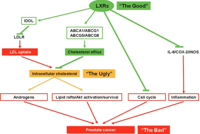
Figure 1. LXRs and prostate physiology: potential beneficial actions of LXRs over prostate cancer. LXR activity in-creases IDOL as well as various ABC transporters, which ultimately dein-creases LDL uptake and inin-creases the efflux of cholesterol, altogether decreasing the intracellular pool of cholesterol. Consequently, this leads to the reduction of androgen synthesis and lipid raft/AKT/survival pathway. LXRs finally induce cell cycle arrests, and by inhibiting the expression of IL-6, COX-2 and iNOS limit the inflammation inside the tumor. Altogether, LXR activation may limit prostate cancer development.
Altogether LXRs demonstrate a critical role in controlling the amount of intracellular choles-terol and in its processing outside the cells. Steroid synthesis: We and others have shown that LXRs could regulate the rate of cholesterol transformation into steroids in various tissues such as testis [27]. A decrease in the amount of circulating testosterone can be detected after LXR activation by the synthetic agonist T0901317 [28]. That well identified mechanism is dependent on the activation by LXRs of Sulfotransferase 2a1 that deactivates andro-gens, and the inhibition by LXRs of the steroid-sulfatase that activates androgens [28]. Interestingly, those hormones have a key role in prostate cancer development. LXRs might thus have also a role to play in this part of the anti-cancer journey.
Cholesterol and prostate cancer: when the Ugly plays with the Bad
Due to its different roles, cholesterol is hence linked to cell proliferation (see above). Indeed, its synthesis increases in tissue with high prolif-eration rate such as in cancer. On the other side, inhibition of HMGCoA-reductase blocks cell growth [29].
Prostate cancer: the Bad at a glance
Prostate cancer (PCa) is the second most diag-nosed cancer and a leading cause of cancer related death [1]. The incidence of PCa is con-stantly increasing due in part to new methods of diagnostic, and also to the increase in life expectancy. Indeed, this cancer has a slow evo-lution and about 85% of diagnosed PCa are in patients older than 65 years old [30]. Interestingly, it is accepted that more men die with PCa than from it. Indeed, an American study performed after autopsy determined that 50% of the men of 50 years old have latent PCa [31]. However the development and the cause of the disease is still poorly understood, and various factors such as genetic/ethnical origin, diet, life style and environmental factors have been suggested to play a role on it [32].
As already stated, great differences in the inci-dence of PCa are observed depending on the ethnical origin or the country of the patients. A Caucasian American has 30% less risk to develop a PCa compared to an African American
[30], but at the same time Asians develop twice less PCa than Americans. These differences are in part due to the ethnical factors, and thus to the genetic background and the lifestyle of the individuals. However it could also show dis-parities in the accessibility of the diagnostic tests and treatments.
Yet, the genetic background cannot explain everything since the first generation of Asian migrants living in the US have a more important risk of PCa than those leaving in Asia [33]. This unexpected observation is credited to be due to factors acting on PCa development rather than on PCa initiation, and presumably on the higher lipid consumption in the USA [34]. Additionally a comparable observation has been done with increased incidence to develop a PCa for Japanese population that moved to America [35]. In this study the authors also compared the migrants according to their age at arrival, and did not find any correlation with the risk to develop PCa. They therefore conclud-ed that PCa risk may be increasconclud-ed by late rath-er than early life style event [35]. These two studies are therefore suggesting a potential lifestyle/diet parameter that can greatly influ-ence the development of PCa.
Role of cholesterol in prostate cancer: the Ugly goes with the Bad!
Cholesterol accumulation in tumors is not a recent observation. White demonstrated in 1909 an accumulation of crystals of lipid nature in tumors [36]. Later Swyer and his coworkers showed for the first time an increase of cholesterol content in zone of the prostate affected by a mild hypertrophy [37] compared to healthy tissues. Afterward, similar observa-tions were obtained on other types of cancer [38-40]. Two mechanisms are generally put for-ward to explain this intracellular cholesterol accumulation: a higher circulating cholesterol uptake, and the increase in the accumulation of the enzymes of the mevalonate pathway [41, 42]
Moreover, increased uptake of LDL particles and therefore exogenous cholesterol attribut-able to a loss of modulation in the LDL receptor expression, and a higher de novo cholesterol synthesis due to the upregulation of the HGM-CoA reductase, have been suggested as key components of that accumulation [17, 43]. The
final result of that process could potentially give sufficient bricks for the membrane to expand and to the tumor to grow and develop [44].
Diet, cholesterol and PCa
Since the late 90’s, multiple lines of evidence have been highlighting the potential influence of diet on PCa appearance. First, intake of products from animal origin is correlated to a higher risk of developing metastatic PCa, but not on the initial development of PCa [45] as shown by the identical prevalence between vegetarians or meat eaters [46]. Second, the presence of dietary fat in the diet was shown to be a risk factor of PCa, although the exact con-tribution of fat was not clearly established [47]. Third, an increase in PCa incidence, angiogen-esis and metastasis was observed in the TRAMP mouse model of PCa fed a western-type diet [48]. Finally aggressiveness of PCa was increased in elders having important dietary fat intake [49]. So far data linking exces-sive consumption of cholesterol, rates of circu-lating cholesterol and risk of PCa have been controversial [50], even though studies sug-gest an impact of cholesterol in the develop-ment of high grade PCa [51-53]. Inversely, Platz et al. pointed out that a “weak” level of circulat-ing cholesterol (< 200mg/dL) was associated with a reduction of the risk of developing a prostate cancer of high grade [54]. Finally, cir-culating cholesterol increases tumor size of LNCaP xenografts in a mouse model, as well as intratumoral synthesis of androgens [55]. This suggests that the androgen dependent tumor growth could be under a deep association with circulating cholesterol. Likewise, high serum HDL is inversely correlated with PCa [53, 56]. Since HDL formation is dependent on the export of cholesterol via the ABCA1 and ABCG1 transporters that are under the positive modu-lation of LXRs, it could be suggested a possible beneficial role to over activating LXRs in PCa, even though this needs to be demonstrated. Modulation of circulating cholesterol and PCa: when reasoning the Ugly can block the Bad Altogether the presented data raise the ques-tion of the molecular mechanisms by which the cholesterol can favor tumor progression. Some observations on cancer development after treatment with statin, a cholesterol-lowering
drug that specifically inhibits the HMG-CoA reductase and therefore the formation of de novo cholesterol, partially answer that ques-tion. Indeed early investigations suggested a potent growth inhibitory effect as well as an anticancer potential of statins in vitro and in vivo [57, 58], partially explained by their ability of inducing apoptosis via the activation of the Caspase-7 [59]. Moreover in the PC3 prostate cancer cell line, statins also prevent the cell migration potential therefore reducing the for-mation of metastatic prostate colonies [60]. Then it seems that these cholesterol-lowering agents can act at different level on PCa pro-gression. The potential use of statins to pre-vent PCa is currently under active investigation mostly on prospective studies. Until now, num-bers of studies have been published and exten-sively reviewed [61]. Statin treatments do not seem to have any beneficial effect on the rate of appearance of prostate cancer conversely to the incidence of advanced PCa [62-64]. Interestingly this effect even increases when statins are used for more than five years [65]. Androgen synthesis is dependent on the amount of circulating cholesterol; besides, PCa is linked to androgen synthesis; moreover statins are cholesterol-lowering drugs. Altogether what could be the potential impact of statins on the hormonal status in prostate cancer? Actually, statins do not seem to decrease the circulating androgen [66], even though a decreased synthesis of androgens cannot be excluded since statins users show a decline in serum PSA levels, an androgen regu-lated gene in prostate [67].
Cholesterol is not only used as a precursor of steroid synthesis. Indeed, it can be found enriched in cell membranes in regions called rafts essential for the activation of the kinase cascade Akt and consequently for tumor sur-vival [68]. Zhuang et al. showed that simvas-tatin decreases the cholesterol content of lipid rafts, leading to a decrease in Akt phosphoryla-tion and activaphosphoryla-tion, and subsequently to an increase of LNCaP cells apoptosis [69]. These results improve our comprehension of the mechanism of statins in cancer progression, and also suggest lipid rafts as new players in PCa development. In accordance with that sug-gestion, the essential component of lipid rafts caveolin 1 is associated with the
aggressive-ness of the PCa tumor and therefore consid-ered as a marker of poor prognosis in PCa [70, 71]. Accordingly, the use of an antibody target-ing the caveolin 1 can block the metastatic pro-cess in PCa [72]. Both observations then con-firm the important role of lipid rafts in PCa progression.
LXRs and prostate cancer: a benefic effect of the Good over the Bad?
LXR activation leads to cell cycle arrest in pros-tate cancer cell lines
Since LXRs control cholesterol homeostasis, these nuclear receptors have been considered as putative pharmacological targets in prostate cancer. Hence, activation of LXRs by natural (22 (R)-hydroxycholesterol, 24 (S)-hydroxychole- sterol) or synthetic (T0901317) agonists led to cycle arrest of LNCaP cells via the lack of deg-radation of p27Kip1, an essential inhibitor of the cell cycle. Moreover, and as expected, treat-ment with LXR agonists also induced the pro-tein accumulation of ABCA1, thus activating cholesterol efflux [73]. Conversely, targeted dis-ruption of ABCA1 increases the proliferation rate of LNCaP cells [74]. Moreover, Chuu et al. observed that LXR-target genes were down-modulated during the tumor progression in mouse, while activation of LXRs by T0901317 delayed the progression of PCa [75]. Altogether, these studies are clearly in favor of an impor-tant protective role of LXRs in prostate cancer progression, even if no data are available in human yet.
How could LXRs be so good?
As presented above, activating LXRs will lead to the modulation of cholesterol concentration by their action on the various pathway involved. LXRs antagonize the development of prostate tumor by interacting with the androgen path-way: Prostate cancer development is tightly associated with androgens. Indeed, it is fre-quent to treat PCa patients with anti-androgens in order to block the androgen response, and therefore the early development of PCa [76]. Interestingly, the androgen receptor (AR) modu-lates the expression of HMG-CoA synthase and reductase, and SREBP2, whose product con-trols genes involved in cholesterol homeostasis such as the LDL-Receptor (LDLR) [77]. The
con-sequences of these modulations are: 1) an increase in intracellular cholesterol due to a higher de novo production and uptake via LDLR; 2) an increase in androgen synthesis from cho-lesterol. This may give an alternative explana-tion to the prostatic tumor growth dependence to cholesterol. Additionally, AR reduces LXR activation in prostate cancer, by competing for their coactivators [78].
There is also a mirror effect as LXRs reduce the proliferation of androgen-dependent cells via androgen deprivation [28], and inhibit tumor growth and slow down the passage of androgen dependent to androgen independent prostate cancer [75]. Furthermore T0901317 has also been suggested to act as an AR antagonist, even though the Kd found is highly question-able [79].
LXRs block cancer development through their transcriptional activity: Controlling the expres-sion of key genes of cholesterol homeostasis is of primary importance to block cancer progres-sion. Modulation of IDOL by LXRs [18] should decrease the amount of LDLR on the cell sur-face and then LDL uptake. Moreover as described previously, LXRs also modulate ABCA1 and ABCG1 two transporters responsi-ble for the export of endogenous cholesterol. Associated to the crucial role of cholesterol on prostate cancer development, LXR activation thus reduces the potential pathogenicity of over accumulation of cholesterol, and therefore may limit the development of PCa.
LXRs induce apoptosis of prostate cancer cell line through lipid raft signaling: Activation of LXRs is associated with an important decrease of phosphorylation of Akt, a key player in the mechanism of cell survival, at the level of lipid rafts [68]. When LXRs are liganded, the pool of cholesterol is decreased in prostate cancer cells in parallel with lipid raft size and number. The consequences are a decrease in activated Akt and the induction of cells apoptosis [80, 81]. Then the effect of LXR activation on lipid rafts and PCa cells is very similar to that observed after statin treatment, thus highlight-ing LXRs as a potential therapeutic target in PCa.
LXRs down-modulate inflammatory molecules correlated with cancer: Cancers are often asso-ciated with increased inflammation inside and
surrounding the tumor [82]. This phenomenon is also characteristic of PCa, since a strong iNOS accumulation is found in tumor compared to peripheral tissue in PCa [83, 84] and is asso-ciated with high Gleason score [83, 85]. Associated to iNOS, COX-2, another pro-inflam-matory enzyme, is highly expressed in tumor associated macrophages [86]. In the mouse model of prostate cancer TRAMP, Cox-2 expres-sion increases with progresexpres-sion of carcinogen-esis and the use of a COX-2 inhibitor increases the survival of mice with prostate cancer [87]. Additionally, IL-6, which promotes tumor growth [88], and activates the PI-3K/Akt signal trans-duction pathway [89], is highly expressed and associated with morbidity in PCa [90-92]. These observations are of particular interest since LXRs are known to down-modulate the accumulation of inflammatory molecules as iNOS, COX-2 and IL-6 [93], another argument making them good targets in PCa.
Pharmacologically targeting LXRs in PCa: Are the Good always trustable?
Considering the various beneficial effect of LXR activation on PCa ex vivo and in mouse models, using LXR modulators to treat patients seems very promising [9, 94]. Unfortunately, most of the LXR agonists also have a consistent delete-rious effect since they lead to transient hyper-triglyceridemia ([95] and reviewed in [96]). The screening of new LXR-ligands is currently under active investigations to limit this inconvenience [97]. The first tissue specific LXR ligand identi-fied has been the (22E)-ergost-22-ene-1α,2βdiol (YT-32). In mice, oral gavage with YT-32 decreased the amount of cholesterol present in the plasma and led to the intestinal accumulation of the LXR-target genes ABCA1, ABCG5 and ABCG8 without any modification of the expression of these genes in the liver [98]. More recently another intestine specific ligand of LXRs, GW6340, leads to LXR- target gene accumulation in the small intestine without increasing neither the liver triglycerides content or the hepatic LXR target genes expression [99]. Besides this side effect due to a hepatic activity of LXRs on the triglyceride synthesis, the fact that LXRs are also highly expressed in many tissues has to be taken into account. Hence, LXR-623 agonist was tested in healthy volunteers [100]. The authors showed a higher accumulation of the expression of ABCA1 and
ABCG1 in blood cells in parallel with lowering the LDL and cholesterol levels in the serum [101]. Unfortunately, an important adverse effect on the central nervous system was observed, which ended the trial [100]. As for the other nuclear receptors targeted in breast (ER) and prostate (AR) cancers, the develop-ment of selective Liver X modulators (SLiM) [96] could be a very promising treatment option in numbers of different pathology where cho-lesterol is involved, including in cancer. Considering the potential important role of LXRs and cholesterol in prostate cancer, the use of SLiM may slow down the evolution into high grade PCa, although more investigations will be necessary.
Acknowledgments
Chester’s lab is supported by grants from Association de Recherche sur les Tumeurs Prostatiques, Ligue contre le Cancer (Allier committee), Fondation pour la Recherche Médicale (FRM), Fondation BNP-Paribas, Association pour la Recherche contre le Cancer (ARC) and Cancéropôle Lyon Rhône-Alpes Auvergne (CLARA). A. Pommier and J. Dufour were funded by MNERT and ARC grants. H. De Boussac was funded by Région Auvergne “Nouveau Chercheur” program.
Declaration of conflicts of interest The authors have nothing to declare.
Address correspondence to: Jean-Marc A. Lobaccaro, “Génétique Reproduction et Développe- ment”, UMR CNRS GReD 6293-Clermont Université-INSERM 1103, 24 avenue des Landais, 63171 BP80026 - Aubière Cedex, France. Phone: 33 4 73 40 74 16; E-mail: j-marc.lobaccaro@univ-bpcler-mont.fr
References
[1] Jemal A, Bray F, Center MM, Ferlay J, Ward E and Forman D. Global cancer statistics. CA Cancer J Clin 2011; 61: 69-90.
[2] Teboul M, Enmark E, Li Q, Wikstrom AC, Pelto-Huikko M and Gustafsson JA. OR-1, a member of the nuclear receptor superfamily that inter-acts with the 9-cis-retinoic acid receptor. Proc Natl Acad Sci U S A 1995; 92: 2096-100. [3] Willy PJ, Umesono K, Ong ES, Evans RM,
Hey-man RA and Mangelsdorf DJ. LXR, a nuclear receptor that defines a distinct retinoid
re-sponse pathway. Genes Dev 1995; 9: 1033-45.
[4] Janowski BA, Willy PJ, Devi TR, Falck JR and Mangelsdorf DJ. An oxysterol signalling path-way mediated by the nuclear receptor LXR al-pha. Nature 1996; 383: 728-31.
[5] Williams S, Bledsoe RK, Collins JL, Boggs S, Lambert MH, Miller AB, Moore J, McKee DD, Moore L, Nichols J, Parks D, Watson M, Wisely B and Willson TM. X-ray crystal structure of the liver X receptor beta ligand binding domain: regulation by a histidine-tryptophan switch. J Biol Chem 2003; 278: 27138-43.
[6] Hoerer S, Schmid A, Heckel A, Budzinski RM and Nar H. Crystal structure of the human liver X receptor beta ligand-binding domain in com-plex with a synthetic agonist. J Mol Biol 2003; 334: 853-61.
[7] Chen M, Bradley MN, Beaven SW and Tontonoz P. Phosphorylation of the liver X receptors. FEBS Lett 2006; 580: 4835-41.
[8] Yamamoto T, Shimano H, Inoue N, Nakagawa Y, Matsuzaka T, Takahashi A, Yahagi N, Sone H, Suzuki H, Toyoshima H and Yamada N. Protein kinase A suppresses sterol regulatory element-binding protein-1C expression via phosphoryla-tion of liver X receptor in the liver. J Biol Chem 2007; 282: 11687-95.
[9] Viennois E, Pommier AJ, Mouzat K, Oumed-dour A, El Hajjaji FZ, Dufour J, Caira F, Volle DH, Baron S and Lobaccaro JM. Targeting liver X receptors in human health: deadlock or prom-ising trail? Expert Opin Ther Targets 2011; 15: 219-32.
[10] Reschly EJ, Ai N, Welsh WJ, Ekins S, Hagey LR and Krasowski MD. Ligand specificity and evo-lution of liver X receptors. J Steroid Biochem Mol Biol 2008; 110: 83-94.
[11] Peet DJ, Turley SD, Ma W, Janowski BA, Lobac-caro JM, Hammer RE and Mangelsdorf DJ. Cholesterol and bile acid metabolism are im-paired in mice lacking the nuclear oxysterol receptor LXR alpha. Cell 1998; 93: 693-704. [12] Janowski BA, Grogan MJ, Jones SA, Wisely GB,
Kliewer SA, Corey EJ and Mangelsdorf DJ. Structural requirements of ligands for the oxy-sterol liver X receptors LXRalpha and LXRbeta. Proc Natl Acad Sci U S A 1999; 96: 266-71. [13] Schultz JR, Tu H, Luk A, Repa JJ, Medina JC, Li
L, Schwendner S, Wang S, Thoolen M, Man-gelsdorf DJ, Lustig KD and Shan B. Role of LXRs in control of lipogenesis. Genes Dev 2000; 14: 2831-8.
[14] Collins JL, Fivush AM, Watson MA, Galardi CM, Lewis MC, Moore LB, Parks DJ, Wilson JG, Tip-pin TK, Binz JG, Plunket KD, Morgan DG, Beau-det EJ, Whitney KD, Kliewer SA and Willson TM. Identification of a nonsteroidal liver X receptor
agonist through parallel array synthesis of ter-tiary amines. J Med Chem 2002; 45: 1963-6. [15] Goldstein JL and Brown MS. Regulation of the
mevalonate pathway. Nature 1990; 343: 425-430.
[16] Alberti S, Schuster G, Parini P, Feltkamp D, Dic-zfalusy U, Rudling M, Angelin B, Bjorkhem I, Pettersson S and Gustafsson JA. Hepatic cho-lesterol metabolism and resistance to dietary cholesterol in LXRbeta-deficient mice. J Clin Invest 2001; 107: 565-73.
[17] Chen Y and Hughes-Fulford M. Human pros-tate cancer cells lack feedback regulation of low-density lipoprotein receptor and its regula-tor, SREBP2. Int J Cancer 2001; 91: 41-5. [18] Zelcer N, Hong C, Boyadjian R and Tontonoz P.
LXR regulates cholesterol uptake through Idol-dependent ubiquitination of the LDL receptor. Science 2009; 325: 100-4.
[19] Repa JJ, Turley SD, Lobaccaro JA, Medina J, Li L, Lustig K, Shan B, Heyman RA, Dietschy JM and Mangelsdorf DJ. Regulation of absorption and ABC1-mediated efflux of cholesterol by RXR heterodimers. Science 2000; 289: 1524-9.
[20] Repa JJ, Liang G, Ou J, Bashmakov Y, Lobac-caro JM, Shimomura I, Shan B, Brown MS, Goldstein JL and Mangelsdorf DJ. Regulation of mouse sterol regulatory element-binding protein-1c gene (SREBP-1c) by oxysterol recep-tors, LXRalpha and LXRbeta. Genes Dev 2000; 14: 2819-30.
[21] Venkateswaran A, Laffitte BA, Joseph SB, Mak PA, Wilpitz DC, Edwards PA and Tontonoz P. Control of cellular cholesterol efflux by the nu-clear oxysterol receptor LXR alpha. Proc Natl Acad Sci U S A 2000; 97: 12097-102.
[22] Venkateswaran A, Repa JJ, Lobaccaro JM, Bronson A, Mangelsdorf DJ and Edwards PA. Human white/murine ABC8 mRNA levels are highly induced in lipid-loaded macrophages. A transcriptional role for specific oxysterols. J Biol Chem 2000; 275: 14700-7.
[23] Laffitte BA, Repa JJ, Joseph SB, Wilpitz DC, Kast HR, Mangelsdorf DJ and Tontonoz P. LXRs control lipid-inducible expression of the apoli-poprotein E gene in macrophages and adipo-cytes. Proc Natl Acad Sci U S A 2001; 98: 507-12.
[24] Wu JE, Basso F, Shamburek RD, Amar MJA, Va-isman B, Szakacs G, Joyce C, Tansey T, Free-man L, Paigen BJ, Thomas F, Brewer HB Jr and Santamarina-Fojo S. Hepatic ABCG5 and ABCG8 overexpression increases hepatobiliary sterol transport but does not alter aortic ath-erosclerosis in transgenic mice. J Biol Chem 2004; 279: 22913-22925.
[25] Yu L, York J, von Bergmann K, Lutjohann D, Co-hen JC and Hobbs HH. Stimulation of
choles-terol excretion by the liver X receptor agonist requires ATP-binding cassette transporters G5 and G8. J Biol Chem 2003; 278: 15565-70. [26] Repa JJ, Berge KE, Pomajzl C, Richardson JA,
Hobbs H and Mangelsdorf DJ. Regulation of ATP-binding cassette sterol transporters ABCG5 and ABCG8 by the liver X receptors al-pha and beta. J Biol Chem 2002; 277: 18793-800.
[27] Volle DH, Mouzat K, Duggavathi R, Siddeek B, Déchelotte P, Sion B, Veyssière G, Benahmed M and Lobaccaro J-MA. Multiple roles of the nuclear receptors for oxysterols liver X receptor to maintain male fertility. Mol Endocrinol 2007; 21: 1014-1027.
[28] Lee JH, Gong H, Khadem S, Lu Y, Gao X, Li S, Zhang J and Xie W. Androgen deprivation by activating the liver X receptor. Endocrinology 2008; 149: 3778-88.
[29] Brown MS and Goldstein JL. Suppression of 3-hydroxy-3-methylglutaryl coenzyme A reduc-tase activity and inhibition of growth of human fibroblasts by 7-ketocholesterol. J Biol Chem 1974; 249: 7306-14.
[30] Gronberg H. Prostate cancer epidemiology. Lancet 2003; 361: 859-64.
[31] Sakr WA, Haas GP, Cassin BF, Pontes JE and Crissman JD. The frequency of carcinoma and intraepithelial neoplasia of the prostate in young male patients. J Urol 1993; 150: 379-85.
[32] Bostwick DG, Burke HB, Djakiew D, Euling S, Ho SM, Landolph J, Morrison H, Sonawane B, Shifflett T, Waters DJ and Timms B. Human prostate cancer risk factors. Cancer 2004; 101: 2371-490.
[33] Cook LS, Goldoft M, Schwartz SM and Weiss NS. Incidence of adenocarcinoma of the pros-tate in Asian immigrants to the United Spros-tates and their descendants. J Urol 1999; 161: 152-5.
[34] Watanabe M, Nakayama T, Shiraishi T, Stem-mermann GN and Yatani R. Comparative stud-ies of prostate cancer in Japan versus the United States. A review. Urol Oncol 2000; 5: 274-283.
[35] Shimizu H, Ross RK, Bernstein L, Yatani R, Henderson BE and Mack TM. Cancers of the prostate and breast among Japanese and white immigrants in Los Angeles County. Br J Cancer 1991; 63: 963-6.
[36] White C. the occurence of crystals in tumours. J Pathol Bacteriol 1909; 13: 3-10.
[37] Swyer G. The cholesterol content of normal and enlarged prostates. Cancer Res 1942; 2: 372-375.
[38] Dessi S, Batetta B, Pulisci D, Spano O, Anchisi C, Tessitore L, Costelli P, Baccino FM, Aroasio E and Pani P. Cholesterol content in tumor
tis-sues is inversely associated with high-density lipoprotein cholesterol in serum in patients with gastrointestinal cancer. Cancer 1994; 73: 253-8.
[39] Rudling M and Collins VP. Low density lipopro-tein receptor and 3-hydroxy-3-methylglutaryl coenzyme A reductase mRNA levels are coordi-nately reduced in human renal cell carcinoma. Biochim Biophys Acta 1996; 1299: 75-9. [40] Yoshioka Y, Sasaki J, Yamamoto M, Saitoh K,
Nakaya S and Kubokawa M. Quantitation by (1)H-NMR of dolichol, cholesterol and choline-containing lipids in extracts of normal and pha-thological thyroid tissue. NMR Biomed 2000; 13: 377-83.
[41] Graziani SR, Igreja FA, Hegg R, Meneghetti C, Brandizzi LI, Barboza R, Amancio RF, Pinotti JA and Maranhao RC. Uptake of a cholesterol-rich emulsion by breast cancer. Gynecol Oncol 2002; 85: 493-7.
[42] Tatidis L, Masquelier M and Vitols S. Elevated uptake of low density lipoprotein by drug resis-tant human leukemic cell lines. Biochem Phar-macol 2002; 63: 2169-80.
[43] Caruso MG, Notarnicola M, Santillo M, Cavalli-ni A and Di Leo A. Enhanced 3-hydroxy-3-meth-yl-glutaryl coenzyme A reductase activity in hu-man colorectal cancer not expressing low density lipoprotein receptor. Anticancer Res 1999; 19: 451-4.
[44] Freeman MR and Solomon KR. Cholesterol and prostate cancer. J Cell Biochem 2004; 91: 54-69.
[45] Michaud DS, Augustsson K, Rimm EB, Stamp-fer MJ, Willet WC and Giovannucci E. A pro-spective study on intake of animal products and risk of prostate cancer. Cancer Causes Control 2001; 12: 557-67.
[46] Key TJ, Fraser GE, Thorogood M, Appleby PN, Beral V, Reeves G, Burr ML, Chang-Claude J, Frentzel-Beyme R, Kuzma JW, Mann J and McPherson K. Mortality in vegetarians and nonvegetarians: detailed findings from a col-laborative analysis of 5 prospective studies. Am J Clin Nutr 1999; 70: 516S-524S.
[47] Kolonel LN, Nomura AM and Cooney RV. Di-etary fat and prostate cancer: current status. J Natl Cancer Inst 1999; 91: 414-28.
[48] Llaverias G, Danilo C, Wang Y, Witkiewicz AK, Daumer K, Lisanti MP and Frank PG. A West-ern-type diet accelerates tumor progression in an autochthonous mouse model of prostate cancer. Am J Pathol 177: 3180-91.
[49] West DW, Slattery ML, Robison LM, French TK and Mahoney AW. Adult dietary intake and prostate cancer risk in Utah: a case-control study with special emphasis on aggressive tu-mors. Cancer Causes Control 1991; 2: 85-94.
[50] Jacobs EJ and Gapstur SM. Cholesterol and cancer: answers and new questions. Cancer Epidemiol Biomarkers Prev 2009; 18: 2805-6. [51] Shafique K, McLoone P, Qureshi K, Leung H,
Hart C and Morrison DS. Cholesterol and the risk of grade-specific prostate cancer inci-dence: evidence from two large prospective cohort studies with up to 37 years’ follow up. BMC Cancer 2012; 12: 25.
[52] Kok DE, van Roermund JG, Aben KK, den Hei-jer M, Swinkels DW, Kampman E and Kiemeney LA. Blood lipid levels and prostate cancer risk; a cohort study. Prostate Cancer Prostatic Dis 2011; 14: 340-5.
[53] Mondul AM, Weinstein SJ, Virtamo J and Al-banes D. Serum total and HDL cholesterol and risk of prostate cancer. Cancer Causes Control 2011; 22: 1545-52.
[54] Platz EA, Till C, Goodman PJ, Parnes HL, Figg WD, Albanes D, Neuhouser ML, Klein EA, Thompson IM and Kristal AR. Men with low se-rum cholesterol have a lower risk of high-grade prostate cancer in the placebo arm of the pros-tate cancer prevention trial. Cancer Epidemiol Biomarkers Prev 2009; 18: 2807-13.
[55] Mostaghel EA, Solomon KR, Pelton K, Freeman MR and Montgomery RB. Impact of circulating cholesterol levels on growth and intratumoral androgen concentration of prostate tumors. PLoS One 2012; 7: e30062.
[56] Ahn J, Lim U, Weinstein SJ, Schatzkin A, Hayes RB, Virtamo J and Albanes D. Prediagnostic to-tal and high-density lipoprotein cholesterol and risk of cancer. Cancer Epidemiol Biomarkers Prev 2009; 18: 2814-21.
[57] Maltese WA, Defendini R, Green RA, Sheridan KM and Donley DK. Suppression of murine neuroblastoma growth in vivo by mevinolin, a competitive inhibitor of 3-hydroxy-3-methylglu-taryl-coenzyme A reductase. J Clin Invest 1985; 76: 1748-54.
[58] Shibata MA, Kavanaugh C, Shibata E, Abe H, Nguyen P, Otsuki Y, Trepel JB and Green JE. Comparative effects of lovastatin on mammary and prostate oncogenesis in transgenic mouse models. Carcinogenesis 2003; 24: 453-9. [59] Marcelli M, Cunningham GR, Haidacher SJ,
Pa-dayatty SJ, Sturgis L, Kagan C and Denner L. Caspase-7 is activated during lovastatin-in-duced apoptosis of the prostate cancer cell line LNCaP. Cancer Res 1998; 58: 76-83. [60] Brown M, Hart C, Tawadros T, Ramani V,
San-gar V, Lau M and Clarke N. The differential ef-fects of statins on the metastatic behaviour of prostate cancer. Br J Cancer 2012; 106: 1689-96.
[61] Roy M, Kung HJ and Ghosh PM. Statins and prostate cancer: role of cholesterol inhibition
vs. prevention of small GTP-binding proteins. Am J Cancer Res 2011; 1: 542-61.
[62] Murtola TJ, Tammela TL, Lahtela J and Auvinen A. Cholesterol-lowering drugs and prostate cancer risk: a population-based case-control study. Cancer Epidemiol Biomarkers Prev 2007; 16: 2226-32.
[63] Shannon J, Tewoderos S, Garzotto M, Beer TM, Derenick R, Palma A and Farris PE. Statins and prostate cancer risk: a case-control study. Am J Epidemiol 2005; 162: 318-25.
[64] Jacobs EJ, Rodriguez C, Bain EB, Wang Y, Thun MJ and Calle EE. Cholesterol-lowering drugs and advanced prostate cancer incidence in a large U.S. cohort. Cancer Epidemiol Biomark-ers Prev 2007; 16: 2213-7.
[65] Platz EA, Leitzmann MF, Visvanathan K, Rimm EB, Stampfer MJ, Willett WC and Giovannucci E. Statin drugs and risk of advanced prostate cancer. J Natl Cancer Inst 2006; 98: 1819-25. [66] Hall SA, Page ST, Travison TG, Montgomery RB, Link CL and McKinlay JB. Do statins affect an-drogen levels in men? Results from the Boston area community health survey. Cancer Epide-miol Biomarkers Prev 2007; 16: 1587-94. [67] Loeb S, Kan D, Helfand BT, Nadler RB and
Cat-alona WJ. Is statin use associated with pros-tate cancer aggressiveness? BJU Int 2010; 105: 1222-5.
[68] Zhuang L, Lin J, Lu ML, Solomon KR and Free-man MR. Cholesterol-rich lipid rafts mediate akt-regulated survival in prostate cancer cells. Cancer Res 2002; 62: 2227-31.
[69] Zhuang L, Kim J, Adam RM, Solomon KR and Freeman MR. Cholesterol targeting alters lipid raft composition and cell survival in prostate cancer cells and xenografts. J Clin Invest 2005; 115: 959-68.
[70] Yang G, Truong LD, Timme TL, Ren C, Wheeler TM, Park SH, Nasu Y, Bangma CH, Kattan MW, Scardino PT and Thompson TC. Elevated ex-pression of caveolin is associated with pros-tate and breast cancer. Clin Cancer Res 1998; 4: 1873-80.
[71] Satoh T, Yang G, Egawa S, Addai J, Frolov A, Kuwao S, Timme TL, Baba S and Thompson TC. Caveolin-1 expression is a predictor of re-currence-free survival in pT2N0 prostate carci-noma diagnosed in Japanese patients. Cancer 2003; 97: 1225-33.
[72] Tahir SA, Yang G, Ebara S, Timme TL, Satoh T, Li L, Goltsov A, Ittmann M, Morrisett JD and Thompson TC. Secreted caveolin-1 stimulates cell survival/clonal growth and contributes to metastasis in androgen-insensitive prostate cancer. Cancer Res 2001; 61: 3882-5.
[73] Fukuchi J, Kokontis JM, Hiipakka RA, Chuu CP and Liao S. Antiproliferative effect of liver X
re-ceptor agonists on LNCaP human prostate cancer cells. Cancer Res 2004; 64: 7686-9. [74] Fukuchi J, Hiipakka RA, Kokontis JM, Hsu S, Ko
AL, Fitzgerald ML and Liao S. Androgenic sup-pression of ATP-binding cassette transporter A1 expression in LNCaP human prostate can-cer cells. Cancan-cer Res 2004; 64: 7682-5. [75] Chuu CP, Hiipakka RA, Kokontis JM, Fukuchi J,
Chen RY and Liao S. Inhibition of tumor growth and progression of LNCaP prostate cancer cells in athymic mice by androgen and liver X receptor agonist. Cancer Res 2006; 66: 6482-6.
[76] Zumsteg ZS and Zelefsky MJ. Short-term an-drogen deprivation therapy for patients with intermediate-risk prostate cancer undergoing dose-escalated radiotherapy: the standard of care? Lancet Oncol 2012; 13: e259-269. [77] Segawa T, Nau ME, Xu LL, Chilukuri RN,
Maka-rem M, Zhang W, Petrovics G, Sesterhenn IA, McLeod DG, Moul JW, Vahey M and Srivastava S. Androgen-induced expression of endoplas-mic reticulum (ER) stress response genes in prostate cancer cells. Oncogene 2002; 21: 8749-58.
[78] Krycer JR and Brown AJ. Cross-talk between the androgen receptor and the liver X receptor: implications for cholesterol homeostasis. J Biol Chem 2011; 286: 20637-47.
[79] Chuu CP, Chen RY, Hiipakka RA, Kokontis JM, Warner KV, Xiang J and Liao S. The liver X re-ceptor agonist T0901317 acts as androgen receptor antagonist in human prostate cancer cells. Biochem Biophys Res Commun 2007; 357: 341-6.
[80] Pommier AJ, Alves G, Viennois E, Bernard S, Communal Y, Sion B, Marceau G, Damon C, Mouzat K, Caira F, Baron S and Lobaccaro JM. Liver X Receptor activation downregulates AKT survival signaling in lipid rafts and induces apoptosis of prostate cancer cells. Oncogene 2010; 29: 2712-23.
[81] Dufour J, Viennois E, De Boussac H, Baron S and Lobaccaro JM. Oxysterol receptors, AKT and prostate cancer. Curr Opin Pharmacol 2012; 12: 724-8.
[82] Vendramini-Costa DB and Carvalho JE. Molec-ular link mechanisms between inflammation and cancer. Current pharmaceutical design 2012; 18: 3831-3852.
[83] Aaltoma SH, Lipponen PK and Kosma VM. In-ducible nitric oxide synthase (iNOS) expression and its prognostic value in prostate cancer. An-ticancer Res 2001; 21: 3101-6.
[84] Klotz T, Bloch W, Volberg C, Engelmann U and Addicks K. Selective expression of inducible nitric oxide synthase in human prostate carci-noma. Cancer 1998; 82: 1897-903.
[85] Baltaci S, Orhan D, Gogus C, Turkolmez K, Tulu-nay O and Gogus O. Inducible nitric oxide syn-thase expression in benign prostatic hyperpla-sia, low- and high-grade prostatic intraepithelial neoplasia and prostatic carcinoma. BJU Int 2001; 88: 100-3.
[86] Tsai CS, Chen FH, Wang CC, Huang HL, Jung SM, Wu CJ, Lee CC, McBride WH, Chiang CS and Hong JH. Macrophages from irradiated tu-mors express higher levels of iNOS, arginase-I and COX-2, and promote tumor growth. Int J Radiat Oncol Biol Phys 2007; 68: 499-507. [87] Gupta S, Adhami VM, Subbarayan M,
MacLen-nan GT, Lewin JS, Hafeli UO, Fu P and Mukhtar H. Suppression of prostate carcinogenesis by dietary supplementation of celecoxib in trans-genic adenocarcinoma of the mouse prostate model. Cancer Res 2004; 64: 3334-43. [88] Steiner H, Godoy-Tundidor S, Rogatsch H,
Berg-er AP, Fuchs D, Comuzzi B, Bartsch G, Hobisch A and Culig Z. Accelerated in vivo growth of prostate tumors that up-regulate interleukin-6 is associated with reduced retinoblastoma pro-tein expression and activation of the mitogen-activated protein kinase pathway. Am J Pathol 2003; 162: 655-63.
[89] Wegiel B, Bjartell A, Culig Z and Persson JL. In-terleukin-6 activates PI3K/Akt pathway and regulates cyclin A1 to promote prostate cancer cell survival. Int J Cancer 2008; 122: 1521-9. [90] Siegsmund MJ, Yamazaki H and Pastan I.
Inter-leukin 6 receptor mRNA in prostate carcino-mas and benign prostate hyperplasia. J Urol 1994; 151: 1396-9.
[91] Twillie DA, Eisenberger MA, Carducci MA, Hseih WS, Kim WY and Simons JW. Interleukin-6: a candidate mediator of human prostate cancer morbidity. Urology 1995; 45: 542-9.
[92] Kuroda K, Nakashima J, Kanao K, Kikuchi E, Miyajima A, Horiguchi Y, Nakagawa K, Oya M, Ohigashi T and Murai M. Interleukin 6 is asso-ciated with cachexia in patients with prostate cancer. Urology 2007; 69: 113-7.
[93] Joseph SB, Castrillo A, Laffitte BA, Mangelsdorf DJ and Tontonoz P. Reciprocal regulation of in-flammation and lipid metabolism by liver X re-ceptors. Nat Med 2003; 9: 213-9.
[94] Chuu CP, Kokontis JM, Hiipakka RA and Liao S. Modulation of liver X receptor signaling as nov-el therapy for prostate cancer. J Biomed Sci 2007; 14: 543-53.
[95] Joseph SB, Laffitte BA, Patel PH, Watson MA, Matsukuma KE, Walczak R, Collins JL, Osborne TF and Tontonoz P. Direct and indirect mecha-nisms for regulation of fatty acid synthase gene expression by liver X receptors. J Biol Chem 2002; 277: 11019-25.
[96] Viennois E, Mouzat K, Dufour J, Morel L, Lobac-caro JM and Baron S. Selective liver X receptor
modulators (SLiMs): what use in human health? Mol Cell Endocrinol 351: 129-41. [97] Jakobsson T, Treuter E, Gustafsson J-Å and
Steffensen KR. Liver X receptor biology and pharmacology: new pathways, challenges and opportunities. Trends in pharmacological sci-ences 2012; 33: 394-404.
[98] Kaneko E, Matsuda M, Yamada Y, Tachibana Y, Shimomura I and Makishima M. Induction of intestinal ATP-binding cassette transporters by a phytosterol-derived liver X receptor agonist. J Biol Chem 2003; 278: 36091-8.
[99] Yasuda T, Grillot D, Billheimer JT, Briand F, Delerive P, Huet S and Rader DJ. Tissue-specif-ic liver X receptor activation promotes macro-phage reverse cholesterol transport in vivo. Arterioscler Thromb Vasc Biol 2010; 30: 781-6.
[100] Katz A, Udata C, Ott E, Hickey L, Burczynski ME, Burghart P, Vesterqvist O and Meng X. Safety, pharmacokinetics, and pharmacody-namics of single doses of LXR-623, a novel liver X-receptor agonist, in healthy participants. J Clin Pharmacol 2009; 49: 643-9.
[101] Quinet EM, Basso MD, Halpern AR, Yates DW, Steffan RJ, Clerin V, Resmini C, Keith JC, Ber-rodin TJ, Feingold I, Zhong W, Hartman HB, Ev-ans MJ, Gardell SJ, DiBlasio-Smith E, Mounts WM, LaVallie ER, Wrobel J, Nambi P and Vlasuk GP. LXR ligand lowers LDL cholesterol in pri-mates, is lipid neutral in hamster, and reduces atherosclerosis in mouse. J Lipid Res 2009; 50: 2358-70.