Activation of PASK by mTORC1 is required for
the onset of the terminal differentiation program
The MIT Faculty has made this article openly available.
Please share
how this access benefits you. Your story matters.
Citation
Kikani, Chintan K. et al. "Activation of PASK by mTORC1 is required
for the onset of the terminal differentiation program." Proceedings
of the National Academy of Sciences of the United States of America
116 (2019): 10382-10391 © 2019 The Author(s)
As Published
10.1073/pnas.1804013116
Publisher
Proceedings of the National Academy of Sciences
Version
Final published version
Citable link
https://hdl.handle.net/1721.1/124691
Terms of Use
Article is made available in accordance with the publisher's
policy and may be subject to US copyright law. Please refer to the
publisher's site for terms of use.
Activation of PASK by mTORC1 is required for the
onset of the terminal differentiation program
Chintan K. Kikania,1, Xiaoying Wua,2, Sarah Fogartya, Seong Anthony Woo Kangb,c, Noah Dephoured,3, Steven P. Gygid, David M. Sabatinib,c,e,f,g, and Jared Ruttera,h,1
aDepartment of Biochemistry, University of Utah School of Medicine, Salt Lake City, UT 84132;bWhitehead Institute for Biomedical Research, Cambridge,
MA 02142;cDepartment of Biology, Massachusetts Institute of Technology, Cambridge, MA 02142;dDepartment of Cell Biology, Harvard Medical School,
Boston, MA 02115;eHoward Hughes Medical Institute, Massachusetts Institute of Technology, Cambridge, MA 02139;fKoch Institute for Integrative Cancer
Research, Massachusetts Institute of Technology, Cambridge, MA 02142;gBroad Institute of Harvard and MIT, Cambridge, MA 02142; andhHoward Hughes
Medical Institute, University of Utah School of Medicine, Salt Lake City, UT 84132
Edited by Tony Hunter, The Salk Institute for Biological Studies, La Jolla, CA, and approved March 12, 2019 (received for review March 7, 2018)
During skeletal muscle regeneration, muscle stem cells (MuSCs) respond to multiple signaling inputs that converge onto mamma-lian target of rapamycin complex 1 (mTORC1) signaling pathways. mTOR function is essential for establishment of the differentiation-committed progenitors (early stage of differentiation, marked by the induction of myogenin expression), myotube fusion, and, ulti-mately, hypertrophy (later stage of differentiation). While a major mTORC1 substrate, p70S6K, is required for myotube fusion and hy-pertrophy, an mTORC1 effector for the induction of myogenin ex-pression remains unclear. Here, we identified Per–Arnt–Sim domain kinase (PASK) as a downstream phosphorylation target of mTORC1 in MuSCs during differentiation. We have recently shown that the PASK phosphorylates Wdr5 to stimulate MuSC differentiation by epigenet-ically activating the myogenin promoter. We show that phosphory-lation of PASK by mTORC1 is required for the activation of myogenin transcription, exit from self-renewal, and induction of the myogene-sis program. Our studies reveal that mTORC1-PASK signaling is re-quired for the rise of myogenin-positive committed myoblasts (early stage of myogenesis), whereas mTORC1-S6K signaling is required for myoblast fusion (later stage of myogenesis). Thus, our discoveries allow molecular dissection of mTOR functions during different stages of the myogenesis program driven by two different substrates.
mTOR
|
PASK|
myogenin|
muscle stem cell|
Pax7S
keletal muscle has a remarkable ability to restore its form and function following nearly complete myofiber destruction due to injury (1). This regenerative potential of skeletal muscle is largely attributed to its resident muscle stem cells (MuSCs) (2). MuSCs occupy a specific niche in the basal lamina, which sup-ports their metabolic and cell cycle quiescence in uninjured muscle. Upon injury to myofibers, disruption of the niche trig-gers the activation of transcriptional, metabolic, and signaling events within MuSCs resulting in cell division. The progenies of these proliferative cells ultimately undergo myogenic differenti-ation and fuse to regenerate the multinuclear myofibers (2–4).Regenerative myogenesis is a well-coordinated program that involves the sequential action of multiple transcription factors working in concert with epigenetic regulators. Following an injury, quiescent paired box 7-positive (Pax7+) MuSCs begin to pro-liferate, and a subset of these MuSCs gain expression of the basic helix–loop–helix transcription factor MyoD. Myogenin (MyoG) is a transcriptional target of MyoD, and MyoD+/MyoG+cells form differentiation-committed myoblasts and initiate the myoblast fu-sion program. Thus, induction of Myog expresfu-sion is a key, irre-versible step that establishes the myogenesis program. Thus, to ensure precise regulation of the Myog promoter activation, the epigenetic regulators, such as histone methyltransferases, demethylases, histone acetyltransferases, and deacetylases, establish the framework for MyoD transcriptional function (5–7). In partic-ular, histone H3 lysine 4 methyltransferase activities of the mixed lineage leukemia (MLL) enzymatic complexes are required for activation of the Myog locus during myogenesis (3, 8, 9). However,
it remains incompletely understood how diverse niche-derived signaling cues impinge upon MLL complexes to regulate tran-scriptional activation of the Myog promoter.
Niche-derived signaling cues, such as Wnt, insulin, insulin-like growth factors (IGFs), and nutrients, are known to regulate MuSC activation, proliferation, commitment, and execution of the myogenesis program (10–12). The establishment of myo-genic commitment is regulated by the PI3K/Akt, mammalian target of rapamycin (mTOR), MAPK, andβ-catenin signaling pathways (11, 13, 14). Of these, the mTOR protein kinase is unique in that it can be activated by nutrients and diverse sig-naling cues present in the regenerating niche (14–17). This ki-nase exists in two functionally distinct complexes, the raptor-containing mTOR complex 1 (mTORC1) and the rictor-containing mTOR complex 2 (mTORC2) (18). The loss of mTOR inhibits both MuSC proliferation and differentiation (16), and this appears to be mostly explained by the loss of the raptor-containing mTORC1 (19). The genetic ablation of rictor in MuSCs, however, appears to be well tolerated, although it may affect MuSC lineage specification (20). In addition to its
Significance
Skeletal muscle harbors a robust, yet quiescent stem cell pop-ulation. These stem cells are activated upon myofiber injury to repair damaged myotome. This regenerative myogenesis is guided by external signaling cues that allow stem cells to acquire various stem cell fates. Here, we describe a growth factor and nutrient-stimulated pathway, whereby mammalian target of rapamycin (mTOR) activates Per–Arnt–Sim domain kinase (PASK) protein kinase in muscle stem cells. This mTOR-dependent acti-vation of PASK is required for the transcriptional induction of the myogenin gene and subsequent terminal differentiation program. Finally, our results suggest that mTOR-PASK and mTOR-S6 Kinase signaling are required for distinct stages of myogenesis program, and link nutrient environment to early stages of the myogenesis program.
Author contributions: C.K.K. and J.R. designed research; C.K.K., X.W., S.F., S.A.W.K., and N.D. performed research; C.K.K., X.W., D.M.S., and J.R. contributed new reagents/analytic tools; C.K.K., X.W., S.P.G., and J.R. analyzed data; and C.K.K. and J.R. wrote the paper. The authors declare no conflict of interest.
This article is a PNAS Direct Submission. Published under thePNAS license.
1To whom correspondence may be addressed. Email: chintan.kikani@utah.edu or rutter@
biochem.utah.edu.
2Present address: Basic Sciences Division, Fred Hutchinson Cancer Research Center, Seattle,
WA 98109.
3Present address: Department of Biochemistry, Weill Cornell Medicine Graduate School of
Medical Sciences, New York, NY 10065.
This article contains supporting information online atwww.pnas.org/lookup/suppl/doi:10. 1073/pnas.1804013116/-/DCSupplemental.
Published online May 9, 2019.
function in regenerative myogenesis, mTORC1 was implicated recently in inducing a Galert state in MuSCs. Galert is a qua-siactivated state of MuSCs in an uninjured, contralateral leg in response to a muscle injury in a distinct leg (17). MuSCs in the Galertstate showed faster cycling times and increased Myog ex-pression. Thus, mTORC1 is critical for MuSC activation and regenerative myogenesis in response to injury. However, despite its importance, it remains unclear how mTORC1 signals to activate the myogenic transcriptional network.
We have recently identified a signaling pathway downstream of the Per–Arnt–Sim domain kinase (PASK) protein kinase, which connects signaling cues to the phosphorylation of Wdr5, a member of MLL, SET1, and other histone-modifying enzymatic complexes, to drive transcriptional activation of Myog and myogenesis (21). Our data show that PASK, via Wdr5 phosphorylation, collabo-rates with MyoD for transcriptional activation of Myog to drive the myogenesis program (21). Thus, we hypothesized that PASK and Wdr5 are intermediates of the signaling pathways that drive myogenesis (21). However, it remained unclear how differen-tiation signaling cues might activate the PASK-Wdr5 pathway. Here, we identify PASK as an interacting partner and a direct substrate of mTORC1 that is a necessary mediator of its myo-genic function. Our data suggest that mTORC1 connects niche-derived nutrient sufficiency and hormonal signals to epigenetic complexes such as MLL via PASK phosphorylation to drive MuSC differentiation.
Results
Nutrients and Insulin Activate PASK in an mTORC1-Dependent
Manner. We have previously reported that PASK expression
was induced several-fold upon skeletal muscle injury and that loss of PASK resulted in severe defects in muscle regeneration (21). We showed that PASK activity was also posttranslationally stimulated during in vitro myogenesis (21). To understand if this activation of PASK is required for its prodifferentiation func-tions, we first asked if PASK is similarly activated during muscle regeneration in vivo. To do so, we generated mice expressing V5-tagged human PASK (hPASK) from the Rosa26 locus (termed Rosa26hPASK-V5). Parenthetically, probably due to the requirement
of posttranslational activation, overexpression of hPASK in mice did not result in any overt skeletal muscle phenotype in uninjured animals. However, during regeneration, Rosa26hPASK-V5 mice showed significantly elevated mRNA and protein levels of MyoG and its target myosin heavy chain (Myh3) (SI Appendix, Fig. S1 A and B) compared with Rosa26 control mice. Strikingly, isolated, and hence activated, MuSC pools derived from the Rosa26hPASK-V5mice showed an increased propensity to differentiate in normal growth media as early as 1 d after isolation in culture media, whereas WT MuSCs remained mononucleated for up to at least 2 d (SI Ap-pendix, Fig. S1C). Using this PASK allele, which is not subjected to transcriptional regulation [SI Appendix, Fig. S1A; compare hPASK vs. mouse PASK (mPASK) mRNA levels], we measured PASK kinase activity during tibialis anterior (TA) muscle regeneration. As shown in Fig. 1A, PASK activity, as assessed by autophosphorylation
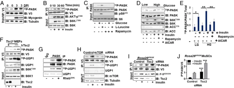
Fig. 1. Nutrient and insulin signaling activates PASK via mTORC1. (A) PASK is activated during skeletal muscle regeneration. TA muscles were isolated from control or BaCl2-injured Rosa26hPASK-V5mice 3 d postinjury (DPI). V5-tagged PASK was immunoprecipitated from tissue extract to assay PASK activation by an
in vitro autophosphorylation assay, which indicates the incorporation of32P into PASK as a function of kinase activity. An immunoblot (IB) of MyoG marks
myogenic regeneration. IP, immunoprecipitation. (B) CHO-K1 cells expressing V5-tagged hPASK were stimulated with 100 nM insulin for the indicated times. PASK was immunoprecipitated using anti-V5 antibody, and an in vitro kinase assay was performed as in A. Activation of PI3K and mTORC1 signaling was demonstrated by the appearance of phospho-AKT and phospho-S6K. (C) HEK293E cells were starved of amino acids and glucose for 8 h, followed by stimulation with either 25 mM glucose or 800μML-leucine for 1 h. Endogenous PASK was purified using anti-PASK antibody from cell extracts, and in vitro kinase activity assay was performed as in A. (D) PASK from HEK293E cells was assayed as in C. Cells were stimulated with 100 nM insulin for 1 h after pre-treatment with DMSO, 100 nM rapamycin, or 25μM 5-aminoimidazole-4-carboxamide ribonucleotide (AICAR). Endogenous PASK was purified using anti-PASK antibody from cell extracts, and an in vitro kinase activity assay was performed as in A. (E) Quantification of D. Phospho-anti-PASK (32P-PASK) and total PASK
from three independent experiments; the ratio was expressed as the fold change in PASK activity under the indicated stimuli. Error bars are± SD. *P < 0.05; **P< 0.01. HG, high glucose; LG, low glucose. (F) V5-PASK was expressed in Tsc2−/−cells with or without complementation with WT hTsc2. Cells were
serum-starved overnight and then stimulated with 100 nM insulin for 1 h. PASK was immunoprecipitated using anti-V5 antibody, and an in vitro kinase assay was performed. The yeast Ugp1 protein, which is robust in an in vitro substrate of PASK, was used as an exogenous substrate. (G) PASK was purified from HEK293T cells with expressing vector control (−) or RhebQ64Land subjected to kinase activity assay as in F. (H) HEK293E cells were transfected with control or
mTOR-targeting siRNA for 24 h. A vector expressing V5-PASK was then transfected; after 24 h, cells were serum-starved overnight and then stimulated with 100 nM insulin for 1 h. PASK was purified and subjected to a kinase activity assay as in F. (I) Primary myoblasts isolated from Rosa26hPASK-V5mice were
transfected with control or mouse Tsc2-targeting siRNA. Twenty-four hours after transfection, cells were switched to 5% serum-containing medium over-night, followed by 4 h of total serum starvation. Cells were then treated with vehicle or 100 nM insulin for 1 h. PASK was then purified and subjected to kinase activity assay as in F. (J) Quantification of PASK kinase activity measurements from three experiments as in I. *P< 0.05; ***P < 0.005.
CELL
(21, 22), was induced 3 d after injury. The increase in PASK activity coincided with the time point when both MyoG and endogenous mouse PASK expression is induced (Fig. 1A and SI Appendix, Fig. S1A).
To understand how niche signals, like nutrients and insulin, tivate PASK during muscle regeneration, we examined PASK ac-tivation by these stimuli in a cell-autonomous manner. As shown in Fig. 1B, PASK was acutely and transiently activated by insulin stimulation in CHO-K1 cells. Similarly, glucose and amino acids such asL-leucine also activate PASK in HEK293E cells (Fig. 1C).
While glucose activated PASK modestly but consistently, we ob-served a strong increase in PASK activity upon addition ofL-leucine
to cell culture media (Fig. 1C). Since mTORC1 is a convergence point in both insulin and amino acid signaling (23), we asked if the mTORC1 activity is required for PASK activation. As shown in Fig. 1C, the addition of the mTORC1 inhibitor rapamycin (24) completely blocked PASK activation by glucose andL-leucine.
To further explore the role of mTORC1 in the regulation of PASK activity downstream of nutrient and insulin stimulation, we analyzed PASK activity in the presence or absence of kinase modulators that either augment or inhibit mTORC1 activity. AMP-activated protein kinase (AMPK) is a negative regulator of mTORC1 kinase function (25). As shown in Fig. 1 D and E, in-sulin activated PASK in the presence of either low or high glucose, and this was suppressed by pretreatment with the AMPK activator 5-aminoimidazole-4-carboxamide ribonucleotide to an extent similar to rapamycin treatment. AMPK phosphorylates and acti-vates Tsc2, which negatively regulates mTORC1 function (26). Consistent with that, Tsc2−/− mouse embryonic fibroblasts (MEFs), complemented with empty vector control but not with human Tsc2 (27), showed mTORC1 hyperactivation, as evidenced by increased phosphorylation of the mTORC1 substrate p70S6K (Fig. 1F). Loss of Tsc2 also increased PASK kinase activity, as shown by increased in vitro autophosphorylation and phos-phorylation of its heterologous substrate Ugp1 (28) (Fig. 1F). Tsc2 functions as a GTPase-activating protein (GAP) for the Rheb GTPase, which stimulates mTORC1 activity (29). Expres-sion of a constitutively activated Rheb (RhebQ64L), which
hyper-activates mTORC1, also resulted in increased PASK activity (Fig. 1G). On the other hand, silencing mTOR resulted in a near-complete block of insulin-stimulated PASK activation (Fig. 1H).
Finally, to test whether mTORC1 contributes to PASK acti-vation in MuSCs, we isolated MuSCs from Rosa26hPASK-V5mice
and assessed PASK activation by insulin after silencing Tsc2. During MuSC isolation, PASK was activated modestly, but it was further activated by insulin stimulation (Fig. 1 I and J). This modest activation of PASK may account for the increased pro-pensity of MuSCs derived from transgenic mice observed in SI Appendix, Fig. S1C. The loss of Tsc2 further activated PASK in the presence or absence of insulin. Thus, our results demonstrate that the mTORC1 complex activates PASK in response to in-sulin and nutrient signaling.
PASK Is Phosphorylated by mTORC1 at Multiple Residues to Stimulate
Its Activity. We hypothesized that mTORC1-dependent
phos-phorylation of one or more residues on PASK might result in its activation by nutrients and insulin. Therefore, we performed metabolic in-cell labeling using radioactive (32P) phosphate in
Tsc2−/−or human Tsc2 (hTsc2)-complemented Tsc2−/−MEFs in the presence or absence of rapamycin. As shown in Fig. 2A, en-dogenous PASK derived from Tsc2−/−MEFs showed significantly increased phosphorylation compared with hTsc2-complemented cells. Furthermore, this increase in PASK phosphorylation was partially suppressible by low-dose rapamycin, consistent with some other mTORC1 substrates (30) (Fig. 2A). We also tested if, similar to PASK activity (Fig. 1G), PASK phosphorylation was induced by constitutively activated Rheb (RhebQ64L) and if that is dependent upon the catalytic activity of PASK. As shown in Fig. 2B, both WT
and kinase-dead (KD) PASK showed enhanced in-cell phos-phorylation in the presence of RhebQ64L. In contrast, the phos-phorylation of PDK1, an upstream activator of Akt that is not an mTORC1 substrate, was not induced by Rheb coexpression. Thus, mTORC1 activation induces the phosphorylation of PASK in cells, and the increased PASK phosphorylation is independent of its own catalytic activity, demonstrating that autophosphorylation is not required.
To identify residues on PASK that are specifically targeted by mTORC1 activity, we performed a domain truncation analysis in the presence or absence of RhebQ64Land rapamycin (Fig. 2C). Rheb stimulated PASK phosphorylation within both the C-terminal kinase domain-containing region that includes resi-dues 941–1,323 and the N-terminal fragment that contains the first 738 residues (Fig. 2D). Surprisingly, these two regions showed differences in sensitivity to rapamycin inhibition, as the ΔC fragment (residues 1–738) showed much more rapamycin sensitivity than the ΔN fragment (residues 941–1,323; Discus-sion). Using mass spectrometry (SI Appendix, Table S1), bioinformatics, and site-directed mutagenesis (Fig. 2E and SI Appendix, Fig. S2 A and B), we identified two clusters of sites that were hyperphosphorylated upon mTORC1 activation. The sites within the N-terminal fragment include Thr640and Thr642
(Fig. 2E), which showed a marked similarity to rapamycin-sensitive sites on another mTORC1 substrate, Grb10 (Fig. 2E). The C-terminal phosphorylation sites include Ser949, Ser953, and
Ser956. Mutation of N-terminal sites alone was not sufficient to block Rheb-stimulated PASK phosphorylation by rapamycin but needed mTOR catalytic inhibition by torin (SI Appendix, Fig. S2 A and B). These results suggest that inhibition of mTOR cata-lytic activity is required for full inhibition of Rheb-stimulated PASK phosphorylation (Fig. 2F and SI Appendix, Fig. S2 A and B). Mutation of all five sites (T640, T642, S949, S953, and S956) to Ala (termed TS[5]A), resulted in essentially complete in-hibition of Rheb-stimulated PASK phosphorylation (Fig. 2F) and kinase activation (Fig. 2G), thus confirming these five sites as targets of mTORC1-stimulated phosphorylation and its effect on PASK activity described in Fig. 1.
mTORC1 activates multiple kinases within the AGC family of protein kinases, such as Akt, p70S6K, and p90RSK. However, our data show that pretreatment with the mTOR inhibitor torin, but not p70S6K inhibitor (PF408671) or pan-AGC kinase in-hibitor (AT13148), abolished Rheb-stimulated PASK phos-phorylation (Fig. 2H). Interestingly, the sequence surrounding the Rheb-stimulated PASK phosphorylation sites appears similar to many of the recently identified mTORC1 substrates (30) (Fig. 2E). To test if mTORC1 can directly phosphorylate these sites, we performed an in vitro kinase assay using purified mTORC1 in the presence of activated Rheb. We used KD PASK to avoid background phosphorylation of WT PASK, which could be fur-ther confounded by an increase in its activity upon mTOR phosphorylation. As shown in Fig. 2I, KD PASK was robustly phosphorylated in vitro by purified mTORC1. Mutation of two N-terminal phosphorylable residues (T640AT642A) only modestly lowered mTORC1 phosphorylation of PASK, and mutation of all five phosphorylation sites was required for complete loss of mTORC1-mediated phosphorylation of PASK. We also utilized a mutagenized peptide array system that was used to identify novel mTORC1 substrates as previously described (30) to pin-point residues targeted by mTORC1. The mutated peptide li-brary was generated by mutating phosphorylable residues within each peptide, except the phosphorylatable residue at position 0 (indicated by the asterisk in SI Appendix, Fig. S2C). These peptides were then used as substrates for in vitro kinase reactions with purified mTORC1. As shown inSI Appendix, Fig. S2 D and E, a peptide representing Thr640/Thr642 showed strong phos-phorylation that could be abrogated by mutation of Thr640(in the site A sequence). Site B was used as a negative control, being a
poor mTORC1 substrate in our experiments. On the other hand, site C peptide showed robust phosphorylation by mTORC1, and mutation of Ser949 was sufficient to signifi-cantly diminish mTORC1-mediated phosphorylation of this peptide (SI Appendix, Fig. S2 B and C) in vitro. Thus, our data suggest that mTORC1-mediated phosphorylation of PASK at multiple residues is required for nutrient and insulin signaling to activate PASK.
PASK Forms a Nutrient-Sensitive Complex with mTORC1. To
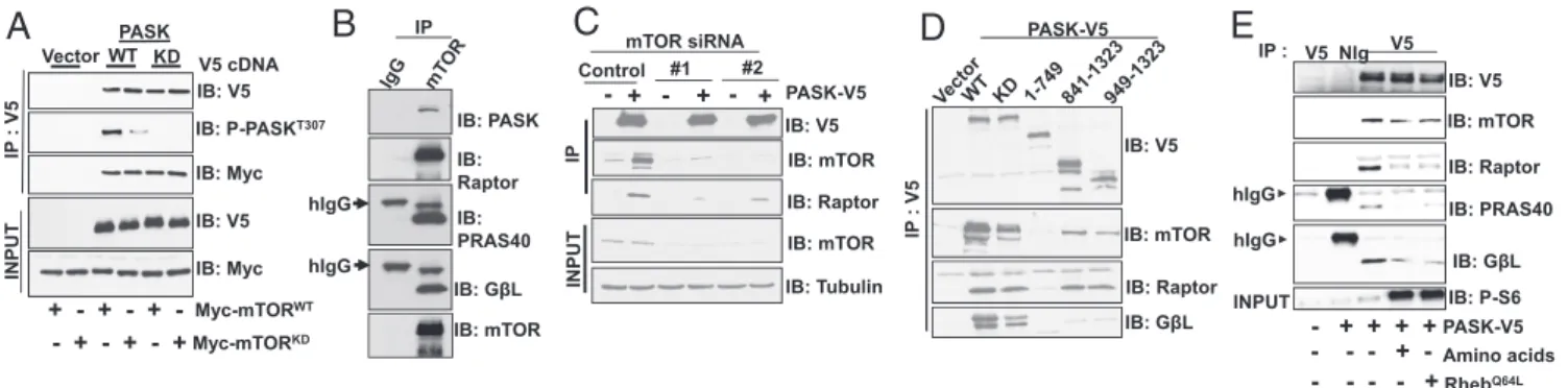
un-derstand the mechanistic basis whereby PASK is phosphorylated and activated by mTORC1, we sought to understand whether PASK associates with mTOR or any of its associated complex 1 proteins. We first immunoprecipitated V5-tagged WT PASK or a K1028R mutant lacking kinase activity (KD) from cells that coexpress either Myc-tagged WT mTOR or a D2357E/V2364I mutant lacking kinase activity (KD). Both WT and KD PASK could be immunoprecipitated with either the WT or KD version of mTOR (Fig. 3A). Interestingly, the WT PASK associated with KD mTOR showed significantly diminished phosphorylation at Thr307, which is an autophosphorylation site that we have shown previously to be a reliable marker of PASK activity (31). Thus, expression of KD mTOR suppresses PASK activity in a domi-nant negative manner in cells. When endogenous mTOR was
isolated from cells, PASK was copurified in addition to the members of the mTORC1 complex (Fig. 3B). Similarly, immu-noprecipitation of PASK-V5 copurified endogenous mTOR and raptor (Fig. 3C). When mTOR was silenced, the association of PASK with mTOR and raptor was significantly reduced, sug-gesting that the PASK-raptor association is likely mediated by mTOR. Using domain truncation analysis, we found that the C-terminal residues 941–1,323 in PASK, which include the kinase domain and surrounding regions, are necessary to interact with endogenous mTORC1 (Fig. 3D). Finally, we found that a mixture ofL-leucine andL-arginine, two of the stimuli that led
to phosphorylation and activation of PASK, as well as expres-sion of RhebQ64L, weakens the PASK-mTOR association (Fig.
3E). Thus, PASK appears to form a nutrient and signaling-sensitive complex with mTORC1, similar to what was pre-viously reported for the mTOR-raptor association (32). These data suggest that PASK dynamically associates with mTORC1, whereby mTOR directly phosphorylates and activates PASK, resulting in its release from the complex.
mTOR Phosphorylation of PASK and p70S6K Regulates Distinct Phases
of Myogenesis.MyoG expression in myoblasts marks an irreversible
commitment to differentiate (33). Hence, activation of Myog transcription is a major point of the control by signaling pathways
32P-PASK IB: PASK Rapamycin - + - +
A
- + + + + IB: V5 RhebQ64L WT KD PDK1 MYC - + - + - + 32P-PASK 32P-PDK1B
WT Rapamycin - + + - + + - + + - - + - - + - - + RhebQ64L IB: V5C
D
RhebQ64LG
- + - + WT TS[5]A 32P-PASK P-UGP1 RhebQ64LH
PAS Kinase 1 PAS 1 1323 738 Kinase 1323 941 IB: V5 UGP1 IB: MYC V5/Myc WT ΔN ΔC ΔC ΔN 32P-PASK hTsc2 __ Tsc2-/-MEFs + Kinase Assa y p32-PASK IB: pS374EBP1 IB: pS235S6 IB: pS473Akt Tubulin IB: V5 Torin PF4708671 AT13418 p32-PASK * KD PASK-K1028R mTORC1 IB:V5 IB: mTOR IB: Raptor IB: HA (Rheb)I
- - + - -- - - + -- - - - + - + + + + + - + + + WT TT->AA S3A TS[5]A IB: V5 Rheb[Q64L] - + - + - + - +E
32P-PASK 3X-Flag-V5-hPASK-KD hPASK V5/Myc IP: IB:F
hGrb10 470NLVGSPSPL478 :*:*: PAS 1 Kinase 1323Fig. 2. PASK is a direct phosphorylation target of mTORC1. (A) Endogenous mouse PASK was immunoprecipitated from Tsc2−/−or hTsc2-complemented Tsc2−/−MEFs after labeling with32P-phosphate for 4 h. Anti-PASK immunoprecipitates were separated by SDS/PAGE and subjected to autoradiography and an immunoblot (IB). (B) HEK293T cells were transfected with vectors expressing V5-tagged WT or K1028R (KD) PASK or Myc-tagged PDK1, as well as either empty vector or a vector expressing RhebQ64L. At 24 h posttransfection, in-cell32P labeling was conducted and the indicated immunoprecipitates were analyzed as in
A. IP, immunoprecipitation. (C) Schematic indicating the domain structure of full-length PASK and the domain truncation mutants used in D. (D) WT PASK or the truncation mutants from C were coexpressed with empty vector or a vector expressing RhebQ64Land were treated with or without 40 nM rapamycin. They
were assessed for in-cell phosphorylation as in A. (E) Schematic of the mTORC1-dependent phosphorylation sites on PASK. (F) WT or TT→AA (T640A T642A), S3A
(S949A S953A S956A) or TS[5]A (T640, T642, S949, S953, S956to Ala) mutants of PASK were expressed in HEK293T cells with or without expression of RhebQ64Land
analyzed for cell phosphorylation as in A. (G) WT or the TS[5]A mutant of PASK was expressed in HEK293T cells with or without coexpression of RhebQ64L, and
kinase activity was measured by autophosphorylation (32P-PASK) and Ugp1 phosphorylation as in Fig. 1F. (H) RhebQ64L-induced PASK in vivo phosphorylation
was measured with or without 50μM AT13148, 50 μM PF4708671, or 100 nM torin pretreatment in HEK293T cells. For Western blot analysis using indicated phosphospecific antibodies, an identical parallel experiment was performed to obtain nonradioactive cell extracts to analyze efficacy of the inhibitor treatment. (I) In vitro kinase assay was performed using purified mTORC1 and KD (K1028R mutant) PASK as described in Materials and Methods. The asterisk indicates the band corresponding to phosphorylated form of raptor in the kinase reaction mixture.
CELL
that regulate myogenesis (34). As a tissue with significant metabolic demand, skeletal muscle homeostasis is tightly linked to nutrient status. This is consistent with the fact that the nutrient-responsive mTORC1 has been shown to regulate myogenesis and myofiber hypertrophy (14–16, 23, 35) although its downstream effectors re-main unknown (14). Because mTORC1 activates PASK and we previously demonstrated that PASK is required for efficient damage-induced myogenesis (21), we asked if mTORC1 and PASK functions converge on a specific mechanism to regulate the myogenesis program. We compared myogenic induction upon loss of PASK and mTORC1 signaling in both MuSCs and C2C12 myoblasts. As shown in SI Appendix, Fig. S4 A and B, both mTORC1 and PASK inhibition (with rapamycin and BioE-1197, respectively) effectively and similarly suppressed MyoG+ conversion and myoblast fusion in isolated MuSCs. This failure to convert to MyoG+ cells appears to be due to impaired in-duction of Myog mRNA in the presence of the inhibitors (SI Appendix, Fig. S4C). To determine specifically how mTORC1 activation affects myogenesis and what role PASK plays in that process in primary myoblasts, we silenced Tsc2 in isolated pri-mary myoblasts and analyzed the mRNA levels of Pax7, Myog, and Acta1, which mark proliferating, committed, and differen-tiated stages, respectively. As shown in SI Appendix, Fig. S4D, loss of Tsc2 resulted in a modest but significant decrease in Pax7 mRNA and an increase in Myog mRNA, suggesting mTORC1 activation increases terminal differentiation commitment. This is further evidenced by increased expression of the muscle-specific actin Acta, which marks terminally differentiated myocytes. Pretreatment with PASK inhibitor (BioE-1197) or mTORC1 inhibitor (rapamycin) resulted in a significant increase in mRNA levels for Pax7, regardless of Tsc2 status. Modest, but significant increase in the mRNA levels of Myog and Acta1 as seen in Tsc2 silenced control myoblasts, was absent when PASK or mTORC1 was inhibited (SI Appendix, Fig. S4D).
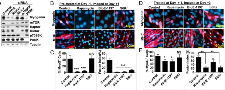
We next sought to identify components of the mTORC1 and mTORC2 complexes that are necessary for MyoG expression and myogenesis. To do so, we silenced mTOR, raptor (member of mTORC1), rictor (member of mTORC2), or the mTORC1 substrate p70S6K (major substrate of mTORC1) or PASK dur-ing insulin-stimulated myogenesis of cultured myoblasts (C2C12). Consistent with a previous report (19), mTORC1, but not mTORC2, is required for MyoG protein expression as loss of raptor, but not rictor, suppressed MyoG induction (Fig. 4A).
Furthermore, silencing of PASK, but not p70S6K, suppressed MyoG expression, suggesting that mTORC1-PASK, but not mTORC1-S6K, signaling is required for induction of the termi-nal differentiation program. These results in cultured myoblasts recapitulated our above-described findings indicating a common role of mTORC1 and PASK in the control of MyoG expression (Fig. 4A andSI Appendix, Fig. S4). However, the mTORC1 is a well-established determinant of skeletal muscle hypertrophy in various animal models, a function that is largely mediated by its substrate p70S6K. To functionally compare mTOR-PASK and mTOR-S6K signaling during myogenesis, we set up a temporal inhibition experiment in which mTORC1, PASK, or p70S6K was inhibited using rapamycin, BioE-1197, or PF408671, respectively, before differentiation initiation (pretreatment at day−1, Fig. 4B) or after differentiation initiation (treatment at day+1, Fig. 4D) in C2C12 cells. Inhibition of mTORC1 and PASK, but not p70S6K, significantly suppressed the rise in MyoG+cell numbers [Fig. 4 B and C, Right (quantified in the latter)]. However, de-spite normal induction of the MyoG, p70S6K inhibition abro-gated the myoblast fusion to the similar extent as mTORC1 or PASK inhibition [Fig. 4 B and C, Left (quantified in the latter)]. Hence, we hypothesized that mTORC1-PASK signaling might be required for MyoG expression and mTORC1-S6K signaling may drive the myoblast fusion event. To test this hypothesis, we stimulated a differentiation program and added mTOR, PASK, or p70S6K inhibitor after 24 h of differentiation (treatment on day+1, Fig. 4D). When imaged at day +3, we noticed that in-hibition mTORC1 and PASK modestly (still significantly) af-fected MyoG expression, whereas MyoG+ cell numbers were comparable in both control and S6K1-inhibited samples [Fig. 4 D and E, Right (quantified in the latter)]. Interestingly, while PASK inhibition only modestly blocked myoblast fusion, inhibition of mTORC1 and S6K nearly completely blocked the fusion pro-gram, despite the overall increase in MHC+myofiber numbers [Fig. 4 D and E, Left (quantified in the latter)]. These results suggest a distinct pathway downstream of mTORC1 during myogenesis, in that mTORC1-PASK signaling drives MyoG ex-pression, whereas mTORC1-S6K signaling is required for the myoblast fusion program.
PASK Phosphorylation by mTORC1 Is Required for the Induction of the
MyoG Expression. MuSCs from Rosa26hPASK-V5 mice show
en-hanced myogenesis compared with control MuSCs, as indicated
IB: V5 IB: Myc IB: Myc IP : V 5 V5 cDNA Myc-mTORWT Myc-mTORKD + + + -- + -- + -- + IB: V5 IB: P-PASKT307 INPUT
A
B
IB: Raptor IB: mTOR IB: V5C
Control #1 #2 IB: Raptor IB: mTOR IP INPUT IB: mTOR IB: Tubulin IB: V5-D
IB: PASK IB: Raptor IB: PRAS40 IB: GβL IB: mTOR hIgG hIgG IPE
IB: P-S6 INPUT IB: PRAS40 hIgG hIgG IP : IB: mTOR IB: Raptor IB: V5 V5 NIg V5 Amino acids - + -Q64L - + + + + PASK-V5 IP : V 5 IB: GβL IB: GβL PASK-V5 PASK-V5 mTOR siRNA + - + - +Fig. 3. PASK associates with mTORC1 in a nutrient-sensitive manner. (A) Vector control or WT or KD (K1028R) PASK was coexpressed with either Myc-tagged WT or D2357E (KD) mTOR in HEK293T cells. Twenty-four hours after transfection, V5-tagged proteins were purified and the presence of Myc-tagged mTOR was detected by Western blotting. Immunoprecipitates were also probed with anti-AKT substrate antibody (RXRXXpS/T), as described in Materials and Methods, to detect PASK-T307phosphorylation. IB, immunoblot; IP, immunoprecipitation. (B) mTOR protein was purified from HEK293T cells, and the
presence of its associated proteins was detected in the immunoprecipitates by Western blotting. (C) mTOR silencing and V5-hPASK expression and IP were performed as described in Fig. 1H. The presence of mTOR and its complex members was detected by Western blotting of the immunoprecipitates. (D) In-dicated V5-tagged PASK truncation mutants were expressed and immunoprecipitated from HEK293T cells. The co-IP of mTORC1 was determined by Western blotting of the immunoprecipitates. (E) Vector or V5-PASK was expressed with RhebQ64Las indicated. For amino acid stimulation, cells were starved of the
amino acidsL-leucine andL-arginine overnight. On the next day, 800μML-leucine and 100μML-arginine were added for 1 h. Cells were lysed, and V5-tagged PASK was purified from HEK293T cells. The relative abundance of mTORC1 was detected by Western blotting of the immunoprecipitates.
by increased MyoG staining (SI Appendix, Fig. S5 A and B) and fusion index (percentage of nuclei inside myotubes/total number of nuclei;SI Appendix, Fig. S5 C and D). Rapamycin treatment effectively reversed this increase in myogenesis in PASK-overexpressing MuSCs (SI Appendix, Fig. S5). We reasoned that since rapamycin is able to suppress myogenesis in MuSCs from Rosa26hPASK-V5mice, mTORC1 is likely already activated during isolation of MuSCs. Therefore, Tsc2 silencing should not have an additive effect on myogenesis. Consistent with that, while the activation of the mTORC1 pathway by Tsc2 silencing resulted in modest stimulation of myogenesis in control cells (SI Appendix, Fig. S5), MuSCs from Rosa26hPASK-V5mice did not show further enhancement of myogenesis. Again, rapamycin treatment inhibited myogenesis regardless of PASK overexpression, suggest-ing the requirement for activated mTORC1 in inducsuggest-ing myogenesis downstream of PASK. Based on these data, and on the fact that BioE-1197 treatment effectively blocked myogenesis downstream of Tsc2 knockdown (SI Appendix, Fig. S4D), we hypothesize that mTOR activation of PASK is required for induction of the myogenesis program.
To test this hypothesis, we retrovirally expressed vector con-trol, WT, KD, or TS[5]A-mutated PASK in isolated MuSCs from PASK−/−mice. As shown in Fig. 5A, the reexpression of WT, but not KD, PASK fully restored MyoG and MHC expression in isolated PASK−/−MuSCs. Expression of TS[5]A-mutated PASK, on the other hand, had very modest effects on MyoG and MHC (Fig. 5A). We also utilized the CRISPR/Cas9 system to delete endogenous mouse PASK in C2C12 myoblasts (CrisprPASK) and reconstituted with either GFP vector or WT, KD, or TS[5]A-mutated PASK. Using these cell lines, we assayed the effec-tiveness of myogenesis in response to insulin. Expression of WT, but not KD PASK, resulted efficient rescue of the number of MyoG+cells in Pask deleted C2C12 cells (Fig. 5 B and C). Ex-pression of TS[5]A-mutated PASK only modestly rescued the defect in MyoG+ cell number compared with WT PASK-expressing cells (Fig. 5 B and C). Similarly, expression of WT
PASK resulted in full restoration of myogenesis, as measured by the fusion index formation (Fig. 5 D and E). In contrast, cells expressing TS[5]A mutant PASK were defective in forming multinucleated myotubes compared with WT PASK-expressing cells (Fig. 5 D and E). We think this defect in the TS[5]A mutant in the later phase of myogenesis is attributable to an overall decline in the number of MyoG+cells that are available for fu-sion. Thus, despite PASK not being critically required for the later phase of myogenesis (myonuclear fusion) once MyoG is sufficiently induced (Fig. 4 B–E), our data suggest that mTORC1 phosphorylation of PASK might be required for generating a sufficient MyoG+ myoblast population that can undergo myo-nuclear fusion to replenish myofibers.
mTOR-PASK-Wdr5 Forms a Signaling Cascade to Regulate the Onset of
the Myogenesis Program. We have previously shown that
phos-phorylation of Wdr5 is necessary and sufficient for the effects of PASK to induce Myog transcription and myogenesis (21). More-over, the physical interaction between PASK and Wdr5 was spe-cifically induced upon insulin treatment to initiate myogenesis (21). Herein, we showed that insulin treatment also stimulated PASK phosphorylation and activation in an mTORC1-dependent manner. Hence, we hypothesized that mTORC1 activation might enhance the PASK-Wdr5 association. To test this, we coexpressed WT or KD PASK with WT-Wdr5 in the presence or absence of RhebQ64Land measured the PASK-Wdr5 association. As expec-ted, expression of RhebQ64Lcaused increased WT PASK in vivo
activity as measured by autophosphorylation at Thr307(Fig. 6A). Activation of mTOR also stimulated interaction between PASK and Wdr5, regardless of PASK activity status (WT vs. KD PASK). Interestingly, the C-terminal residues where mTORC1 phos-phorylates PASK are adjacent to the Wdr5 binding region in PASK that we identified previously (21) (Fig. 6B). To more pre-cisely map the Wdr5 binding region in PASK, we mutagenized the conserved residues within this stretch and examined effects on the interaction with Wdr5 (Fig. 6C). As shown in Fig. 6D, mutation of Pre-treated at Day -1, Imaged at Day +1
Control Rapamycin BioE-1197 S6Ki
Myogenin
MHC MHC
Myogenin
Treated at Day + 1, Imaged at Day +3
% My oG + Cel ls Control Rapa myci n BioE-11 97 S6Ki
B
C
Control Rapamycin BioE-1197
*** *** *** * * ** ** S6Ki 100μm 40μm 100μm 40μm
D
E
Myogenin mTOR Raptor Rictor p70S6K PASK Tubulin siRNAA
NS NS *Fig. 4. PASK and p70S6K are required for distinct phases of myogenesis downstream of mTORC1. (A) Indicated components of mTORC1 and mTORC2 were silenced in C2C12 myoblasts. Forty-eight hours after silencing, cells were stimulated to differentiate using 100 nM insulin. Myogenin protein expression was used as an indication of differentiation for each cell population. (B and C) C2C12 cells were pretreated with 100 nM rapamycin, 50μM BioE-1197, or 40 μM PF4708671 (S6Ki) at day−1. Cells were allowed to attain confluency (24 h) and induced for differentiation at day 0 in the continued presence of inhibitors. Twenty-four hours following differentiation, at day+1, cells were fixed with 4% paraformaldehyde and the induction of the myogenesis program (by antimyogenin staining) and myoblast fusion (by anti-MHC staining) was quantified (in D). ***P< 0.005 (control vs. inhibitors). NS, not significant vs. control. (D and E) C2C12 cells were allowed to attain confluency and induced for differentiation in the absence of inhibitors. Twenty-four hours following differ-entiation, at day+1, cells were treated with 100 nM rapamycin, 50 μM BioE-1197, or 40 μM S6Ki in differentiation media. Cells were fixed at day 3, and induction of the myogenesis program (by antimyogenin staining) and myoblast fusion (by anti-MHC staining) was measured and quantified (in E). *P< 0.05; **P< 0.005 (control vs. inhibitors).
CELL
the highly conserved C924 and W926 PASK residues to alanine resulted in a significantly weakened interaction with Wdr5. As these residues are adjacent to the mTORC1 phosphorylation site on PASK (Fig. 6C), we hypothesized that mTORC1-mediated PASK phosphorylation might augment Wdr5 binding. Indeed, we found that the TS[5]A mutant, lacking mTORC1-mediated phosphorylation, failed to show RhebQ64L-dependent induction of the interaction between PASK and Wdr5 (Fig. 6E). Taken to-gether, these data suggest that mTORC1-mediated phosphoryla-tion of PASK stimulates the PASK-Wdr5 associaphosphoryla-tion. We have shown previously that this interaction correlates strongly with PASK phosphorylation of Wdr5 at Ser49, which orchestrates epi-genetic changes at the Myog promoter to enable gene expression. To test if Wdr5 phosphorylation at Ser49by PASK is a mechanism whereby mTORC1 signals to induce myogenesis, we first tested whether expression of the phosphomimetic Wdr5 mutant (Wdr5S49E) might rescue the defect in myogenesis resulting from mTORC1 inhibition. As shown in Fig. 6F, rapamycin completely pre-vented the induction of Myog and Mylff in response to differ-entiation cues. These defects were completely reversed by expression of the phosphomimetic Wdr5 mutant Wdr5S49E, while the expression of Wdr5WT or Wdr5S49A had no effect (Fig. 6F). Consistent with the mRNA data, Western blot analysis showed that Wdr5S49E, but not Wdr5S49A, also restored MyoG and MHC protein induction in rapamycin-treated cells (Fig. 6G). Intriguingly, while the expression of Wdr5S49E
res-cues the MyoG expression (Fig. 6 F and G), it does not appear to completely rescue the rapamycin-inhibited myoblast fusion program (Fig. 6 H and I). The myotubes are smaller and thinner and have fewer myonuclei in Wdr5S49E-expressing cells in
rapamycin-treated cells compared with DMSO-treated samples (Fig. 6H and quantified in Fig. 6I). This result is consistent with our data that mTOR-PASK signaling is required for efficient MyoG induction (which is rescued by Wdr5S49E), whereas mTOR-S6K1 signaling is required for an efficient myoblast fusion program (only partially rescued by Wdr5S49E). Taken together, our results have identified a signaling pathway that transmits nutrient and hormonal signals via mTORC1 phos-phorylation and activation of PASK to induce Myog expression and commitment to differentiate through phosphorylation of the Wdr5 epigenetic regulator (Fig. 7) and shows functional partitioning of the mTORC1 function during myogenesis. Discussion
mTORC1 integrates multiple signals from the regenerating niche, including nutrients as well as hormones, such as insulin, IGF1, or Wnt, and is required for myogenesis and muscle growth and hypertrophy. In this study, we show that PASK is a substrate of mTORC1 downstream of these same niche signals, particu-larly insulin and nutrients. mTORC1-dependent phosphorylation and activation of PASK activate Myog transcription, and thereby establish the commitment to myogenic differentiation (Fig. 7). mTORC1 activation of p70S6K simultaneously activates the protein synthesis that is required for rapid myotube hypertrophy, resulting in the culmination of the myogenesis program. Thus, mTORC1 coordinately enables both aspects of myogenesis via activation of distinct protein kinase signaling pathways. mTORC1, PASK, and Wdr5 are widely expressed in stem cells. As PASK is required for the differentiation of multiple stem cell lineages (21), this model could represent a common mechanism by which nutrient IB: PASK IB: Myogenin IB: MHC TS[5]A IB: Tubulin PASK
-/-B
WT cDNAMHC+DAPI MHC+DAPI MHC+DAPI
C
E
KDA
WT KD TS[5]A GFP Vector MHC+DAPI FLAG-PASKMyog+DAPI Myog+DAPI Myog+DAPI Myog+DAPI GFP Vector 40μm 40μm WT KD TS[5]A CrisprPASK * # Control Vec tor W T-PASK KD -PA SK TS[5 ]A-PAS K Fusion Index (% ) CrisprPASK * # Control Ve cto r WT-PASKKD-PASK TS[5]A-PASK % M y o G + Cells FLAG-PASK
D
Fig. 5. PASK phosphorylation at mTORC1 sites is required for efficient myogenesis. (A) PASK+/+or PASK−/−MuSCs were isolated from TA muscles of mice with the respective genotype. Twenty-four hours after isolation, PASK−/−cells were infected with retroviruses expressing Flag-tagged WT, K1028R, or TS[5]A PASK. Forty-eight hours after infection, PASK+/+and PASK−/−cells were stimulated to differentiate with 100 nM insulin. Protein extracts were prepared from all cells, and myogenesis was measured by Western blotting using the indicated antibodies. IB, immunoblot. (B and C) C2C12 myoblasts with CRISPR/Cas9-deleted PASK were infected with the retroviruses containing indicated cDNAs. Forty-eight hours after retroviral infection, C2C12 cells were induced to differentiate using 100 nM Insulin. Forty-eight hours after induction of differentiation, cells were fixed and stained with anti-MyoG antibody to determine MyoG induction efficiency (in C) as described in Fig. 4B. *P< 0.05 (between TS[5]A and vector or KD PASK, significantly better rescue);#P< 0.005 (between TS[5]A and WT
hPASK, significantly worse rescue). (D) C2C12 myoblasts with CRISPR/Cas9-deleted PASK were infected with the retroviruses containing indicated cDNAs. Forty-eight hours after retroviral infection, C2C12 cells were induced to differentiate using 100 nM insulin. Myogenesis was determined by immunofluo-rescence microscopy using antibodies against MHC. (E) Fusion index was calculated as in Fig. 4B. *P< 0.05 between TS[5]A and vector or KD PASK;#P<
0.005 between TS[5]A and WT hPASK.
and hormonal signaling could establish the commitment to differ-entiate via mTORC1 signaling. This might be particularly relevant for cell types that are highly metabolically active, like muscle cells and adipocytes (36).
The role of the mTOR protein kinase in the regulation of myogenesis appears to be multidimensional (14). mTOR has been
shown to regulate the myogenesis program using two different mechanisms, only one of which depends upon its catalytic activity (14, 35). Moreover, mTOR not only regulates the early stages of myogenesis but also controls the remodeling of myotubes after differentiation (16, 37). Despite considerable interest, it remains unclear how mTOR signals to establish the early steps of myogenic
Vect or Vector WT S49AS49E 0 2 4 6 Myog Mylpf Flag-WDR5 Flag-WDR5 IB: MyoG IB: MHC IB: Pax7 IB: Tubulin DMSO Rapamycin + - -- + + + Flag-WDR5 Flag-WDR5 + + + + + + - + - + - + IP: V5 LacZ WT KD PASK-V5 IB:V5 IB:FLAG RhebQ64L Phospho-(PASKThr307) IB:Flag
A
INPUT PAS Kinase 1 1323 Wdr5 binding regionmTOR phosphorylation sites
B
C
949 924 926D
E
IP: V5 INPUT IB:V5 IB:V5 IB:FLAG IB:Flag LacZ WT TS[5]A V5:PASKFlag-WDR5 + + + + + - - + - + RhebQ64L IP: V5 INPUT V5 FLAG Flag V5 Flag-WDR5 + + + Rapamycin Rapamycin
F
G
DMSO Rapamy cinVector+ MHC WT+MHC S49A+MHC S49E+MHC
H
Flag-WDR5
Flag-WDR5
841 941
mTOR phosphorylation sites
953 956
*** ***
I
*
hPASK
Fig. 6. mTOR-PASK-Wdr5 signaling cascade regulates the myogenin expression. (A) V5-LacZ as a control or WT or KD PASK was coexpressed with Flag-tagged WT-Wdr5 in the presence or absence of RhebQ64L. Twenty-four hours after transfection, V5-tagged proteins were immunoprecipitated and the
abundance of Flag-Wdr5 was determined by Western blotting. Activation status of PASK by RhebQ64Lwas measured by Western blotting of the
im-munoprecipitates with anti-AKT substrate antibody (Materials and Methods). IB, immunoblot; IP, immunoprecipitation. (B) Domain arrangement of hPASK indicating relative positions of mTORC1-dependent phosphorylation sites and the Wdr5-interacting region on PASK. (C) Alignment of a region of PASK encompassing the Wdr5 binding region and mTOR phosphorylation sites from different species. Conserved residues are marked by black boxes. (D) V5-tagged LacZ or WT or C924A/W926A PASK was coexpressed with Flag-Wdr5 in HEK293T cells. The association between Wdr5 and various PASK proteins
was measured by probing immunoprecipitate using the indicated antibodies. (E ) V5-tagged LacZ or WT or TS[5]A mutant PASK was coexpressed with Flag-Wdr5 in the presence or absence of RhebQ64L. Western blotting to detect relative enrichment of Flag-Wdr5 was performed as in A. (F ) C2C12 myoblasts
were infected with retroviruses expressing control or WT, S49A, or S49E Wdr5. Twenty-four hours after infection, cells were treated with DMSO or 40 nM
rapamycin for 24 h, followed by induction of differentiation by 100 nM insulin in the presence or absence of rapamycin as indicated. Normalized levels of mRNA for Myog and Mylpf (Mhc) were determined using quantitative RT-PCR with 18s rRNA used as a normalizer. Blue bars indicate the extent of normal myogenesis in the absence of rapamycin inhibition for comparison. (G) Western blot analysis from an experiment as in F. (H and I) Immunofluorescence microscopic examination of myogenesis of WT and the indicated Wdr5 mutants in DMSO or rapamycin. The fusion index was calculated as described in Materials and Methods. (Scale bars, 40μM.)
CELL
commitment in MuSCs. Multiple genetic studies and our data (shown in Fig. 4E) suggest the requirement of the raptor-containing mTORC1, but not the rictor-containing mTORC2, to mediate the early steps of myogenic commitment (17, 19, 20, 38, 39). One of the major substrates of mTORC1, p70S6K1, is dispensable for the early steps of the myogenesis program (14, 40) (Fig. 4E). S6K1 or S6K2, however, plays an important role in myotube remodeling. Our re-sults suggest that PASK may be the missing downstream effector of mTORC1 signaling that initiates myogenesis. mTORC1 phos-phorylates PASK within two distinct regions: the N-terminal resi-dues T640and T642and the C-terminal residues S949, S953, and S956.
These residues are highly conserved, suggesting the possible con-servation of mTORC1-PASK signaling across metazoan species. Interestingly, similar to what is observed for several other mTORC1 substrates, the two regions of PASK sites have variable rapamycin sensitivity. Consistent with being more robust sites, phosphorylation at S949, S953, and S956is resistant to rapamycin. These sites are adjacent to the Wdr5 binding motif, and we showed that phosphorylation augments PASK interaction with Wdr5, and presumably activation of myogenic gene expression. It remains unclear what role phosphorylation of the N-terminal residues plays in PASK. However, it is noteworthy that rapamycmediated in-hibition of mTORC1, while not sufficient to block all in vivo phosphorylation of PASK (SI Appendix, Fig. S2 A and B), is suffi-cient to block an amino acid- and insulin-dependent increase in PASK activity (Fig. 1 C and D). Thus, two islands of mTORC1 phosphorylation sites on PASK may have a slightly distinct func-tional role. It is possible that due to the juxtaposition of these sites to the N-terminal PAS domain of PASK, they may impact the intra-molecular interaction between PAS and the kinase domains (41) and regulate PASK catalytic activity. On the other hand, the
juxta-position of C-terminal sites to the Wdr5 binding region may regulate interactions between PASK and Wdr5. Thus, mTORC1 might function to integrate nutrient and hormonal signals to increase PASK activity and Wdr5 phosphorylation to coordinate myogenesis via multisite phosphorylation.
PASK expression was induced several-fold during regener-ative myogenesis as early as day 3 postinjury and coincides with the induction of Myog expression (17). Our data presented here suggest that mTORC1-mediated posttranslational acti-vation of PASK might provide another layer of control over the critical decision of commitment to differentiate. We pro-pose that such precise signaling control of the epigenetic network is required to regulate myogenesis in accordance with the environmental status. As such, multiple pharmacological and genetic approaches have established that hyperactivation of PI3K/mTORC1 signaling leads to MuSC exhaustion and exacerbates age-associated muscle wasting, presumably due to premature myogenesis and loss of self-renewal potential. Similarly, we show that overexpression of WT PASK (Fig. 5 A– D andSI Appendix, Fig. S1B) or Wdr5S49Eis sufficient to ac-tivate the myogenesis program even in the absence of differ-entiation signaling. Thus, we propose that PASK activity and expression are both controlled to prevent the precocious dif-ferentiation of MuSCs. By integrating nutrient information with epigenetic changes via Wdr5 phosphorylation, mTORC1-PASK signaling can precisely and appropriately control the myogenesis program.
Materials and Methods
Cell Lines and Treatments. HEK293E, HEK293T, and C2C12 myoblasts were obtained from the American Type Culture Collection (ATCC). Parental Tsc2−/− and hTsc2-complemented MEFs were a gift from Brendan Manning, Dana-Farbar Harvard Cancer Center, Massachusetts, Boston. These cells were all cultured in DMEM supplemented with 10% FBS and 1% penicillin and streptomycin. For amino acid stimulation, custom DMEM was ordered from Invitrogen to lackL-leucine orL-leucine andL-arginine. For all nu-trient and insulin stimulation experiments, C2C12 cells, HEK293E cells, or MEFs were used, as these cells respond to insulin, and were prepared as described inSI Appendix, Extended Methods and Material. HEK293T cells were used for an experiment involving RhebQ64Loverexpression. All cells
were routinely checked for the presence of mycoplasma and were kar-yotyped by the ATCC. All cells were kept at 37 °C with 5% CO2.
Isolation of Primary Myoblasts. MuSCs were isolated from 10- to 12-wk-old WT and PASK−/− littermates according to published protocols (42). Briefly, TA muscles from hind limbs of WT or PASK−/−mice were isolated, minced in DMEM, and enzymatically digested with 0.1% Pronase for 1 h. After repeated trituration, the cell suspension was filtered through a 100-μM filter. Cells were plated on Matrigel-precoated plates and allowed to grow for 4 d. The differentiation of these MuSC-derived myoblasts was stimulated by the addition of 100 nM insulin in serum-free DMEM.
Cell Lysis and Immunoprecipitation. For immunoprecipitation, cells were lysed in a native lysis buffer containing 40 mM Hepes (pH 7.4), 150 mM NaCl, 2 mM KCl, 1 mM EDTA, 1 mM EGTA, 100 mM sodium pyrophosphate, 10 mM β-glycerophosphate, and protease and phosphatase inhibitor mixtures. Lysates were incubated on ice for 15 min, followed by high-speed centrifugation to pellet insoluble debris. The soluble fraction was subjected to immunoprecipitation using the antibodies as indicated. For mTOR-PASK coimmunoprecipitation ex-periments, cells were lysed in mTOR lysis buffer as described inSI Appendix,
Extended Methods and Material.
In Vitro Kinase Assay. The in vitro kinase assays for PASK were performed as described previously (21, 22). Briefly, endogenous or overexpressed PASK was purified from cells using an anti-PASK antibody or anti-V5 antibody utilizing a native cell lysis buffer as described above. The kinase reaction was performed by washing immunoprecipitated PASK with kinase buffer without ATP [20 mM Hepes (pH 7.4), 10 mM MgCl2, 50μM ATP, 1 mM DTT].
The kinase reaction was initiated by adding 100 ng of purified Wdr5 or Ugp1 as substrate and 1μCi of [γ-32P]ATP (PerkinElmer Life Sciences) per
reaction. The reaction was terminated after 10 min by adding denaturing Fig. 7. Partitioning of mTORC1 functions during myogenesis by PASK and
p70S6K activation. mTORC1 controls both the early (establishment of com-mitted myoblasts) and later (myonuclei fusion and remodeling) stages of myogenesis. Our findings show that distinct mTORC1 substrates are critical in these two stages. Stem cell-enriched PASK is a necessary downstream ef-fector of mTORC1 function in MuSCs that is required for the transition from Pax7+stem cells to MyoG+committed progenitors. PASK expression declines once stable MyoG expression is induced and the nascent myogenesis program is underway. Subsequently, p70S6K1-driven translational up-regulation results in myotube maturation, hypertrophy, and the metabolic adaptation necessary for muscle function.
SDS sample buffer. For mTORC1, an in vitro kinase assay was performed as described inSI Appendix, Extended Methods and Materialusing purified Flag-raptor+ mTOR complex and purified KD PASK.
Metabolic in Vivo Labeling. Metabolic in vivo labeling was performed in cells expressing various PASK plasmids with or without constitutively active Rheb (RhebQ64L). Twenty-four hours after transfection, cells were washed twice
with phosphate-free DMEM (Invitrogen), followed by incubation with 1.0 mCi of32P. Cells were washed with phosphate-free DMEM to remove
unincorporated 32P, lysed using lysis buffer, and immunoprecipitated as
described above. Immunocomplexes were washed with buffer (20 mM Na2HPO4, 0.5% Triton X-100, 0.1% SDS, 0.02% NaN3) containing high salt
(1 M NaCl, 0.1% BSA) followed by low salt (150 mm NaCl) in the same buffer. Immunoprecipitated PASK was released by SDS/PAGE sample loading buffer
and separated by SDS/PAGE, followed by transfer to nitrocellulose mem-branes and autoradiographic imaging.
Immunofluorescence Microscopy. Primary myoblasts (isolated from Rosa26hPASK-V5
or WT mice) or C2C12 myoblasts growing on coverslips were fixed with 4% paraformaldehyde, permeabilized with 0.2% Triton X-100, and incubated with the respective primary antibodies overnight in a humidified chamber as described in Materials and Methods. Corresponding fluorescence-conjugated secondary antibodies were added for 1 h, followed by mounting coverslips on frosted slides in mounting media containing DAPI. Image quantification and analysis were performed as described inSI Appendix, Extended Methods and Material. ACKNOWLEDGMENTS. We thank Drs. Rick Lundberg, Eddine Saiah, Sarah Mahoney, and Lisa Molz (Navitor Inc.) for providing Flag-raptor cell lines, purified Rheb, and procotols necessary to perform mTORC1 in vitro kinase assay.
1. Baghdadi MB, Tajbakhsh S (2017) Regulation and phylogeny of skeletal muscle re-generation. Dev Biol 433:200–209.
2. Chang NC, Rudnicki MA (2014) Satellite cells: The architects of skeletal muscle. Curr Top Dev Biol 107:161–181.
3. Buckingham M, Rigby PW (2014) Gene regulatory networks and transcriptional mechanisms that control myogenesis. Dev Cell 28:225–238.
4. Relaix F, Zammit PS (2012) Satellite cells are essential for skeletal muscle regeneration: The cell on the edge returns centre stage. Development 139:2845–2856. 5. Segales J, Perdiguero E, Munoz-Canoves P (2014) Epigenetic control of adult skeletal
muscle stem cell functions. FEBS J 282:1571–1588.
6. Blum R, Dynlacht BD (2013) The role of MyoD1 and histone modifications in the ac-tivation of muscle enhancers. Epigenetics 8:778–784.
7. Saccone V, Puri PL (2010) Epigenetic regulation of skeletal myogenesis. Organogenesis 6: 48–53.
8. Aziz A, Liu QC, Dilworth FJ (2010) Regulating a master regulator: Establishing tissue-specific gene expression in skeletal muscle. Epigenetics 5:691–695.
9. Rampalli S, et al. (2007) p38 MAPK signaling regulates recruitment of Ash2L-containing methyltransferase complexes to specific genes during differentiation. Nat Struct Mol Biol 14:1150–1156.
10. Bentzinger CF, von Maltzahn J, Rudnicki MA (2010) Extrinsic regulation of satellite cell specification. Stem Cell Res Ther 1:27.
11. Dayanidhi S, Lieber RL (2014) Skeletal muscle satellite cells: Mediators of muscle growth during development and implications for developmental disorders. Muscle Nerve 50:723–732.
12. Kuang S, Gillespie MA, Rudnicki MA (2008) Niche regulation of muscle satellite cell self-renewal and differentiation. Cell Stem Cell 2:22–31.
13. Dumont NA, Wang YX, Rudnicki MA (2015) Intrinsic and extrinsic mechanisms regu-lating satellite cell function. Development 142:1572–1581.
14. Ge Y, Chen J (2012) Mammalian target of rapamycin (mTOR) signaling network in skeletal myogenesis. J Biol Chem 287:43928–43935.
15. von Maltzahn J, Bentzinger CF, Rudnicki MA (2011) Wnt7a-Fzd7 signalling directly activates the Akt/mTOR anabolic growth pathway in skeletal muscle. Nat Cell Biol 14: 186–191.
16. Zhang P, et al. (2015) mTOR is necessary for proper satellite cell activity and skeletal muscle regeneration. Biochem Biophys Res Commun 463:102–108.
17. Rodgers JT, et al. (2014) mTORC1 controls the adaptive transition of quiescent stem cells from G0 to G(Alert). Nature 510:393–396.
18. Laplante M, Sabatini DM (2012) mTOR signaling in growth control and disease. Cell 149:274–293.
19. Bentzinger CF, et al. (2008) Skeletal muscle-specific ablation of raptor, but not of rictor, causes metabolic changes and results in muscle dystrophy. Cell Metab 8:411–424. 20. Hung CM, et al. (2014) Rictor/mTORC2 loss in the Myf5 lineage reprograms brown fat
metabolism and protects mice against obesity and metabolic disease. Cell Rep 8: 256–271.
21. Kikani CK, et al. (2016) Pask integrates hormonal signaling with histone modification via Wdr5 phosphorylation to drive myogenesis. eLife 5:e17985.
22. Kikani CK, et al. (2010) Structural bases of PAS domain-regulated kinase (PASK) ac-tivation in the absence of acac-tivation loop phosphorylation. J Biol Chem 285: 41034–41043.
23. Saxton RA, Sabatini DM (2017) mTOR signaling in growth, metabolism, and disease. Cell 168:960–976.
24. Yip CK, Murata K, Walz T, Sabatini DM, Kang SA (2010) Structure of the human mTOR complex I and its implications for rapamycin inhibition. Mol Cell 38:768–774. 25. Inoki K, Kim J, Guan KL (2012) AMPK and mTOR in cellular energy homeostasis and
drug targets. Annu Rev Pharmacol Toxicol 52:381–400.
26. Inoki K, Zhu T, Guan KL (2003) TSC2 mediates cellular energy response to control cell growth and survival. Cell 115:577–590.
27. Onda H, Lueck A, Marks PW, Warren HB, Kwiatkowski DJ (1999) Tsc2(+/−) mice de-velop tumors in multiple sites that express gelsolin and are influenced by genetic background. J Clin Invest 104:687–695.
28. Smith TL, Rutter J (2007) Regulation of glucose partitioning by PAS kinase and Ugp1 phosphorylation. Mol Cell 26:491–499.
29. Inoki K, Li Y, Xu T, Guan KL (2003) Rheb GTPase is a direct target of TSC2 GAP activity and regulates mTOR signaling. Genes Dev 17:1829–1834.
30. Kang SA, et al. (2013) mTORC1 phosphorylation sites encode their sensitivity to starvation and rapamycin. Science 341:1236566.
31. Wu X, et al. (2014) PAS kinase drives lipogenesis through SREBP-1 maturation. Cell Rep 8:242–255.
32. Kim DH, et al. (2002) mTOR interacts with raptor to form a nutrient-sensitive complex that signals to the cell growth machinery. Cell 110:163–175.
33. Olguin HC, Yang Z, Tapscott SJ, Olwin BB (2007) Reciprocal inhibition between Pax7 and muscle regulatory factors modulates myogenic cell fate determination. J Cell Biol 177:769–779.
34. Mok GF, Sweetman D (2011) Many routes to the same destination: Lessons from skeletal muscle development. Reproduction 141:301–312.
35. Erbay E, Chen J (2001) The mammalian target of rapamycin regulates C2C12 myogenesis via a kinase-independent mechanism. J Biol Chem 276:36079–36082.
36. Martin SK, et al. (2015) Brief report: The differential roles of mTORC1 and mTORC2 in mesenchymal stem cell differentiation. Stem Cells 33:1359–1365.
37. Risson V, et al. (2009) Muscle inactivation of mTOR causes metabolic and dystrophin defects leading to severe myopathy. J Cell Biol 187:859–874.
38. Wu H, et al. (2015) The mammalian target of rapamycin signaling pathway regulates myocyte enhancer factor-2C phosphorylation levels through integrin-linked kinase in goat skeletal muscle satellite cells. Cell Biol Int 39:1264–1273.
39. Mao X, et al. (2006) APPL1 binds to adiponectin receptors and mediates adiponectin signalling and function. Nat Cell Biol 8:516–523.
40. Ohanna M, et al. (2005) Atrophy of S6K1(-/-) skeletal muscle cells reveals distinct mTOR effectors for cell cycle and size control. Nat Cell Biol 7:286–294.
41. Amezcua CA, Harper SM, Rutter J, Gardner KH (2002) Structure and interactions of PAS kinase N-terminal PAS domain: Model for intramolecular kinase regulation. Structure 10:1349–1361.
42. Danoviz ME, Yablonka-Reuveni Z (2012) Skeletal muscle satellite cells: Background and methods for isolation and analysis in a primary culture system. Methods Mol Biol 798:21–52.
CELL