HAL Id: hal-01602032
https://hal.archives-ouvertes.fr/hal-01602032
Submitted on 31 May 2020
HAL is a multi-disciplinary open access
archive for the deposit and dissemination of
sci-entific research documents, whether they are
pub-lished or not. The documents may come from
teaching and research institutions in France or
abroad, or from public or private research centers.
L’archive ouverte pluridisciplinaire HAL, est
destinée au dépôt et à la diffusion de documents
scientifiques de niveau recherche, publiés ou non,
émanant des établissements d’enseignement et de
recherche français ou étrangers, des laboratoires
publics ou privés.
Copyright
Arsenite Oxidase from Ralstonia sp. 22:
characterization of the enzyme and its interaction with
soluble cytochromes.
Aurélie Lieutaud, Robert van Lis, Simon Duval, Line Capowiez, Daniel
Muller, Régine Lebrun, Sabrina Lignon, Marie-Laure Fardeau, Marie-Claire
Lett, Wolfgang Nitschke, et al.
To cite this version:
Aurélie Lieutaud, Robert van Lis, Simon Duval, Line Capowiez, Daniel Muller, et al.. Arsenite Oxidase
from Ralstonia sp. 22: characterization of the enzyme and its interaction with soluble cytochromes..
Journal of Biological Chemistry, American Society for Biochemistry and Molecular Biology, 2010, 285
(27), pp.20433-20441. �10.1074/jbc.M110.113761�. �hal-01602032�
Arsenite Oxidase from Ralstonia sp. 22
CHARACTERIZATION OF THE ENZYME AND ITS INTERACTION WITH SOLUBLE
CYTOCHROMES
□SReceived for publication, February 12, 2010, and in revised form, April 16, 2010Published, JBC Papers in Press, April 26, 2010, DOI 10.1074/jbc.M110.113761
Aure´lie Lieutaud‡1, Robert van Lis‡, Simon Duval‡2, Line Capowiez‡, Daniel Muller§3, Re´gine Lebrun¶,
Sabrina Lignon¶, Marie-Laure Fardeau储, Marie-Claire Lett§, Wolfgang Nitschke‡, and Barbara Schoepp-Cothenet‡4
From the‡Laboratoire de Bioe´nerge´tique et Inge´nierie des Prote´ines UPR 9036, IFR88, CNRS, F-13402 Marseille Cedex 20, France,
§Ge´ne´tique Mole´culaire, Ge´nomique, Microbiologie, UMR7156 CNRS/Universite´ Louis Pasteur, F-67083 Strasbourg Cedex, France, ¶Plate-forme Prote´omique de l’IMM, Marseille Prote´omique (IBiSA), CNRS, F-13402 Marseille Cedex 20, France,储IRD/ESIL, LMBEC,
Universite´s de Provence et de la Me´diterrane´e, 163 Avenue de Luminy, F-13288 Marseille Cedex 9, France
We characterized the aro arsenite oxidation system in the novel strain Ralstonia sp. 22, a-proteobacterium isolated from soil samples of the Salsigne mine in southern France. The induc-ible aro system consists of a heterodimeric membrane-associ-ated enzyme reacting with a dedicmembrane-associ-ated soluble cytochrome c554. Our biochemical results suggest that the weak association of the enzyme to the membrane probably arises from a still unknown interaction partner. Analysis of the phylogeny of the aro gene cluster revealed that it results from a lateral gene transfer from a species closely related to Achromobacter sp. SY8. This consti-tutes the first clear cut case of such a transfer in the Aro phylog-eny. The biochemical study of the enzyme demonstrates that it can accommodate in vitro various cytochromes, two of which, c552 and c554, are from the parent species. Cytochrome c552
belongs to the sox and not the aro system. Kinetic studies fur-thermore established that sulfite and sulfide, substrates of the sox system, are both inhibitors of Aro activity. These results reinforce the idea that sulfur and arsenic metabolism are linked.
Arsenic is most commonly found in an insoluble, and thereby not toxic, form associated with more than 200 rock and mineral species. However, in natural environments such as geothermal springs and in sites contaminated by industries (1) or by bioleaching of arsenic minerals (see Oremland and Stolz, Ref. 2), high amounts of soluble forms can be accumulated. These forms, arsenate (AsV)5 and arsenite (AsIII) are both toxic to
complex life. AsV, a phosphate analog, interferes with normal
phosphorylation processes by replacing phosphate, whereas AsIIIbinds to sulfhydryl groups of cysteine residues in proteins,
thereby inactivating them. AsIIIis considered to be 100⫻ more toxic than AsV. AsIIIcan be oxidized to AsVeither chemically or
microbially (3). Since the first report of bacterial AsIIIoxidation
by Green (4) in 1918, an exponential number (see Refs. 5–12) of phylogenetically diverse AsIII-oxidizing bacteria have been
iso-lated from different environments. These bacteria can be divided into two groups: (i) chemolithoautotrophs (aerobes or anaerobes, using AsIIIas the electron donor and CO
2/HCO3⫺
as the sole carbon source) or (ii) heterotrophs (growing in the presence of organic matter) (for recent reviews, see Refs. 13 and 14).
Apart from the two cases of Ectothiorhodospiraceae, Alka-lilimnicola ehrlichii str. MLHE-1 (15) and PHS-1 (16), the enzyme identified as responsible for AsIII oxidation has been
shown to be AsIII oxidase. Whereas Aox was the name first
given to the gene cluster coding for the enzyme (17), it presents a drawback in denoting the molybdopterin subunit as AoxB in conflict with the general dimethyl sulfoxide reductase super-family (to which the enzyme belongs; see below) nomenclature and in which the catalytic molybdopterin subunit invariably is called A. The name Aro, which was introduced later (18), is admittedly similar to a denomination already in use since the 1970s for aromatic amino acid synthesis enzymes (19) but has the advantages of i) following the dimethyl sulfoxide reductase superfamily nomenclature and ii) explicitly matching the name of AsVreductase. Aso, introduced by Silver (13), is used only
scarcely. We have, therefore, chosen to use Aro in the following text.
Aro can be found either in the periplasm (20, 21) or associ-ated to the cytoplasmic membrane (5, 13, 22–24). AroA (90 – 100 kDa) carries a molybdopterin cofactor together with a [3Fe-4S] center and characterizes the enzyme as a member of the dimethyl sulfoxide reductase superfamily. AroB (14 kDa), is a member of the Rieske proteins superfamily by virtue of its [2Fe-2S] center (see 25 and 66, accompanying article) and is
consid-□S The on-line version of this article (available at http://www.jbc.org) contains
supplemental Figs. S1 and S2.
The nucleotide sequence(s) reported in this paper has been submitted to the
DDBJ/GenBankTM/EBI Data Bank with accession number(s) EU304284,
EU304273, and GQ904715.
1Supported by a Ph.D. grant from ADEME/BRGM.
2Supported by a Ministe`re Franc¸ais de la Recherche Ph.D. grant.
3Present address: Ecologie Microbienne/UMR CNRS 5557, Universite´ Claude
Bernard-Lyon 1, F-69622 Villeurbanne Cedex, France.
4To whom correspondence should be addressed: Laboratoire de
Bioe´nerge´-tique et Inge´nierie des Prote´ines UPR 9036, IFR88, CNRS, 13402 Marseille Cedex 20, France. Tel.: 33-4-91164672; Fax: 33-4-91164578; E-mail: schoepp@ifr88.cnrs-mrs.fr.
5The abbreviations used are: AsV, arsenate; Aro, arsenite oxidase; 3As,
Thio-monas sp. 3As; AMPSO,
3-((1,1-dimethyl-2-hydroxyethyl)amino)-2-hy-droxypropanesulfonic acid; AsIII, arsenite; CAPS,
N-cyclohexyl-3-aminopro-panesulfonic acid; CDM, chemically defined medium; cyt, cytochrome; DCPIP, 2,4-dichlorophenolindophenol; FPLC, fast protein liquid chro-matography; HPLC-ICP AES, high performance liquid
chromatography-inductively coupled plasma atomic emission spectrophotometry; MES, 2-(N-morpholino)ethanesulfonic acid; MOPS, 3-(N-morpholino)pro-panesulfonic acid; NT-14, Hydrogenophaga NT-14; NT-26, Rhizobium NT-26; S22, Ralstonia sp. 22; SY8, Achromobacter sp. SY8; MALDI-TOF, matrix-assisted laser desorption ionization time-of-flight.
THE JOURNAL OF BIOLOGICAL CHEMISTRY VOL. 285, NO. 27, pp. 20433–20441, July 2, 2010 © 2010 by The American Society for Biochemistry and Molecular Biology, Inc. Printed in the U.S.A.
at INRA Institut National de la Recherche Agronomique on May 14, 2019
http://www.jbc.org/
ered to be responsible for a possible membrane attachment (23, 24). Only scant data have, so far, been reported on the enzymol-ogy of Aro. Because AsIIIis a two-electron donating substrate,
the catalytic turnover is assumed to start with the oxidation of AsIIIby the molybdenum center (which can accept up to two electrons). Several facts indicate that the catalytic cycle of AsIII
oxidation results in most cases in the reduction of a soluble cyt. First, cytochromes (cyts) have been copurified with the enzyme (21, 22). Secondly, cyt-encoding genes are often present in the arogene clusters (10, 24, 26 –28). And Finally, the AsIII
oxida-tion process in Ochrobactrum tritici requires the cyt encoded in the aro operon (28). No detailed studies have been presented, however, addressing the electron transfer reaction between Aro and cyt. The only enzymatic data presently available on Aro have been obtained using 2,4 dichlorophenolindolphenol (DCPIP) or azurin, two nonphysiological electron acceptors of the Alcaligenes faecalis enzyme (22). Enzymatic properties of Aro deduced from these studies, therefore, do not necessarily reflect the physiological reaction. In this paper, we describe the purification and characterization of Aro from the novel strain Ralstoniasp. 22 (S22). This-proteobacterium has been iso-lated from soil samples of the Salsigne mine in southern France. In addition to the enzyme, we also purified two cyts among them is the likely physiological electron acceptor of Aro, cyt c554, and we characterized the reaction of Aro with both cyts.
The presented results therefore are the first detailed enzymatic data on an Aro reacting with cyts. Moreover, the use of cyts in the activity assays allowed us to screen the sensitivity of purified Aro toward sulfur compounds. The observed inhibitory effects support the idea that arsenic and sulfur metabolisms are func-tionally related. As stated above, a number of Aro enzymes have been studied in the past with respect to specific properties. However, in none of these cases, a complete characterization of the enzyme determining biochemical, biophysical (see Ref. 66, accompanying article), and enzymatic parameters has been obtained, hampering a comprehensive understanding of the enzyme and its comparison with other members of the dimethyl sulfoxide reductase superfamily. The present work fills in these gaps by presenting an exhaustive description of Aro in S22.
EXPERIMENTAL PROCEDURES
S22 Isolation and Growth Conditions—The strain S22 was isolated from arsenic-contaminated soil collected near the gold mine of Salsigne, Aude, France. Soil samples were inoculated at 25⫾ 2 °C into a liquid chemically defined medium (CDM) (described by Muller et al. (17) for Herminiimonas arsenicoxy-dans), supplemented with 1.33 mMAsIIIto develop enriched cultures. A pure culture was obtained by successive isolation of colonies at 25⫾ 2 °C on CDM, solidified by addition of 20 g⫺1 liter⫺1of agar-agar (Difco).
S22 was grown aerobically at 28 °C in 5-liter bottles of CDM. When included in the medium, 5 mMAsIII(NaAsO2) or 20 mM
thiosulfate (Na2O3S2,5H2O) were added. The final pH was⬃ 7.
Cultures were harvested during the late exponential phase. Preparation of Spheroplast and Periplasmic Fractions— Spheroplasts were prepared as published previously (29) with some modifications. Bacteria were incubated for 1 h at 30 °C
(instead of 30 min) with lysozyme 1 mg⫺1ml⫺1(instead of 0.5 mg⫺1ml⫺1). After incubation, cells were centrifuged at 4000⫻ g for 30 min, and spheroplasts were retrieved in the pellet, whereas the supernatant constitutes the periplasmic fraction. Spheroplasts were resuspended in 100 mMphosphate buffer at pH 7.4 for subsequent characterization.
Purification of the Aro—Cells were suspended in 50 mM Tricine at pH 8 (buffer A) and broken by passing twice through a French press. Unbroken cells were eliminated by centrifuga-tion at 10,000 ⫻ g, and a subsequent ultracentrifugation (280,000 ⫻ g) separated the “total soluble fraction” (in the supernatant) from the “membrane fraction” (in the pellet). Enzyme purification was performed from the total soluble frac-tion at 4 °C. The sample, once oxidized with ferricyanide, was loaded on a DEAE Sephacel column equilibrated with buffer A. Aro eluted at 50 mMNaCl from this column. The sample was then dialyzed to eliminate NaCl and subsequently loaded on a monoQ DEAE column (fast protein liquid chromatography (FPLC) system) equilibrated with buffer A. This second DEAE was eluted at 1 ml⫺1min⫺1with a 0 –100 mMNaCl gradient and Aro eluted at⬃30 mMNaCl. The Aro fraction was then con-centrated by centrifugation in Amicon Ultra-5 concentrators. The sample was then loaded onto a Superdex 200 gel filtration column (FPLC system), which was equilibrated with buffer A/NaCl 100 mMand eluted at 0.4 ml⫺1 min⫺1. Only freshly purified enzyme was used for enzymatic analyses.
Purification of Cytochromes—The fraction containing almost all the soluble cyts eluted during washing of the DEAE Sephacel used for the Aro purification. This “DEAE-cyt fraction,” was then loaded on a CM-52 column equilibrated with buffer A. Because binding of the cyt c552on the CM column depends on
its oxidation state, we systematically oxidized the fraction before loading. Cyts c551and c554eluted together during
wash-ing of the CM. The c552eluted from the CM column at 25 mM
NaCl. Both cyt fractions were then separately concentrated by centrifugation in an Amicon Ultra-5 concentrator and loaded separately onto a Superdex 75 gel filtration column (FPLC sys-tem), which was equilibrated with buffer A/NaCl 100 mMand eluted at 0.4 ml⫺1min⫺1. Pure cyts c554and c552were obtained
after this step. The enriched cyt c551obtained from this step was not further purified.
Aro Activity Assays—Aro activity was routinely measured optically in 50 mMMES, pH 6, at 37 °C, using 200Msodium AsIIIas an electron donor, 150
MDCPIP as an electron accep-tor, and 20Mphenazine methosulfate as an electron mediator between Aro and DCPIP. The activity was followed as the reduction of DCPIP, i.e. decreasing absorption monitored at 600 nm (⑀600 pH 6 experimentally determined at 12 mM⫺1 cm⫺1). The reaction was initiated by addition of AsIII. In the
specific enzymatic studies, sulfite (sodium sulfite) and thiosul-fate (sodium thiosulthiosul-fate) were tested as electron donors, whereas azurin from Pseudomonas aeruginosa and cyt c from bovine heart (commercially available) and cyt c555purified from
Aquifex aeolicusas described in Ref. 30, or cyts c552and c554
purified from S22 (see above) were tested as electron acceptors. The pH optimum was assayed by using mixed buffers MES/ MOPS/Tricine/AMPSO/CAPS at 15 mMeach. Finally NaN3,
sulfite, sulfide, thiosulfate, and AsVwere tested as potential
at INRA Institut National de la Recherche Agronomique on May 14, 2019
http://www.jbc.org/
inhibitors. In these cases, the kinetics were followed as reduc-tion of cyt, i.e. increasing absorpreduc-tion monitored at the␣ band maximum. Aro activity was also detected on native polyacryl-amide gels. Total soluble fraction and membrane fraction from French press treatment on one side or periplasm and sphero-plasts from lysozyme treatment on the other side were analyzed by native gel electrophoresis. Equivalent samples (⬃30g of total proteins) from each cell-breaking treatment were loaded on the gel. The electrophoresis was done on a native 10% poly-acrylamide Laemmli gel system (31) containing 0.1% Triton X-100. The gel was then equilibrated in 50 mMMES, pH 6, for 15 min and subsequently incubated for 30 min in the dark in the same buffer supplemented with 300MDCPIP and 100M phenazine methosulfate. Addition of 200 M sodium AsIII allowed the detection of the Aro band by its destaining activity. Biochemical Protein Analyses—Protein concentrations were determined by the BCA method using bovine serum albumin as standard. The subunit composition was determined by SDS-PAGE following the procedure of Laemmli (31) on a 5–15% gradient polyacrylamide gel. The cyt composition of the “total soluble fraction” (see above) was analyzed by electrophoresis following the procedure of Judd (32) on a 18% polyacrylamide gel. The molecular weight of native Aro and cyts were estimated by gel filtration in buffer A/100 mMNaCl on Superdex 200 or 75, respectively, using apoferrin, amylase, alcohol dehydrogen-ase, bovine serum albumin, carbonic anhydrdehydrogen-ase, and bovine heart cyt as molecular weight standards.
DNA Works and Sequencing—DNA manipulations were car-ried out according to standard protocols as described by Sam-brook et al. (33). Total DNA of strain S22 was isolated using the Wizard Genomic DNA purification kit (Promega). 16S rDNA fragments were amplified by PCR on DNA extract using the eubacterial universal primers specific for 16S rDNA (P8, 5⬘-AGAGATTTGATCCTGGCTCAG-3⬘ and Pc1544, 5⬘-AAGGAGGTGATCCAGCCGCA-3⬘). The amplified 16S rDNA fragment was purified via phenol extraction and 2-pro-panol precipitation and sequenced. Based on the aro operon of Achromobacter sp. SY8 (SY8) (GenBankTM accession no.
EF523515), oligonucleotides were designed that amplify a DNA stretch of 3508 bp using PCR, covering the aroA, aroB, and aroC(cyt c554) genes of S22, in three overlapping fragments. For
fragment 1 (1190 bp), forward primer 5 ⬘-CGTCCGAAAGC-TACTTGG-3⬘ and reverse primer 5⬘-GCAGTGAACAT-CAGCTTCC-3⬘ were used; for fragment 2 (1246 bp), forward primer 5⬘-TTCTCCTGTTTCGACCACG-3⬘ was used; for reverse primer, 5⬘- CCTTTAGCGTGTTGGCG-3⬘ was used; and for fragment 3 (1241 bp), forward primer 5 ⬘-ACGCATC-CGCCTATCTC-3⬘ and reverse primer 5⬘-CATTAAGCGC-GAACCCG-3⬘ were used. PCR amplification was done using Pfu DNA polymerase (Promega) on DNA extract, and the amplified DNA fragments were cloned into vector pSTBlue-1 using the Perfectly Blunt cloning kit (Novagen). Then, DNA sequencing of the vector inserts was performed (GATC Bio-tech) using the T7 and SP6 primers, and the resulting sequences were assembled into the complete 3508-bp sequence.
Sequence Analyses—ClustalX (34) was used to obtain multi-ple sequence alignments of proteins or 16S rDNA. Phylogenetic trees were reconstructed from these alignments using the NJ
algorithm implemented in ClustalX or using the parsimony method (PHYLYP package). The nucleotide sequences described in this study have been deposited in GenBankTMwith
the following accession numbers: EU304284 (S22 16S rRNA), EU304273 (partial aoxB gene), and GQ904715 (total 3508-bp arocluster).
Determination of Arsenic Speciation in the Growth Medium and Minimal Inhibitory Concentration—Qualitative AsIII
oxi-dation activity from bacteria was followed by visualizing the AsIIIconcentration in the growth medium by a colorimetric
method as described by Simeonova et al. (35). When needed, arsenic species were more precisely quantified by HPLC-ICP AES as described by Weeger et al. (36). Minimal inhibitory con-centration of AsIIIand AsVwere determined by following the
procedure described by Lim and Cooksey (37).
Protein Identification Techniques—Intact protein mass anal-yses were performed on a MALDI-TOF mass spectrometer UltraflexII from Bruker Daltonik. External calibration was made on the singly charged ion [M⫹ H]⫹at 16,951.56 of apo-myoglobin, at 12,361.96 of cyt, and the doubly charged ion [M ⫹ 2H]2⫹/2 at 8476.28 of apomyoglobin. N-terminal sequence determination was performed by Edman degradation using an automatic sequencer model Procise 494 from Applied Biosystems on bands from blotted protein onto 0.2m polyvi-nylidene fluoride membrane stained with Ponceau Red.
Optical spectra were recorded on a Carry 5E spectrophoto-meter. Redox titrations were performed on purified cyto-chromes at 15 °C as described by Dutton (38) in the presence of the following redox mediators at 10M: 1,4-p-benzoquinone, 2,5-dimethyl-p-benzoquinone, and 2-hydroxy-1,2-naphtho-quinone. Reductive titrations were carried out using sodium dithionite, and oxidative titrations were carried out using ferricyanide.
RESULTS
Isolation and Phylogenetic Characterization of the Novel Strain Ralstonia sp. 22—A soil sample contaminated by gold mine wastes rich in arsenic (Salsigne, Aude, France) was cul-tured on CDM supplemented with AsIII(1.33 m
M). A pure cul-ture was obtained by successive isolation of colonies at 25⫾ 2 °C on AsIII-supplemented CDM. Isolate S22 showed AsIII
oxi-dase activity when grown on CDM supplemented with 1.33 mM AsIII. HPLC-ICP AES experiments demonstrated the
progres-sive disappearance of AsIIIin parallel to the appearance of AsV
in the supernatant of the strain S22 culture (data not shown). These findings suggested the oxidation of AsIIIby strain S22.
Phylogenetic analyses based on the 16S rDNA (1,455 bp) sequence indicated that the strain belongs to the class of the -proteobacteria and that the nearest phylogenetic relatives are members of the Burkholderiaceae family in the order Burk-holderiales. Analysis of binary similarity data showed that the 16S rRNA sequence of strain S22 displays 97% identity to sequences of representatives of Ralstonia genera (solanacea-rumspecies), suggesting that the strain S22 is a new species member of the genus Ralstonia, a group of bacteria frequently found in soils.
The minimal inhibitory concentration value, defined as the ion concentration that inhibited confluent growth on plates
Characterization of Arsenite Oxidase from Ralstonia sp. 22
at INRA Institut National de la Recherche Agronomique on May 14, 2019
http://www.jbc.org/
after 3 days at 30 °C, was determined for AsIII
and AsV. Inter-estingly, S22 showed resistance up to 30 mMAsIII, which is a 5⫻ higher resistance level than that exhibited by the arsenic oxidiz-ing bacterium H. arsenicoxydans (17) (Table 1) and comparable to the recently characterized SY8, Pseudomonas sp. TS44 and O. triticibacteria (13, 23, and 50 mM, respectively) (10, 28). The Aro enzyme from S22 subsequently was further characterized. Inducibility and Cellular Localization of the Aro—In contrast to A. faecalis (22), H. arsenicoxydans (36), and Rhizobium NT-26 (NT-26) (27), S22 shows a basic, although weak, activity when grown in the absence of AsIII, as already observed for
Thiomonas3As (3As) (24), but features a 20⫻ enhanced activ-ity when grown with 5 mMAsIII(data not shown).
Because conflicting results have been published previously concerning the localization of the Aro enzyme, we addressed this question for the S22 enzyme after treatment of the bacteria with French press or lysozyme. As found previously for A. fae-calis (22), H. arsenicoxydans (17), Chloroflexus aurantiacus (23), and 3As (24), the Aro of S22 is membrane-associated as evidenced by the detection of the major part of the activity in the fraction of spheroplasts on native gel (Fig. 1). However, as already observed for A. faecalis (22), the enzyme could be released to the soluble fraction by changing the cell rupture method (Fig. 1). As previously published (20), the Aro from NT-26 is dominantly retrieved from the soluble fraction, even after lysozyme treatment (data not shown), whereas the Aro from H. arsenicoxydans, even after French press treatment, remained up to 65% membrane-associated.6The “localization”
of the enzyme, therefore, seems to depend not only on the breaking conditions but also on the specific organism.
Purification and Preliminary Characterization of Aro—As almost all the Aro from S22 is found in the soluble fraction after French press treatment, this fraction was used for purification. As first noticed for A. faecalis (22), Aro is particularly thermo-stable in all organisms tested so far (39). The enzyme from S22
retained 95% activity after heating to 60 °C for 5 min (data not shown). This property allowed us to routinely measure Aro activity at 37 °C with a better signal to noise ratio (see “Experi-mental Procedures”).
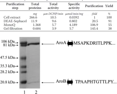
Two anion-exchange chromatographic steps and one gel fil-tration chromatography step were applied to achieve the puri-fication of the enzyme. This protocol led to a 145-fold final enrichment of the enzyme with a yield of 38% (Table 2). As shown in Fig. 2, the obtained pure enzyme consists of two sub-units with apparent molecular masses of 97 kDa and 16 kDa. They correspond to AroA and AroB, respectively, as confirmed by N-terminal sequencing (Fig. 2). The native molecular mass of the isolated enzyme, based on gel filtration chromatography, was found to be 110 kDa (data not shown) suggesting a␣11
configuration, similar to what was reported for the A. faecalis enzyme (22).
The complete DNA sequence of the aroA and aroB genes has been obtained. The deduced amino acid sequence of AroA displays 99, 81, 72, and 71% identity with AroA from SY8, A. faecalis, H. arsenicoxydans, and Burkholderia mul-tivorans, respectively (supplemental Fig. S1). The deduced amino acid sequence of AroB displays 99, 69, 57, and 54% identity with SY8, A. faecalis, B. multivorans, and H. arseni-coxydansAroB, respectively (supplemental Fig. S2).
Purification and Characterization of Two Cyts Able to React
with Aro—During the first DEAE chromatographic step, we
detected the presence of c-type cyts in the unretained fraction. Because in vitro enzymatic experiments revealed that purified Aro was able to reduce this fraction in the presence of AsIII, we decided to purify its constituent heme proteins. We isolated three cyts using a CM-52 cation exchange column and a gel filtration column successively and named them cyts c551, c552, and c554according to their respective␣-bands in the reduced
state. In contrast to cyt c551, cytochromes c552and c554were
6S. Duval and B. Schoepp-Cothenet, unpublished data.
FIGURE 1. Polyacrylamide gel electrophoresis and in-gel Aro activity staining of subcellular fractions of S22. Lane 1, spheroplasts; lane 2, periplasm; lane 3, membrane fraction after French press treatment; lane 4, soluble fraction after French press treatment.
FIGURE 2. Coomassie Blue-stained SDS-PAGE on purified Aro from S22. Lane 1, molecular weight standards; lane 2, purified enzyme. Identification of subunits was confirmed by N-terminal sequencing.
TABLE 1
Comparison of S22 to H. arsenicoxydans for AsIIIand AsVMinimal Inhibitory Concentrations H. arsenicoxydans Ralstonia sp. 22 MIC AsIII 6 m M 30 mM MIC AsV 200 m M 200 mM TABLE 2
Purification of Aro from S22
All kinetics were performed using DCPIP as electron acceptor. Purification step Total proteins Total activity Specific
activity Purification Yield
mg m DCPIP/min mol/min/mg -fold %
Cell extract 266.6 10.5 0.0392 1 100
DEAE-Sephacel 11.9 9.6 0.802 20.5 91
MonoQ 1.368 5.7 4.189 106.9 55
Gel filtration 0.684 3.9 5.7 145.4 38
at INRA Institut National de la Recherche Agronomique on May 14, 2019
http://www.jbc.org/
both able to react with isolated Aro (Fig. 3) and were therefore further characterized.
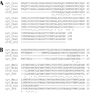
The N-terminal sequence (54 residues) of cyt c552(Fig. 4A)
revealed a high identity (75%) with cyts of Cupriavidus necator (formerly Ralstonia eutropha) H16, Cupriavidus taiwanensis and Cupriavidus metallidurans (formerly Ralstonia metalli-durans) CH34 (for which genome sequence are available) encoded by genes located in the sox cluster. The N-terminal sequence (56 residues) of cyt c554(Fig. 4B) revealed this protein
to be the product of the aroC gene located in the S22 aro cluster. Its sequence is astonishingly similar to that translated from the cyt gene present in the arsenite oxidase aox operon (10) of SY8 (99%) and shares only 45 and 43% identity with cyts c551/552 of Burkholderia multivorans and Burkholderia cenocepacia, respectively. Between each other, the two cyts c554and c552
from S22 are⬍40% identical.
The molecular masses, determined by MALDI-TOF mass spectrometry, of cyts c552and c554are 9615⫾ 2 and 9648 ⫾ 2
Da, respectively. The calculated molecular masses of the cyts retrieved from the Cupriavidus genomes and from the S22 aro operon, however, are both higher, around 11,600 Da. Indeed, in both the c552and c554precursor sequences, a 20-amino acid
stretch was predicted, using the SignalP program (40), to be a
signal peptide characteristic of the Sec secretion pathway (see (41) for review). The prediction of the cleavage site was in per-fect agreement with the determined N-terminal sequences VDA and APD (Fig. 4) for c552and c554, respectively.
Both cyts have been purified in the monomeric state as judged from size exclusion chromatography (data not shown). The redox potentials Em of cyts c552 and c554 at pH 8 were
determined at⫹230 ⫾ 5 mV and ⫹250 ⫾ 5 mV, respectively (data not shown), close to the value determined for cyt c552in NT-26 (27).
The fact that the aroC gene in the aro operon of S22 codes for cyt c554strongly argued in favor of c554, rather than c552, being
involved in AsIIIoxidation. However, both cyts were found in
approximately equal amounts when purified from cells grown on 5 mMAsIII. We therefore analyzed which of the cyts was induced by the presence of AsIII. Because the cyt c
552homologs
in the sequenced Cupriavidus genomes are encoded by genes localized in the sulfur oxidation sox cluster, we also analyzed the cyt contents of cells grown with and without thiosulfate (known to be the regulator of the sox operon (42– 45)). Cyt contents under these conditions were then compared with those obtained from cells grown with 5 mMAsIII. SDS-PAGE (Fig. 5A) detected an increased total amount of 10-kDa cyts in the cells grown either on thiosulfate or AsIIIbut could not
dis-criminate between cyt c552and c554(Fig. 5A, lanes 5 and 6), due to their very similar molecular masses, as mentioned above. We therefore spectroscopically quantified each of the cyts con-FIGURE 3. Spectra of c552and c554cytochromes enzymatically reduced by
purified Aro. Oxidized cyts (black lines) were incubated for 3 min with the enzyme (dotted lines); c552(A) and c554(B). Insets show the␣-bands of
cyto-chrome c552and c554absorbing at 552 and 554 nm, respectively.
FIGURE 4. Sequence alignment of cytochromes purified from S22. The N-terminal sequence of purified cyt c552(c552_S22; A) is compared with the
sequences of cyts from Cupriavidus necator H16 (cyt_Cnec; GenBankTM
acces-sion no. YP_727996), C. taiwanensis (cyt_Ctai; GenBankTMaccession no.
YP_002007005) and C. metallidurans CH34 (cyt_Cmet; GenBankTMaccession
no. YP_585565), whereas the N-terminal sequence of c554(c554_S22; B) is
compared with the sequences of cyts from Achromobacter sp. SY8 (cyt_SY8; GenBankTMaccession no. ABP63661), Burkholderia multivorans (cyt_Bmul;
GenBankTMaccession no. YP_001578392) and B. cenocepacia (cyt_Bcen;
Gen-BankTMaccession no. YP_002229329).
Characterization of Arsenite Oxidase from Ralstonia sp. 22
at INRA Institut National de la Recherche Agronomique on May 14, 2019
http://www.jbc.org/
tained in the DEAE-cyt fraction, under the tested growth con-ditions (Fig. 5B). In the absence of AsIIIor thiosulfate, cyt c552
represents 75% of the DEAE-cyt fraction content (again spec-troscopically quantified), whereas cyt c554amounts to⬃5% and
c554accounts for⬍2%. In the presence of 5 mMAsIII, the c552 fraction fell to 39% of the DEAE-cyt fraction content, whereas the c554complement increased to at least 43%. The relative
increase of cyt c554 quantity in response to AsIII therefore
approximately parallels that observed for Aro activity (see above). In the presence of 20 mMthiosulfate, the contribution of c552fell to 67% in relative cyt content but increased in absolute
quantity as compared with the control conditions (Fig. 5A, lane 4 versus lane 2, and Fig. 5B, Thio versus N.A.). The proportion of c552decreased due to the increase of c551from 2 to 12%.
Alto-gether, these results established not only that the accumulation of c551is linked to the AsIIIoxidation process but also suggested
that production of c552and c551is linked to the thiosulfate
oxi-dation pathway.
Enzymatic Study of the AsIIIOxidation System—As detailed
above, the enzyme from S22 was able to reduce cyts c552and
c554from S22 (Fig. 3). Several further cyts were tested for their reactivity with Aro. Bovine heart cyt c or Rhodobacter spha-eroidescyt c2were not reduced by the S22 enzyme (data not
shown), whereas cyt c555 from A. aeolicus and azurin from P. aeruginosawere (data not shown). Due to the limited quan-tities of purified c552 and c554, in-depth kinetic studies were
performed using cyt c555from A. aeolicus and yielded a Kmof 6
Mfor AsIII(Table 3) as well as substrate inhibition at concen-trations higher than 100M. We therefore analyzed the affinity for the reacting cyts at a concentration of 100MAsIII. Kinetic data, analyzed by reciprocal plots, yielded a Kmvalue of 50M
for cyt c555(Table 3). With the aim to compare the efficiency of cyts and DCPIP as electron acceptors, we first measured kinet-ics at pH 6, i.e. the pH value determined by Anderson et al. (22) and ourselves (data not shown) to be optimal with this electron acceptor. The Vmobtained using c555 at this pH value is an
order of magnitude higher than the Vm determined using DCPIP at the same pH. However, kinetics measured at other pH values yielded the range of pH 8 –9 as optimal for electron
transfer from Aro to the cyts and led to a further doubling of Vm. Preliminary kinetic studies with cyts c552and c554purified from S22 yielded Kmvalues of 13Mand 7M, respectively, at
a pH of 8.5.
Because no data have been reported to identify potential inhibitors of Aro, we screened the effect of selected chemicals on the Aro activity. We first tested the product of the physio-logical reaction, AsV, as potential inhibitor and found no effect.
We observed any inhibitory effect by NaN3, a well known
inhib-itor of several other molybdopterin enzymes (46, 47). Using cyts as electron acceptors allowed us to test sulfur compounds that otherwise would reduce DCPIP directly. Because H2S has been
reported to strongly inhibit AsIIIoxidation of
Hydrogenobacu-lum whole cells (48), we tested this compound on Aro and indeed found an inhibitory effect on our enzyme. We did not perform a detailed enzymatic analysis in the presence of sulfide but observed an I50(sulfide concentration yielding 50%
inhibi-tion) of⬃70M. A further sulfur compound, i.e. sulfite (but not thiosulfate), appeared to strongly inhibit Aro. A more detailed enzymatic analysis in the presence of sulfite showed a “mixed mode of inhibition” (data not shown), i.e. with not only an effect on affinity (Km) but also on catalysis (Km/Vm), with an I50of 10
M. Precise kinetics parameters were obtained with the A. ae-olicuscyt but were always verified using the S22 cyts.
As cyt c552potentially participates in thiosulfate oxidation,
we assayed whether the Aro featured measurable thiosulfate or sulfite oxidase activity. This was not the case (data not shown).
DISCUSSION
As mentioned in the introduction, many different aspects of the Aro enzymes have been studied in a variety of species, although an exhaustive characterization of a single case is still lacking. We took advantage of the availability of a new species of AsIIIoxidizer, the-proteobacterium S22, to perform a com-prehensive study of its Aro covering its phylogenetic position-ing, expression properties, biochemical and biophysical (see Ref. 66, accompanying article), as well as enzymatic parameters and its interaction with potential redox partners.
Aro from S22, a Clear Cut Case of Lateral Gene Transfer— Previous phylogenetic studies on the molybdopterin subunit of Aro (23, 49) suggested this enzyme to have evolved with its parent species. These conclusions have later been confirmed by the study of its Rieske subunit (50). Analysis of binary similarity data showed the 16S rDNA sequence of S22 to be more closely related to H. arsenicoxydans (91% identity) and B. multivorans (92%) species than to SY8 (86%) and A. faecalis (89%) species. As described above, however, both the sequences of the molyb-FIGURE 5. A, 3,3⬘,5,5⬘-tetramethyl-benzidine- stained SDS-PAGE of
cyto-chromes present in the S22 periplasm under different growing conditions. Lane 1, molecular weight standards; lane 2, cyts expressed without any addi-tion; lane 3, cyts expressed in the presence of 5 mMAsIII; lane 4, cyts expressed
in the presence of 20 mMthiosulfate; lane 5, purified cyt c554; lane 6, purified
cyt c552. B, schematic representation of cyt content of the DEAE-cyt fraction
under different growing conditions. Lane N.A., cyt content without any addi-tion; lane As, cyt content with 5 mMAsIII; lane Thio, cyt content with 20 mM
thiosulfate. Note that the DEAE-cyt fraction represent only part of the total cyt fraction (see “Experimental Procedures”) and that⬃20% of cyt content in this DEAE-cyt fraction remains unidentified under each of the growth conditions.
TABLE 3
Kinetic properties of the Aro from S22
*Vmaxis expressed asMAsoxidized⫻ min⫺1mg⫺1. It is noteworthy that 1
mole of AsIII
reduces 1 mole of DCPIP but 2 moles of cyt. N.D., not determined.
e⫺donor AsIII
Km(M) 6
Inhibitors Sulfite Sulfide
I50(M) 10 70
e⫺acceptor c555 c552 c554 DCPIP
Km(M) 50 13 7 N.D.
Vmax* 152 140 140 5.7
at INRA Institut National de la Recherche Agronomique on May 14, 2019
http://www.jbc.org/
dopterin and the Rieske subunits of Aro from S22 cluster with those of SY8 and A. faecalis enzymes instead of those from B. multivoransand H. arsenicoxydans species (see “Results”). This evolutionary relationship is further corroborated by the sequence that we determined for cytochrome c554. The
deduced identity of the AroA, AroB, and AroC sequences from S22 with the AoxB, AoxA, and AoxC sequences from SY8 were so suspiciously high (99%) that we were led to verify by 16S rDNA sequencing that our S22 strain was pure even when grown in 5 mMAsIII. These results therefore suggest that the entire aro operon of S22 has been acquired from an Achro-mobacter-related species by horizontal gene transfer. A detailed examination of the AroA-based phylogenetic tree (Fig. 2 in Ref. 49) pinpoints several abnormal branches as compared with the phylogenetic tree of the parent species. In some of these cases, discrepancies may be due to misalignments. The high homology between the SY8 and S22 sequences, however, does not allow for such a possibility for S22. This case, there-fore, constitutes the first unambiguous example of lateral gene transfer in the evolutionary pathway of Aro. It is noteworthy, though, that this lateral gene transfer occurred within the -proteobacterial subclass and thus does not significantly blur the picture of an overall coevolution of the enzyme and its par-ent species.
Is Aro a Membrane-bound Enzyme?—Muller et al. (17) observed a twin arginine translocation signal peptide in the H. arsenicoxydansAroB protein, and the presence of such a signal has now been detected in all available AroB sequences (18 and Fig. 6). This observation raises the question of whether this signal peptide is cleaved after the protein has been trans-ported across the cytoplasmic membrane or whether it serves to anchor Aro to the membrane. Cleavage sites were predicted (via the method developed by Bendtsen et al. (51)) for all Aro-Rieske proteins but not for the Aro-Rieske subunit of Aro-Rieske-cyt b complexes (a homologous protein, see Ref. 66, accompanying article). The latter result is in line with the following. i) All Rieske/cyt b Rieskes characterized so far have been demon-strated to be membrane bound with their uncleaved twin argi-nine translocation signal peptide serving as an anchor (52). ii) The predicted cleavage sites in Aro-Rieskes correspond to the N terminus determined in the isolated enzymes. Nevertheless, three types of Aro can be found. The C. aurantiacus and 3As enzymes correspond to the “rather membranous type” i.e. retained in the membrane fraction even after harsh treatment (23, 24). The NT-26 and Hydrogenophaga NT-14 (NT-14) enzymes belong to the class of “rather soluble type” Aros pres-ent in the soluble fraction even after mild treatmpres-ent (18, 21). Intermediate between these extreme cases are the H.
arseni-coxydans, A. faecalis, Arthrobacter, and S22 cases, for which the relative abundance in the membrane and the soluble fractions varies as a function of the harshness of cell disruption (5, 17, 22 and present work).
A scenario reconciling all results obtained so far, already pro-posed by Santini and vanden Hoven (18), consists in the attach-ment of AroAB to the membrane via another protein. This attachment must be of variable strength to explain the ensem-ble of the data and structurally specific as the Rieske protein has been observed to be oriented in a defined geometry (23) on Chloroflexusmembranes. This question is reminiscent of the problem arising from the study of the AsVreductase enzyme
(49). Some of the representatives of this family have been iso-lated as membrane-associated and others as soluble. However, in all characterized AsVreductase enzymes, a
membrane-asso-ciated component, which can be variable proteins, has been identified (53–55). We can imagine a similar scenario for the Aro enzyme, with the corresponding membrane-attached part-ner still to be identified. To address this question, we are cur-rently studying the case of the H. arsenicoxydans Aro, as this enzyme can indeed be easily obtained in a membrane-associ-ated form (see “Results”).
Aro Displays Strong Selectivity toward Its Electron Transfer Partners—Whereas the Aro enzyme may be more or less tightly membrane-associated in different species, it invariably appears to reduce soluble periplasmic electron carrier proteins. To the exception of one previous study (27), nonphysiological electron acceptors have been employed in activity tests, and the physio-logical electron acceptors have been deduced merely from genetic arguments. To place conclusions concerning the inter-action of Aro with its redox partners on firm ground, we have studied coexpression profiles and electron transfer activities of soluble cyts interacting with Aro in S22. The obtained results strongly suggest the physiological electron carrier of the S22 Aro to be cyt c554, i.e. the cyt present in the aro cluster. Spec-troscopic quantification clearly established a strong increase of the content of cyt when cells were grown on AsIII. This result suggests that c554gene expression is induced by As
III. It is of
note that only four species were shown to have a cyt gene co-transcribed with the aroA and aroB genes and hence induced by AsIII: Agrobacterium tumefaciens, H. arsenicoxydans, 3As, and
O. tritici(26, 28, 56, 57).
As we have shown, cyt c554is not the only cyt reacting with Aro in S22. Another cyt, c552, dominant in the absence of As
III
but still as abundant as c554 in the presence of As III
, accepts electrons from Aro and therefore mediates high turnover of the enzyme. Although no equivalent study has been performed on other bacteria, this result correlates well with circumstantial observations made in other organisms. For example, the⌬c552
mutant of NT-26 is still capable of autotrophic growth on AsIII
(27), suggesting that at least one other carrier may accept elec-trons from Aro. Finally, the recent sequencing and character-ization of the aro operon in 3As identified two cyts as being co-transcribed with aroAB (57). The Aro enzyme therefore appears to often be able to interact with several different elec-tron carriers in the same organism and the production of these carriers can either be regulated by AsIIIor not. In stark contrast
to this stands the clear cut and strong discrimination against a FIGURE 6. Prediction of twin arginine translocation signal cleavage site in
the N-terminal sequences of the Rieske subunit from Aros and Rieske-cyt
b complex. Prediction has been performed using the method developed by
Bendtsen et al. (51). The first residue after the cleavage site is colored in gray, whereas the observed first residue in the purified enzyme is boxed.
Characterization of Arsenite Oxidase from Ralstonia sp. 22
at INRA Institut National de la Recherche Agronomique on May 14, 2019
http://www.jbc.org/
specific subgroup of type I cyt as exemplified by horse heart or R. sphaeroidescyts. This selectivity is striking and not related to the redox potential of the carrier because all of the above cited carriers have EmpH 8 values⬃⫹240 mV. We therefore have
initiated an in-depth study of this phenomenon in a range of different Aro enzymes. The corresponding results and a struc-tural rationalization for this selectivity, observed in all exam-ined Aros, will be published elsewhere.6
Inhibitory Effects of Sulfur Compounds and Metabolic Signif-icance Thereof—Arsenic and sulfur often coexist in the envi-ronment and share similar microbial transformations. The study of the effect of sulfur compounds on AsIIIoxidation is
therefore indispensable to understand how both metabolisms interact. Our work revealed inhibitory effects of both sulfide and sulfite, with apparent I50of 70Mand 10M, respectively.
Are these compounds true enzymatic inhibitors? A concentra-tion of 60Msulfide has been observed to completely stop AsIII oxidation in whole cells of Hydrogenobaculum at low pH (48), whereas sulfide has been reported to strongly enhance the AsIII
oxidation in Mono Lake samples at high pH (58). However, did sulfide act on the Aro directly in these cases? Sulfide has been shown to react with AsIII, forming orpiment at low pH (59) and
thioarsenic species at high pH (58, 60). In these three cited works, sulfide was added in equal if not higher quantities com-pared with AsIII, allowing the product of the reaction between
AsIIIand sulfide to significantly modify the AsIIIquantity
avail-able for Aro-mediated oxidation. The observed effects are therefore potentially nonenzymatic. In our case, sulfide shows significant inhibitory effect at concentrations where, whatever the sulfide/AsIIIreaction product, free AsIIIis always saturating.
Our work is therefore the first to clearly establish a true inhib-itory effect of sulfide on the enzymatic AsIIIoxidation.
Sulfite is another sulfur compound revealed by our work to be a strong inhibitor of the Aro enzyme from S22. A conflicting result was published by Phillips (61) on A. faecalis whole cells, but we observed a similar inhibition also on the NT-26 enzyme. In both cases, the inhibition appears to be of a mixed-type inhi-bition, i.e. both competitive and noncompetitive. The compet-itive character, i.e. the effect on the Kmfor As
IIIsuggests that
sulfite reacts at the molybdopterin center. Because dialysis restores the activity, the binding of sulfite seems to be transient. This effect appears, therefore, to be distinct from the irreversi-ble effect described for sulfite oxidase (a molybdopterin enzyme) in presence of AsIII(62).
An increasing number of studies find relationships between sulfur and arsenic metabolism. Interest has been focused on the interaction between AsVand sulfate reduction (see Refs. 63, 64
for original works). More recently, the interaction between AsIIIoxidation and sulfur metabolism was addressed (48, 58,
65), but it has not been studied at the molecular level prior to this work. Although ability of Aro to use cyt c552as electron acceptor may be interpreted as a possible cross-talk between sulfur and arsenic metabolisms, this merging of electron trans-fer pathways is not obligatory because Aro possesses a second, dedicated, acceptor, i.e. cyt c554. The more straightforward link
between AsIII and sulfur oxidation processes revealed by our
work consists in the sensitivity of Aro to two substrates of the soxsystem, i.e. sulfite and sulfide. The sox system, well
charac-terized in Paracoccus pantotrophus and coded by 15 genes, cat-alyzes thiosulfate-, sulfite-, sulfur-, and sulfide-dependent cyt c reduction (43, 44). We can therefore imagine that the sox sys-tem, will enhance the AsIIIoxidation rate of S22, by promoting
the consumption of sulfite and sulfide.
Acknowledgments—We are indebted to Maya Belghazi from CAPM-IFR Jean Roche for access to the Ultraflex II mass spectrometer. We furthermore thank Pascale Infonsi for the gift of A. aeolicus cyt c555.
REFERENCES
1. Nriagu, J. O. (1994) Arsenic in the Environment. John Wiley and Sons, Inc, New York
2. Oremland, R. S., and Stolz, J. F. (2005) Trends Microbiol. 13, 45– 49 3. Inskeep, W. P., McDermott, T. R., and Fendorf, S. (2002) Environmental
Chemistry of Arsenic(Frankenberger, W. T., ed) pp. 183–215, Marcel Dek-ker, Inc. New York
4. Green, H. H. (1918) S. Afr. J. Sci. 14, 465– 467
5. Prasad, K. S., Subramanian, V., and Paul, J. (2009) Biometals 22, 711–721 6. Hamamura, N., Macur, R. E., Korf, S., Ackerman, G., Taylor, W. P., Ko-zubal, M., Reysenbach, A. L., and Inskeep, W. P. (2009) Environ. Microbiol. 11,421– 431
7. Chang, J. S., Yoon, I. H., Lee, J. H., Kim, K. R., An, J., and Kim, K. W. (2010) Environ. Geochem. Health 32,95–105
8. Valenzuela, C., Campos, V. L., Yan˜ez, J., Zaror, C. A., and Mondaca, M. A. (2009) Bull. Environ. Contam. Toxicol. 82, 593–596
9. Sun, W., Sierra-Alvarez, R., Fernandez, N., Sanz, J. L., Amils, R., Legatzki, A., Maier, R. M., and Field, J. A. (2009) FEMS. Microbiol. Ecol. 68, 72– 85 10. Cai, L., Rensing, C., Li, X., and Wang, G. (2009) Appl. Microbiol.
Biotech-nol. 83,715–725
11. Cai, L., Liu, G., Rensing, C., and Wang, G. (2009) BMC Microbiol. 9, 4 12. Handley, K. M., He´ry, M., and Lloyd, J. R. (2009) Int. J. Sys. Evol. Microbiol.
59,886 – 892
13. Silver, S., and Phung, L. T. (2005) Appl. Environ. Microbiol. 71, 599 – 608 14. Stolz, J. F., Basu, P., Santini, J. M., and Oremland, R. S. (2006) Annu. Rev.
Microbiol. 60,107–130
15. Hoeft, S. E., Blum, J. S., Stolz, J. F., Tabita, F. R., Witte, B., King, G. M., Santini, J. M., and Oremland, R. S. (2007) Int. J. Syst. Evol. Microbiol. 57, 504 –512
16. Kulp, T. R., Hoeft, S. E., Asao, M., Madigan, M. T., Hollibaugh, J. T., Fisher, J. C., Stolz, J. F., Culbertson, C. W., Miller, L. G., and Oremland, R. S. (2008) Science 321, 967–970
17. Muller, D., Lie`vremont, D., Simeonova, D. D., Hubert, J. C., and Lett., M. C. (2003) J. Bacteriol. 185, 135–141
18. Santini, J. M., and vanden Hoven, R. N. (2004) J. Bacteriol. 186, 1614 –1619 19. Nasser, D., and Nester, E. W. (1967) J. Bacteriol. 94, 1706 –1714 20. Santini, J. M., Sly, L. I., Schnagl, R. D., and Macy, J. M. (2000) Appl. Environ.
Microbiol. 66,92–97
21. vanden Hoven, R. N., and Santini, J. M. (2004) Biochim. Biophys. Acta 1656,148 –155
22. Anderson, G. L., Williams, J., and Hille, R. (1992) J. Biol. Chem. 267, 23674 –23682
23. Lebrun, E., Brugna, M., Baymann, F., Muller, D., Lie`vremont, D., Lett, M. C., and Nitschke, W. (2003) Mol. Biol. Evol. 20, 686 – 693
24. Duquesne, K., Lieutaud, A., Ratouchniak, J., Muller, D., Lett, M. C., and Bonnefoy, V. (2008) Environ. Microbiol. 10, 228 –237
25. Ellis, P. J., Conrads, T., Hille, R., and Kuhn, P. (2001) Structure 9, 125–132 26. Kashyap, D. R., Botero, L. M., Franck, W. L., Hassett, D. J., and
McDer-mott, T. R. (2006) J. Bacteriol. 188, 1081–1088
27. Santini, J. M., Kappler, U., Ward, S. A., Honeychurch, M. J., vanden Hoven, R. N., and Bernhardt, P. V. (2007) Biochim. Biophys. Acta 1767, 189 –196 28. Branco, R., Francisco, R., Chung, A. P., and Morais, P. V. (2009) App.
Environ. Microbiol. 75,5141–5147
29. Kaback, H. L. (1971) Methods Enzymol. 22, 99 –122
30. Baymann, F., Tron, P., Schoepp-Cothenet, B., Aubert, C., Bianco, P.,
at INRA Institut National de la Recherche Agronomique on May 14, 2019
http://www.jbc.org/
ter, K. O., Nitschke, W., and Schu¨tz, M. (2001) Biochemistry 40, 13681–13689
31. Laemmli, U. K. (1970) Nature 227, 680 – 685
32. Judd, R. C. (2002) in The Protein Protocols Handbook, (Walker, J. M., ed) 2nd Ed., pp. 73–76, Humana Press, Inc., Ottawa, Ontario, Canada 33. Sambrook, J., Fritsch, E. F., and Maniatis, T. (1989) in Molecular Cloning:
A Laboratory Manual, 2nd Ed., Cold Spring Harbor Laboratory Press, Cold Spring Harbor, NY
34. Thompson, J. D., Higgins, D. G., and Gibson, T. J. (1994) Nucleic Acids Res. 22,4673– 4680
35. Simeonova, D. D., Lie`vremont, D., Lagarde, F., Muller, D. A., Groudeva, V. I., and Lett, M. C. (2004) FEMS Microbiol. Lett. 237, 249 –253 36. Weeger, W., Lie`vremont, D., Perret, M., Lagarde, F., Hubert, J. C., Leroy,
M., and Lett, M. C. (1999) Biometals 12, 141–149
37. Lim, C. K., and Cooksey, D. A. (1993) J. Bacteriol. 175, 4492– 4498 38. Dutton, P. L (1971) Biochim. Biophys. Acta 226, 63– 80
39. Duval, S. (2008) Les enzymes du me´tabolisme de l’arsenic chez les pro-caryotes, aspects fonctionnels, phylogénétiques et structuraux.Ph.D. thesis, Universite´ de Provence, Marseilles, France
40. Bendtsen, J. D., Nielsen, H., von Heijne, G., and Brunak, S. (2004) J. Mol. Biol. 340,783–795
41. Pugsley, A. P. (1993) Microbiol. Rev. 57, 50 –108
42. Kelly, D. P., Shergill, J. K., Lu, W. P., and Wood, A. P. (1997) Antonie Leeuwenhoek 71,95–107
43. Friedrich, C. G., Quentmeier, A., Bardischewsky, F., Rother, D., Kraft, R., Kostka, S., and Prinz, H. (2000) J. Bacteriol. 182, 4677– 4687
44. Rother, D., Henrich, H. J., Quentmeier, A., Bardischewsky, F., and Friedrich, C. G. (2001) J. Bacteriol. 183, 4499 – 4508
45. Hensen, D., Sperling, D., Tru¨per, H. G., Brune, D. C., and Dahl, C. (2006) Mol. Microbiol. 62,794 – 810
46. Enoch, H. G., and Lester, R. L. (1975) J. Biol. Chem. 250, 6693– 6705 47. Forget, P. (1971) Eur. J. Biochem. 18, 442– 450
48. Donahoe-Christiansen, J., D’Imperio, S., Jackson, C. R., Inskeep, W. P., and McDermott, T. R. (2004) Appl. Environ. Microbiol. 70, 1865–1868 49. Duval, S., Ducluzeau, A. L., Nitschke, W., and Schoepp-Cothenet, B.
(2008) BMC Evol. Biol. 8, 206 –219
50. Lebrun, E., Santini, J. M., Brugna, M., Ducluzeau, A. L., Ouchane, S., Scho-epp-Cothenet, B., Baymann, F., and Nitschke, W. (2006) Mol. Biol. Evol. 23,1180 –1191
51. Bendtsen, J. D., Nielsen, H., Widdick, D., Palmer, T., and Brunak, S. (2005) BMC Bioinformatics 6,167–175
52. Kallas, T. (1994) in The Molecular Biology of Cyanobacteria (Bryant, D. A., ed.) pp. 259 –317, Kluwer Academic Publishers, the Netherlands 53. Saltikov, C. W., and Newman, D. K. (2003) Proc. Natl. Acad. Sci. U.S.A.
100,10983–10988
54. Malasarn, D., Keeffe, J. R., and Newman, D. K. (2008) J. Bacteriol. 190, 135–142
55. Murphy, J. N., and Saltikov, C. W. (2007) J. Bacteriol. 189, 2283–2290 56. Muller, D. (2004) Analyse génétique et moléculaire du stress arsenic de
souches bactériennes isolées d’environments contaminés par l’arsenic. Ph.D. thesis, Universite´ de Strasbourg, France
57. Slyemi, D., Ratouchniak, J., and Bonnefoy, V. (2007) Adv. Mat. Res. 20 –21, 427– 430
58. Fisher, J. C., Wallschla¨ger, D., Planer-Friedrich, B., and Hollibaugh, J. T. (2008) Environ. Sci. Technol. 42, 81– 85
59. Rochette, E. A., Bostick, B. C., Li, G. C., and Fendorf, S. (2000) Environ. Sci. Technol. 34,4714 – 4720
60. Stauder, S., Raue, B., and Sacher, F. (2005) Environ. Sci. Technol. 39, 5933–5939
61. Phillips, S. E., and Taylor, M. L. (1976) Appl. Environ. Microbiol. 32, 392–399
62. Gardlik, S., and Rajagopalan, K. V. (1991) J. Biol. Chem. 266, 16627–16632 63. Dowdle, P. R., Laverman, A. M., and Oremland, R. S. (1996) Appl. Environ.
Microbiol. 62,1664 –1669
64. Newman, D. K., Kennedy, E. K., Coates, J. D., Ahmann, D., Ellis, D. J., Lovley, D. R., and Morel, F. M. (1997) Arch. Microbiol. 168, 380 –388 65. Langner, H. W., Jackson, C. R., McDermott, T. R., and Inskeep, W. P.
(2001) Environ. Sci. Technol. 35, 3302–3309
66. Duval, S., Santini, J. M., Nitschke, W., Hille, R., and Schoepp-Cothenet, B. (2010) J. Biol. Chem. 285, 20442–20451
Characterization of Arsenite Oxidase from Ralstonia sp. 22
at INRA Institut National de la Recherche Agronomique on May 14, 2019
http://www.jbc.org/
and Barbara Schoepp-Cothenet
Lebrun, Sabrina Lignon, Marie-Laure Fardeau, Marie-Claire Lett, Wolfgang Nitschke
Aurélie Lieutaud, Robert van Lis, Simon Duval, Line Capowiez, Daniel Muller, Régine
ENZYME AND ITS INTERACTION WITH SOLUBLE CYTOCHROMES
doi: 10.1074/jbc.M110.113761 originally published online April 26, 2010 2010, 285:20433-20441.
J. Biol. Chem.
10.1074/jbc.M110.113761 Access the most updated version of this article at doi:
Alerts:
When a correction for this article is posted •
When this article is cited •
to choose from all of JBC's e-mail alerts Click here
Supplemental material:
http://www.jbc.org/content/suppl/2010/04/26/M110.113761.DC1 http://www.jbc.org/content/285/27/20433.full.html#ref-list-1This article cites 59 references, 22 of which can be accessed free at
at INRA Institut National de la Recherche Agronomique on May 14, 2019
http://www.jbc.org/