Chromosome Dynamics of the Early Meiotic Cell Cycle
in S. cerevisiae
by
Hannah G. Blitzblau Sc.B. Biochemistry Brown University, 1999Submitted to the Department of Biology in partial fulfillment of the requirements for the degree of
Doctor of Philosophy in Biology at the
Massachusetts Institute of Technology February 2008
C 2008 Hannah G. Blitzblau. All rights reserved The author hereby grants to MIT permission to reproduce and distribute publicly paper and electronic copies of this thesis in whole or in part
Signature of Author: ' D _" Department of Biology January 11, 2008 Certified by: Stephen P. Bell Professor of Biology Thesis Supervisor Accepted by: . . . , - -Stephen P. Bell Professor of Biology Chair, Committee for Graduate Students
O TEOHNOLOGY
FEB 12 2008
LIBRARIES
Chromosomi~e Dynamics of the Early Meiotic Cell Cycle in S. cerevisiae
By
Hannah G. Blitzblau
Submitted to the Department of Biology in partial fulfillment of the requirements for the degree of Doctor of Philosophy in Biology
Abstract
In every cell cycle the genetic material must be duplicated and transmitted to the daughter cells. Meiosis is a developmental program that allows a diploid cell to produce haploid progeny. The reduction in chromosome number obtained during meiosis requires specialized mechanisms that are absent during the canonical mitotic cell cycle. Although previous studies found strong similarities between pre-mitotic and pre-meiotic DNA replication, pre-meiotic S phase is longer than pre-mitotic S phase, suggesting that meiosis-specific events regulate the rate of DNA replication. Additionally, after DNA replication, homologous recombination is initiated by the introduction of hundreds DNA double-strand breaks (DSBs) into the genome to produce physical DNA exchnges, or crossovers, between homologous chromosomes. To investigate the chromosome dynamics of the early meiotic cell cycle, I performed comprehensive analysis of pre-meiotic DNA replication and DSB formation in budding yeast.
Genome-wide studies of pre-meiotic DNA replication confirmed that the same replication origins are selected and activated in pre-meiotic and pre-mitotic cells, although replication was delayed at a large number of origins. These results indicate that the regulation of DNA replication is similar in the meiotic and mitotic cell cycles, but that the replication-timing program differs. Elimination of meiosis-specific cohesion or homologous recombination had no
effect on the number or identity of early pre-meiotic origins. Analysis of cells sporulated in the presence of the replication inhibitor HU revealed a Cln3-dependent inhibition of meiotic entry.
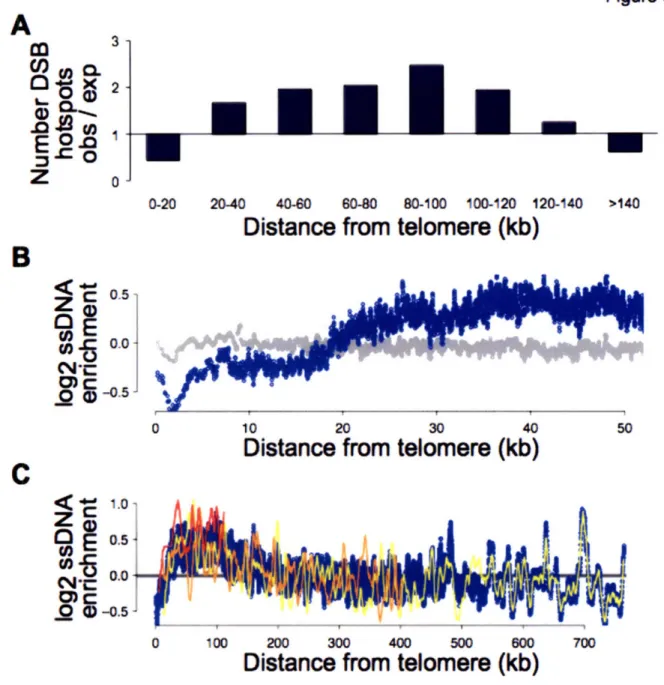
To map the locations of meiotic DSBs, I developed a method to detect meiotic ssDNA. Examination of the sites of ssDNA enrichment indicated that DSBs occur mainly in the promoters of active genes, consistent with previous studies of individual DSB sites. Global analysis of the most common DSB sites revealed a non-random distribution of DSB "hotspots." In particular, DSB hotspots are over-enriched close to chromosome ends, which could explain why small chromosomes have a higher DSB density than large chromosomes. This mechanism could help ensure that all homologous chromosomes receive at least one crossover and segregate properly in meiosis. These studies also indicated that suppression of recombination at telomeres, centromeres and around the rDNA occurs by 3 distinct mechanisms.
To Sister, Anjakat and Menalie, for their inspiration and support
Acknowledgements
This work could not have been completed without the help and support of a large number of people. Thank you to the past and present members of the Bell Lab family for creating a stimulating environment for scientific discovery and extracurricular hobbies. I am also indebted to past and present members of the Amon lab, particularly those in the meiosis group, for providing me with reagents, protocols, ideas and genuine friendship. Thank you to Andreas, as our collaboration has been great fun and the kind of success that the faculty told us would magically happen if we spent enough time together in the Pit (and Starbucks). My classmates in Biograd 2000 have become friends and colleagues in pursuits inside the lab, as well as dim sum, supper club, girls' night, river walks etc. My friends and family outside of MIT have constantly reminded me that I am part of a larger world that requires an excess of love, patience and creative vision to go 'round. I cannot thank my roommates, sister, Phil and Anna, enough for being there for me through this process as only the best of friends and family can.
I am extremely grateful to past and present members of my thesis committee, Anja-Katrin Bielinsky, Frank Solomon, Terry Orr-Weaver and Rick Young, who have always imparted scientific and life advice when it was needed.
I would like to specially acknowledge Angelika Amon, who has supported me
ceaselessly as a mentor, second advisor, role model and friend.
Thank you especially to Steve, who, from start to finish, entrusted me with the means and freedom to follow my whims, wherever they took us. I have enjoyed his sound advice, stimulating scientific discussions, ability to be understanding, and generous nature, which make the Bell Lab a great place to work.
Table of Contents
Sum m ary ... 3
Acknowledgements ... ... 7
Table of Contents ... 9
Chapter I: Introduction... 11
Meiosis: a specialized cell division ... ... 13
Regulation of cell cycle entry ... 17
Regulation of DNA replication ... 20
The intra-S phase checkpoint... 23
Regulation of replication timing ... ... ... 24
Pre-meiotic DNA replication ... 27
Initiation of homologous recombination ... ... ... 28
Repair of meiotic DSBs... ... 31
Regulation of crossover formation... 32
Co-regulation of DNA replication and recombination... ... 35
Thesis Summary ... 37
References... 38
Chapter II: Characterization of a slow S phase: pre-meiotic DNA replication in yeast .... 45
Sum m ary ... 47
Introduction... 49
R esults ... 52
Differential pre-RC formation at a subset of pre-meiotic origins ... 52
The same origins are active in the meiotic and mitotic cell cycles ... 55
Replication initiation is delayed for some pre-meiotic origins ... 58
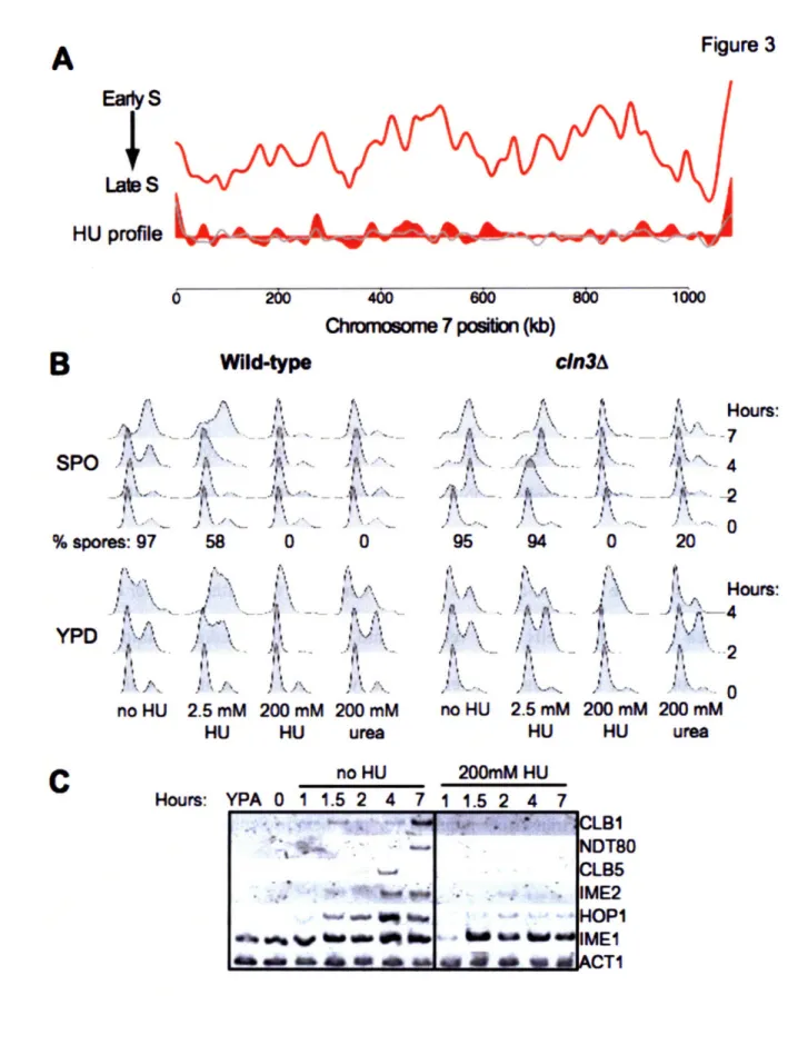
Hydroxyurea inhibits meiotic cell cycle entry... ... . 60
Initiation timing differs in the meiotic and mitotic cell cycles... 64
Replication timing, cohesion and homolgous recombination ... 67
D iscussion ... 70
Transcription regulated pre-RC assembly at a subset of origins... 70
HU inhibits meiotic entry in a Cln3-dependent manner ... 72
Fewer early replicating origins during pre-meiotic S phase ... 73
Pre-meiotic and pre-mitotic DNA replication kinetics differ... 74
Why a slow S phase? ... 77
Materials and Methods ... ... 78
Supplemental Data ... ... 80
Chapter III: Mapping of meiotic ssDNA reveals double-strand break hotspots near
centromeres and telomeres ... 107
Sum m ary ... 109
Introdu ction ... 111
R e su lts ... 1 13 Labeling of ssDNA reveals DSB sites ... 113
Hotspots mapped by ssDNA versus Spo 11l localization... 117
DSBs are enriched in the promoters of active genes ... 120
Strong hotspots in the pericentromeric regions ... 123
Hotspot distribution near the rDNA and telomeres ... 125
D iscussion ... ... ... 133
Acknowledgements ... 139
Materials and Methods ... 140
Supplemental Data ... 142
R eferences... ... 169
Chapter IV: Discussion and Future Directions... ... 173
K ey conclusions ... 175
Regulation of replication complex assembly ... 176
Regulation of replication timing in meiosis ... 177
The intra-S phase checkpoint in meiosis ... 179
Global distribution of DSB activity... 181
Suppression of homologous recombination ... 182
R eferences ... .. ... 184
Chapter I
Meiosis: a specialized cell division
The survival of all species depends on their ability to reproduce and adapt to a changing environment. Sexually reproducing organisms have an evolutionary advantage because they can give rise to a genetically distinct offspring in each reproductive cycle through the fusion of gametes with half the genetic material of the parents. The reduction in chromosome number required for gamete production is carried out during a specialized cell division called meiosis, in which a single round of DNA replication precedes two successive chromosome segregation events. The most common problem encountered during meiosis is the failure of chromosomes to properly segregate, which leads to gametes with an inappropriate chromosome number and results in aneuploid offspring. In humans, more than 20% of conceptions fail because of meiotic errors and the rate of such errors increases with age (Hassold and Hunt 2001). Both genetic and environmental factors have been implicated in meiotic chromosome non-disjunction (Hunt and Hassold 2002). However, a mechanistic understanding of these effects has been impeded by the relative lack of understanding of mammalian meiosis. In contrast, many key insights into meiotic chromosome segregation have been revealed by recent work in both budding and fission yeast. The high degree of conservation of many meiotic regulators across eukaryotic species implies that understanding meiosis in the relatively simple yeast system will contribute to our understanding of human fertility.
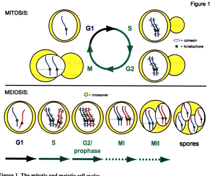
Meiosis shares many characteristics with the well-studied mitotic cell division cycle, but instead of conserving chromosome number, meiosis allows cells to reduce the chromosome number by half (Figure 1). In mitosis, a single DNA replication event precedes the segregation of replicated chromosomes, termed sister chromatids, away from each other. Mitosis produces two
cells with identical genetic material to the progenitor. Meiosis is comprised of a single DNA replication event, followed by two consecutive chromosome segregations. In the first meiotic division, homologous chromosomes are separated and in the second division, sister chromatids segregate. DNA replication and the second meiotic division closely resemble the mitotic cell cycle and are regulated by many of the same factors. In contrast, the segregation of homologous chromosomes is unique to meiosis and requires specialized machinery and regulation.
Figure
1
MITOSIS:
0 =kinet choreMEIOSIS:
'-ý crosoverG1
S
G21
MI
MII
prophase
N
gnomon----.--.,--spores
Figure 1. The mitotic and meiotic cell cycles
The major events of each stage of the mitotic and meiotic cell cycles are depicted. Red and blue indicate homologous chromosomes.
The alignment and faithful segregation of chromosomes is highly regulated to prevent lethal chromosome loss events. Chromosome segregation requires the attachment of the kinetochores of the two chromosomes to spindles emanating from opposite poles (Figure 1, mitosis). Cells sense the bipolar attachment of chromosomes because the chromosomes are physically attached to each other, such that when microtubules from opposite poles provide a pulling force on the chromosomes, tension is generated. In mitotically dividing cells, sister chromatids are segregated away from each other. Sister chromatids are bound together by the cohesion complex (cohesin) as they are replicated (Michaelis, Ciosk et al. 1997; Uhlmann and Nasmyth 1998) (Figure 1). Thus, when the kinetochores attach to microtubules from opposite poles in mitosis, the existing physical connection between sister chromatids allows tension to be generated and chromosomes can be faithfully segregated. In contrast, homologous chromosomes have no predetermined interaction and must become properly aligned and physically connected before they can satisfy the tension requirement of the spindle checkpoint in meiosis. The alignment and attachment of homologous chromosomes is accomplished by the process of meiotic homologous recombination. During recombination, at least one set of sister chromatids from each homologous chromosome pair undergoes a physical DNA exchange, or crossover (Figure 1, yellow star).
The faithful segregation of homologous chromosomes also requires meiosis-specific cohesin and kinetochore-associated factors. Because the sister chromatids of each homolog are held together by cohesion after they are replicated, the cohesin complexes distal to the crossover physically hold chromosomes together and allow tension to be generated when homologous chromosome attach to the meiotic spindle in metaphase I (Figure 1, meiotic G2/prophase).
Furthermore, special protein complexes located at the kinetochores ensure that the sister chromatids of each homolog attach to spindles from the same pole, so that homologs, but not sisters, are separated (Toth, Rabitsch et al. 2000; Rabitsch, Petronczki et al. 2003; Lee, Kiburz et al. 2004) (Figure 1, meiosis I). Following segregation of homologs in meiosis I, sister chromatids are segregated without either an intervening DNA replication event or new establishment of cohesion (meiosis II). Cohesion distal to the crossovers must be removed for chromosomes to separate in meiosis I, but for the second division to occur properly, cohesion at centromeres must be retained between sister chromatids until they have properly aligned at metaphase II. This step-wise removal of cohesin differs from that of mitosis and is carried out, in part, through the use of distinct cohesin complexes in meiosis (Buonomo, Clyne et al. 2000) and specialized proteins that protect cohesin around centromeres (Kerrebrock, Moore et al. 1995; Kitajima, Kawashima et al. 2004; Lee, Kiburz et al. 2004; Marston, Tham et al. 2004; Kiburz, Reynolds et al. 2005; Watanabe 2005).
Following the segregation of homologous chromosomes in meiosis I, sister chromatids are bioriented on the meiosis II spindle (Figure 1, meiosis II). Segregation of sister chromatids produces four haploid nuclei, which are packaged into spores in yeast. Cells then exit the cell cycle and meiosis is completed. The need for both homologous recombination and special regulation of cohesin and kinetochore components obliges meiotic cells to undertake a discrete and highly regulated developmental program. In this thesis, I will focus on the chromosome dynamics that occur early in the meiotic program. Specifically, I will focus on measuring pre-meiotic DNA replication and the initiation of recombination.
Regulation of cell cycle entry
The decision to undergo the meiotic program is regulated by external cellular signals in all organisms. In higher eukaryotes, meiosis is reserved for the production of gametes and occurs only in specialized organs. In this case, temporal and spatially restricted signals control the meiotic entry of a specific subset cells called germ cells. In yeast, the decision to enter either the mitotic or meiotic cell cycles is nutritionally regulated (Figure 2). When nutrients are plentiful, cells grow until they reach a sufficient size and then enter the mitotic cell cycle. In contrast, when nutrients are limiting, yeast can either enter GO and cease cell division or, if they are diploid, enter the meiotic cell cycle. Yeast sporulate when they are starved of fermentable carbon and nitrogen. The spores that result from yeast meiosis enter a dormant state in which they can survive nutritional deprivation for long periods of time.
The meiotic developmental program is determined by altered transcription and protein synthesis. Altogether, -1600 genes (>25% of the yeast genome) are differentially expressed between sporulating and non-sporulating cells (Chu, DeRisi et al. 1998; Primig, Williams et al. 2000). These changes in gene expression prevent mitosis-specific events such as budding and promote meiotic processes necessary for segregation of homologous chromosomes and spore formation. Meiosis-specific transcriptional activators implement the altered transcription program. Entry into meiosis is regulated by the synthesis and stability of the Imel transcription factor, which activates genes necessary for DNA replication and homologous recombination (Smith, Su et al. 1990). After homologous recombination, the Ndt80 transcription factor activates the expression of genes necessary for meiotic chromosome segregation (Chu and Herskowitz
Figure 2
mitotic cell cycle entry
CIn31
Cinl,2/1
•
budding
CDK
CDK
I
"Y
Sic1
+ nutrients-.-
DNA replication
DNA
replication/
recombination
- nutrientsImel
l-
lme2
meiotic
cell cycle entry
Sic1
Nchromosome
segregationFigure 2: Cell cycle entry
The processes of mitotic and meiotic cell cycle entry are depicted. The presence or absence of fermentable carbon and nitrogen sources determines whether cells will enter the mitotic or meiotic cell cycle. Different mechanisms of activating Clb/CDK are shown.
In yeast, both the mitotic and meiotic cell cycles are regulated by the activity of the single cyclin-dependent kinase (CDK), Cdc28. Cdc28 promotes all DNA replication and chromosome segregation events in a complex with one of the six B-type cyclins, or Clbs. Clb5 and Clb6 are synthesized in G1, but remain inactive due to the presence of the B-type CDK inhibitor Sicl
(Schwob, Bohm et al. 1994). At the G1/S phase transition, Sicl is degraded and Clb-CDK is activated. Clb5/Clb6-associated CDK activity promotes DNA replication (Kuhne and Linder 1993; Schwob and Nasmyth 1993). Prior to mitosis, four other B type cyclins, Clbsl-4, are synthesized and their activity regulates the single chromosome segregation (Surana, Robitsch et al. 1991; Fitch, Dahmann et al. 1992). CDK regulation is similar in sporulating cells, with a few exceptions. Clb5 and Clb6 are essential for both DNA replication and homologous recombination in early meiosis (Dirick, Goetsch et al. 1998; Stuart and Wittenberg 1998; Benjamin, Zhang et al. 2003). Clbsl, 3, 4 and 5 are synthesized prior to the two meiotic chromosome segregation events, but expression of CLB2 is highly repressed in meiosis (Grandin and Reed 1993; Dahmann and Futcher 1995). After both mitosis and meiosis, the complete inactivation of Clb-CDK leads to spindle disassembly and cell cycle exit, allowing cells to reenter the next Gl phase. Therefore, the proper regulation of CDK activity is critical to all aspects of cell cycle progression in yeast.
The decision to activate CDK and enter either the meiotic or mitotic cell cycle occurs during the Gl phase when cells sense nutrient availability (Figure 2). When nutrients are plentiful, the mitotic cell cycle is initiated by the association of Cdc28 with the Gl-specific cyclins Clnl, Cln2 and Cln3. Cln-CDK activity drives cell cycle progression by promoting both budding and Clb-CDK activation (Dirick, Bohm et al. 1995). Phosphorylation of Sicl by Cln-CDK targets Sicl for degradation, activating Clb-Cln-CDK and stimulating entry into S phase/DNA synthesis. Cln3 is the first cyclin to be activated during mitotic Gl and it both stimulates Clnl, Cln2, Clb5 and Clb6 synthesis and prevents cells from entering meiosis (Colomina, Gari et al. 1999). When nutrients are limiting, Cln3 is never activated. Instead, Imel induces transcription
of the gene for the meiosis-specific kinase, Ime2. Ime2 is homologous to Cdc28, but does not require a cyclin subunit for activation. Ime2 substrate specificity differs from Cdc28 (Clifford, Marinco et al. 2004; Holt, Hutti et al. 2007), allowing it to differentially regulate events of the early meiotic cell cycle. For example, Ime2 does not promote mitosis specific events such as budding, whereas Ime2 specifically promotes meiosis-specific functions such as activation of the Ndt80 transcription factor (Benjamin, Zhang et al. 2003; Honigberg and Purnapatre 2003). In some cases, Ime2 performs the same function as Cln-CDK, although it does so by phosphorylating substrates on distinct residues. For example, Ime2 is required to target Sicl for degradation and thereby promote Clb-CDK activation in meiosis (Benjamin, Zhang et al. 2003), but it does so through the utilization of alternate sites on Sicl than CDK uses (Sedgwick, Rawluk
et al. 2006; Sawarynski, Kaplun et al. 2007).
Regulation of DNA replication
Activation of Clb-CDK drives cells into S phase. In both mitotic and meiotic cell cycles it is critical that the DNA is fully duplicated prior to its segregation into daughter cells. This ensures that each progeny receives a full complement of the genetic material. Additionally, faithful chromosome segregation in mitosis and meiosis require cohesion between sister chromatids that is established during S phase as the DNA is replicated (Uhlmann and Nasmyth 1998). The regulation of DNA replication during the mitotic cell cycle has been relatively well-characterized. The conservation of many of the key regulators of DNA replication in meiosis suggests that similar mechanisms probably function in pre-meiotic DNA replication.
The duplication of large eukaryotic genomes requires the coordination of hundreds of replication forks distributed throughout the genome. DNA replication begins at specific sites in the DNA termed origins of replication, and bidirectional replication forks proceed away from each origin. The region duplicated by the forks emanating from a single origin is termed a replicon. The duplication of large eukaryotic genomes that contain multiple chromosomes requires many replicons. The average eukaryotic replicon is -40-100 kb, which is less than the length of even the smallest chromosome in yeast. Replicon size is limited both by stability of the replication fork assemblies and cell growth rate. Because replication usually initiates from origins, if forks collapse they are generally not able to reinitiate (Labib, Tercero et al. 2000). Additionally, utilization of multiple origins of replication allows DNA replication to be completed in a timely manner, providing a growth advantage to cells. However, replication forks must be coordinated such that each piece of DNA in the cell is duplicated only once per cell cycle, since over- or under-replication leads to genome instability and death (DePamphilis, Blow et al. 2006).
The initiation of DNA replication occurs in two steps; selection of potential replication origins and initiation of DNA replication (Figure 3) (Diffley and Labib 2002). Origins are selected during the Gl phase of the cell cycle by the association of a complex containing the replicative helicase with all potential origins of replication. Helicase loading is initiated by the multifunctional origin recognition complex (ORC), which binds to yeast replication origins in a sequence-specific manner throughout the cell cycle (Bell and Dutta 2002). When cells enter G1, the Cdc6 and Cdtl proteins associate with ORC, and together ORC, Cdc6 and Cdtl load the replicative helicase, the Mcm2-7 complex, onto DNA (Bowers, Randell et al. 2004; DePamphilis
2005; Randell, Bowers et al. 2006). The resulting "pre-replicative" complex (pre-RC), marks all potential replication origins. When cells progress into S phase, two kinases, Clb-CDK and Dbf4-Cdc7 (DDK), are activated, which promotes the origin association of additional proteins, including polymerases and regulatory factors (Zou and Stillman 2000). These proteins assemble into a pair of bidirectional replication forks that initiate the replication process. Existing pre-RCs are dismantled during initiation or when their associated DNA is replicated. CDK activation also prevents further pre-RC formation for the remainder of the cell cycle by multiple mechanisms involving phosphorylation of pre-RC components (Dahmann, Diffley et al. 1995; Nguyen, Co et al. 2001). The separation of pre-RC formation in G1 and replication initiation in S phase ensures that each origin is replicated only once, (Diffley 2004).
Figure 3
ORIGIN SELECTION
ORIGIN ACTIVATION
CAt-l
pre-RC replication forks
Figure 3: Cell cycle regulation of DNA replication initiation
During G1 with Clb/CDK activity is low (blue arrow), Cdc6 and Cdt associate with ORC and load the Mcm2-7 complex onto origin DNA. When cells enter S phase and Clb/CDK increases (green arrow), polymerases and accessory factors associate with origins (grey pentagons) and repliation initiates.
When cells enter S phase, DNA replication proceeds in an ordered fashion. Each origin initiates replication with a particular efficiency (the percentage of cells in which the origin initiates) and at a certain time during S phase. Forks progress away from origins at a relatively constant rate, leading to a reproducible temporal program of genomic replication. Multiple techniques have been used to measure local and genome-wide replication timing in mitotic cells and have led to a model in which different origins initiate replication at distinct times throughout S phase (Raghuraman, Winzeler et al. 2001; Yabuki, Terashima et al. 2002). It is unclear why all origins do not initiate replication as soon as cells enter S phase. Current models postulate either a limited supply of replication initiation factors or an advantage of reserving initiation from some origins until later in S phase in case existing forks stall or encounter DNA damage.
The intra-S phase checkpoint
When cells encounter problems during DNA replication, such as DNA damage or limiting nucleotide pools, they initiate the intra-S phase checkpoint to arrest cell cycle progression until DNA replication is completed. This checkpoint is distinct from other DNA damage checkpoints, because DNA replication intermediates share characteristics with other types of damage (e.g. the presence of ssDNA) and thus a different threshold must be set for checkpoint response during S phase (Shimada, Pasero et al. 2002). It is thought that the sensors
of the checkpoints are components of the replication fork and both fork stalling and excessive exposure of ssDNA are signals of replication stress (Paulovich and Hartwell 1995; Foiani, Pellicioli et al. 2000; Tercero, Longhese et al. 2003). The function of the checkpoint is carried out through the activation of the ATM-like kinase Mecl, and subsequently the Chk2-related kinase Rad53 (Cocker, Piatti et al. 1996; Santocanale and Diffley 1998; Shirahige, Hori et al.
1998). The intra-S phase checkpoint is distinct from the DNA damage checkpoint that prevents entry into mitosis in a Chkl-dependent manner (Sanchez, Bachant et al. 1999; Melo and Toczyski 2002). The Rad53 kinase can also be activated by the DNA damage checkpoint factors during S phase (Branzei and Foiani 2005). Rad53 activation leads to stabilization of stalled replication forks, inhibition of further initiation of DNA replication, and inhibition of spindle formation and progression into mitosis (Lopes, Cotta-Ramusino et al. 2001; Tercero and Diffley 2001; Branzei and Foiani 2005).
Regulation of replication timing
Activation of the intra-S phase checkpoint regulates of the kinetics of DNA replication at the levels of both the initiation and elongation of replication. Two well-characterized replication inhibitors have been used to study the effects of replication stress on replication progression: (1) hydroxyurea (HU) is an inhibitor if ribonucleotide reductase (RNR) that exerts an effect by decreasing intracellular dNTP levels; and (2) methylmethane sulfonate (MMS), which inhibits replication by methylating the DNA bases. Under both conditions, the effect of the inhibitor is sensed during S phase when forks stall due to either low nucleotide levels or DNA damage (Tercero, Longhese et al. 2003). The stalled forks activate the intra-S phase checkpoint, which leads to a decrease in the rate of DNA replication through both the inhibition of replication
initiation from origins that have not yet begun replication, as well as slowed replication fork progression (Shirahige, Hori et al. 1998; Tercero and Diffley 2001). When synchronized cells enter S phase in the presence of HU or MMS, origins that have already initiated replication before activation of the checkpoint are termed "early" origins (Yabuki, Terashima et al. 2002). These are the same origins that normally initiate replication early and efficiently in an unperturbed S phase (Raghuraman, Winzeler et al. 2001; Yabuki, Terashima et al. 2002). Therefore, the intra-S phase checkpoint has been used to separate origins that initiate DNA replication at different times in S phase into two functional classes; early origins that initiate DNA replication efficiently in the presence of replication inhibitors and late origins that do not.
Decreased levels of replication factors that function in both pre-RC formation and replication initiation can also lead to altered replication kinetics. Insufficient pre-RC formation leads to inefficient origin activation and protracted S phase progression due to the utilization of a smaller number of origins. Similarly, reduction of CDK levels through the deletion of the Clb5 cyclin inhibits replication initiation from late origins and therefore slows S phase progression (Donaldson, Raghuraman et al. 1998).
Chromatin structure has also been implicated in the regulation of replication timing. Elimination of the histone deacetylase Rpd3 was reported to alter replication efficiency and the timing of replication initiation (Vogelauer, Rubbi et al. 2002; Aparicio, Viggiani et al. 2004). However, gene expression is dramatically altered in rpd3A cells, thus, it is unclear whether the effect of Rpd3 removal on replication timing is through changes in origin proximal chromatin structure or because the expression of genes necessary for replication initiation is reduced in the
mutant background. A more direct test of the role of local chromatin structure was provided by experiments in which a histone acetylase was targeted to a late initiating origin of replication. Intriguingly, this modification resulted in an earlier time of replication intiation at that site, suggesting that local chromatin structure can influence origin activity (Vogelauer, Rubbi et al. 2002). Despite these findings, genome-wide analysis of histone occupancy and modification have not yet uncovered any correlation between local chromatin structure and the timing or efficiency of replication initiation.
The rate of DNA replication is also regulated during development. During the early cell divisions of Drosophila or Xenopus embryos when little transcription of the genome is occurring, DNA replication occurs very rapidly through the utilization of a large number of closely spaced origins (Coverley and Laskey 1994). Under these conditions of very rapid cell growth, there appears to be no S phase or DNA damage checkpoint, indicating that it is advantageous for cells to divide as quickly as possible at the expense of fidelity. Such a mechanism is tolerated because these organisms generate many embryos per mating. It is also likely that when replicon size is small, forks are less likely to stall because they do not need to process as far. As development proceeds and the transcription of many more genes occurs, the cell cycle length increases dramatically, fewer origins of replication are utilized and the intra-S phase checkpoint functions to ensure proper replication prior to chromosome segregation (Hyrien, Maric et al. 1995). Meiosis is an obligate developmental program in all sexually reproducing species. Interestingly, in every organism studied to date, pre-meiotic DNA replication lasts longer than mitotic DNA replication (Zickler and Kleckner 1999). Meiosis is not a proliferative cycle, but rather a single cell division that produces gametes that must fuse with each other or germinate before they can
proliferate. Thus there is little cost to the organism if the meiotic cell cycle is longer than the mitotic cell cycle, but there could be an important benefit to decreasing the rate of S phase during meiosis. This observation suggests that meiosis-specific events may regulate the timing of pre-meiotic DNA replication.
Pre-meiotic DNA replication
Pre-meiotic S phase in yeast takes approximately twice as long as mitotic S phase (Williamson, Johnston et al. 1983; Padmore, Cao et al. 1991), although the mechanism of DNA replication seems to be largely conserved. DNA replication mainly initiates at the same origins during both cell cycles (Collins and Newlon 1994; Mori and Shirahige 2007) and is dependent on the same members of the pre-RC, including ORC, Mcm2-7 and Cdc6 (Murakami and Nurse 2001; Lindner, Gregan et al. 2002; Ofir, Sagee et al. 2004; Hochwagen, Tham et al. 2005). Additionally, S phase in meiosis requires Clb5-CDK activation (Dirick, Goetsch et al. 1998; Stuart and Wittenberg 1998; Benjamin, Zhang et al. 2003). In contrast, DDK has been reported to be dispensable for pre-meiotic DNA replication (Schild and Byers 1978; Hollingsworth and Sclafani 1993; Wan, Zhang et al. 2006), although these results may be due to incomplete inactivation of the Cdc7 kinase. Because the basic mechanisms of DNA replication are similar in meiosis and mitosis, the longer pre-meiotic S phase could be the effect of decreases in the timing or efficiency of replication initiation or a slower replication fork rate during sporulation.
Obvious candidates for the meiosis-specific events that could influence DNA replication timing are the cohesin and recombination factors that are present only in the meiotic cell cycle. Cohesion is established between sister chromatids during pre-meiotic DNA replication, however,
the composition of the cohesin complex is altered in meiosis. The Rec8 protein replaces the Scc 1 subunit in most cohesin complexes. Rec8-containing cohesin is essential for both meiotic recombination and the step-wise loss of cohesion during chromosome segregation (Klein, Mahr et al. 1999; Toth, Rabitsch et al. 2000). Similarly, some of the proteins required for homologous recombination begin to associate with chromosomes during pre-meiotic S phase (Smith and Roeder 1997; Leu, Chua et al. 1998; Blat, Protacio et al. 2002; Tsubouchi and Roeder 2002). Rec8 and Spol 1, the endonuclease that makes DSBs during homologous recombination, have been implicated in the regulation of S phase length in sporulating yeast (Cha, Weiner et al. 2000), although the mechanism of such an effect is unknown. Studies of these effects have been hampered by the difficulty in measuring the length of pre-meiotic S phase in yeast. Entry into the meiotic cell cycle is not easily synchronized, therefore, within a population, indvidual cells start and finish S phase at different times. Although the average length of S phase in the population can be estimated from FACS measurements of DNA content, the length of
pre-meiotic S phase in individual cells has not been calculated.
Initiation of homologous recombination
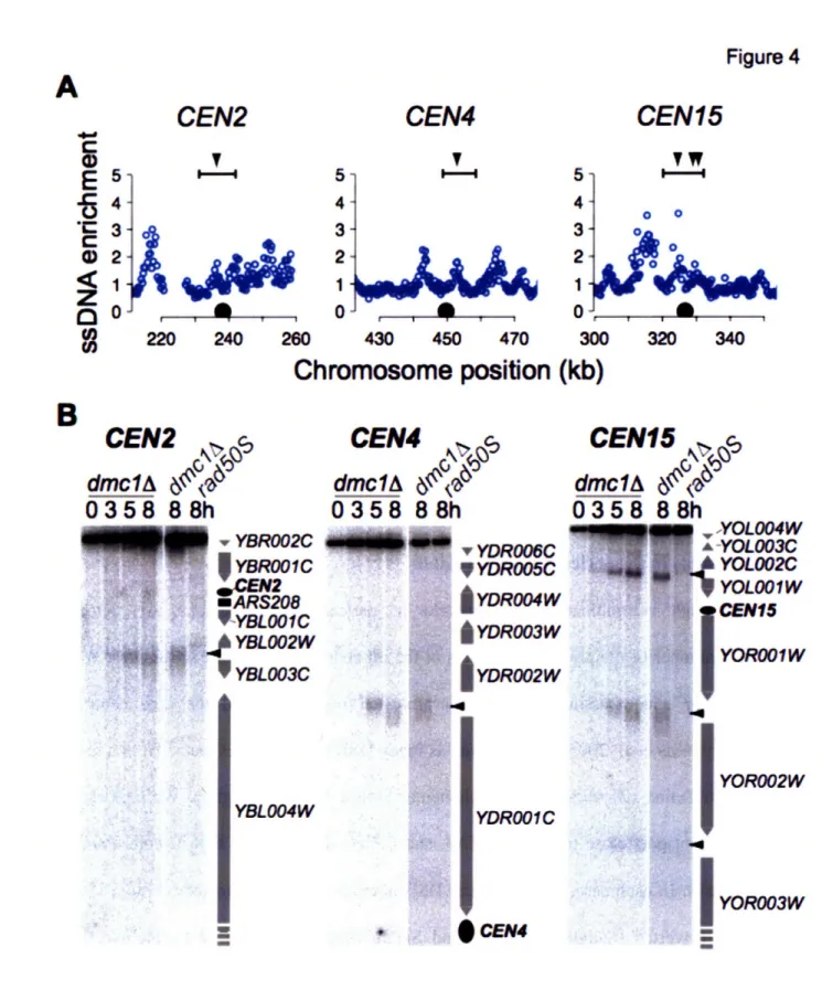
Directly after pre-meiotic DNA replication, the process of homologous recombination begins. The ultimate goal of homologous recombination is to create a physical DNA linkage between at least one set of sister chromatids for every homologous chromosome pair, because this serves to hold the chromosomes together during the first meiotic division. Homologous recombination proceeds in several steps. Double-strand breaks (DSBs) are formed across the genome, homologous chromosomes pair and form stable interactions and mature recombination
products are formed (Figure 4). The cytological results of the physical DNA exchanges between homologous chromosomes are chiasmata.
Homologous recombination is initiated during meiotic G2/prophase by the regulated introduction of at least 200 DSBs into the 16 chromosomes of the yeast genome. A large number of proteins are required for DSB formation in yeast (Keeney 2001; Arora, Kee et al. 2004; Prieler, Penkner et al. 2005), and a subset of these factors begin to associate with chromosomes prior to DSB formation. During meiotic G2/prophase, chromosomes begin to condense and some potential DSB sites undergo a "chromatin transition" and become sensitive to nucleolytic cleavage (Ohta, Shibata et al. 1994). This finding indicates that DSB site selection occurs prior to DNA cleavage. DSB formation also requires the activity of CDK and DDK (Hochwagen, Tham et al. 2005; Wan, Zhang et al. 2006).
DSBs are made by a topoisomerase-VI-related protein, Spol l, along with several accessory factors (Keeney 2001). Spo Il is widely conserved, with orthologs present in a wide variety of eukaryotic species including yeast, plants and humans. Spo Il is thought to cleave the DNA through a covalent intermediate, in which the 5' end of the DNA is attached to a catalytic tyrosine via a phosphodiester bond, similar to topoisomerases (Bergerat, de Massy et al. 1997) (Figure 4). A non-null mutation in the protein Rad50, termed rad50S, and null alleles of SAE2/COM1 block cells at this stage of DSB formation (Alani, Padmore et al. 1990; McKee and Kleckner 1997; Prinz, Amon et al. 1997). In a wild-type cell, Spoll is removed from the chromosome by cleavage of the DNA strand attached to Spo 11l, allowing for repair of the DSB (Neale, Pan et al. 2005).
3. x 3'
'3'
*1
Rad50, See2CIomn
Rec8. Ho2. ind1
Figure 4
Cytological stage
DSB formation and
resectionof 5' ends Lept
Unstable interaction with
- " homologous chmmatid
NCO pathway CO pathways
DNA synt
Mms47
\
Z
ZyZi
" _-- _ _
invasion
extended end 4 annealing
- '- I -n -~
Annealia of DNA synthesis Pacd~
and ligtation
D syn Resolution of Double
and igation Holliday Junction
_ Diffusi I _ and Di r7 s Itene tene Stage Iotene Chromoome Structure Axes form Bridges SCs form SCs stabilized SCs disassemble A C *a-a
Chiasmata
(Adapted from Hochwagen and Amon, 2006)
Figure 4: Control of meiotic homologous recombination
The mechanism and key regulators of homologous recombination are depicted on the left panel. The molecular reactions occur during the cytological stage indicated. The accompanying changes in chromosome structure that occur during meiotic Gs/prophase are shown on the far fight.
J
Repair of meiotic DSBs
In meiosis, DSB repair occurs almost exclusively through recombination, instead of non-homologous end joining or other repair pathways that are observed in other cell types. The first intermediate produced is ssDNA, via the 5'-3' resection of one strand of DNA on either side of the DSB site (Figure 4). Approximately -600 bp of ssDNA are generated on either side of the DSB site, which nucleates the binding of several repair factors including the recA homologs, Rad51 and Dmcl. Once loaded on the DNA, these factors form a nucleoprotein filament and initiate the search for homologous DNA with which to repair the break. In mitotic cells, the Rad51 protein normally directs repair to the sister-chromatid. In meiosis, the Dmcl protein directs the strand invasion step of repair to the homolog instead. Cells lacking Dmcl or Rad51 arrest with long stretches (>1kb) of unrepaired ssDNA at DSB sites.
In the meiotic cell cycle, the repair of DSBs occurs preferentially from the homologous chromosome, with only 20% of repair events occurring from the sister chromatid (Schwacha and Kleckner 1997). Repair from the homolog can lead to either a non-crossover (NCO) or crossover (CO) recombination product, depending on how the repair intermediates are resolved (Figure 4). The traditional model for homologous recombination depicts a double Holliday junction (DHJ), which has an equal likelihood to be resolved by one of two recombination events to produce CO or NCOs. Analysis of mutants that specifically affect CO or NCO production indicate that in vivo, the production of NCOs occurs before mature CO recombinants are produced and NCOs arise via a transient interaction with the homologous chromosome, in which there is no stable strand exchange (Storlazzi, Xu et al. 1995; Hunter and Kleckner 2001)(Allers and Lichten 2001). Stable strand exchange and the production of DHJs occurs only during crossover production
(Figure 4). An alternate pathway to crossover formation that may not require DHJ formation exists, and this pathway is not regulated by crossover interference (see below).
DSB repair facilitates the interaction between homologous chromosomes, although chromosome pairing can occur slowly in the absence of DSB formation (Weiner and Kleckner 1994). Homologous chromosomes establish a stable interaction through the process of CO recombination and a protein complex known as the synaptonemal complex (SC) associates along their length. The exact role of the SC is not well-understood. SC formation is nucleated at CO sites, indicating that its assembly occurs after the CO decision is made. Mutants in SC components, such as the Zipl protein, show reduced levels of CO formation, indicating that the SC may stabilize stable strand exchange and CO designation until DHJs are resolved. In G2/prophase, the telomeres of all chromosomes congress in a few discrete clusters known as the bouquet. Bouquet formation is dependent on the protein Ndj 1 and in its absence, chromosome pairing is delayed, indicating that the bouquet also plays an ill-defined role in chromosome synapsis (Trelles-Sticken, Dresser et al. 2000).
Regulation of crossover formation
Ultimately, each pair of homologous chromosomes should have at least one CO to be properly segregated during the first meiotic division. The regulation of CO distribution in the genome is regulated in at least two ways. First, DSBs must occur on all chromosomes. Second, a mechanism exists to distribute crossovers to all chromosomes. Only -90 COs occur per cell in yeast and these must be distributed to all of the 16 chromosomes (Mortimer, Schild et al. 1991).
The phenomenon of crossover interference prevents multiple CO recombinants from occurring in close proximity on the same chromosome.
DSBs occur more frequently at some genomic loci termed "hotspots." Studies of several individual DSB sites indicated that DSBs occur most frequently in intergenic regions that contain promoters (Baudat and Nicolas 1997). Some DSB hotspots contain binding sites for transcription factors and their activity is increased by transcription factor binding, but other hotspots do not depend on transcription factors (Petes 2001). Some of the determinants of hotspots activity seem to be encoded in cis, since a DSB hotspot that was moved to a "cold" region retained some hotspot activity, albeit at a lower level than at the endogenous locus (Wu and Lichten 1995). Deletion experiments indicated that no single sequence within a DSB hotspot was essential for hotspot activity, and at individual hotspots all break do not occur in the same location, consistent with the idea that there is no simple consensus sequence that determines hotspot activity (De Massy, Baudat et al. 1994). Furthermore, the movement of hotspots in the genome indicated that altering the DSB activity at one locus can increase or decrease DSB activity at nearby hotspots. These experiments indicate that large scale chromosome organization or competition may also regulate DSB activity.
Alleles that arrest DSB formation with Spol 1 linked to the DNA have allowed the genome-wide mapping of DSB hotspots in yeast, via the purification of Spol 1-associated DNA (Gerton, DeRisi et al. 2000; Borde, Lin et al. 2004). Spol 1 sites were found to be distributed across all chromosomes. No sequence or chromatin determinants of DSB site selection could be identified. A slight correlation with elevated GC-content was observed, although GC-content
was calculated for relatively large (5kb) windows and the breaks did not necessarily occur in the GC-rich sequences themselves. It is worth noting that all of these experiments were all carried out in the rad50OS or sae2A/comlA background. The efficiency of DSBs at specific loci is altered in the rad50OS background compared to wild-type cells (Borde, Goldman et al. 2000), suggesting that not all DSB hotspots were identified in the Spo 11l localization studies.
Although DSBs occur preferentially in some loci, other genomic regions are repressed for homologous recombination through multiple mechanisms. DSBs are under-represented near repeat-rich regions such as telomeres and the rDNA. Homologous recombination within repetitive DNA at chromosome ends could lead to CO formation with the incorrect chromosome. Additionally, since cohesin distal to the CO is required to hold chromosomes together during metaphase I in meiosis, a CO at the very end of the DNA molecule could cause chromosome non-disjunction because of the lack of cohesin distal to the CO. Homologous recombination within the rDNA is also prevented, because unequal exchanges between homologs can lead to rDNA loss and death. Finally, CO formation close to centromeres is prevented, and genome-wide studies of Spol l localization indicated that DSBs are under-represented in these regions (Gerton, DeRisi et al. 2000). CO formation close to centromeres is associated with increased levels of non-disjunction, suggesting that crossovers in this region are detrimental to faithful chromosome segregation (Lambie and Roeder 1988; Sears, Hegemann et al. 1995; Lamb, Sherman et al. 2005; Rockmill, Voelkel-Meiman et al. 2006). It is thought that CO formation within this region disrupts the specialized structure necessary for chromosome segregation in meiosis.
Alternate mechanisms exist to distribute at least one CO to all chromosomes. Some species have a far larger number of CO than chromosomes, and in this case CO are randomly distributed. These species also lack SC, suggesting that the SC plays a role in CO distribution. For other organisms, such as yeast, it has been observed that COs are limiting in number, non-randomly distributed and do not occur close together, a phenomenon termed CO interference. Members of the SC including Zipl have been implicated in CO interference, because genetically they appear to lack CO interference, although they probably function downstream of the initial CO selection to stablize CO intermediates (Borner, Kleckner et al. 2004; Fung, Rockmill et al. 2004). Not all crossovers are subject to crossover interference, however, indicating that at least two pathways exist to regulate CO formation. For example, cells containing a version of Spo 11 that makes fewer DSBs than average maintain a wild-type level of COs and exhibit fewer NCO events (Martini, Diaz et al. 2006). The COs that are not affected by interference are regulated by distinct factors (Hollingsworth and Brill 2004). This suggests that a basal level of CO are produced on all chromosomes in the absence of interference, and that additional CO are
distributed through an interference-dependent mechanism.
Co-regulation of DNA replication and recombination
The proper execution of any cell cycle requires that cells complete one process before beginning the next, for example completing DNA replication before attempting to segregate chromosomes. Several checkpoints operate in meiosis to ensure the integrity of the DNA prior to chromosome segregation. Because the initiation of homologous recombination involves the introduction of hundreds of DSBs into the yeast genome, it can be viewed as a form of severe DNA damage. If this occurred prior to or during DNA replication, it would lead to activation of
the intra-S phase checkpoint and replication arrest. Additionally, intermediates produced during errors in DNA replication and during recombination share many structural similarities (such as ssDNA), and the cell might not differentiate between them. Therefore, cells must complete DNA replication, at least on a local level, before DSBs are formed. To test this coordination, a form of chromosome III was created that experiences severely delayed replication on one arm, due to the deletion of several origins. Both the chromatin transition and DSB formation occur later on the delayed replicating chromosome arm than on the wild-type arm, indicating that DSB formation is delayed in response to the delay in the replication of the local DNA (Borde, Goldman et al. 2000; Murakami, Borde et al. 2003). Although these studies indicate that replication and DSB formation are separated, the mechanism of coupling is unknown.
One candidate for ensuring complete replication before the initiation of recombination is the intra-S phase checkpoint, which can monitor the process of DNA replication. The intra-S phase checkpoint has been reported to function during meiosis, similar to during mitotic cell division (Stuart and Wittenberg 1998). Studies indicate that neither mitotic nor meiotic cells monitor DNA replication by counting the number of chromosomes, but instead by sensing ongoing DNA replication by the presence of replication forks or replication intermediates such as ssDNA. Cells lacking Cdc6 that never initiate DNA replication progress in the cell cycle in both mitosis and meiosis, and these cells make DSBs in the absence of DNA replication. A basic understanding of pre-meiotic DNA replication and recombination is be required to better
Thesis summary
In this thesis, I describe the characterization of pre-meiotic DNA replication and DSB formation across the S. cerevisiae genome. I present a model for the protracted pre-meiotic S phase arising as a result of both inefficient initiation of DNA replication and slower fork progression rates. I find no evidence that the altered replication kinetics during pre-meiotic DNA replication are regulated by or related to either cohesion or recombination, suggesting that the link between pre-meiotic DNA replication and homologous recombination affects only the kinetics of DSB formation. Analysis of DSB distribution revealed significant differences in the mechanisms of inhibition of recombination at telomeres, centromeres and near the rDNA. Additionally, this study revealed a non-random distribution of DSB hotspots, which explains how chromosomes of all sizes receive a sufficient number of DSBs to form at least one crossover during homologous recombination.
References
Alani, E., R. Padmore, et al. (1990). "Analysis of Wild-Type and rad50 Mutants of Yeast Suggests an Intimate Relationship between Meiotic Chromosome Synapsis and Recombination." Cell 61: 419-436.
Allers, T. and M. Lichten (2001). "Differential Timing and Control of Noncrossover and Crossover Recombination during Meiosis." Cell 106: 47-57.
Aparicio, J. G., C. J. Viggiani, et al. (2004). "The Rpd3-Sin3 histone deacetylase regulates replication timing and enables intra-S origin control in Saccharomyces cerevisiae." Mol Cell Biol 24(11): 4769-80.
Arora, C., K. Kee, et al. (2004). "Antiviral protein Ski8 is a direct partner of Spol l in meiotic DNA break formation, independent of its cytoplasmic role in RNA metabolism." Mol Cell 13(4): 549-59.
Baudat, F. and A. Nicolas (1997). "Clustering of meiotic double-strand breaks on yeast chromosome III." Proc Natl Acad Sci U S A 94(10): 5213-8.
Bell, S. P. and A. Dutta (2002). "DNA replication in eukaryotic cells." Annu Rev Biochem 71: 333-74.
Benjamin, K. R., C. Zhang, et al. (2003). "Control of landmark events in meiosis by the CDK Cdc28 and the meiosis-specific kinase Ime2." Genes Dev 17(12): 1524-39.
Bergerat, A., B. de Massy, et al. (1997). "An Atypical Topoisomerase II from Archaea with Implications for Meiotic Recombination." Nature 386: 414-417.
Blat, Y., R. U. Protacio, et al. (2002). "Physical and functional interactions among basic chromosome organizational features govern early steps of meiotic chiasma formation."
Cell 111(6): 791-802.
Borde, V., A. S. Goldman, et al. (2000). "Direct coupling between meiotic DNA replication and recombination initiation." Science 290(5492): 806-9.
Borde, V., W. Lin, et al. (2004). "Association of Mrel ip with double-strand break sites during yeast meiosis." Mol Cell 13(3): 389-401.
Borner, G. V., N. Kleckner, et al. (2004). "Crossover/noncrossover differentiation, synaptonemal complex formation, and regulatory surveillance at the leptotene/zygotene transition of meiosis." Cell 117(1): 29-45.
Bowers, J. L., J. C. Randell, et al. (2004). "ATP hydrolysis by ORC catalyzes reiterative Mcm2-7 assembly at a defined origin of replication." Mol Cell 16(6): 96Mcm2-7-Mcm2-78.
Branzei, D. and M. Foiani (2005). "The DNA damage response during DNA replication." Curr Opin Cell Biol 17(6): 568-75.
Buonomo, S. B., R. K. Clyne, et al. (2000). "Disjunction of homologous chromosomes in meiosis I depends on proteolytic cleavage of the meiotic cohesin Rec8 by separin." Cell 103(3): 387-98.
Cha, R. S., B. M. Weiner, et al. (2000). "Progression of meiotic DNA replication is modulated by interchromosomal interaction proteins, negatively by Spol lp and positively by Rec8p." Genes Dev 14(4): 493-503.
Chu, S., J. DeRisi, et al. (1998). "The transcriptional program of sporulation in budding yeast." Science 282(5389): 699-705.
Chu, S. and I. Herskowitz (1998). "Gametogenesis in Yeast Is Regulated by a Transcriptional Cascade Dependent on Ndt80." Mol. Cell 1: 685-696.
Clifford, D. M., S. M. Marinco, et al. (2004). "The meiosis-specific protein kinase Ime2 directs phosphorylation of replication protein A." J Biol Chem 279(7): 6163-70.
Cocker, J. H., S. Piatti, et al. (1996). "An essential role for the Cdc6 protein in forming the pre-replicative complexes of budding yeast." Nature 379(6561): 180-2.
Collins, I. and C. S. Newlon (1994). "Chromosomal DNA replication initiates at the same origins in meiosis and mitosis." Mol Cell Biol 14(5): 3524-34.
Colomina, N., E. Gari, et al. (1999). "Gl cyclins block the Imel pathway to make mitosis and meiosis incompatible in budding yeast." Embo J 18(2): 320-9.
Coverley, D. and R. A. Laskey (1994). "Regulation of eukaryotic DNA replication." Annu Rev Biochem 63: 745-76.
Dahmann, C., J. F. Diffley, et al. (1995). "S-phase-promoting cyclin-dependent kinases prevent re-replication by inhibiting the transition of replication origins to a pre-replicative state." Curr Biol 5(11): 1257-69.
Dahmann, C. and B. Futcher (1995). "Specialization of B-type cyclins for mitosis or meiosis in S. cerevisiae." Genetics 140(3): 957-63.
De Massy, B., F. Baudat, et al. (1994). "Initiation of recombination in Saccharomyces cerevisiae haploid meiosis." Proc Natl Acad Sci U S A 91(25): 11929-33.
DePamphilis, M. L. (2005). "Cell cycle dependent regulation of the origin recognition complex." Cell Cycle 4(1): 70-9.
DePamphilis, M. L., J. J. Blow, et al. (2006). "Regulating the licensing of DNA replication origins in metazoa." Curr Opin Cell Biol 18(3): 231-9.
Diffley, J. F. (2004). "Regulation of early events in chromosome replication." Curr Biol 14(18): R778-86.
Diffley, J. F. and K. Labib (2002). "The chromosome replication cycle." J Cell Sci 115(Pt 5): 869-72.
Dirick, L., T. Bohm, et al. (1995). "Roles and regulation of Cln-Cdc28 kinases at the start of the cell cycle of Saccharomyces cerevisiae." Embo J 14(19): 4803-13.
Dirick, L., L. Goetsch, et al. (1998). "Regulation of meiotic S phase by Ime2 and a Clb5,6-associated kinase in Saccharomyces cerevisiae." Science 281(5384): 1854-7.
Donaldson, A. D., M. K. Raghuraman, et al. (1998). "CLB5-dependent activation of late replication origins in S. cerevisiae." Mol Cell 2(2): 173-82.
Fitch, I., C. Dahmann, et al. (1992). "Characterization of four B-type cyclin genes of the budding yeast Saccharomyces cerevisiae." Mol Biol Cell 3(7): 805-18.
Foiani, M., A. Pellicioli, et al. (2000). "DNA damage checkpoints and DNA replication controls in Saccharomyces cerevisiae." Mutat Res 451(1-2): 187-96.
Fung, J. C., B. Rockmill, et al. (2004). "Imposition of crossover interference through the nonrandom distribution of synapsis initiation complexes." Cell 116(6): 795-802.
Gerton, J. L., J. DeRisi, et al. (2000). "Inaugural article: global mapping of meiotic recombination hotspots and coldspots in the yeast Saccharomyces cerevisiae." Proc Natl Acad Sci U S A 97(21): 11383-90.
Grandin, N. and S. I. Reed (1993). "Differential function and expression of Saccharomyces cerevisiae B-type cyclins in mitosis and meiosis." Mol Cell Biol 13(4): 2113-25.
Hassold, T. and P. Hunt (2001). "To err (meiotically) is human: the genesis of human aneuploidy." Nat Rev Genet 2(4): 280-91.
Hochwagen, A., W. H. Tham, et al. (2005). "The FK506 binding protein Fpr3 counteracts protein phosphatase 1 to maintain meiotic recombination checkpoint activity." Cell 122(6): 861-73.
Hollingsworth, N. M. and S. J. Brill (2004). "The Mus81 solution to resolution: generating meiotic crossovers without Holliday junctions." Genes Dev 18(2): 117-25.
Hollingsworth, R. E., Jr. and R. A. Sclafani (1993). "Yeast pre-meiotic DNA replication utilizes mitotic origin ARS 1 independently of CDC7 function." Chromosoma 102(6): 415-20. Holt, L. J., J. E. Hutti, et al. (2007). "Evolution of Ime2 phosphorylation sites on Cdkl substrates
provides a mechanism to limit the effects of the phosphatase Cdc 14 in meiosis." Mol Cell 25(5): 689-702.
Honigberg, S. M. and K. Purnapatre (2003). "Signal pathway integration in the switch from the mitotic cell cycle to meiosis in yeast." J Cell Sci 116(Pt 11): 2137-47.
Hunt, P. A. and T. J. Hassold (2002). "Sex matters in meiosis." Science 296(5576): 2181-3. Hunter, N. and N. Kleckner (2001). "The single-end invasion: an asymmetric intermediate at the
double-strand break to double-holliday junction transition of meiotic recombination." Cell 106(1): 59-70.
Hyrien, O., C. Maric, et al. (1995). "Transition in specification of embryonic metazoan DNA replication origins." Science 270(5238): 994-7.
Keeney, S. (2001). "Mechanism and control of meiotic recombination initiation." Curr Top Dev Biol 52: 1-53.
Kerrebrock, A. W., D. P. Moore, et al. (1995). "Mei-S332, a Drosophila protein required for sister-chromatid cohesion, can localize to meiotic centromere regions." Cell 83(2): 247-56.
Kiburz, B. M., D. B. Reynolds, et al. (2005). "The core centromere and Sgol establish a 50-kb cohesin-protected domain around centromeres during meiosis I." Genes Dev 19(24): 3017-30.
Kitajima, T. S., S. A. Kawashima, et al. (2004). "The conserved kinetochore protein shugoshin protects centromeric cohesion during meiosis." Nature 427(6974): 510-7.
Klein, F., P. Mahr, et al. (1999). "A Central Role for Cohesins in Sister Chromatid Cohesion, Formation of Axial Elements and Recombination during Yeast Meiosis." Cell 98: 91-103. Kuhne, C. and P. Linder (1993). "A new pair of B-type cyclins from Saccharomyces cerevisiae
that function early in the cell cycle." Embo J 12(9): 3437-47.
Labib, K., J. A. Tercero, et al. (2000). "Uninterrupted MCM2-7 function required for DNA replication fork progression." Science 288(5471): 1643-7.
Lamb, N. E., S. L. Sherman, et al. (2005). "Effect of meiotic recombination on the production of aneuploid gametes in humans." Cytogenet Genome Res 111(3-4): 250-5.
Lambie, E. J. and G. S. Roeder (1988). "A yeast centromere acts in cis to inhibit meiotic gene conversion of adjacent sequences." Cell 52(6): 863-73.
Lee, B. H., B. M. Kiburz, et al. (2004). "Spol3 maintains centromeric cohesion and kinetochore coorientation during meiosis I." Curr Biol 14(24): 2168-82.
Leu, J.-Y., P. R. Chua, et al. (1998). "The Meiosis-Specific Hop2 Protein of S. cerevisiae Ensures Synapsis between Homologous Chromosomes." Cell 94: 375-386.
Lindner, K., J. Gregan, et al. (2002). "Essential role of MCM proteins in premeiotic DNA replication." Mol Biol Cell 13(2): 435-44.
Lopes, M., C. Cotta-Ramusino, et al. (2001). "The DNA replication checkpoint response stabilizes stalled replication forks." Nature 412(6846): 557-61.
Marston, A. L., W. H. Tham, et al. (2004). "A genome-wide screen identifies genes required for centromeric cohesion." Science 303(5662): 1367-70.
Martini, E., R. L. Diaz, et al. (2006). "Crossover homeostasis in yeast meiosis." Cell 126(2): 285-95.
McKee, A. H. and N. Kleckner (1997). "A general method for identifying recessive diploid-specific mutations in Saccharomyces cerevisiae, its application to the isolation of mutants blocked at intermediate stages of meiotic prophase and characterization of a new gene SAE2." Genetics 146(3): 797-816.
Melo, J. and D. Toczyski (2002). "A unified view of the DNA-damage checkpoint." Curr Opin Cell Biol 14(2): 237-45.
Michaelis, C., R. Ciosk, et al. (1997). "Cohesins: Chromosomal Proteins that Prevent Premature Separation of Sister Chromatids." Cell 91: 35-45.
Mori, S. and K. Shirahige (2007). "Perturbation of the activity of replication origin by meiosis-specific transcription." J Biol Chem 282(7): 4447-52.
Mortimer, R. K., D. Schild, et al. (1991). "Genetic and physical maps of Saccharomyces cerevisiae." Methods Enzymol 194: 827-63.
Murakami, H., V. Borde, et al. (2003). "Correlation between premeiotic DNA replication and chromatin transition at yeast recombination initiation sites." Nucleic Acids Res 31(14): 4085-90.
Murakami, H. and P. Nurse (2001). "Regulation of premeiotic S phase and recombination-related double-strand DNA breaks during meiosis in fission yeast." Nat Genet 28(3): 290-3. Neale, M. J., J. Pan, et al. (2005). "Endonucleolytic processing of covalent protein-linked DNA
double-strand breaks." Nature 436(7053): 1053-7.
Nguyen, V. Q., C. Co, et al. (2001). "Cyclin-dependent kinases prevent DNA re-replication through multiple mechanisms." Nature 411(6841): 1068-73.
Ofir, Y., S. Sagee, et al. (2004). "The role and regulation of the preRC component Cdc6 in the initiation of premeiotic DNA replication." Mol Biol Cell 15(5): 2230-42.
Ohta, K., T. Shibata, et al. (1994). "Changes in chromatin structure at recombination initiation sites during yeast meiosis." Embo J 13(23): 5754-63.
Padmore, R., L. Cao, et al. (1991). "Temporal comparison of recombination and synaptonemal complex formation during meiosis in S. cerevisiae." Cell 66(6): 1239-56.
Paulovich, A. G. and L. H. Hartwell (1995). "A checkpoint regulates the rate of progression through S phase in S. cerevisiae in response to DNA damage." Cell 82(5): 841-7.
Petes, T. D. (2001). "Meiotic recombination hot spots and cold spots." Nat Rev Genet 2(5): 360-9.
Prieler, S., A. Penkner, et al. (2005). "The control of Spoll's interaction with meiotic recombination hotspots." Genes Dev 19(2): 255-69.
Primig, M., R. M. Williams, et al. (2000). "The Core Meiotic Transcriptome in Budding Yeasts." Nat. Genet. 26: 415-423.
Prinz, S., A. Amon, et al. (1997). "Isolation of COM1, a new gene required to complete meiotic double-strand break-induced recombination in Saccharomyces cerevisiae." Genetics
146(3): 781-95.
Rabitsch, K. P., M. Petronczki, et al. (2003). "Kinetochore recruitment of two nucleolar proteins is required for homolog segregation in meiosis I." Dev Cell 4(4): 535-48.
Raghuraman, M. K., E. A. Winzeler, et al. (2001). "Replication dynamics of the yeast genome." Science 294(5540): 115-21.