HAL Id: hal-01209318
https://hal.archives-ouvertes.fr/hal-01209318
Submitted on 27 Sep 2018HAL is a multi-disciplinary open access archive for the deposit and dissemination of sci-entific research documents, whether they are pub-lished or not. The documents may come from teaching and research institutions in France or abroad, or from public or private research centers.
L’archive ouverte pluridisciplinaire HAL, est destinée au dépôt et à la diffusion de documents scientifiques de niveau recherche, publiés ou non, émanant des établissements d’enseignement et de recherche français ou étrangers, des laboratoires publics ou privés.
Molecular Mobility in Dense Protein Systems: An
Investigation through 1H NMR Relaxometry and
Diffusometry
Antoine Bouchoux, Diane Schorr, Awa Daffé, Mireille Cambert, Genevieve
Gesan-Guiziou, François Mariette
To cite this version:
Antoine Bouchoux, Diane Schorr, Awa Daffé, Mireille Cambert, Genevieve Gesan-Guiziou, et al.. Molecular Mobility in Dense Protein Systems: An Investigation through 1H NMR Relaxometry and Diffusometry. Journal of Physical Chemistry B, American Chemical Society, 2012, 116 (38), pp.11744-11753. �10.1021/jp306078k�. �hal-01209318�
1
Molecular Mobility in Dense Protein Systems: An Investigation through
1H
NMR Relaxometry and Diffusometry
Antoine Bouchoux†,‡,*, Diane Schorr,§,|| Awa Daffé,§,|| Mireille Cambert,§,|| Geneviève
Gésan-Guiziou,†,‡ François Mariette§,||
† INRA, UMR 1253 Science et Technologie du Lait et de l'Œuf, F-35042 Rennes, France
‡ AGROCAMPUS OUEST, UMR1253 STLO, F-35042 Rennes, France
§Irstea, UR TERE, 17 Avenue de Cucillé, CS 64427, F-35044 Rennes, France
|| Université européenne de Bretagne, France
*Corresponding author. Tel: +33.561.55.77.58; Fax: +33.561.55.61.39;
[email protected]; Present address: CNRS, Laboratoire de Génie Chimique,
118 Route de Narbonne, F-31062 Toulouse, France.
Abstract
Understanding how proteins behave in highly concentrated systems is a major issue in many fields of research, including biology, biophysics, and chemical engineering. In this paper, we provide a comprehensive 1H NMR study of molecular mobility in dilute to highly
concentrated dispersions of the exact same protein (casein) but organized in two distinct supramolecular forms: sponge-like casein micelles or soft casein aggregates. Both relaxometry and diffusometry experiments were performed, so that three different parameters are reported: spin-spin relaxation rates of non-water protons (1/T2,ne), spin-spin relaxation
rates of water protons (1/T2,e+w), and water self-diffusion coefficients (Dw).The results are
2
supramolecular organization on each mobility indicator. We also examine if connections exist between the observed changes in molecular mobility and the already documented changes in rheological and osmotic properties of casein dispersions as concentration is increased.
Keywords NMR, protein, relaxation time, water self-diffusion, glass transition, jamming, gel,
osmotic stress, casein micelle, sodium caseinate
INTRODUCTION
Understanding how proteins behave at high concentrations (> ~100 g.L-1) and how the
general properties of protein dispersions are affected by concentration are questions of great interest in many fields of research including biophysics, pharmaceutics, food science, chemical engineering. As a first example, high protein concentrations are encountered in an increasing number of pharmaceutical preparations (monoclonal antibodies injections for instance) for which it is a true challenge to control the viscosity and/or to reduce both reversible and irreversible protein self-association phenomena.1,2 In physiological conditions,
situations with high concentrations of proteins, and perhaps more generally, with proteins being immersed in a "crowded" environment, are also frequently observed. This leads to specific confinement-induced effects that can be of considerable importance with regards to protein structure and activity.3,4 In food science, knowing how concentration affects the
structural, mechanical, and diffusional properties of new (e.g., high protein nutrition bars) or old and common (e.g., cheese) food products is a decisive question as well.5,6 Finally, the
performances of a fair number of industrial operations in which proteins get concentrated (drying, filtration, centrifugation) also directly rely on the way the proteins interact, organize themselves, and affect the overall properties of the processed solution. A typical example is the purification of proteins through ultra- or microfiltration, which leads to the formation of a
3
concentrated layer of proteins at the filter surface, and that directly impacts the performance of the operation in terms of both selectivity and productivity.7,8
Some specific aspects of the behavior of proteins at high concentrations are now well documented. This mainly includes direct measurements of protein-protein interactions through osmotic stress or scattering techniques,9-13 reports on the viscosity and viscoelastic
properties of protein dispersions at varying concentrations,14-17 studies about possible protein
self-association phenomena (clustering, aggregation) induced at high concentrations,18-20 and
investigations of the protein diffusional properties (self- and collective diffusion) in crowded solutions.21-23 In this paper, we focus on a quite different aspect which is the effect of protein
concentration/self-crowding on a variety of 1H molecular mobility parameters as determined
through nuclear magnetic resonance (NMR). To our knowledge, still little academic attention has been devoted to this point,24-26 even though such "local" mobility indicators can
potentially give important atomic-scale information about the effect of protein crowding. More precisely, the indicators we provide in this paper are the results of 1H relaxation and
diffusion NMR measurements; with values of (i) protein proton and water proton spin-spin relaxation times,27 and (ii) water diffusivities,28 both as a function of protein concentration. In
brief, NMR relaxation times and diffusion coefficients are measures of the rotational and translational mobility of a given molecule, respectively. Both parameters depend on the environment that surrounds the molecule, and are then expected to give interesting indications on the effect of the restrictions imposed by a strong increase in protein concentration. NMR measurements were performed over a wide range of protein concentration, i.e., from the dilute regime (~20 g.L-1) to the highly concentrated regime (~500 g.L-1) where almost half of the
volume of the dispersion is occupied by proteins. Such high concentrations were attained through osmotic stress, a technique in which the composition and characteristics of the solvent are maintained during the concentration process.10,29
4
As for the "model" protein used in this work, the non-globular casein protein was chosen. Besides the fact that caseins are available in large quantities (which was a prerequisite for this study) and are used in an increasing number of industrial applications (milk transformation, new foods and pharmaceutical), the main reason for this choice is that dense dispersions of caseins are now well characterized in terms of protein-protein interactions, rheological properties, and structuration.10,13,15,30-35 Moreover, caseins have interesting associating
properties that make them adopt very distinct supramolecular organizations depending on their environment (Figure 1):
Figure 1. A schematic view of the supramolecular organization of caseins in dispersions of casein micelles (native phosphocaseinate, NPC) and sodium caseinate (SC) as a function of casein concentration. For the casein micelle representation, the CaP nanoclusters are depicted as small black dots. The dashed contour lines are a way to illustrate the fact that the micellar entity is preserved upon compression and that neighboring micelles do not interpenetrate.
- Casein micelle form (native phosphocaseinate, NPC): in "fresh" bovine milk, caseins (as1,
as2, b, and k caseins, in proportion of 3:1:3:1 in mass) are organized into so-called "casein
micelles". Those micelles are complex associative colloids of about 100 nm in size that contain 8% in mass of phosphate and calcium ions in the form of ~4 nm nanoclusters that are distributed into their core.36 Strong associative interactions exist between those calcium
phosphate nanoclusters (termed CaP nanoclusters in the following) and the calcium-sensitive,
Casein Micelle
(NPC)
Sodium Caseinate
(SC)
5
highly phosphorylated as1- and as2-caseins that are present in the micelle.37 The casein
micelles also contain ~3.7 g of water per gram of casein,38 which allows to consider them as
natural and highly hydrated microgels.
- Sodium caseinate form (SC): the mineral content of the casein micelles can be simply removed through acidification and precipitation at pH 4.6. After a subsequent increase in pH to a physiological value of ~7 through the addition of sodium hydroxide, a dispersion of sodium caseinate is obtained. In such a dispersion, the casein molecules are no longer organized into micelles but are present in the form of fragile star-like aggregates of ~20 nm in diameter containing ~12 g of water per gram of casein.39-42
Obviously, these strong structural differences remain as casein concentration increases (Figure 1 gives a schematic view of the concentration process), and this results in very distinct osmotic and mechanical properties between dense NPC and SC dispersions.10,15 Whether or
not this distinction is also visible when looking at the "local" indicators given by NMR spectroscopy was another objective of this study. NMR measurements were then performed on both types of dispersion. Note here that a few NMR relaxometry and diffusometry studies have already been devoted to casein dispersions (SC and/or NPC).43-48 However, all of them
are limited to the "native" concentration of casein in milk (~25 g.L-1) or to the dilute to
semi-dilute concentration regime (< 200 g.L-1), i.e., where changes in the osmotic, structural, and
rheological properties of the dispersions are still minor.
To summarize, we provide here a comprehensive 1H NMR study of molecular mobility in
dilute to highly concentrated dispersions of the exact same protein (casein) but organized in two distinct supramolecular forms. The results obtained are discussed in an effort to understand the respective effects of protein crowding and protein supramolecular organization on each mobility indicator (proton relaxation times and diffusivities). Importantly, we also examine if connections exist between the observed changes in molecular mobility and the
6
already documented changes in rheological and osmotic properties of casein dispersions as concentration is increased. Altogether, we believe that the results presented in this paper provide useful information about the properties of dense dispersions of proteins in general (and casein in particular), and can have potential implications on a growing number of industrial applications.
EXPERIMENTAL SECTION
Proteins. Most of the experiments were performed with dispersions made from casein
powders (NPC or SC powder) dissolved in a aqueous solvent made from the ultrafiltration of skimmed milk (UF). We chose to use such a "physiological" solvent because the rheological, structural and interaction properties of the resulting dispersions are now well documented for a wide range of concentrations, including very dense systems.10,15,30-32,34 Note that the use of
UF for making NPC dispersions also ensures that the caseins are organized into casein micelles that are close to their so-called "native" state.49,50
Two different batches of native phosphocaseinate (NPC) powder were used in this work. They were all prepared in our laboratory following a protocol given by Pierre et al. and Schuck et al,51,52 and summarized in a previous work.10 The sodium caseinate (SC) powder,
was purchased from Armor Protéines (Saint-Brice-en-Coglès, France) and was produced according to a protocol that we also describe elsewhere.10 In all powders, the caseins - and
their associated minerals for NPC - represent >90% of the total solid content.
The UF solvent was prepared through membrane ultrafiltration (5-kDa cutoff) of fresh skimmed milk. Its average ionic composition is: ~20 mM Na+, ~40 mM K+, ~10 mM Ca2+,
~30 mM Cl-, ~10 mM phosphate and ~10 mM citrate.53 UF also contains lactose (150 mM)
7
both purchased from Sigma-Aldrich (Lyon, France), were added to UF as preservatives at 0.02% and 0.1% (w/w), respectively.
Sample preparation. Dispersions at casein concentrations from ~20 to ~150 g.L-1 were
prepared by simply dispersing the casein powders in a given volume of UF. The dispersions were then stirred overnight at 35-50°C to ensure full dissociation of the powder and complete equilibration. At 20°C, the pH of all the dispersions matched the average pH of milk, i.e., pH 6.7 ± 0.1. Note that a few dispersions were also prepared using D2O in place of UF for
performing specific NMR relaxation experiments (Figure 2). Note that the use of pure D2O
solvent in place of the UF solvent has only small effects on the way caseins organize themselves. In particular, caseins are still organized into casein micelles in NPC dispersions made with pure D2O. Those micelles have characteristics that are close to native, with only
minor changes in size (5-10% decrease) and mineral composition (5-8% decrease in Ca and P content).49,54,55
Dispersions at higher casein concentrations were prepared through osmotic stress, a concentration technique based on water exchange between the sample and a polymer solution of controlled osmotic pressure.10 The sample is placed in a dialysis bag that, in turn, is
immersed in a reservoir containing the polymer solution. The cutoff of the dialysis bag is chosen so that it only retains the polymer and the colloidal matter of the sample. Conversely the solvent, i.e., water, ions, and small organic molecules can exchange between the two compartments.
In our case, a poly(ethylene glycol) (PEG) with a molar mass of 35 kDa (Fluka, Buchs, Switzerland) was used as the "stressing" polymer. Polymer solutions were prepared at different concentrations in order to reach different levels of casein concentration at the end of the process.10 SC or NPC samples at low initial concentration (~50 g.L-1) were placed into
8
were immersed in the PEG solutions. During the concentration process, the bags were then refilled regularly with casein dispersion in order to obtain a sufficient amount of concentrated sample. Osmotic equilibrium was reached in about 20 days. This technique made it possible to prepare ~1-2 mL of homogeneous samples with casein concentrations varying from ~200 to ~500 g.L-1.
The total solid content in all the prepared samples were determined through drying at 105°C. It was then converted into casein concentrations in gram of casein per gram of water ( ) or in gram of casein per total volume (g.L-1) by using equivalence relationships that
we properly determined beforehand from density measurements performed with SC and NPC dispersions of known casein concentrations.
NMR. The NMR measurements were all performed using a 20 MHz minispec Bruker
spectrometer equipped with a pulsed field gradient probe. Typical NMR tubes of 8 mm in diameter were used and filled with the - either liquid or solid - casein samples to a height of ~1 cm. All experiments were carried out at a controlled temperature of 25°C
The relaxation times were measured using a Carr-Purcell-Meiboom-Gill (CPMG) sequence.56,57 The short relaxation times (between 11 µs and 100 µs) were obtained from the
FID signal acquired after the first 90° pulse of the sequence, at a sampling rate of 25 MHz. Higher relaxation times were measured using the whole CPMG sequence; the echo train being recorded with a time spacing of 0.1 ms between the 90° and the first 180° pulse. The number of scans was 16. The number of points and the recycle delay (at 5*T1) were adjusted for each
sample. The NMR intensity signal I(t) was fitted to the following exponential expression using the Levenberg-Marquardt method :
(1) 1 water g.g -1 2 2, 2, ( ) exp exp + æ ö æ ö = ç- ÷+ ç- ÷ è neø è e wø t t I t I T I T
9
with (I1,T2,ne) and (I2,T2,e+w) the magnitude and transverse relaxation time of two relaxation
components, i.e., a component for non-exchangeable protein protons (ne) and a component for exchangeable protein protons and water protons (e+w). In the following, the relaxation results are presented in terms of transverse relaxation rates, i.e., 1/T2, in ms-1.
The water self-diffusion coefficients Dw were measured using a pulsed-gradient spin-echo
(PGSE) sequence.58 In brief, D
w is calculated through the signal attenuation I/Ig=0, where I and
Ig=0 are the signals obtained in the presence of a gradient pulse of magnitude g and in the
absence of a gradient, respectively:
(2)
with g the proton gyromagnetic ratio (= 26.752 × 10⁷ T.s−1.rad−1), d the duration of the
gradient pulse, and D is the diffusion time. d and D were adjusted in order to have the same echo attenuation for all samples. For samples with casein concentrations lower than 400 g.L-1,
d was taken as 0.5 ms and D as 7.5 ms. For the more concentrated samples, d and D were 1 and 4 ms, respectively. The maximum gradient strength, gmax, was 3.11 T.m-1. All the results
are given is terms of ratios Dw/Dw,0 between the water diffusivity measured in the casein
sample and the one measured in pure solvent.
RESULTS AND DISCUSSION
Proton relaxation 1: non-exchangeable protein protons. Figure 2 gives the variation
with casein concentration of a first mobility indicator determined though NMR relaxometry, which is the transverse relaxation time T2,ne for the "non-exchangeable" casein protons. As it
is commonly done in NMR, the evolution of the relaxation rate 1/T2,ne rather than the
relaxation time is represented in Figure 2. This rate corresponds to the more rapid component of the 1H NMR relaxation signal. A first simple reason for which we attribute this component
(
)
(
2 2 2)
0 exp 3 = = -g d D -d w g I g D I10
to nonwater protons is the especially high relaxation rates obtained (dozens of ms-1) as
compared to typical values for "free" water protons in the two limiting cases of pure water (~4.10-4 ms-1),59 and of a protein powder containing about 10% of water (~3 ms-1 for a dairy
powder for instance, Mariette et al., unpublished data). A second reason comes from the results of experiments performed in pure D2O rather than in the aqueous "UF" solvent. In that
case, a single 1H relaxation component - which obviously corresponds to nonwater protons -
is observed, and the obtained relaxation rates are similar to those measured in UF (Figure 2, open symbols). A third reason, but that we will not develop here, is the fact that the proportion of the 1/T2,ne component in the double exponential fits (eq 1) increases at a
consistent and predicable rate with casein concentration. That point is fully discussed in part A of the Supporting Information file.
Figure 2. NMR relaxation of non-exchangeable protein protons. The relaxation rate 1/T2,ne is plotted
as a function of casein concentration for both NPC (squares) and SC (circles) dispersions. For low concentrations, measurements were performed with dispersions prepared with either UF (solid symbols) or heavy water D2O (open symbols) as solvent. The inset shows the concentration
dependence of the specific viscosity for SC dispersions, as measured through steady shear rheometry in a Couette geometry.15 In both graphs, the limits of the three concentration regimes are drawn as
vertical dashed lines.
10 100 1000 0 10 20 30 40 50 10 100 1000 10-1 100 101 102 103 104 105 SC (3) (2) Relaxation rat e, 1/T 2,ne (m s -1 ) Casein concentration (g.L-1) (1) NPC SC S p . vis co sit y, hsp Concentration (g.L-1)
11
As just mentioned, the relaxation rates of Figure 2 characterize the non-exchangeable protons that are present in the protein chain. As a consequence, those 1/T2,ne values are related
to both the rotational and the segmental motion of the protein itself, at a given concentration and in a given "structural form". For NPC dispersions, 1/T2,ne is comprised between ~35-45
ms-1 in the whole range of concentrations investigated and does not show much variation with
casein concentration C until C ≈ 180 g.L-1. At C > 180 g.L-1, a slight but clear increase is
observed. The results for SC dispersions are quite different, with comparatively low values (5-10 ms-1) at C £ 80 g.L-1 and a sudden increase in 1/T2,ne in the 80-100 g.L-1 range. At C ³ 100
g.L-1, the relaxation rates are somewhat lower but relatively similar to those measured with
NPC dispersions.
These results necessarily reflect the differences in structure/organization between the two kinds of casein dispersions, as well as the effect of concentration on this organization. We can tentatively interpret them by considering three distinct concentration regimes in which NPC and SC caseins respond differently to an increase in concentration:
(1) Diluted regime, C < 80 g.L-1: in this first concentration regime, both NPC and SC
dispersions contain the well-defined objects that we briefly described in the introduction section. When concentration increases, the casein micelles (NPC) and the casein star-like aggregates (SC) get closer to each other but without coming in direct contact (Figure 1).15 As
a consequence, the restrictions that are locally experienced by the casein chains are not affected by the increase in concentration. This readily explains why the relaxation rates measured for both dispersions do not change significantly with concentration. On the other hand, the strong difference in 1/T2,ne between SC and NPC is to be related to the very distinct
properties of the involved casein particles. In SC dispersions, the caseins are organized into soft and fragile star-like aggregates that are formed through weak attractive interactions between casein molecules (as in self-associating polymer solutions).39-42 Conversely, the NPC
12
dispersions contain casein micelles that are stabilized through stronger interactions between the casein chains (electrostatic and hydrophobic) and, perhaps more importantly, between the phosphoseryl residues of the as1- and as2-casein chains and the CaP nanoclusters.36,37,60 As a
result, casein micelles are objects that are more packed and less hydrated than sodium caseinate particles. In average, the movements of the casein chains are then much more constrained in the casein micelles than in the soft and "vaporous" casein aggregates found in the SC dispersions. That explains, at least qualitatively, why the corresponding NMR relaxation is faster in NPC than in SC dispersions (we do not attempt here to quantitatively interpret the difference in T2,ne between the two types of dispersions since it requires, to our
opinion, too many approximations and conjectures).
(2) Intermediate regime, 80 g.L-1 < C < 180 g.L-1: starting from 80 g.L-1, the SC particles
are progressively forced to get into contact and to interpenetrate/entangle (Figure 1).15,41 This
obviously makes the motion of the casein chains more and more difficult; which in turn explains this sudden and great increase in 1/T2,ne (circles, Figure 2). Incidentally, this
phenomenon nicely correlates with the huge increase in viscosity for SC dispersions at C > 80 g.L-1 (inset of Figure 2), which was also interpreted through the transition towards a
"jammed" or "hyperentangled" regime.15,41 On the other hand, the relaxation rate measured for
dispersions of casein micelles remains unchanged in this second concentration regime. The casein micelles have indeed a much lower specific volume than the SC particles, and a concentration as high as 180 g.L-1 is necessary to force them to get into direct contact.10,15,30
The micelles are therefore still separated from each other in the range 80-180 g.L-1, and the
corresponding relaxation rate, which is linked to the motion of the casein chains into the micelle structure, is therefore still independent of concentration.
(3) Dense regime, C > 180 g.L-1: starting from 180 g.L-1, the casein micelles are
13
dispersions progressively turn into soft solids10,15). As a result, both SC and NPC dispersions
can now be considered as dense and "uniform" dispersions of caseins (Figure 1), i.e., dispersions where the casein chains experience the same degree of entanglement. This explains why the measured relaxation rates are then very similar (Figure 2). Note however that slightly higher relaxation rates are obtained for NPC dispersions, where CaP nanoclusters are present. This suggests that 1/T2,ne is not only related to "entanglement constraints", but is
also marginally sensitive to the motional restrictions induced by the specific interactions between the as1- and as2-casein chains and the CaP nanoclusters.
When C is further increased, the casein micelles are forced to deform and deswell, i.e., water is removed from within their interior.30 Quite expectedly, we then observe an increase
in 1/T2,ne as the motion of the caseins that constitute the micelles get more and more
constrained (we lack the data for SC dispersions at very high concentrations but it is very likely that such an increase in 1/T2,ne occurs in that case as well). Interestingly, this increase is
relatively moderate compared to the one observed at the transition to the hyperentangled regime for SC dispersions (regime 2). This suggests that 1/T2,ne are less sensitive to packing
constraints when those constraints exceed a certain level.
Proton relaxation 2: exchangeable protein protons and water protons. The relaxation
rates that correspond to the second and slowest component of the measured 1H NMR
relaxation signal are given in Figures 3A and 3B. Quite naturally, this relaxation signal is attributed to the protons that were not considered in the previous section, namely, the water protons and the protein protons that are in exchange with surrounding water. Figure 3A is in linear scale and shows the relaxation rate, noted 1/T2,e+w, as a function of casein concentration
expressed in gram of casein per total volume (as in Figure 2). In such a representation, 1/T2,e+w appears to increase monotonously with C for both NPC and SC casein dispersions. In
14
rate when moving from one concentration regime to another. The same results are plotted in a logarithmic scale in Figure 3B as a function of casein concentration, this time expressed in gram per gram of water. With this representation, it appears that a direct and simple linear relationship exists between 1/T2,e+w and the concentration in . This suggests even more
strongly that one unique mechanism is responsible for the regular increase in relaxation rate over the whole range of casein concentration.
Figure 3. NMR relaxation of both exchangeable protein protons and water protons. (A) Relaxation rate 1/T2,e+w plotted as a function of casein concentration in g.L-1 for NPC solutions (squares) and SC
solutions (circles). The dashed lines are guides for the eye. (B) Same data plotted in a log-log scale and as a function of casein concentration in gram per gram of water. For comparison, values available for solutions of the globular protein BSA are plotted as well: data of Olechowicz et al. (stars, pH 5.3 in pure water),24 and Venturi et al. (triangles, pH 6.8 in 0.2 M NaCl).25 All the dashed lines have slope 1.
1 water g.g -0 100 200 300 400 500 600 0.00 0.05 0.10 0.15 0.20 A SC NPC Relaxation r ate, 1 /T 2,e+ w (m s -1 ) Casein concentration (g.L-1) 0.01 0.1 1 1E-3 0.01 0.1 BSA B SC NPC Relaxation r ate, 1 /T 2,e+ w (m s -1 ) Casein concentration (g.g-1 water)
15
The other striking feature in Figure 3B is the large and persistent relative difference that exists between NPC and SC. Over the entire range of casein concentrations, the relaxation rates measured for the two casein dispersions indeed differ by almost half an order of magnitude (~ ×3.7); the larger values being obtained with NPC. Note that this strong dependence of 1/T2,e+w with the way the caseins are organized has already been reported by
Mariette et al., but only in the dilute regime, i.e., at ~25 g.L-1.46 In their paper, a relative
difference of ×4 is for instance observed between dispersions of casein micelles and dispersions where the micelles have been dissociated through the addition of EDTA and the subsequent release of calcium and phosphate from the micelle's interior.
In order to discuss these results, let us first start with the general three-component model that considers the 1H nucleus to be potentially present in three different environments a, b or c.61 The measured relaxation rate is then expressed through:
(3)
where subscript a refers to "free water" protons, b refers to hydration water protons, the mobility of which depends on the motion of the hydrated species, and c refers to water protons in exchange with exchangeable protein protons. Pi is the proportion of protons
belonging to each category, and Kbc is the rate of chemical exchange from state b to state c.
In our case, the measured relaxation rates are always much larger than 1/T2a (≈ 4.10-4 ms-1).
This implies that the contribution of "free water" protons (= component a) can be reasonably neglected in eq 3. Additionally, one can show that the proportions Pi can be simply expressed
in the following form:59
(4)
where ni is the mass of water in state i per 1 g of dry protein. As a result, eq 3 becomes: 1 2, 2 2 2 1 a b c e w a b c bc P P P T + =T +T +T +K -1 . water i i g g P n C»
-16
(5)
This last equation gives a direct linear relationship between 1/T2,e+w and C in g of protein
per g of water, which is in perfect qualitative agreement with our data (Figure 3B). This suggests that the three-component model is well adapted to describe water relaxation in both NPC and SC dispersions and in the range of concentrations studied. In other words, the results of Figures 3A and 3B can be simply explained through the increase in the relative proportion of water protons in states b (protein hydration) and c (chemical exchange) as more proteins are present in the dispersion. Note that this linear relationship between 1/T2,e+w and C is a
known feature for casein and protein dispersions at low to moderate concentrations (C < 0.25 ).44,62 But it is the first time - to our knowledge - that such a dependence is observed to
extend up to very high concentrations where dramatic "macroscopic" effects take place (jamming/gel transition, mechanical strengthening) and could potentially affect the values of 1/T2b and/or . (We discuss the special case of BSA (Figure 3B), reported in the
papers of Venturi et al.25 and Olechnowicz et al.,24 in the last paragraph of this section).
Let us now discuss the relative difference in 1/T2,e+w between dispersions of casein
micelles and dispersions of sodium caseinate (Figures 3A and 3B). According to eq 5, this difference may originate from variations in ni, Pi and/or Kij between NPC and SC. Because
both dispersions contain the same casein molecules, nc is likely to be identical in the two
cases. If we now consider the parameter nb, which is an indirect measure of protein hydration,
all the observations made so far on casein systems indicate that caseins are naturally more hydrated in SC dispersions than in NPC dispersions, i.e., nb,SC ³ nb,NPC.10,15,30,36,39,41 Then, by
virtue of eq 5, it comes that the difference in 1/T2,e+w between NPC and SC can only be
explained through a strong variation of 1/T2b and/or between the two dispersions.
We will not attempt here to assess the relative importance of each relaxation term: chemical
1 1 . 2, 2 2 1 water b c g g e w b c bc n n C T + - T T K -é ù » ê + ú + ë û 1 water g.g -1 2 1Tc+Kbc -1 2 1Tc+Kbc
-17
exchange versus protein hydration (historical and diverging opinions exist on that point 46,59,62-64). But the fact that one and/or the other of these relaxation rates strongly depend on the way
the caseins are organized is qualitatively explainable. Indeed, the mobility of the protons in states b (hydration water protons) and c (water protons in exchange with exchangeable protein protons) are related to the rotational and the segmental motion of the protein itself. It is then not surprising that the corresponding relaxation rates 1/T2b and/or are higher in
NPC dispersions where the casein micelles are stabilized through strong casein-casein and casein-CaP interactions, than in SC dispersions where such interactions are lacking.
As already mentioned, an interesting point here is that the linear relationship of Figure 3B requires that 1/T2b and are constants that do not change with casein concentration
(eq 5). This is in contrast to the non-water proton relaxation results of Figure 2 where 1/T2,ne
clearly varies with C. This suggests that casein entanglement/self-crowding induced by concentration has no direct effect on the relaxation of the water protons that are associated to the casein chains, neither on the chemical exchange rate Kbc. Instead, the relaxation of
hydration and exchangeable water protons, as well as possibly the chemical exchange rate
Kbc, appear to strongly depend on the loss of molecular mobility induced by direct
casein-casein and casein-casein-CaP interactions (NPC vs SC difference).
As for comparison, we provide in Figure 3B the 1/T2 values obtained by other authors for
dense dispersions of the globular protein BSA.24,25 At C < 0.5 , 1/T
2 is also found to
increase linearly with concentration. The absolute values of 1/T2 are however much lower
than those measured in SC and NPC dispersions. Following the discussion above, this difference is probably due the absence of any protein-protein association phenomena in BSA solutions as compared to casein dispersions. At C > 0.5 (i.e., C > ~300 g.L-1), the 1/T
2
values obtained by Venturi et al.25 are much higher than expected and linearity is lost. To our
opinion, this is here the "classical" sign of protein aggregation or clustering,65 which is known
1 2 1Tc+Kbc -1 2 1Tc+Kbc -1 water g.g -1 water g.g
-18
to occur in highly concentrated dispersions of globular proteins.18,19 On the whole, these NMR
relaxation results formerly obtained with BSA appear to be in full accordance with our own casein data and with the interpretations we propose. Of course, a rigorous structural investigation of the behavior of BSA at very high concentrations still remains to be done to fully support that point.
Water self-diffusion. In this last section, we now focus on how water displacement (rather
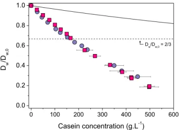
than water spin-spin relaxation) is affected by the presence of caseins, their concentration, and their spatial organization. For that purpose, we consider the water diffusion data of Figure 4, plotted as a function of casein concentration and given for both NPC and SC casein dispersions. For simplicity, the results are reported as self-diffusion coefficients, Dw, relative
to the water self-diffusion coefficient in pure solvent (UF), Dw,0 ≈ 1.98 × 10⁻⁹ m2.s-1. Note that
these results were obtained through pulsed-gradient NMR experiments that involved diffusion times of ~4-7 ms. This implies that the average distance traveled by the water molecules during one NMR measurement is, at least, in the order of a few microns.
For both dispersions, Figure 4 clearly indicates that water diffusivity decreases in a regular fashion with casein concentration. This suggests that there is no direct connection between Dw
and the abrupt changes in the mechanical/rheological/osmotic properties of the dispersions when entering the successive concentration regimes depicted in Figure 1. There is, moreover, no noticeable difference between the diffusivities measured in NPC and SC dispersions (Figure 4). This in turn suggests that Dw is not (or too poorly) sensitive to the organization
adopted by the casein chains and does only depend on the total amount of proteins present in the volume of the dispersion (a conclusion that was already drawn by Mariette et al.,45 but
only in the dilute to semi-dilute concentration regime). Note finally that the observed decrease in water diffusivity with casein concentration is very significant since Dw/Dw,0values as low as ~0.2 are measured in very dense dispersions.
19
Figure 4. Concentration dependence of water self-diffusion in casein dispersions. The diffusion coefficient Dw relative to its value in pure solvent Dw,0 is plotted as a function of casein concentration for NPC (squares) and SC dispersions (circles). The full line is obtained through a simple obstruction model (eqs 6-7), considering that all the water molecules present in the dispersion are able to diffuse and that the casein chains are the only impenetrable matter (veff* = 0.73 cm3.g-1 in eq. 7). The obstruction model of eqs 6-7 is unable to predict the diffusion data located below the dashed horizontal line, i.e., Dw/Dw,0 < 2/3.
Two distinct approaches can be used to discuss these results. The first one consists in assuming that water molecules are located in either water-rich or water-poor regions that have distinct diffusional properties. The effective diffusivity of water in the whole dispersion is then a complex function of the volume occupied by each region and of the corresponding water concentrations and diffusivities. This is the well-known "cell-diffusion model", initially proposed by Jönsson et al.,66 and recently used by Mariette's and McGrath's groups for
discussing water diffusion in casein systems at low to moderate concentrations.45,48 Although
interesting, we decided not to use this approach in the present paper. The first reason is that the cell-diffusion model involves a fair number of parameters which are often difficult to estimate and/or discuss in the case of casein systems as compared to "model" colloidal systems: how to define the water-rich and water-poor regions in SC and NPC dispersions?, what concentrations and diffusivities do we use for water in these regions? Another important
0 100 200 300 400 500 600 0.0 0.2 0.4 0.6 0.8 1.0 D w /D w,0 Casein concentration (g.L-1) Dw/Dw,0 = 2/3
20
reason is that the cell-diffusion model is in fact unable to predict the correct diffusivities at concentrations C > 200 g.L-1. For a detailed discussion about the cell-diffusion model and its
potential relevance to casein systems, we refer the reader to part B of the Supporting Information provided with this article.
The second and probably simpler approach is to consider that the dispersions are always homogeneous with regards to water diffusion. The local diffusivity of water is then assumed to be the same in all places in the dispersion, whatever the concentration of the proteins and their spatial organization. For casein systems, this proposition is of course questionable; especially at low concentrations (C < C*) where the SC and NPC particles are clearly
separated by regions of pure solvent. On the other hand, the distance traveled by each water molecule during our NMR measurements is always much larger than the size of both inter- and intra-particular heterogeneities in the system (a few microns versus a few dozens of nanometers). As a consequence, all the individual water molecules "seen" by the NMR instrument have about the same average diffusion. Hence it seems very reasonable to use such a "homogeneous" approach for modeling the diffusion data obtained in this work.
Following this, the first models that can be examined are those based on obstruction effects exclusively. In these models, initially developed for polymer solutions, the diffusing molecule is considered as the only mobile entity in the system. The polymer chains are in turn treated as fixed and impenetrable segments immersed in the dispersion; the presence of which causes a direct increase in the average path length between two points in the system.67 When dealing
with water diffusion, as in our case, the size of the diffusant is often not taken into account since it is generally much smaller than the characteristic dimensions of the system. A basic obstruction model is then:66
(6) ,0 1 1 2 w eff w D D = +f
21
with feff the effective volume occupied by impenetrable and motionless matter, which can be
simply related to the casein concentration through:
(7) where veff* is the specific volume of impenetrable matter
Figure 4 shows the predicted values of Dw/Dw,0 for veff* = 0.73 ml.g-1, i.e., the specific
volume of anhydrous casein.68 This assumes that all the volume that is not occupied by
proteins is filled with water molecules that are able to diffuse freely. In that case, the Dw/Dw,0
values are clearly overestimated compared to the experimental measurements (Figure 4). From this result, the first idea that comes to mind is that the available diffusional volume is in fact much lower than expected; some water molecules being strongly associated to the casein chains and thus acting as immobile matter that contributes to the obstruction effect. This simple and a priori reasonable idea, which is equivalent to adjusting veff* to higher values, is
however not sufficient to explain our results. The obvious reason for this is that eqs 6-7 are unable to predict diffusion coefficients lower than the limiting value Dw/Dw,0 = 2/3,
calculated for feff = 1 (Figure 4). So even when assuming strong "hydration" effects, diffusion
models based on obstruction only are totally unable to describe water self-diffusion at very high protein concentrations.
Another diffusion model that we can now consider is the one proposed by Wang et al. in their pioneering work on water self-diffusion in pure protein solutions.72 In this model, the
obstruction component is complemented with a "slowing down" component that takes into account the hydration phenomena. The following expression is used:72,73
(8) where is a shape factor that accounts for the shape and orientation of the diffusant relative to its displacement and f is the fraction of hydration or "immobile" water bound to the protein
* eff v Ceff f =
(
)
(
)
,0 1 1 w eff w D f D = -af -a22
surface. feff is defined as previously as the volume fraction occupied by motionless species.
feff and f can both be expressed as a function of casein concentration using eq 7 and:
(9)
where vprot* = 0.73 ml.g-1 is the specific volume of anhydrous casein and veff* is the specific
volume of the protein plus its associated water molecules.
For dispersions of a globular protein, is taken as the shape factor of an equivalent ellipsoid with dimensions a/b that match the size of the hydrated protein. This gives values ranging from 1.5 to 1.6 for ovalbumin for instance.72 For barnacle muscle, which is obviously
a very a different type of system, Clark et al. propose to use the shape factor of smooth elongate rods, i.e., = 1.667. In our case, we have no rationale for choosing a precise value for since caseins are non-globular proteins that self-associate. We therefore decided to use = 1.5, which is the theoretical value for a sphere and is in the range of the values cited above. Note also that the small error we probably make by using = 1.5 has only minor repercussions on the calculated values of Dw/Dw,0 (see the paper of Wang et al.72 for a detailed
analysis of the sensitivity of the model towards ).
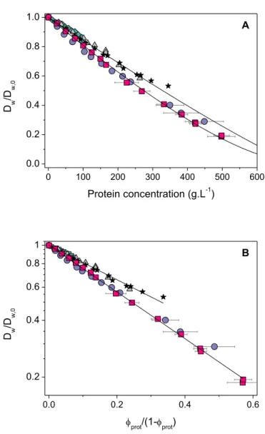
As shown in Figure 5A, the fit of the Wang's model to our data is very satisfactory. The only adjustable parameter here is veff*, and we obtain veff* = 1.05 mL.g-1. From this specific
volume, we can calculate a parameter H which is the mass of "non-mobile" water per gram of casein:
(10)
with dw is the water density taken as dw = 1 g.mL-1. * * * 1 eff prot prot v v f v C -= -a a a a a a * * * 1 prot w prot eff v H d v v æ ö = çç - ÷÷ è ø
23
Figure 5. Concentration dependence of water self-diffusion: comparison with other protein solutions and fits to theoretical models. Data obtained in this work: NPC solutions (squares) and SC solutions (circles). Data from other works: ovalbumin (triangles, Bourret et al.,69 Wang et al.70), lysozyme
(diamonds, Bourret et al.69), whey proteins (stars, Colsenet et al.71). (A) Combined obstruction and
slowing-down model: the full lines are fits of eq 8 to the experimental data. (B) Free volume theory: the full lines are fits of eq 11 to the data. Results are presented as a function of fprot/(1-fprot) and in a semi-log plot to highlight the exponential dependence of eq 11.
We obtain H = 0.22 gwater.gcasein-1, a value that is close to the standard "full" hydration of a
typical globular protein in water, which is about 0.3-0.4 g.g-1 according to Perez et al. or
Steinbach et al. for instance74,75. Note that we are not saying that H is the actual hydration of
the protein, but it seems reasonable to find H values that are close to this actual hydration.
0 100 200 300 400 500 600 0.0 0.2 0.4 0.6 0.8 1.0 A D w /D w,0 Protein concentration (g.L-1) 0.0 0.2 0.4 0.6 0.2 0.4 0.6 0.8 1 B D w /D w,0 fprot/(1-fprot)
24
For comparison, the water self-diffusion values obtained with solutions of other proteins, namely ovalbumin,69,70 lysozyme,69 and whey proteins,71 are also shown in Fig. 5A. Water
diffusion is about the same in all these solutions of globular proteins, and is noticeably higher than the diffusion measured in casein systems. In the context of the Wang's model, it suggests that lysozyme, ovalbumin, and whey proteins, "immobilize" less water molecules than caseins (the corresponding H value is 0.13-0.15 g.g-1). This is very acceptable since caseins are
intrinsically unstructured/disordered proteins that are less hydrophobic in nature and consequently more exposed to solvent than globular proteins like ovalbumin, lysozyme, and the proteins that compose whey (b-lactoglobulin, a-lactalbumin, serum albumin).
Note that many other diffusion models are proposed in the literature (see the review of Masaro et al.67). It is clearly beyond the purpose of this work to discuss each of them. We
propose however to briefly examine the model suggested by Yasuda et al., which is based on the free volume theory.76 This model is indeed known to be particularly suitable for the
analysis of the diffusion of small entities in semi-dilute polymer systems.67,77 It gives:
(11)
where B is a constant, a* is the cross-section area of the diffusant (i.e., the minimum hole size
required for its displacement), is the free volume per molecule of solvent, and fprot is the
volume fraction occupied by the anhydrous protein.
By varying the ratio Ba*/ , we are able to obtain very good fit of this model to our data
and to those measured with other protein solutions (Figure 5B). The free volume theory then appears appropriate for describing - at least qualitatively - water self-diffusion in dense dispersions of proteins. We do not go further here as we believe it is quite challenging to quantitatively interpret the values obtained for Ba*/ in each case.
* * ,0 exp 1 prot w w V prot D Ba D f f f é æ öù = ê- çç ÷÷ú -ê è øú ë û * V f * V f * V f
25
CONCLUSION
How is molecular mobility affected in dense protein systems? Does this mobility depend on the supramolecular organization of the proteins? Are there any connections with the macroscopic properties of the dispersion? In an effort to address these issues, careful 1H NMR
relaxometry and diffusometry experiments were performed with model dispersions of caseins over a wide range of concentrations. Three distinct properties were measured: the spin-spin relaxation rate of non-water protons 1/T2,ne, the spin-spin relaxation rate of water protons
1/T2,e+w and the relative self-diffusion of water Dw/Dw,0. Quite remarkably, we find that these
three NMR indicators have very different behaviors towards both the concentration of the proteins and their organization:
. The spin-spin relaxation rate of non-water protons 1/T2,ne is found to be directly related to
the average "compactness" of the casein chains in the dispersion. At low concentrations, relaxation is much faster when the caseins are organized into compact micelles (NPC) rather than in soft and fragile casein aggregates (SC). When water is progressively removed from the dispersion, relaxation does not change as long as the NPC or SC particles are not brought into direct contact. When the concentration for close-packing is approached, the relaxation rate then increases significantly. This phenomenon appears to be directly correlated with the abrupt changes in viscosity of the dispersions. In very dense dispersions, the NPC and SC particles fill all the available volume. They either deform/compress or interpenetrate as concentration increases, and 1/T2,ne increases accordingly. On the other hand, 1/T2,ne is found
to be the same for both NPC and SC dispersions; suggesting it is poorly sensitive to the strong local interactions that exist between caseins and mineral nanoclusters in the casein micelles.
. In contrast, the spin-spin relaxation rate of water protons 1/T2,e+w is not correlated to the
macroscopic properties of the dispersion and is simply proportional to the relative amount of water that is associated to the caseins. However, water relaxation is much faster in dispersion
26
of casein micelles; which we interpret as an indirect effect of the strong local interactions between the casein chains and the CaP nanoclusters. In SC dispersions, i.e., where self-association phenomena are much less important, water relaxation is close to the relaxation values measured in dispersions of globular proteins.
. Finally, water self-diffusion Dw/Dw,0 does only depend on casein concentration. It is not
related to the spatial organization of the caseins (micelles or star-like aggregate), and it is not related to the osmotic/rheological properties of the dispersion. However, there is a significant difference between Dw/Dw,0 in casein systems and Dw/Dw,0 in dispersions of globular proteins.
By using basic obstruction/hydration diffusion models, we find that this is probably explained by the fact that caseins are natively disordered proteins that are less hydrophobic than well-folded globular proteins.
In conclusion, the NMR technique, combined with the osmotic stress method, is able to provide interesting and - to our knowledge - new information about the general properties of dense dispersions of proteins. Of course, further investigations needs to be done to fully understand the reported variations of complex properties such as spin-spin NMR relaxation times or diffusivities. As an example, we are now in the process of measuring the self-diffusion coefficients of tracers of different sizes in dense casein systems (as in the work of Le Feunteun et al.78 for instance). There is no doubt that such complementary results may help
in better apprehending the basis of molecular transport in "self-crowding" protein systems.
ASSOCIATED CONTENT
Supporting Information. The supporting material provides some discussions about the
relaxation signal of the non-exchangeable casein protons (part A), as well as an analysis of the cell-diffusion model of Jönsson et al.66 and its potential relevance to casein systems (part B).
27
REFERENCES
[1] Saluja, A.; Badkar, A. V.; Zeng, D. L.; Nema, S.; Kalonia, D. S. Biophys. J. 2007, 92, 234-244. [2] Yadav, S.; Shire, S. J.; Kalonia, D. S. J. Pharm. Sci. 2010, 99, 4812-4829.
[3] Ellis, R. J.; Minton, A. P. Nature 2003, 425, 27-28.
[4] Zhou, H. X.; Rivas, G.; Minton, A. P. Annu. Rev. Biophys. 2008, 37, 375-397.
[5] Purwanti, N.; van der Goot, A. J.; Boom, R.; Vereijken, J. Trends Food Sci. Technol 2010, 21, 85-94.
[6] Floury, J.; Madec, M. N.; Waharte, F.; Jeanson, S.; Lortal, S. Food Chem. 2012, 133, 551-556. [7] Belfort, G.; Davis, R. H.; Zydney, A. L. J. Membr. Sci. 1994, 96, 1-58.
[8] Kelly, S. T.; Zydney, A. L. J. Membr. Sci. 1995, 107, 115-127.
[9] Ianeselli, L.; Zhang, F.; Skoda, M. W. A.; Jacobs, R. M. J.; Martin, R. A.; Callow, S.; Prévost, S.; Schreiber, F. J. Phys. Chem. B 2010,
[10] Bouchoux, A.; Cayemitte, P. E.; Jardin, J.; Gésan-Guiziou, G.; Cabane, B. Biophys. J. 2009, 96, 693-706.
[11] Vilker, V. L.; Colton, C. K.; Smith, K. A. J. Colloid Interface Sci. 1981, 79, 548-566. [12] Yousef, M. A.; Datta, R.; Rodgers, V. G. J. J. Colloid Interface Sci. 2001, 243, 321-325. [13] Farrer, D.; Lips, A. Int. Dairy J. 1999, 9, 281-286.
[14] Lefebvre, J. Rheol. Acta 1982, 21, 620-625.
[15] Bouchoux, A.; Debbou, B.; Gesan-Guiziou, G.; Famelart, M. H.; Doublier, J. L.; Cabane, B. J.
Chem. Phys. 2009, 131, 165106-165111.
[16] Brownsey, G. J.; Noel, T. R.; Parker, R.; Ring, S. G. Biophys. J. 2003, 85, 3943-3950.
[17] Parker, R.; Noel, T. R.; Brownsey, G. J.; Laos, K.; Ring, S. G. Biophys. J. 2005, 89, 1227-1236. [18] Stadler, A. M.; Schweins, R.; Zaccai, G.; Lindner, P. J. Phys. Chem. Lett. 2010, 1805-1808. [19] Stradner, A.; Cardinaux, F.; Schurtenberger, P. J. Phys. Chem. B 2006, 110, 21222-21231.
[20] Shukla, A.; Mylonas, E.; Di Cola, E.; Finet, S.; Timmins, P.; Narayanan, T.; Svergun, D. I. Proc.
Natl. Acad. Sci. U.S.A. 2008, 105, 5075-5080.
[21] Heinen, M.; Zanini, F.; Roosen-Runge, F.; Fedunova, D.; Zhang, F.; Hennig, M.; Seydel, T.; Schweins, R.; Sztucki, M.; Antalik, M. et al Soft Matter 2012, 8, 1404-1419.
[22] Li, C.; Wang, Y.; Pielak, G. J. J. Phys. Chem. B 2009, 113, 13390-13392.
[23] Roosen-Runge, F.; Hennig, M.; Zhang, F.; Jacobs, R. M. J.; Sztucki, M.; Schober, H.; Seydel, T.; Schreiber, F. Proc. Natl. Acad. Sci. U.S.A. 2011, 108, 11815-11820.
28
[24] Olechnowicz, R.; Masierak, W.; Bodurka, J.; Gutsze, A. Magn. Reson. Chem. 1999, 37, S147-S149.
[25] Venturi, L.; Woodward, N.; Hibberd, D.; Marigheto, N.; Gravelle, A.; Ferrante, G.; Hills, B. P.
Appl. Magn. Reson. 2008, 33, 213-234.
[26] Bernado, P.; de la Torre, J. G.; Pons, M. J. Mol. Recognit. 2004, 17, 397-407. [27] Kimmich, R.; Anoardo, E. Prog. Nucl. Magn. Reson. Spectrosc. 2004, 44, 257-320.
[28] Ardelean, I. and R. Kimmich. 2003. Principles and Unconventional Aspects of NMR Diffusometry. In Annual Reports on NMR Spectroscopy, Vol. 49, Academic Press, 45-115.
[29] Bonnet-Gonnet, C.; Belloni, L.; Cabane, B. Langmuir 1994, 10, 4012-4021.
[30] Bouchoux, A.; Gésan-Guiziou, G.; Pérez, J.; Cabane, B. Biophys. J. 2010, 99, 3754-3762.
[31] Dahbi, L.; Alexander, M.; Trappe, V.; Dhont, J. K. G.; Schurtenberger, P. J. Colloid Interface
Sci. 2010, 342, 564-570.
[32] Dalgleish, D. G.; Corredig, M. Annu. Rev. Food Sci. Technol. 2012, 3, 449-467.
[33] Mezzenga, R.; Schurtenberger, P.; Burbidge, A.; Michel, M. Nat. Mater. 2005, 4, 729-740. [34] Pignon, F.; Belina, G.; Narayanan, T.; Paubel, X.; Magnin, A.; Gésan-Guiziou, G. J. Chem. Phys. 2004, 121, 8138-8146.
[35] Qu, P.; Gésan-Guiziou, G.; Bouchoux, A. J. Membr. Sci. 2012, 417-418, 10-19.
[36] Dalgleish, D. G. Soft Matter 2011, 7, 2265-2272.
[37] Horne, D. S. Curr. Opin. Colloid Interface Sci. 2006, 11, 148-153. [38] De Kruif, C. G. J. Dairy Sci. 1998, 81, 3019-3028.
[39] HadjSadok, A.; Pitkowski, A.; Nicolai, T.; Benyahia, L.; Moulai-Mostefa, N. Food
Hydrocolloids 2008, 22, 1460-1466.
[40] Pitkowski, A.; Durand, D.; Nicolai, T. J. Colloid Interface Sci. 2008, 326, 96-102. [41] Thomar, P.; Durand, D.; Benyahia, L.; Nicolai, T. Faraday Discuss. 2012,
[42] Lucey, J. A.; Srinivasan, M.; Singh, H.; Munro, P. A. J. Agric. Food Chem. 2000, 48, 1610-1616. [43] Hikichi, K.; Saito, H.; Tokita, M.; Niki, R. Polymer J. 1991, 23, 1265-1267.
[44] Le Dean, A.; Mariette, F.; Marin, M. J. Agric. Food Chem. 2004, 52, 5449-5455.
[45] Mariette, F.; Topgaard, D.; Jonsson, B.; Soderman, O. J. Agric. Food Chem. 2002, 50, 4295-4302.
[46] Mariette, F.; Tellier, C.; Brule, G.; Marchal, P. J. Dairy Res. 1993, 60, 175-188.
[47] Metais, A.; Cambert, M.; Riaublanc, A.; Mariette, F. J. Agric. Food Chem. 2004, 52, 3988-3995. [48] Tan, H. L.; McGrath, K. M. J. Colloid Interface Sci. 2010, 342, 399-406.
29
[49] Famelart, M. H.; Lepesant, F.; Gaucheron, F.; Le Graet, Y.; Schuck, P. Lait 1996, 76, 445-460. [50] Ouanezar, M.; Guyomarc'h, F.; Bouchoux, A. Langmuir 2012, 28, 4915-4919.
[51] Pierre, A.; Fauquant, J.; Le Graët, Y.; Piot, M.; Maubois, J. L. Lait 1992, 72, 461-474.
[52] Schuck, P.; Piot, M.; Méjean, S.; Le Graët, Y.; Fauquant, J.; Brulé, G.; Maubois, J. L. Lait 1994,
74, 375-388.
[53] Jenness, R.; Koops, J. Neth. Milk Dairy J. 1962, 16, 153-164.
[54] Hansen, S.; Bauer, R.; Lomholt, S. B.; Quist, K. B.; Pedersen, J. S.; Mortensen, K. Eur. Biophys.
J. 1996, 24, 143-147.
[55] Jackson, A. J.; McGillivray, D. J. Chem. Commun. 2011, 47, 487-489. [56] Carr, H. Y.; Purcell, E. M. Phys. Rev. 1954, 94, 630-638.
[57] Levitt, M. H.; Freeman, R. J. Magn. Reson. 1981, 43, 65-80. [58] Stejskal, E. O.; Tanner, J. E. J. Chem. Phys. 1965, 42, 288-292.
[59] Daszkiewicz, O.; Hennel, J. W.; Lubas, B.; Szczepkowski, T. W. Nature 1963, 200, 1006-1007. [60] Horne, D. S. Int. Dairy J. 1998, 8, 171-177.
[61] Fedotov, V. D.; Miftakhutdinova, F. G.; Murtazin, F. Biofizika 1969, 14, 873-882. [62] Hills, B. P.; Takacs, S. F.; Belton, P. S. Food Chem. 1990, 37, 95-111.
[63] Venu, K.; Denisov, V. P.; Halle, B. J. Am. Chem. Soc. 1997, 119, 3122-3134.
[64] Moller, S. M.; Whittaker, A. K.; Stokes, J. R.; Gidley, M. J.; Andersen, U.; Bertram, H. C. J.
Agric. Food Chem. 2011, 59, 10097-10103.
[65] Hills, B. P.; Takacs, S. F.; Belton, P. S. Mol. Phys. 1989, 67, 919-937.
[66] Jönsson, B.; Wennerström, H.; Nilsson, P. G.; Linse, P. Colloid. Polym. Sci. 1986, 264, 77-88. [67] Masaro, L.; Zhu, X. X. Prog. Polym. Sci. 1999, 24, 731-775.
[68] Morris, G. A.; Foster, T. J.; Harding, S. E. Biomacromolecules 2000, 1, 764-767. [69] Bourret, D.; Parello, J. J. Phys. Colloques 1984, 45, C7-255.
[70] Wang, J. H.; Anfinsen, C. B.; Polestra, F. M. J. Am. Chem. Soc. 1954, 76, 4763-4765. [71] Colsenet, R.; Mariette, F.; Cambert, M. J. Agric. Food Chem. 2005, 53, 6784-6790. [72] Wang, J. H. J. Am. Chem. Soc. 1954, 76, 4755-4763.
[73] Clark, M. E.; Burnell, E. E.; Chapman, N. R.; Hinke, J. A. Biophys. J. 1982, 39, 289-299. [74] Perez, J.; Zanotti, J. M.; Durand, D. Biophys. J. 1999, 77, 454-469.
30
[76] Yasuda, H.; Lamaze, C. E.; Ikenberry, L. D. Makromol. Chem. 1968, 118, 19-35. [77] Amsden, B. Macromolecules 1998, 31, 8382-8395.
31
Table of Contents (TOC) Image
10 100 1000 0 10 20 30 40 50 SC Protei n rela xati on rate, 1/T 2 (ms -1) Casein concentration (g.L-1) NPC Micellar casein (NPC) Non-micellar casein (SC)